
Influenza Virus Infection Induces a Narrow Antibody …Influenza Virus Infection Induces a Narrow...
15
Influenza Virus Infection Induces a Narrow Antibody Response in Children but a Broad Recall Response in Adults Philip Meade, a,b,c,d Guillermina Kuan, e,f Shirin Strohmeier, b,c,d Hannah E. Maier, g Fatima Amanat, a,b,c,d Angel Balmaseda, f,h Kimihito Ito, i Ericka Kirkpatrick, a,b,c,d Andres Javier, b Lionel Gresh, f Raffael Nachbagauer, b,c,d Aubree Gordon, c,g,j Florian Krammer b,c,d a Graduate School of Biomedical Sciences, Icahn School of Medicine at Mount Sinai, New York, New York, USA b Department of Microbiology, Icahn School of Medicine at Mount Sinai, New York, New York, USA c Centers of Excellence for Influenza Research and Surveillance (CEIRS) d Center for Research on Influenza Pathogenesis (CRIP), New York, New York, USA e Sustainable Sciences Institute, Managua, Nicaragua f Centro de Salud Sócrates Flores Vivas, Ministry of Health, Managua, Nicaragua g Department of Epidemiology, School of Public Health, University of Michigan, Ann Arbor, Michigan, USA h Laboratorio Nacional de Virología, Centro Nacional de Diagnóstico y Referencia, Ministry of Health, Managua, Nicaragua i Division of Bioinformatics, Hokkaido University Research Center for Zoonosis Control, Kitaku, Japan j St. Jude Center of Excellence for Influenza Research and Surveillance, Memphis, Tennessee, USA ABSTRACT In contrast to influenza virus vaccination, natural infection induces long- lived and relatively broad immune responses. However, many aspects of the anti- body response to natural infection are not well understood. Here, we assessed the immune response after H1N1 influenza virus infection in children and adults in a Ni- caraguan household transmission study using an influenza virus protein microarray (IVPM). This technology allows us to simultaneously measure IgG and IgA antibody responses to hemagglutinins of many different virus strains and subtypes quantita- tively with a high throughput. We found that children under 6 years of age re- sponded to natural infection with a relatively narrow response that targeted mostly the hemagglutinin of the strain that caused the infection. Adults, however, have a much broader response, including a boost in antibodies to many group 1 subtype hemagglutinins. Also, a strong recall response against historic H1 hemagglutinins that share the K133 epitope with the pandemic H1N1 virus was observed. Of note, some children, while responding narrowly within H1 and group 1 hemagglutinins, induced a boost to H3 and other group 2 hemagglutinins when infected with H1N1 when they had experienced an H3N2 infection earlier in life. This is an interesting phenomenon providing evidence for immune imprinting and a significant new in- sight which might be leveraged in future universal influenza virus vaccine strategies. Finally, preexisting immunity to pandemic H1 hemagglutinins was significantly asso- ciated with protection from infection in both children and adults. In adults, preexist- ing immunity to non-H1 group 1 hemagglutinins was also significantly associated with protection from infection. IMPORTANCE It is known since Thomas Francis, Jr. published his first paper on orig- inal antigenic sin in 1960 that the first infection(s) with influenza virus leaves a spe- cial immunological imprint which shapes immune responses to future infections with antigenically related influenza virus strains. Imprinting has been implicated in both protective effects as well as blunting of the immune response to vaccines. De- spite the fact that this phenomenon was already described almost 60 years ago, we have very little detailed knowledge of the characteristics and breadth of the im- mune response to the first exposure(s) to influenza virus in life and how this com- pares to later exposure as adults. Here, we investigate these immune responses in Citation Meade P, Kuan G, Strohmeier S, Maier HE, Amanat F, Balmaseda A, Ito K, Kirkpatrick E, Javier A, Gresh L, Nachbagauer R, Gordon A, Krammer F. 2020. Influenza virus infection induces a narrow antibody response in children but a broad recall response in adults. mBio 11:e03243-19. https://doi.org/10.1128/ mBio.03243-19. Editor Stacey Schultz-Cherry, St. Jude Children's Research Hospital Copyright © 2020 Meade et al. This is an open-access article distributed under the terms of the Creative Commons Attribution 4.0 International license. Address correspondence to Aubree Gordon, [email protected], or Florian Krammer, fl[email protected]. Received 9 December 2019 Accepted 11 December 2019 Published RESEARCH ARTICLE Clinical Science and Epidemiology January/February 2020 Volume 11 Issue 1 e03243-19 ® mbio.asm.org 1 21 January 2020 on February 27, 2020 by guest http://mbio.asm.org/ Downloaded from
Transcript of Influenza Virus Infection Induces a Narrow Antibody …Influenza Virus Infection Induces a Narrow...
Influenza Virus Infection Induces a Narrow Antibody Responsein Children but a Broad Recall Response in Adults
Philip Meade,a,b,c,d Guillermina Kuan,e,f Shirin Strohmeier,b,c,d Hannah E. Maier,g Fatima Amanat,a,b,c,d Angel Balmaseda,f,h
Kimihito Ito,i Ericka Kirkpatrick,a,b,c,d Andres Javier,b Lionel Gresh,f Raffael Nachbagauer,b,c,d Aubree Gordon,c,g,j
Florian Krammerb,c,d
aGraduate School of Biomedical Sciences, Icahn School of Medicine at Mount Sinai, New York, New York, USAbDepartment of Microbiology, Icahn School of Medicine at Mount Sinai, New York, New York, USAcCenters of Excellence for Influenza Research and Surveillance (CEIRS)dCenter for Research on Influenza Pathogenesis (CRIP), New York, New York, USAeSustainable Sciences Institute, Managua, NicaraguafCentro de Salud Sócrates Flores Vivas, Ministry of Health, Managua, NicaraguagDepartment of Epidemiology, School of Public Health, University of Michigan, Ann Arbor, Michigan, USAhLaboratorio Nacional de Virología, Centro Nacional de Diagnóstico y Referencia, Ministry of Health, Managua, NicaraguaiDivision of Bioinformatics, Hokkaido University Research Center for Zoonosis Control, Kitaku, JapanjSt. Jude Center of Excellence for Influenza Research and Surveillance, Memphis, Tennessee, USA
ABSTRACT In contrast to influenza virus vaccination, natural infection induces long-lived and relatively broad immune responses. However, many aspects of the anti-body response to natural infection are not well understood. Here, we assessed theimmune response after H1N1 influenza virus infection in children and adults in a Ni-caraguan household transmission study using an influenza virus protein microarray(IVPM). This technology allows us to simultaneously measure IgG and IgA antibodyresponses to hemagglutinins of many different virus strains and subtypes quantita-tively with a high throughput. We found that children under 6 years of age re-sponded to natural infection with a relatively narrow response that targeted mostlythe hemagglutinin of the strain that caused the infection. Adults, however, have amuch broader response, including a boost in antibodies to many group 1 subtypehemagglutinins. Also, a strong recall response against historic H1 hemagglutininsthat share the K133 epitope with the pandemic H1N1 virus was observed. Of note,some children, while responding narrowly within H1 and group 1 hemagglutinins,induced a boost to H3 and other group 2 hemagglutinins when infected with H1N1when they had experienced an H3N2 infection earlier in life. This is an interestingphenomenon providing evidence for immune imprinting and a significant new in-sight which might be leveraged in future universal influenza virus vaccine strategies.Finally, preexisting immunity to pandemic H1 hemagglutinins was significantly asso-ciated with protection from infection in both children and adults. In adults, preexist-ing immunity to non-H1 group 1 hemagglutinins was also significantly associatedwith protection from infection.
IMPORTANCE It is known since Thomas Francis, Jr. published his first paper on orig-inal antigenic sin in 1960 that the first infection(s) with influenza virus leaves a spe-cial immunological imprint which shapes immune responses to future infectionswith antigenically related influenza virus strains. Imprinting has been implicated inboth protective effects as well as blunting of the immune response to vaccines. De-spite the fact that this phenomenon was already described almost 60 years ago, wehave very little detailed knowledge of the characteristics and breadth of the im-mune response to the first exposure(s) to influenza virus in life and how this com-pares to later exposure as adults. Here, we investigate these immune responses in
Citation Meade P, Kuan G, Strohmeier S, MaierHE, Amanat F, Balmaseda A, Ito K, Kirkpatrick E,Javier A, Gresh L, Nachbagauer R, Gordon A,Krammer F. 2020. Influenza virus infectioninduces a narrow antibody response inchildren but a broad recall response in adults.mBio 11:e03243-19. https://doi.org/10.1128/mBio.03243-19.
Editor Stacey Schultz-Cherry, St. JudeChildren's Research Hospital
Copyright © 2020 Meade et al. This is anopen-access article distributed under the termsof the Creative Commons Attribution 4.0International license.
Address correspondence to Aubree Gordon,[email protected], or Florian Krammer,[email protected].
Received 9 December 2019Accepted 11 December 2019Published
RESEARCH ARTICLEClinical Science and Epidemiology
January/February 2020 Volume 11 Issue 1 e03243-19 ® mbio.asm.org 1
21 January 2020
on February 27, 2020 by guest
http://mbio.asm
.org/D
ownloaded from
detail using an influenza virus protein microarray. While our findings are mostly de-scriptive in nature and based on a small sample size, they provide a strong basis forfuture large-scale studies to better understand imprinting effects.
KEYWORDS influenza virus, natural infection, imprinting, heterosubtypic immunity,cross-reactivity, influenza
Influenza virus infections are a major global public health problem. Current vaccineswork when well matched to circulating pathogenic strains but induce narrow and
often short-lived antibody responses (1). In contrast, it has been shown that naturalinfection can induce long-lived (potentially lifelong) and broader immune responses(1). However, many aspects of the humoral immune response to natural infection arestill not well understood. Typically, hemagglutination inhibition (HI) titers against asmall panel of viruses of the same subtype that caused the infection are assessed todefine immune responses, but little attention is given to hemagglutinin (HA)-bindingantibodies. The true breadth of the immune response induced by natural infection withrespect to antibodies binding to historic strains and heterosubtypic hemagglutinins(HAs), including group 1 (H1, H2, H5, H6, H8, H9, H11, H12, H13, H16, H17, and H18) andgroup 2 (H3, H4, H7, H10, H14, and H15) HAs is unknown (Fig. 1A). However, it hasrecently been shown that HA-binding antibodies are an independent correlate ofprotection (2). Of note, many antibodies with antiviral functions, including thoseagainst the conserved stalk domain of HA, are not detected using traditional assays (1).Another important aspect is how the exposure history to influenza viruses shapes thebreadth of the immune response to infection. The first exposure(s) to influenza virus inlife leaves an immunological imprint in humans, historically referred to as originalantigenic sin (3). This imprinting has recently been shown to play major roles in shapingthe antibody response after both natural infection as well as vaccination (4–11). In thisrespect, it is also unclear if children (who have very little immune history but mightalready be imprinted by their first infection) have a different response and breadth ofresponse to infection from those of adults (who have extensive preexposure historiesto influenza virus, including imprinting events early in their lives). In the past, we haveused enzyme-linked immunosorbent assays (ELISAs) and generated antigenic land-scapes to explore these questions (12). However, performing individual ELISAs againstrecombinant HAs of many different virus strains and subtypes is tedious and time-consuming and can be sample intensive. Recently, we therefore developed influenzavirus protein microarrays (IVPMs) (13). For this technology, we print a library ofrecombinant HA proteins, including all HA subtypes, on microarrays which are thenprobed with serial dilutions of serum (see Fig. S1 in the supplemental material). Thisallows us to quantitatively assess the breadth of the antibody response to a largenumber of HAs. Here, we used this technology to investigate the immune response ina household influenza transmission study in Nicaragua. Pre- and postexposure samplesof children and adults infected (PCR�) with pandemic H1N1 virus were characterizedalongside exposed but noninfected (PCR�) children and adults to learn more about thebreadth of immune responses to natural influenza virus infection.
RESULTSStudy design. In this study, we tested sera from a Nicaraguan household influenza
transmission study that were collected during November and December 2015 (Fig. 1B).Briefly, influenza index cases were identified and their households enrolled (Fig. 1C)(14). Blood samples were collected from all household members at enrollment. House-holds were then intensively monitored for 10 to 14 days, during which time nasal/oropharyngeal swabs were collected every 2 to 3 days to be tested by reversetranscription-PCR (RT-PCR) for virus, according to CDC protocols (15). Follow-up bloodsamples were collected 30 to 45 days after enrollment. While index cases might havealready had clinical signs and virus replication, they presented to the study clinic within1 day on average of reported symptom onset, and it was likely before the onset of the
Meade et al. ®
January/February 2020 Volume 11 Issue 1 e03243-19 mbio.asm.org 2
on February 27, 2020 by guest
http://mbio.asm
.org/D
ownloaded from
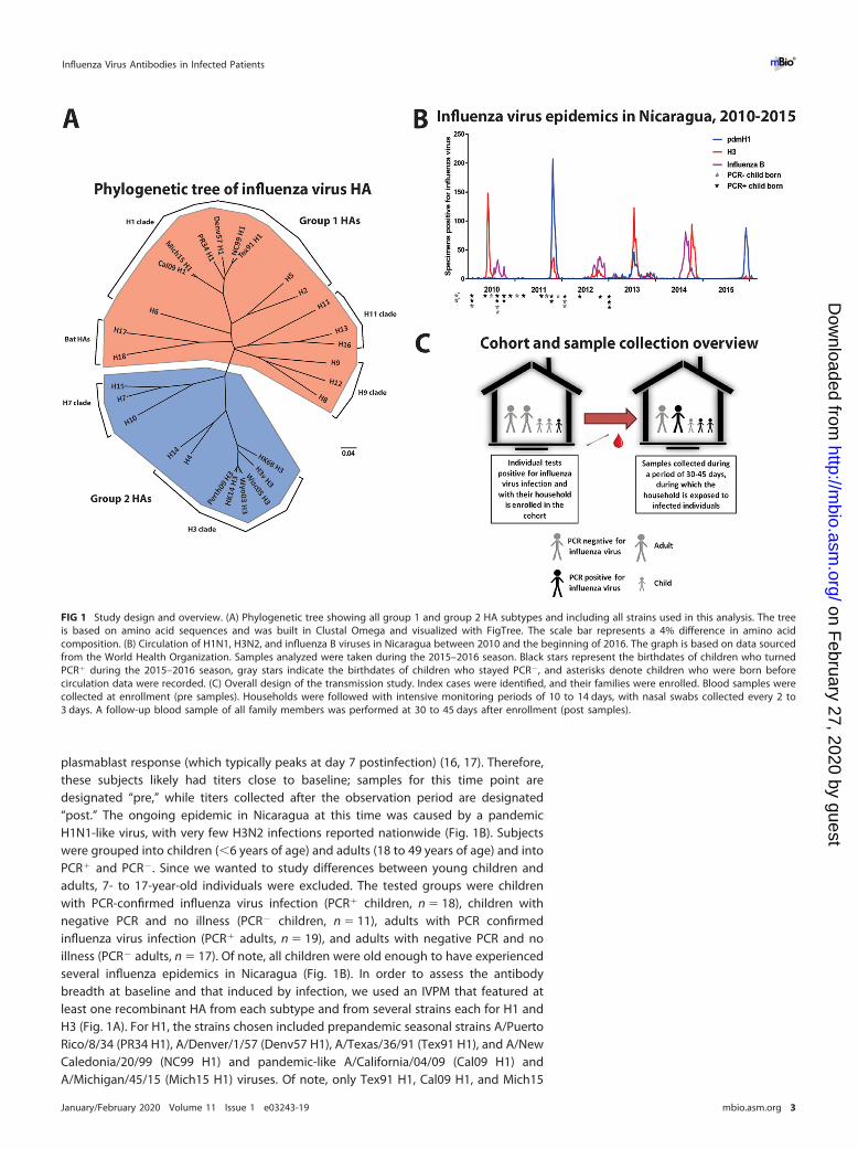
plasmablast response (which typically peaks at day 7 postinfection) (16, 17). Therefore,these subjects likely had titers close to baseline; samples for this time point aredesignated “pre,” while titers collected after the observation period are designated“post.” The ongoing epidemic in Nicaragua at this time was caused by a pandemicH1N1-like virus, with very few H3N2 infections reported nationwide (Fig. 1B). Subjectswere grouped into children (�6 years of age) and adults (18 to 49 years of age) and intoPCR� and PCR�. Since we wanted to study differences between young children andadults, 7- to 17-year-old individuals were excluded. The tested groups were childrenwith PCR-confirmed influenza virus infection (PCR� children, n � 18), children withnegative PCR and no illness (PCR� children, n � 11), adults with PCR confirmedinfluenza virus infection (PCR� adults, n � 19), and adults with negative PCR and noillness (PCR� adults, n � 17). Of note, all children were old enough to have experiencedseveral influenza epidemics in Nicaragua (Fig. 1B). In order to assess the antibodybreadth at baseline and that induced by infection, we used an IVPM that featured atleast one recombinant HA from each subtype and from several strains each for H1 andH3 (Fig. 1A). For H1, the strains chosen included prepandemic seasonal strains A/PuertoRico/8/34 (PR34 H1), A/Denver/1/57 (Denv57 H1), A/Texas/36/91 (Tex91 H1), and A/NewCaledonia/20/99 (NC99 H1) and pandemic-like A/California/04/09 (Cal09 H1) andA/Michigan/45/15 (Mich15 H1) viruses. Of note, only Tex91 H1, Cal09 H1, and Mich15
FIG 1 Study design and overview. (A) Phylogenetic tree showing all group 1 and group 2 HA subtypes and including all strains used in this analysis. The treeis based on amino acid sequences and was built in Clustal Omega and visualized with FigTree. The scale bar represents a 4% difference in amino acidcomposition. (B) Circulation of H1N1, H3N2, and influenza B viruses in Nicaragua between 2010 and the beginning of 2016. The graph is based on data sourcedfrom the World Health Organization. Samples analyzed were taken during the 2015–2016 season. Black stars represent the birthdates of children who turnedPCR� during the 2015–2016 season, gray stars indicate the birthdates of children who stayed PCR�, and asterisks denote children who were born beforecirculation data were recorded. (C) Overall design of the transmission study. Index cases were identified, and their families were enrolled. Blood samples werecollected at enrollment (pre samples). Households were followed with intensive monitoring periods of 10 to 14 days, with nasal swabs collected every 2 to3 days. A follow-up blood sample of all family members was performed at 30 to 45 days after enrollment (post samples).
Influenza Virus Antibodies in Infected Patients ®
January/February 2020 Volume 11 Issue 1 e03243-19 mbio.asm.org 3
on February 27, 2020 by guest
http://mbio.asm
.org/D
ownloaded from
H1 contained the K133 epitope (10), and Mich15 H1 was antigenically closest to thestrains circulating in Nicaragua in 2015. Recombinant HAs for H3 strains includedA/Hong Kong/1/68 (HK68 H3), A/Wyoming/03/03 (Wyo03 H3), A/Wisconsin/67/05(Wisc05 H3), A/Perth/16/09 (Perth09 H3), and A/Hong Kong/4801/14 (HK14 H3), as wellas the H3 variant strain A/Indiana/10/11 (H3v). These HAs were printed on epoxysilaneglass microarray slides and probed with sera, and then IgG and IgA signals were readin parallel from the same arrays using two differently labeled secondary antibodies(Fig. S1). Protein spots were printed in triplicate for each array, and samples wereassayed in three different dilutions. The reported data are based on the area under thecurve (AUC) calculated from the average signal for each protein at each dilution. Thismethod was chosen because it is more quantitative than assaying one serum dilutiononly. We have also shown in the past (13) and here that the method correlates well withELISA results, can detect antibodies with high specificity, and can detect broadlyneutralizing antibodies that bind to fragile and conformational epitopes (Fig. S2).
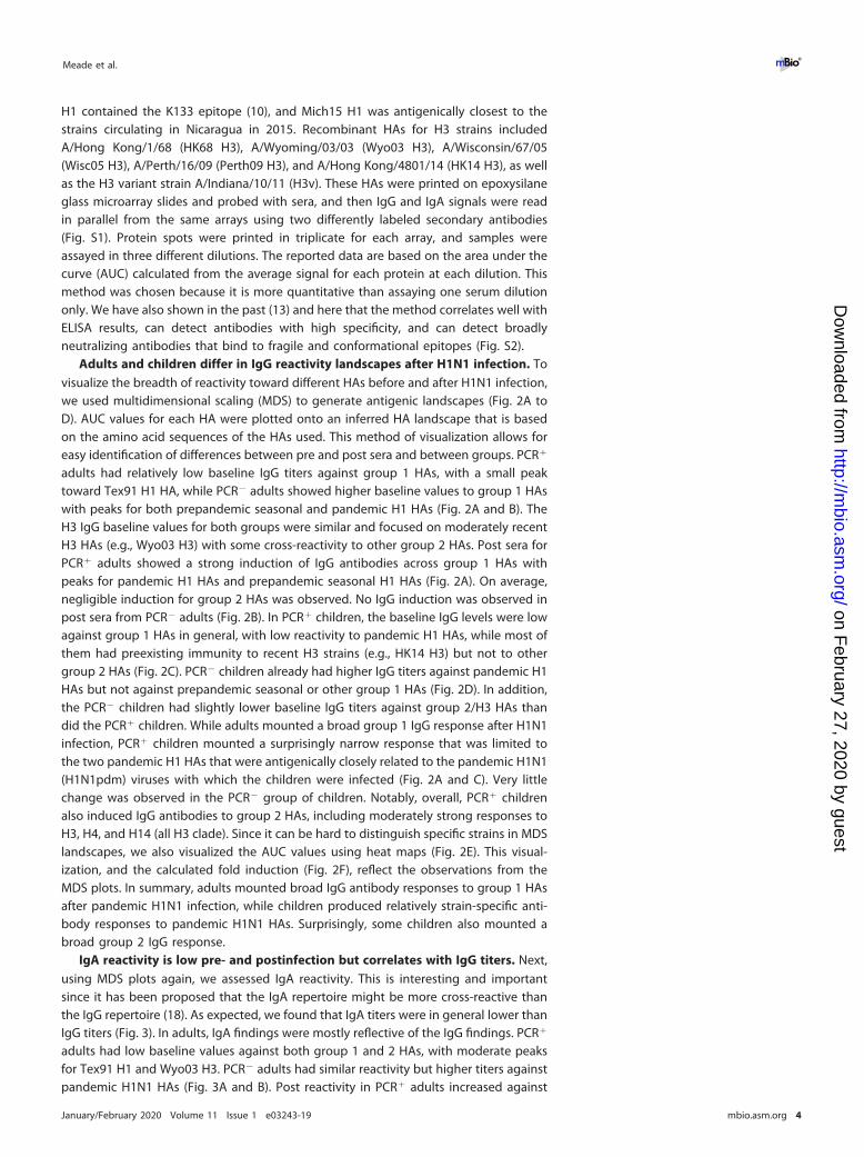
Adults and children differ in IgG reactivity landscapes after H1N1 infection. Tovisualize the breadth of reactivity toward different HAs before and after H1N1 infection,we used multidimensional scaling (MDS) to generate antigenic landscapes (Fig. 2A toD). AUC values for each HA were plotted onto an inferred HA landscape that is basedon the amino acid sequences of the HAs used. This method of visualization allows foreasy identification of differences between pre and post sera and between groups. PCR�
adults had relatively low baseline IgG titers against group 1 HAs, with a small peaktoward Tex91 H1 HA, while PCR� adults showed higher baseline values to group 1 HAswith peaks for both prepandemic seasonal and pandemic H1 HAs (Fig. 2A and B). TheH3 IgG baseline values for both groups were similar and focused on moderately recentH3 HAs (e.g., Wyo03 H3) with some cross-reactivity to other group 2 HAs. Post sera forPCR� adults showed a strong induction of IgG antibodies across group 1 HAs withpeaks for pandemic H1 HAs and prepandemic seasonal H1 HAs (Fig. 2A). On average,negligible induction for group 2 HAs was observed. No IgG induction was observed inpost sera from PCR� adults (Fig. 2B). In PCR� children, the baseline IgG levels were lowagainst group 1 HAs in general, with low reactivity to pandemic H1 HAs, while most ofthem had preexisting immunity to recent H3 strains (e.g., HK14 H3) but not to othergroup 2 HAs (Fig. 2C). PCR� children already had higher IgG titers against pandemic H1HAs but not against prepandemic seasonal or other group 1 HAs (Fig. 2D). In addition,the PCR� children had slightly lower baseline IgG titers against group 2/H3 HAs thandid the PCR� children. While adults mounted a broad group 1 IgG response after H1N1infection, PCR� children mounted a surprisingly narrow response that was limited tothe two pandemic H1 HAs that were antigenically closely related to the pandemic H1N1(H1N1pdm) viruses with which the children were infected (Fig. 2A and C). Very littlechange was observed in the PCR� group of children. Notably, overall, PCR� childrenalso induced IgG antibodies to group 2 HAs, including moderately strong responses toH3, H4, and H14 (all H3 clade). Since it can be hard to distinguish specific strains in MDSlandscapes, we also visualized the AUC values using heat maps (Fig. 2E). This visual-ization, and the calculated fold induction (Fig. 2F), reflect the observations from theMDS plots. In summary, adults mounted broad IgG antibody responses to group 1 HAsafter pandemic H1N1 infection, while children produced relatively strain-specific anti-body responses to pandemic H1N1 HAs. Surprisingly, some children also mounted abroad group 2 IgG response.
IgA reactivity is low pre- and postinfection but correlates with IgG titers. Next,using MDS plots again, we assessed IgA reactivity. This is interesting and importantsince it has been proposed that the IgA repertoire might be more cross-reactive thanthe IgG repertoire (18). As expected, we found that IgA titers were in general lower thanIgG titers (Fig. 3). In adults, IgA findings were mostly reflective of the IgG findings. PCR�
adults had low baseline values against both group 1 and 2 HAs, with moderate peaksfor Tex91 H1 and Wyo03 H3. PCR� adults had similar reactivity but higher titers againstpandemic H1N1 HAs (Fig. 3A and B). Post reactivity in PCR� adults increased against
Meade et al. ®
January/February 2020 Volume 11 Issue 1 e03243-19 mbio.asm.org 4
on February 27, 2020 by guest
http://mbio.asm
.org/D
ownloaded from

FIG 2 IgG antigenic landscapes of adults and children pre- and postexposure. Influenza virus protein microarray (IVPM)AUC values and amino acid sequences were used to generate antigenic landscapes using multidimensional scaling. The
(Continued on next page)
Influenza Virus Antibodies in Infected Patients ®
January/February 2020 Volume 11 Issue 1 e03243-19 mbio.asm.org 5
on February 27, 2020 by guest
http://mbio.asm
.org/D
ownloaded from

pandemic and seasonal H1 HAs, while no change was seen for PCR� adults. Interest-ingly, the breadth of the IgA response in PCR� adults was lower than that for IgG, withrelatively little induction of group 1 HA titers detected. IgA titers in pre and post serafrom PCR� children were negligible, and PCR� children had slightly elevated IgA titersagainst H1 in the pre sera that did not change over time (Fig. 3C and D). Again, to makereactivity to specific strains more visible, we also visualized the data as a heat map(Fig. 3E), with induction shown in Fig. 3F. We further performed a correlation analysisfor IgG versus IgA in adults and children and found low but significant correlation inboth cases (Fig. 3G and H). In summary, IgA titers were lower in all four groups and lessbroad than the IgG titers in adults but showed moderate correlation with IgG titers inadults and children.
H3N2 preexposed children mount a response to pandemic H1 and group 2 HAsafter H1N1 infection. To better understand the responses mounted by PCR� adultsand children, we compared the reactivity profiles of individuals and grouped them intofour categories (Fig. 4). The majority of the PCR� adults (n � 12) showed an inductionto H1 HAs related to the strain causing the infection, to prepandemic seasonal H1 HAs,as well as to other group 1 HAs (Fig. 4A). Specifically, antibody titers to other membersof the H1 clade (H2, H5, and H6) and the H9 clade (H8, H9, and H12) were boosted, withless induction of reactivity to the H11 clade (H11, H13, and H16) and the bat HAs (H17and H18). In this group of individuals, no induction of cross-group antibodies (in thiscase to group 2 HAs) was observed. A minority of PCR� adults also showed inductionof cross-group antibodies after pandemic H1N1 infection in addition to the broadgroup 1 induction (Fig. 4B). Curiously, this group 2 induction was not necessarilyfocused on recent H3 strains but on HAs from older H3 strains, as well as other subtypesfrom the H3 (H3, H4, and H14) and H7 (H7, H10, and H15) clades. The majority of PCR�
children had a narrow response specific to pandemic H1 HA with negligible cross-reactivity to other H1 HAs or group 1 HAs. Six of these PCR� children (33%) had a verynarrow response to the pandemic H1 HAs, with slightly higher titers to the better-matched Mich15 H1 HA than to Cal09 H1 HA (Fig. 4C). Another six PCR� children (33%)showed a very distinct response. In addition to the narrow response to the pandemicH1 HAs, they mounted a strong response to group 2 HAs with a preference to older H3strains, H4 and H14 (Fig. 4D). In this group, in comparison to the pandemic H1HA-only-reacting group of PCR� children, the preexisting baseline reactivity to H3 washigher, which might have played a role in this peculiar response. The remainingchildren were low H1 responders (n � 3), had a low response to H1 but responded wellto group 2 (n � 2), or responded broadly to H1 and group 2 (n � 1).
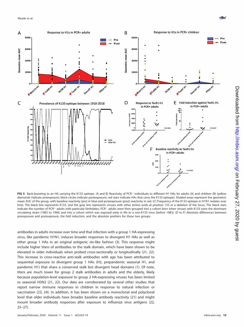
A recall response against the H1 K133 epitopes is dominant in adults. Asdescribed above, pandemic H1N1 infection caused an induction of antibodies toprepandemic seasonal H1 HAs in adults but not in children. Responses to one H1 HA,Tex91 H1, stood out specifically, as they were often higher than the responses to theHA of the infecting virus (Fig. 5A and B). It has been described that pandemic H1N1infection induces antibodies that target the K133 epitope but only for age groups thatwere imprinted with seasonal H1N1 viruses carrying K133 as well (mainly 1983 to 1996;Fig. 5C) (10). Besides the two pandemic H1N1 HAs (Cal09 H1 and Mich15 H1), onlyTex91 H1 on the IVPM array carries the K133 amino acid, which is absent (the wholeposition is deleted) from the other tested H1 HAs. When we had a closer look at H1N1reactivity of all PCR� adults, we clearly saw that the highest reactivity in pre- andpostinfection sera was toward Tex91 H1 for a majority of individuals, and the induction
FIG 2 Legend (Continued)z axis represents reactivity to a substrate (AUC as geometric mean titer), and the x and y axes represent amino aciddifferences between HAs used as the substrate. The gray plane under each of the red and blue planes represents thepreexposure reactivity, the blue plane represents the postexposure group 1 reactivity, and the red plane represents thepostexposure group 2 reactivity. The different strains/subtypes are indicated by colored spheres labeled with the substratename. (A) PCR� adults. (B) PCR� adults. (C) PCR� children. (D) PCR� children. (E and F) Shown are the same data in a heatmap and the fold induction in a heat map format (F) (as geometric mean induction).
Meade et al. ®
January/February 2020 Volume 11 Issue 1 e03243-19 mbio.asm.org 6
on February 27, 2020 by guest
http://mbio.asm
.org/D
ownloaded from
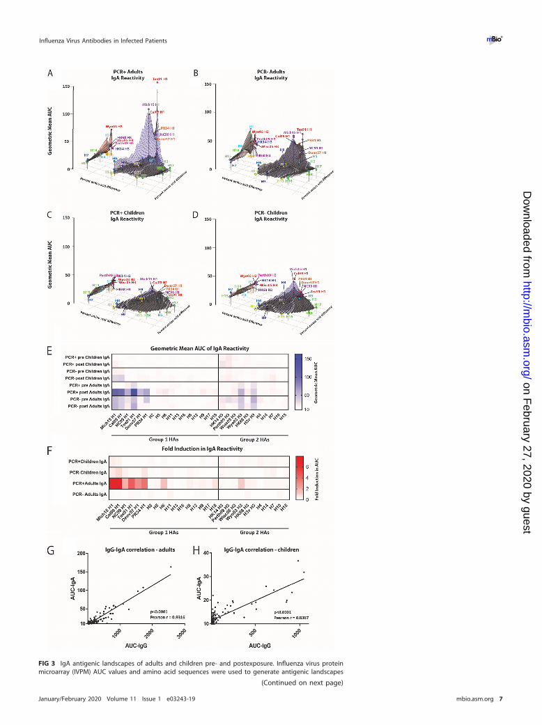
FIG 3 IgA antigenic landscapes of adults and children pre- and postexposure. Influenza virus proteinmicroarray (IVPM) AUC values and amino acid sequences were used to generate antigenic landscapes
(Continued on next page)
Influenza Virus Antibodies in Infected Patients ®
January/February 2020 Volume 11 Issue 1 e03243-19 mbio.asm.org 7
on February 27, 2020 by guest
http://mbio.asm
.org/D
ownloaded from
for this H1 was higher than for all other prepandemic seasonal H1 HAs, suggesting aclear back-boost (Fig. 5A). This was not observed for PCR� children (Fig. 5B). Wetherefore mapped the birth years of the PCR� adults on a timeline that represents thefrequency of the K133 epitope between 1918 and 2018 (Fig. 5C). We then divided thePCR� adults into a group that likely was imprinted with a K133 epitope bearing H1N1(1983 to 1996) and compared them to the group that was likely imprinted by a virusin which the K133 epitope was absent (born before 1983). Typically, the first exposureto influenza virus happens in the first few years of life. Given an attack rate ofapproximately 23% in unvaccinated children (19), it is very likely that the first exposurehappens close to birth. We found no significant differences in change of reactivity(Fig. 5D), fold induction (Fig. 5E), or baseline reactivity (Fig. 5F) to Tex91 between thetwo groups, although there was a trend toward higher baseline reactivity in adults whowere likely imprinted with a K133 virus.
Higher preexisting reactivity to group 1 HA, the H1 subtype, and pandemic H1HA correlates with protection. Finally, we wanted to assess the relationship betweenpreexisting HA antibody levels and the risk of pandemic H1N1 infection. First, weplotted the averaged preexisting antibody AUCs for pandemic H1 HAs, prepandemicseasonal H1 HAs, and non-H1 group 1 HAs for PCR� and PCR� individuals. Especiallythe non-H1 group 1 titers are a representation of the “antigenic altitude” of a subjectin the MDS plots. This analysis was performed for adults and children separately. Forboth adults and children, preexisting antibody levels to pandemic H1 HA were higherin the PCR� groups (Fig. 6A). For adults, the same applied to reactivity to prepandemicH1 HA, where PCR� individuals had higher titers than did PCR� individuals (Fig. 6B).This was also the case in children but at lower reactivity in general (Fig. 6B) and with3 children actually showing high reactivity in the PCR� group. For adults, preexistingantibodies to non-H1 group 1 HAs were higher in PCR� individuals (Fig. 6C), againsuggesting the greater breadth of response in adults. This was not the case in children,who had lower preexisting antibodies to non-H1 group 1 HAs in general (Fig. 6C). Next,we examined protective effects associated with a 2-fold increase in antibody levelsadjusted for age and sex. We found that in adults, preexisting antibodies to pandemicH1 HAs (odds ratio [OR], 0.19; confidence interval [CI], 0.06 to 0.66) and non-H1 group1 HAs (OR, 0.48; CI, 0.23 to 0.98) were significantly associated with protection fromPCR-confirmed infection (Fig. 6D). In children, preexisting antibodies to pandemic H1HAs were significantly associated with protection (OR, 0.28; CI, 0.09 to 0.88), butantibodies to non-H1 group 1 HAs were not (Fig. 6D). In both children and adults,antibodies to prepandemic H1s were not associated with protection (Fig. 6D).
DISCUSSION
Using the IVPM technology, we have made several interesting observations in thisstudy. The most intriguing finding is the difference in the breadth of the antibodyresponses in children and adults. Children showed a very narrow antibody responseafter infection that, for group 1 HAs, targeted only pandemic H1N1-like HAs, which areantigenically related to the virus causing the infection. In contrast, adults experienceda back-boost to a broad range of seasonal H1 and other group 1 HAs. This observationsuggests that the initial exposure of children to influenza viruses causes a biasedresponse to immunodominant and strain-specific epitopes, a phenomenon that canalso be observed in naive mice (12, 20). It further suggests that levels of cross-reactive
FIG 3 Legend (Continued)using multidimensional scaling. The z axis represents reactivity to a substrate (AUC as geometric meantiter), and the x and y axes represent amino acid differences between HAs used as the substrate. The grayplane under each of the red and blue planes represents the preexposure reactivity, the blue planerepresents the postexposure group 1 reactivity, and the red plane represents the postexposure group 2reactivity. The different strains/subtypes are indicated by colored spheres labeled with the substratename. (A) PCR� adults. (B) PCR� adults. (C) PCR� children. (D) PCR� children. (E and F) Shown are thesame data in a heat map and the fold induction in a heat map format (F). (G and H) Correlation analysisbetween IgG and IgA titers for adults (G) and children (H).
Meade et al. ®
January/February 2020 Volume 11 Issue 1 e03243-19 mbio.asm.org 8
on February 27, 2020 by guest
http://mbio.asm
.org/D
ownloaded from

FIG 4 IgG reactivity profiles. PCR� adults and children were binned into categories each based on theirreactivity profile. The y axis of these plots shows the geometric mean AUC of the group, and the different HAsare plotted on the x axis. (A) Adults who induce a predominant group 1 response. (B) Profile of adults whoinduce IgG against both group 1 and group 2 HAs. (C) Reactivity profile of children who mount a narrowpandemic H1 HA response. (D) Reactivity of children who mount a narrow pandemic H1 HA response plus aresponse to group 2 HAs.
Influenza Virus Antibodies in Infected Patients ®
January/February 2020 Volume 11 Issue 1 e03243-19 mbio.asm.org 9
on February 27, 2020 by guest
http://mbio.asm
.org/D
ownloaded from
antibodies in adults increase over time and that infection with a group 1 HA-expressingvirus, like pandemic H1N1, induces broader responses to divergent H1 HAs as well asother group 1 HAs in an original antigenic sin-like fashion (3). This response mightinclude higher titers of antibodies to the stalk domain, which have been shown to beelevated in older individuals when probed cross-sectionally or longitudinally (21, 22).This increase in cross-reactive anti-stalk antibodies with age has been attributed tosequential exposure to divergent group 1 HAs (H2, prepandemic seasonal H1, andpandemic H1) that share a conserved stalk but divergent head domains (1). Of note,titers are much lower for group 2 stalk antibodies in adults and the elderly, likelybecause population-level exposure to group 2 HA-expressing viruses has been limitedto seasonal H3N2 (21, 22). Our data are corroborated by several other studies thatreport narrow immune responses in children in response to natural infection orvaccination (23, 24). In addition, it has been shown on a monoclonal and polyclonallevel that older individuals have broader baseline antibody reactivity (21) and mightmount broader antibody responses after exposure to influenza virus antigens (22,25–27).
FIG 5 Back-boosting to an HA carrying the K133 epitope. (A and B) Reactivity of PCR� individuals to different H1 HAs for adults (A) and children (B) (yellowdiamonds indicate preexposure; black circles indicate postexposure; red stars indicate HAs that carry the K133 epitope). Shaded areas represent the geometricmean AUC of the group, with baseline reactivity (pre) in blue and postexposure (post) reactivity in red. (C) Frequency of the K133 epitope in H1N1 isolates overtime. The black line represents K133, and the gray line represents viruses with other amino acids at position 133 or a deletion of the locus. The black starsindicate the number of PCR� adults with particular birthdates. PCR� adults were then grouped into a cohort born when viruses with K133 were the dominantcirculating strain (1983 to 1996) and into a cohort which was exposed early in life to a non-K133 virus (before 1983). (D to F) Absolute differences betweenpreexposure and postexposure, the fold induction, and the absolute pretiters for these two groups.
Meade et al. ®
January/February 2020 Volume 11 Issue 1 e03243-19 mbio.asm.org 10
on February 27, 2020 by guest
http://mbio.asm
.org/D
ownloaded from

Another phenomenon that was observed was strong reactivity toward Tex91 inadults after exposure to pandemic H1N1. Tex91 and pandemic H1N1 HAs share aconserved epitope in the head domain centered around K133 (10). It has been shownpreviously that individuals born when a K133-carrying virus was circulating mounted aK133-focused response to the pandemic H1N1 HA (10). Individuals that were bornwhen non-K133-expressing viruses circulated did not show this focused immuneresponse to the K133 epitope of pandemic H1N1 (10). These differences were notobserved in our study, as K133- and non-K133-imprinted adults had a similar back-boost to Tex91. However, baseline titers to Tex91 were higher in K133-imprintedindividuals. Differences between this and other studies might be caused by thedifferent assays and methodologies used. As an alternative explanation, the number ofsubjects might have been too small to detect differences between the groups. How-ever, the magnitude of baseline cross-reactivity to Tex91 and of the boost afterpandemic H1N1 exposure validates the importance of this epitope for immunity toH1N1 viruses.
As described above, children mounted a very narrow response to the H1 HAs closelyrelated to the infecting strain and did not induce cross-reactive antibodies to other H1or group 1 HAs. However, a third of the children also mounted a response toward group2 HAs, especially older H3 HAs, H4, and H14. Of note, this group of children had beenpreexposed to H3N2 and had higher titers to recent H3 HAs than those of the groupthat mounted an HA response specific to pandemic H1N1. It is unclear what caused thisstrong cross-reactivity. Initially, we did consider that coinfections with H3N2 could haveoccurred. However, circulation of H3N2 in the analyzed season in Nicaragua wasnegligible; therefore, this scenario is unlikely to have occurred in a third of theH1N1-infected children. Another scenario could be that these children came in contactwith avian influenza viruses, e.g., H4 or H14, which could have caused this induction orwhich could have caused an imprinting pattern that then triggered this peculiarresponse after pandemic H1N1 infection. However, it is unlikely that this occurred in33% of PCR� children. Another possibility is that a strong H3N2 priming throughnatural infection can leave an imprint that also influences the response to group 1
FIG 6 Preexisting group 1 anti-HA titers by infection status and correlates of protection. (A to C) Mean preexisting anti-HA titers (AUC) by PCR status and agefor the 2 pandemic H1 strains (A), all prepandemic seasonal H1 strains (B), and all non-H1 group 1 strains (C). (D) Odds ratios for infection based on preexposuretiters against pandemic H1 HA, prepandemic seasonal H1 HAs, and non-H1 group 1 HAs for adults and children. Unadjusted and models adjusted for age andsex are presented. Odds ratios are for a 2-fold increase in titer.
Influenza Virus Antibodies in Infected Patients ®
January/February 2020 Volume 11 Issue 1 e03243-19 mbio.asm.org 11
on February 27, 2020 by guest
http://mbio.asm
.org/D
ownloaded from

viruses such as pandemic H1N1. It is possible that this response is driven by anti-stalkantibodies since cross-group-reactive antibodies have been reported (28–30). Mostcross-group stalk-reactive antibodies would likely target all group 1 and group 2 HAsand not specifically pandemic H1 plus group 2 HAs (28–30). However, recently, threestudies reported VH3-53 germ line anti-stalk antibodies after H7N9 vaccination thatshowed the same pattern of targeting pandemic H1N1 plus broad group 2 binding(31–33). Another class of antibodies that could be involved in this phenomenon areantibodies that bind the head trimer interface and have recently been reported inhumans as well (34).
The observed phenomena including the back-boost in adults against group 1 HAsand against the K133 epitope as well as the H1-group 2 cross-reactivity in children areevidence for the complex response of the immune system to (sequential) exposure toinfluenza viruses (5, 6, 9). Our findings certainly warrant follow-up studies with largernumbers of subjects and longitudinal analyses to shed more light on mechanismsbehind back-boosting and imprinting effects. Analysis of the antibody response on amonoclonal antibody level would also help assess which specific class of antibody isresponsible for the observed cross-reactivity.
Another interesting observation was the difference between IgG and IgA serumresponses. The IgG response was higher in signal strength. While this could beinfluenced by the different secondary antibodies used in the analysis, it is likely also areflection of the larger amount of IgG than IgA in serum. Surprisingly, the IgG responsein adults was broader than the IgA response, which is in contrast to assumptions madebased on human B-cell analysis (18). Furthermore, the IgA response to infection wasrelatively low in children, which is unexpected since antigen presented on mucosalsurfaces is expected to drive stronger IgA responses (which might still be the case forlocal mucosal immunity, which was not assessed in our study). However, IgG and IgAresponses still correlated moderately in both children and adults.
Finally, we analyzed if preexisting immunity would be predictive of the risk ofgetting infected with pandemic H1N1. In adults, preexisting antibody titers to pan-demic H1 HAs and non-H1 group 1 HAs were associated with significant protectionfrom infection; antibodies to prepandemic H1 strains were somewhat higher amongPCR� adults but not associated with significant protection. In children, only preexistingantibodies to the pandemic H1 HAs were associated with significant protection frominfection. However, it needs to be kept in mind that children had very low levels ofantibody to seasonal H1 and non-H1 group 1 HAs. This finding indicates that bindingantibodies as measured in the IVPM might serve as correlate of protection. We havealso shown this recently with ELISA data against a specific H1 HA and the stalk ofH1 (2).
The small number of subjects tested and the relatively low number of HAs probeddo not allow us to draw firm conclusions. However, this study serves to generatehypotheses regarding imprinting and the evolution of antibody responses duringsequential exposure to natural infection with influenza viruses. In conclusion, we showthat children mount a much narrower antibody response to pandemic H1N1 infectionthan do adults, who respond broadly to group 1 HAs. Notably, a subpopulation ofchildren induces pandemic H1 HA plus group 2 HA reactivity, a new phenomenon thatmight have significant implications for our understanding of imprinting and futurevaccine design. Furthermore, we show strong back-boosting in adults to an HA thatcarries the K133 epitope, and we provide evidence the IVPM binding data might serveas correlate of protection. While the current study is descriptive in nature and haslimitations in terms of sample size, our findings are highly significant since they informthe design of future studies to elucidate imprinting effects and the longitudinaldynamics of antibody responses to influenza virus infection. These insights might openup new avenues for broadly protective of even universal influenza virus vaccinestrategies.
Meade et al. ®
January/February 2020 Volume 11 Issue 1 e03243-19 mbio.asm.org 12
on February 27, 2020 by guest
http://mbio.asm
.org/D
ownloaded from
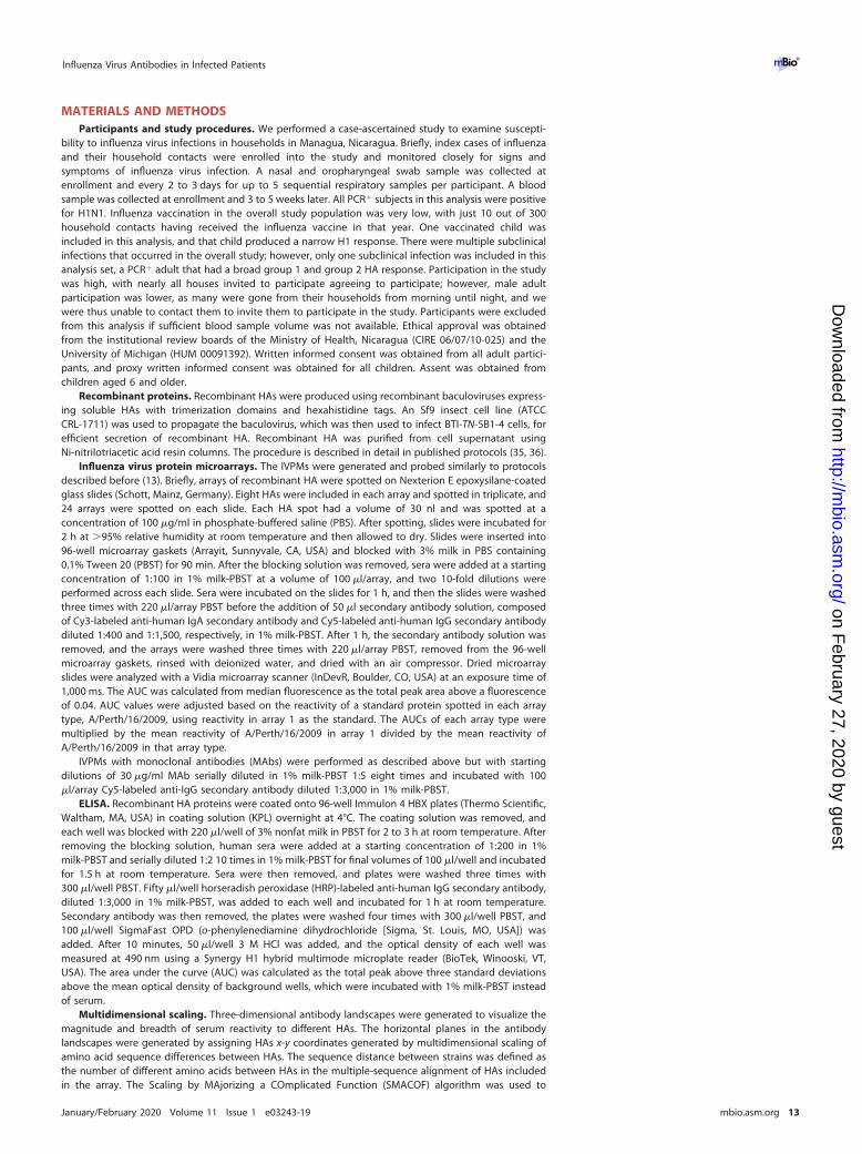
MATERIALS AND METHODSParticipants and study procedures. We performed a case-ascertained study to examine suscepti-
bility to influenza virus infections in households in Managua, Nicaragua. Briefly, index cases of influenzaand their household contacts were enrolled into the study and monitored closely for signs andsymptoms of influenza virus infection. A nasal and oropharyngeal swab sample was collected atenrollment and every 2 to 3 days for up to 5 sequential respiratory samples per participant. A bloodsample was collected at enrollment and 3 to 5 weeks later. All PCR� subjects in this analysis were positivefor H1N1. Influenza vaccination in the overall study population was very low, with just 10 out of 300household contacts having received the influenza vaccine in that year. One vaccinated child wasincluded in this analysis, and that child produced a narrow H1 response. There were multiple subclinicalinfections that occurred in the overall study; however, only one subclinical infection was included in thisanalysis set, a PCR� adult that had a broad group 1 and group 2 HA response. Participation in the studywas high, with nearly all houses invited to participate agreeing to participate; however, male adultparticipation was lower, as many were gone from their households from morning until night, and wewere thus unable to contact them to invite them to participate in the study. Participants were excludedfrom this analysis if sufficient blood sample volume was not available. Ethical approval was obtainedfrom the institutional review boards of the Ministry of Health, Nicaragua (CIRE 06/07/10-025) and theUniversity of Michigan (HUM 00091392). Written informed consent was obtained from all adult partici-pants, and proxy written informed consent was obtained for all children. Assent was obtained fromchildren aged 6 and older.
Recombinant proteins. Recombinant HAs were produced using recombinant baculoviruses express-ing soluble HAs with trimerization domains and hexahistidine tags. An Sf9 insect cell line (ATCCCRL-1711) was used to propagate the baculovirus, which was then used to infect BTI-TN-5B1-4 cells, forefficient secretion of recombinant HA. Recombinant HA was purified from cell supernatant usingNi-nitrilotriacetic acid resin columns. The procedure is described in detail in published protocols (35, 36).
Influenza virus protein microarrays. The IVPMs were generated and probed similarly to protocolsdescribed before (13). Briefly, arrays of recombinant HA were spotted on Nexterion E epoxysilane-coatedglass slides (Schott, Mainz, Germany). Eight HAs were included in each array and spotted in triplicate, and24 arrays were spotted on each slide. Each HA spot had a volume of 30 nl and was spotted at aconcentration of 100 �g/ml in phosphate-buffered saline (PBS). After spotting, slides were incubated for2 h at �95% relative humidity at room temperature and then allowed to dry. Slides were inserted into96-well microarray gaskets (Arrayit, Sunnyvale, CA, USA) and blocked with 3% milk in PBS containing0.1% Tween 20 (PBST) for 90 min. After the blocking solution was removed, sera were added at a startingconcentration of 1:100 in 1% milk-PBST at a volume of 100 �l/array, and two 10-fold dilutions wereperformed across each slide. Sera were incubated on the slides for 1 h, and then the slides were washedthree times with 220 �l/array PBST before the addition of 50 �l secondary antibody solution, composedof Cy3-labeled anti-human IgA secondary antibody and Cy5-labeled anti-human IgG secondary antibodydiluted 1:400 and 1:1,500, respectively, in 1% milk-PBST. After 1 h, the secondary antibody solution wasremoved, and the arrays were washed three times with 220 �l/array PBST, removed from the 96-wellmicroarray gaskets, rinsed with deionized water, and dried with an air compressor. Dried microarrayslides were analyzed with a Vidia microarray scanner (InDevR, Boulder, CO, USA) at an exposure time of1,000 ms. The AUC was calculated from median fluorescence as the total peak area above a fluorescenceof 0.04. AUC values were adjusted based on the reactivity of a standard protein spotted in each arraytype, A/Perth/16/2009, using reactivity in array 1 as the standard. The AUCs of each array type weremultiplied by the mean reactivity of A/Perth/16/2009 in array 1 divided by the mean reactivity ofA/Perth/16/2009 in that array type.
IVPMs with monoclonal antibodies (MAbs) were performed as described above but with startingdilutions of 30 �g/ml MAb serially diluted in 1% milk-PBST 1:5 eight times and incubated with 100�l/array Cy5-labeled anti-IgG secondary antibody diluted 1:3,000 in 1% milk-PBST.
ELISA. Recombinant HA proteins were coated onto 96-well Immulon 4 HBX plates (Thermo Scientific,Waltham, MA, USA) in coating solution (KPL) overnight at 4°C. The coating solution was removed, andeach well was blocked with 220 �l/well of 3% nonfat milk in PBST for 2 to 3 h at room temperature. Afterremoving the blocking solution, human sera were added at a starting concentration of 1:200 in 1%milk-PBST and serially diluted 1:2 10 times in 1% milk-PBST for final volumes of 100 �l/well and incubatedfor 1.5 h at room temperature. Sera were then removed, and plates were washed three times with300 �l/well PBST. Fifty �l/well horseradish peroxidase (HRP)-labeled anti-human IgG secondary antibody,diluted 1:3,000 in 1% milk-PBST, was added to each well and incubated for 1 h at room temperature.Secondary antibody was then removed, the plates were washed four times with 300 �l/well PBST, and100 �l/well SigmaFast OPD (o-phenylenediamine dihydrochloride [Sigma, St. Louis, MO, USA]) wasadded. After 10 minutes, 50 �l/well 3 M HCl was added, and the optical density of each well wasmeasured at 490 nm using a Synergy H1 hybrid multimode microplate reader (BioTek, Winooski, VT,USA). The area under the curve (AUC) was calculated as the total peak above three standard deviationsabove the mean optical density of background wells, which were incubated with 1% milk-PBST insteadof serum.
Multidimensional scaling. Three-dimensional antibody landscapes were generated to visualize themagnitude and breadth of serum reactivity to different HAs. The horizontal planes in the antibodylandscapes were generated by assigning HAs x-y coordinates generated by multidimensional scaling ofamino acid sequence differences between HAs. The sequence distance between strains was defined asthe number of different amino acids between HAs in the multiple-sequence alignment of HAs includedin the array. The Scaling by MAjorizing a COmplicated Function (SMACOF) algorithm was used to
Influenza Virus Antibodies in Infected Patients ®
January/February 2020 Volume 11 Issue 1 e03243-19 mbio.asm.org 13
on February 27, 2020 by guest
http://mbio.asm
.org/D
ownloaded from
minimize the sum of squared errors between the Euclidean distance in the two-dimensional (2D) planeand the HA sequence distance. For each HA, an antibody landscape surface was generated fromgeometric mean AUC values using multilevel B-splines for pre- and postexposure sera (12).
Statistical analysis and viral sequence analysis. Statistical analysis was performed in GraphPadPrism 7.0 and R. Antigenic altitudes were compared using an unpaired t test, Pearson correlation analysesand linear regressions were used to compare IVPM and ELISA data and to compare IgA and IgG IVPMdata, respectively, and AUC analyses were performed on IVPM and ELISA data. Logistic regression wasused to measure the correlates of protection from preexisting antibodies. Crude models and modelsadjusted for age and sex were run; this was done using the “glm” function in R. Plots in Fig. 6 werecreated using ggplot2.
Sequences for K133 epitope analysis were collected from the Global Initiative for Sharing all InfluenzaData (GISAID) from the years 1918 to 2019. They were then sorted by the year of isolation. Within eachyear, a subset of randomly selected sequences was compiled so that were 100 sequences or fewer if therewere not 100 isolates for a specific collection year. Sequences for each year were aligned using MUSCLE.The residue at position 133 was then examined for the presence or absence of K133. The percentprevalence of K133 was calculated by the number of K133 residues in the yearly data set divided by thetotal isolates included for that year.
SUPPLEMENTAL MATERIALSupplemental material is available online only.FIG S1, DOCX file, 1.2 MB.FIG S2, DOCX file, 0.3 MB.
ACKNOWLEDGMENTSWork in the Krammer laboratory was supported by the NIAID Centers of Excellence for
Influenza Research and Surveillance (CEIRS; grant HHSN272201400008C). The work in theGordon group and Nicaragua was supported through the National Institute for Allergy andInfectious Diseases (award R01 AI120997 and contract number HHSN272201400006C).
We declare no conflicts of interest.
REFERENCES1. Krammer F. 2019. The human antibody response to influenza A virus
infection and vaccination. Nat Rev Immunol 19:383–397. https://doi.org/10.1038/s41577-019-0143-6.
2. Ng S, Nachbagauer R, Balmaseda A, Stadlbauer D, Ojeda S, Patel M,Rajabhathor A, Lopez R, Guglia AF, Sanchez N, Amanat F, Gresh L, KuanG, Krammer F, Gordon A. 2019. Novel correlates of protection againstpandemic H1N1 influenza A virus infection. Nat Med 25:962–967. https://doi.org/10.1038/s41591-019-0463-x.
3. Francis T. 1960. On the doctrine of original antigenic sin. Proc Am PhilosSoc 104:572–578.
4. Guthmiller JJ, Wilson PC. 2018. Harnessing immune history to combatinfluenza viruses. Curr Opin Immunol 53:187–195. https://doi.org/10.1016/j.coi.2018.05.010.
5. Lewnard JA, Cobey S, Lewnard J, Cobey S. 2018. Immune history andinfluenza vaccine effectiveness. Vaccines 6:28. https://doi.org/10.3390/vaccines6020028.
6. Gostic KM, Ambrose M, Worobey M, Lloyd-Smith JO. 2016. Potentprotection against H5N1 and H7N9 influenza via childhood hemag-glutinin imprinting. Science 354:722–726. https://doi.org/10.1126/science.aag1322.
7. Tesini BL, Kanagaiah P, Wang J, Hahn M, Halliley JL, Chaves FA, NguyenPQT, Nogales A, DeDiego ML, Anderson CS, Ellebedy AH, Strohmeier S,Krammer F, Yang H, Bandyopadhyay S, Ahmed R, Treanor JJ, Martinez-Sobrido L, Golding H, Khurana S, Zand MS, Topham DJ, Sangster MY.2019. Broad hemagglutinin-specific memory B cell expansion by sea-sonal influenza virus infection reflects early-life imprinting and adapta-tion to the infecting virus. J Virol 93:e00169-19. https://doi.org/10.1128/JVI.00169-19.
8. Hensley SE. 2014. Challenges of selecting seasonal influenza vaccinestrains for humans with diverse pre-exposure histories. Curr Opin Virol8:85– 89. https://doi.org/10.1016/j.coviro.2014.07.007.
9. Cobey S, Hensley SE. 2017. Immune history and influenza virus suscep-tibility. Curr Opin Virol 22:105–111. https://doi.org/10.1016/j.coviro.2016.12.004.
10. Li Y, Myers JL, Bostick DL, Sullivan CB, Madara J, Linderman SL, Liu Q,Carter DM, Wrammert J, Esposito S, Principi N, Plotkin JB, Ross TM,Ahmed R, Wilson PC, Hensley SE. 2013. Immune history shapes specific-
ity of pandemic H1N1 influenza antibody responses. J Exp Med 210:1493–1500. https://doi.org/10.1084/jem.20130212.
11. Linderman SL, Chambers BS, Zost SJ, Parkhouse K, Li Y, Herrmann C,Ellebedy AH, Carter DM, Andrews SF, Zheng NY, Huang M, Huang Y,Strauss D, Shaz BH, Hodinka RL, Reyes-Terán G, Ross TM, Wilson PC,Ahmed R, Bloom JD, Hensley SE. 2014. Potential antigenic explanationfor atypical H1N1 infections among middle-aged adults during the2013–2014 influenza season. Proc Natl Acad Sci U S A 111:15798 –15803.https://doi.org/10.1073/pnas.1409171111.
12. Nachbagauer R, Choi A, Hirsh A, Margine I, Iida S, Barrera A, Ferres M,Albrecht RA, García-Sastre A, Bouvier NM, Ito K, Medina RA, Palese P,Krammer F. 2017. Defining the antibody cross-reactome directed againstthe influenza virus surface glycoproteins. Nat Immunol 18:464 – 473.https://doi.org/10.1038/ni.3684.
13. Meade P, Latorre-Margalef N, Stallknecht DE, Krammer F. 2017. Devel-opment of an influenza virus protein microarray to measure the humoralresponse to influenza virus infection in mallards. Emerg Microbes Infect6:e110. https://doi.org/10.1038/emi.2017.98.
14. Gordon A, Tsang TK, Cowling BJ, Kuan G, Ojeda S, Sanchez N, Gresh L,Lopez R, Balmaseda A, Harris E. 2018. Influenza transmission dynamics inurban households, Managua, Nicaragua, 2012–2014. Emerg Infect Dis24:1882–1888. https://doi.org/10.3201/eid2410.161258.
15. Novel Swine-Origin Influenza A (H1N1) Virus Investigation Team, Da-wood FS, Jain S, Finelli L, Shaw MW, Lindstrom S, Garten RJ, Gubareva LV,Xu X, Bridges CB, Uyeki TM. 2009. Emergence of a novel swine-origininfluenza A (H1N1) virus in humans. N Engl J Med 360:2605–2615.https://doi.org/10.1056/NEJMoa0903810.
16. Wrammert J, Smith K, Miller J, Langley WA, Kokko K, Larsen C, Zheng NY,Mays I, Garman L, Helms C, James J, Air GM, Capra JD, Ahmed R, WilsonPC. 2008. Rapid cloning of high-affinity human monoclonal antibodiesagainst influenza virus. Nature 453:667– 671. https://doi.org/10.1038/nature06890.
17. Chen YQ, Wohlbold TJ, Zheng NY, Huang M, Huang Y, Neu KE, Lee J, WanH, Rojas KT, Kirkpatrick E, Henry C, Palm AE, Stamper CT, Lan LY, TophamDJ, Treanor J, Wrammert J, Ahmed R, Eichelberger MC, Georgiou G,Krammer F, Wilson PC. 2018. Influenza infection in humans induces
Meade et al. ®
January/February 2020 Volume 11 Issue 1 e03243-19 mbio.asm.org 14
on February 27, 2020 by guest
http://mbio.asm
.org/D
ownloaded from
broadly cross-reactive and protective neuraminidase-reactive antibod-ies. Cell 173:417– 429.e10. https://doi.org/10.1016/j.cell.2018.03.030.
18. He W, Mullarkey CE, Duty JA, Moran TM, Palese P, Miller MS. 2015.Broadly neutralizing anti-influenza virus antibodies: enhancement ofneutralizing potency in polyclonal mixtures and IgA backbones. J Virol89:3610 –3618. https://doi.org/10.1128/JVI.03099-14.
19. Somes MP, Turner RM, Dwyer LJ, Newall AT. 2018. Estimating the annualattack rate of seasonal influenza among unvaccinated individuals: asystematic review and meta-analysis. Vaccine 36:3199 –3207. https://doi.org/10.1016/j.vaccine.2018.04.063.
20. Angeletti D, Gibbs JS, Angel M, Kosik I, Hickman HD, Frank GM, Das SR,Wheatley AK, Prabhakaran M, Leggat DJ, McDermott AB, Yewdell JW.2017. Defining B cell immunodominance to viruses. Nat Immunol 18:456 – 463. https://doi.org/10.1038/ni.3680.
21. Nachbagauer R, Choi A, Izikson R, Cox MM, Palese P, Krammer F. 2016.Age dependence and isotype specificity of influenza virus hemaggluti-nin stalk-reactive antibodies in humans. mBio 7:e01996-15. https://doi.org/10.1128/mBio.01996-15.
22. Miller MS, Gardner TJ, Krammer F, Aguado LC, Tortorella D, Basler CF,Palese P. 2013. Neutralizing antibodies against previously encounteredinfluenza virus strains increase over time: a longitudinal analysis. SciTransl Med 5:198ra107. https://doi.org/10.1126/scitranslmed.3006637.
23. Nakajima S, Nobusawa E, Nakajima K. 2000. Variation in responseamong individuals to antigenic sites on the HA protein of humaninfluenza virus may be responsible for the emergence of drift strainsin the human population. Virology 274:220 –231. https://doi.org/10.1006/viro.2000.0453.
24. Islam S, Mohn KG, Krammer F, Sanne M, Bredholt G, Jul-Larsen Å, TeteSM, Zhou F, Brokstad KA, Cox RJ. 2017. Influenza A haemagglutininspecific IgG responses in children and adults after seasonal trivalent liveattenuated influenza vaccination. Vaccine 35:5666 –5673. https://doi.org/10.1016/j.vaccine.2017.08.044.
25. Henry C, Zheng NY, Huang M, Cabanov A, Rojas KT, Kaur K, Andrews SF,Palm AE, Chen YQ, Li Y, Hoskova K, Utset HA, Vieira MC, Wrammert J,Ahmed R, Holden-Wiltse J, Topham DJ, Treanor JJ, Ertl HC, Schmader KE,Cobey S, Krammer F, Hensley SE, Greenberg H, He XS, Wilson PC. 2019.Influenza virus vaccination elicits poorly adapted B cell responses inelderly individuals. Cell Host Microbe 25:357–366.e6. https://doi.org/10.1016/j.chom.2019.01.002.
26. Fonville JM, Wilks SH, James SL, Fox A, Ventresca M, Aban M, Xue L,Jones TC, Le NM, Pham QT, Tran ND, Wong Y, Mosterin A, Katzelnick LC,Labonte D, Le TT, van der Net G, Skepner E, Russell CA, Kaplan TD,Rimmelzwaan GF, Masurel N, de Jong JC, Palache A, Beyer WE, Le QM,Nguyen TH, Wertheim HF, Hurt AC, Osterhaus AD, Barr IG, Fouchier RA,Horby PW, Smith DJ. 2014. Antibody landscapes after influenza virusinfection or vaccination. Science 346:996 –1000. https://doi.org/10.1126/science.1256427.
27. Lessler J, Riley S, Read JM, Wang S, Zhu H, Smith GJ, Guan Y, Jiang CQ,Cummings DA. 2012. Evidence for antigenic seniority in influenza A(H3N2) antibody responses in southern China. PLoS Pathog 8:e1002802.https://doi.org/10.1371/journal.ppat.1002802.
28. Dreyfus C, Laursen NS, Kwaks T, Zuijdgeest D, Khayat R, Ekiert DC, Lee JH,Metlagel Z, Bujny MV, Jongeneelen M, van der Vlugt R, Lamrani M, KorseHJ, Geelen E, Sahin Ö, Sieuwerts M, Brakenhoff JP, Vogels R, Li OT, PoonLL, Peiris M, Koudstaal W, Ward AB, Wilson IA, Goudsmit J, Friesen RH.2012. Highly conserved protective epitopes on influenza B viruses.Science 337:1343–1348. https://doi.org/10.1126/science.1222908.
29. Corti D, Voss J, Gamblin SJ, Codoni G, Macagno A, Jarrossay D, VachieriSG, Pinna D, Minola A, Vanzetta F, Silacci C, Fernandez-Rodriguez BM,Agatic G, Bianchi S, Giacchetto-Sasselli I, Calder L, Sallusto F, Collins P,Haire LF, Temperton N, Langedijk JP, Skehel JJ, Lanzavecchia A. 2011. Aneutralizing antibody selected from plasma cells that binds to group 1and group 2 influenza A hemagglutinins. Science 333:850 – 856. https://doi.org/10.1126/science.1205669.
30. Wu Y, Cho M, Shore D, Song M, Choi J, Jiang T, Deng YQ, Bourgeois M,Almli L, Yang H, Chen LM, Shi Y, Qi J, Li A, Yi KS, Chang M, Bae JS, LeeH, Shin J, Stevens J, Hong S, Qin CF, Gao GF, Chang SJ, Donis RO. 2015.A potent broad-spectrum protective human monoclonal antibody cross-linking two haemagglutinin monomers of influenza A virus. Nat Com-mun 6:7708. https://doi.org/10.1038/ncomms8708.
31. Andrews SF, Joyce MG, Chambers MJ, Gillespie RA, Kanekiyo M, Leung K,Yang ES, Tsybovsky Y, Wheatley AK, Crank MC, Boyington JC, Prabha-karan MS, Narpala SR, Chen X, Bailer RT, Chen G, Coates E, Kwong PD,Koup RA, Mascola JR, Graham BS, Ledgerwood JE, McDermott AB. 2017.Preferential induction of cross-group influenza A hemagglutininstem–specific memory B cells after H7N9 immunization in humans. SciImmunol 2:eaan2676. https://doi.org/10.1126/sciimmunol.aan2676.
32. Stadlbauer D, Nachbagauer R, Meade P, Krammer F. 2017. Universalinfluenza virus vaccines: what can we learn from the human immuneresponse following exposure to H7 subtype viruses? Front Med 11:471– 479. https://doi.org/10.1007/s11684-017-0602-z.
33. Henry Dunand CJ, Leon PE, Huang M, Choi A, Chromikova V, Ho IY, TanGS, Cruz J, Hirsh A, Zheng NY, Mullarkey CE, Ennis FA, Terajima M,Treanor JJ, Topham DJ, Subbarao K, Palese P, Krammer F, Wilson PC.2016. Both neutralizing and non-neutralizing human H7N9 influenzavaccine-induced monoclonal antibodies confer protection. Cell HostMicrobe 19:800 – 813. https://doi.org/10.1016/j.chom.2016.05.014.
34. Lee J, Boutz DR, Chromikova V, Joyce MG, Vollmers C, Leung K, HortonAP, DeKosky BJ, Lee CH, Lavinder JJ, Murrin EM, Chrysostomou C, Hoi KH,Tsybovsky Y, Thomas PV, Druz A, Zhang B, Zhang Y, Wang L, Kong WP,Park D, Popova LI, Dekker CL, Davis MM, Carter CE, Ross TM, EllingtonAD, Wilson PC, Marcotte EM, Mascola JR, Ippolito GC, Krammer F, QuakeSR, Kwong PD, Georgiou G. 2016. Molecular-level analysis of the serumantibody repertoire in young adults before and after seasonal influenzavaccination. Nat Med 22:1456 –1464. https://doi.org/10.1038/nm.4224.
35. Margine I, Palese P, Krammer F. 2013. Expression of functional recom-binant hemagglutinin and neuraminidase proteins from the novel H7N9influenza virus using the baculovirus expression system. J Vis Exp6:e51112. https://doi.org/10.3791/51112.
36. Krammer F, Margine I, Tan GS, Pica N, Krause JC, Palese P. 2012. Acarboxy-terminal trimerization domain stabilizes conformationalepitopes on the stalk domain of soluble recombinant hemagglutininsubstrates. PLoS One 7:e43603. https://doi.org/10.1371/journal.pone.0043603.
37. Wrammert J, Koutsonanos D, Li G-M, Edupuganti S, Sui J, Morrissey M,McCausland M, Skountzou I, Hornig M, Lipkin WI, Mehta A, Razavi B, DelRio C, Zheng N-Y, Lee J-H, Huang M, Ali Z, Kaur K, Andrews S, Amara RR,Wang Y, Das SR, O’Donnell CD, Yewdell JW, Subbarao K, Marasco WA,Mulligan MJ, Compans R, Ahmed R, Wilson PC. 2011. Broadly cross-reactive antibodies dominate the human B cell response against 2009pandemic H1N1 influenza virus infection. J Exp Med 208:181–193.https://doi.org/10.1084/jem.20101352.
Influenza Virus Antibodies in Infected Patients ®
January/February 2020 Volume 11 Issue 1 e03243-19 mbio.asm.org 15
on February 27, 2020 by guest
http://mbio.asm
.org/D
ownloaded from