INFLUENZA IMAGES. Influenza A particles showing the variety of forms the virus can take. The...
10
INFLUENZA IMAGES
-
Upload
dennis-elliott -
Category
Documents
-
view
217 -
download
0
Transcript of INFLUENZA IMAGES. Influenza A particles showing the variety of forms the virus can take. The...

INFLUENZA IMAGES

Influenza A particles showing the variety of forms the virus can take. The particles are surrounded by spikes consisting of haemagglutinin and neuraminidase proteins (purple) embedded in the viral membrane. The internal helical component appears as a compressed spring-like structure where the particle has been ruptured (centre top). This internal component carries the M protein, which
specifies the virus as type A or B. It is only type A, however, that undergoes the large and very rapid changes leading to new viral subtypes known as antigenic shift.CC BY NC ND R Dourmashkin/Wellcome Images
Influenza A

BIGPICTUREEDUCATION.COM
Drawing of the 1918 Spanish influenza virus, showing congestion of vessels, infiltration of round cells and thickening of membrane.CC BY NC Wellcome Library, London
Original drawing of the 1918 Spanish flu virus
BIGPICTUREEDUCATION.COM
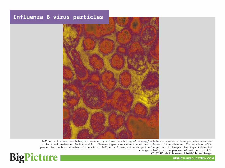
Influenza B virus particles, surrounded by spikes consisting of haemagglutinin and neuraminidase proteins embedded in the viral membrane. Both A and B influenza types can cause the epidemic forms of the disease; flu vaccines offer protection to both strains of the virus. Influenza B does not undergo the large, rapid changes that type A does
but changes slowly by the process of antigenic drift.CC BY NC ND R Dourmashkin/Wellcome Images
Influenza B virus particles

BIGPICTUREEDUCATION.COM
H1N1 influenza virus particles; surface proteins on the particles are shown in black.CC BY NIAID/Flickr
H1N1 influenza virus particles

BIGPICTUREEDUCATION.COM
A model of the H1N1 virus, which causes ‘swine flu’. The name H1N1 refers to the types of haemagglutinin and neuraminidase proteins on the surface of the virus. There are roughly ten times as many haemagglutinin proteins (red) as neuraminidase (yellow). In the centre is the single-stranded viral RNA (purple).
CC BY NC ND Anna Tanczos/Wellcome Images
H1N1 virus

BIGPICTUREEDUCATION.COM
Annotated image of the influenza virus:1. Haemagglutinin (H), the structure that enables the virus to bind to host cells.
2. M2, an ion channel.3. Neuraminidase (N), an enzyme that helps to release new virus particles from the cell.
Credit:Adapted from Sebastian Kaulitzki/iStockphoto
Annotated graphic of the H1N1 virus

BIGPICTUREEDUCATION.COM
Influenza viruses (blue) attaching to the cells of the upper respiratory tract. Viruses floating in the air are breathed in and bind to the hair-like microvilli and cilia on the surface of the cells that line the trachea. They then enter the cells and start to proliferate, eventually causing the cells to die.
CC BY NC NDR Dourmashkin/Wellcome Images
Influenza viruses infecting cells

Influenza viruses being uncoated in a cell
BIGPICTUREEDUCATION.COM
Influenza viruses (the dark particles around the upper surface of the endosome) in an infected cell. These viruses are in the process of being uncoated before replicating in the host cell.
CC BY NC ND R Dourmashkin/Wellcome Images
Reusing our imagesImages and illustrations• All images, unless otherwise indicated, are from Wellcome Images.• Contemporary images are free to use for educational purposes (they have a
Creative Commons Attribution, Non-commercial, No derivatives licence ). Please make sure you credit them as we have done on the site; the format is ‘Creator’s name, Wellcome Images’.
• Historical images have a Creative Commons Attribution 4.0 licence : they’re free to use in any way as long as they’re credited to ‘Wellcome Library, London’.
• Flickr images that we have used have a Creative Commons Attribution 4.0 licence, meaning we – and you – are free to use in any way as long as the original owner is credited.
• Cartoon illustrations are © Glen McBeth. We commission Glen to produce these illustrations for ‘Big Picture’. He is happy for teachers and students to use his illustrations in a classroom setting, but for other uses, permission must be sought.
• We source other images from photo libraries such as Science Photo Library, Corbis and iStock and will acknowledge in an image’s credit if this is the case. We do not hold the rights to these images, so if you would like to reproduce them, you will need to contact the photo library directly.
• If you’re unsure about whether you can use or republish a piece of content, just get in touch with us at [email protected].