Influence of ceramic material, thickness of restoration and cement … · 2019-03-13 · Influence...
10
ORIGINAL RESEARCH Dental Materials João Paulo Mendes TRIBST (a) Amanda Maria de Oliveira DAL PIVA (a) Marcela Moreira PENTEADO (a) Alexandre Luiz Souto BORGES (a) Marco Antonio BOTTINO (a) (a) Universidade Estadual Paulista – Unesp, Instituto de Ciência e Tecnologia, Department of Dental Materials and Prosthodontics, Post-Graduate Program in Restorative Dentistry (Prosthodontic), São José dos Campos, SP, Brazil. Declaration of Interest: The authors certify that they have no commercial or associative interest that represents a conflict of interest in connection with the manuscript. Corresponding Author: Amanda Maria de Oliveira Dal Piva E-mail: [email protected] https://doi.org/10.1590/1807-3107bor-2018.vol32.0118 Submitted: May 15, 201 Accepted for publication: September 29, 2018 Last revision: October 17, 2018 Influence of ceramic material, thickness of restoration and cement layer on stress distribution of occlusal veneers Abstract: The aim of this study was to evaluate stress distribution in an occlusal veneer according to the restorative material, restoration thickness, and cement layer thickness. A tridimensional model of a human maxillary first molar with an occlusal veneer preparation was constructed using a modeling software of finite element analysis. The model was replicated 9 times to evaluate the factors: restoration thickness (0.6, 1.2, and 1.8 mm) and cement layer thickness (100, 200, and 300 μm). Then, each model received different restorative materials (High Translucency Zirconia – [YZHT], Lithium Disilicate – [LD], Zirconia Reinforced Lithium Silicate – [ZLS], Feldspathic – [F], and Hybrid Ceramic – [HC]), totaling forty-five groups. An axial load (600 N) was applied on the occlusal face for static structural analysis. Solids were considered isotropic, homogeneous, and linearly elastic. Contacts were considered perfectly bonded. Fixation occurred in the dental root and a mechanical static structural analysis was performed. Descriptive statistical analysis and one-way ANOVA ( α =10%) were performed for tensile stress peak values in the restoration and cement layer. The difference between groups was compared using the Tukey’s test with 10% significance to match the percentage of the mesh convergence test. According to the results, the cement layer thickness did not influence stress distribution in the restoration (p ≥ 0.10). The thicker the restoration, the higher the tensile stress concentration in the restoration. The graphs showed higher stress concentration in the YZHT, followed by LD, F, ZLS, and HC. Also, the restorative material influenced stress concentration on the cement layer, which decreased according to the sequence HC>YZHT>ZLS>LD>F. HC stood out for causing the least stress concentration in the restoration. Cement layer thickness did not interfere in the mechanical performance of the restorations. Keywords: Finite Element Analysis; Ceramics; Dental Veneer; Tooth Wear. Introduction Tabletop or ultrathin occlusal veneers are a contemporary restorative approach indicated for teeth with occlusal wear. They consist of an important therapeutic modality to recover the occlusal vertical dimension of patients with great occlusal wear related to a parafunctional habit 1 or physiological processes such as erosions 2 . The main advantage of occlusal 1 Braz. Oral Res. 2018;32:e118
Transcript of Influence of ceramic material, thickness of restoration and cement … · 2019-03-13 · Influence...

Original research
Dental Materials
João Paulo Mendes TRIBST(a) Amanda Maria de Oliveira DAL PIVA(a) Marcela Moreira PENTEADO(a) Alexandre Luiz Souto BORGES(a) Marco Antonio BOTTINO(a)
(a) Universidade Estadual Paulista – Unesp, Instituto de Ciência e Tecnologia, Department of Dental Materials and Prosthodontics, Post-Graduate Program in Restorative Dentistry (Prosthodontic), São José dos Campos, SP, Brazil.
Declaration of Interest: The authors certify that they have no commercial or associative interest that represents a conflict of interest in connection with the manuscript.
Corresponding Author: Amanda Maria de Oliveira Dal Piva E-mail: [email protected]
https://doi.org/10.1590/1807-3107bor-2018.vol32.0118
Submitted: May 15, 201 Accepted for publication: September 29, 2018 Last revision: October 17, 2018
Influence of ceramic material, thickness of restoration and cement layer on stress distribution of occlusal veneers
Abstract: The aim of this study was to evaluate stress distribution in an occlusal veneer according to the restorative material, restoration thickness, and cement layer thickness. A tridimensional model of a human maxillary first molar with an occlusal veneer preparation was constructed using a modeling software of finite element analysis. The model was replicated 9 times to evaluate the factors: restoration thickness (0.6, 1.2, and 1.8 mm) and cement layer thickness (100, 200, and 300 μm). Then, each model received different restorative materials (High Translucency Zirconia – [YZHT], Lithium Disilicate – [LD], Zirconia Reinforced Lithium Silicate – [ZLS], Feldspathic – [F], and Hybrid Ceramic – [HC]), totaling forty-five groups. An axial load (600 N) was applied on the occlusal face for static structural analysis. Solids were considered isotropic, homogeneous, and linearly elastic. Contacts were considered perfectly bonded. Fixation occurred in the dental root and a mechanical static structural analysis was performed. Descriptive statistical analysis and one-way ANOVA (α =10%) were performed for tensile stress peak values in the restoration and cement layer. The difference between groups was compared using the Tukey’s test with 10% significance to match the percentage of the mesh convergence test. According to the results, the cement layer thickness did not influence stress distribution in the restoration (p ≥ 0.10). The thicker the restoration, the higher the tensile stress concentration in the restoration. The graphs showed higher stress concentration in the YZHT, followed by LD, F, ZLS, and HC. Also, the restorative material influenced stress concentration on the cement layer, which decreased according to the sequence HC>YZHT>ZLS>LD>F. HC stood out for causing the least stress concentration in the restoration. Cement layer thickness did not interfere in the mechanical performance of the restorations.
Keywords: Finite Element Analysis; Ceramics; Dental Veneer; Tooth Wear.
Introduction
Tabletop or ultrathin occlusal veneers are a contemporary restorative approach indicated for teeth with occlusal wear. They consist of an important therapeutic modality to recover the occlusal vertical dimension of patients with great occlusal wear related to a parafunctional habit1 or physiological processes such as erosions2. The main advantage of occlusal
1Braz. Oral Res. 2018;32:e118

Inf luence of ceramic material, thickness of restoration and cement layer on stress distribution of occlusal veneers
veneers is the recovery of the masticatory function with maximum preservation of dental structure2,3 being a conservative option to traditional onlays4 and complete coverage crowns.2,5 Other advantages are the possibility to predict the final result with temporary restorations6 and the easiness of cementation.7
Although direct composite resins restorations are commonly made,5,6 the use of indirect ceramic materials may provide greater predictability to the treatment in recovering the occlusal vertical dimension during a prolonged time.4,5 However, multiple factors interfere in restoration dynamics such as the final appearance of the dental preparation, restoration geometry and thickness, as well as the mechanical performance of the ceramic material associated with the adhesive technique.8
With the advances in CAD/CAM (computer aided design/Computer aided manufacturing) materials and resin cements,9 the loss of dental structure can be minimized using conservative preparations for occlusal veneers.2,6 Several studies have evaluated fracture3,5,6 and fatigue resistance5 of restorations made in ceramics or composite resin4,5 of different thicknesses.6 The authors observed that thickness is not as influential as the material under a compressive load, thus allowing tabletop veneers to resist loads higher than masticatory ones.6 Until now, no clinical trial or case report has evaluated the most common type of failure of occlusal veneers. However, according to laboratorial fatigue tests, cracks in the restoration and debonding are the most common failure types.5,10 Therefore, it is important to understand how stress from masticatory forces is distributed4,11 in occlusal veneers. Computational simulations from modeling the structures to be evaluated4,11,12,13 allows the visualization of stress concentration regions. As assessed in in vitro studies, defects in stress regions are the origin of fractures.
This study aimed to evaluate the stress distribution in an occlusal veneer restoration according to the restorative material, restoration thickness, and cement thickness. The hypotheses were: a) there would be differences in stress distribution in the restoration and cement layer according to the occlusal veneer thickness; b) a thicker cement layer could negatively influence the mechanical response of the
occlusal veneer and resin cement; and c) different ceramic materials would exhibit different mechanical behaviors under the same conditions.
Methodology
Finite element analysis pre-processingFor finite element (FE) analysis (FEA), a tridimensional
(3D) model of a human maxillary first molar was generated according to anatomical references containing enamel, dentin and periodontal ligament. The pulp chamber and root canals were generated as an empty space in the dentin4,11 without elastic modulus. This 3D-FE model was inserted in a fixation cylinder that simulated bone tissue.11,12 Next, the tooth was replicated in 9 identical models with occlusal wear characteristic of patients with severe dental erosion. Three levels of wear were simulated:4 0.6, 1.2, and 1.8 mm. For that, the occlusal preparation followed the cusps convergence according to Magne et al.,5 simulating the rehabilitation with veneers of respective thicknesses for each model. The geometry of the occlusal preparation was based on a previous study whose minimum restoration thickness was 0.6 mm at the center and 1.2 mm at the cusps5 (Figure 1). From this definition, the restoration thickness was increased 2- and 3-fold, resulting in 1.2 mm and 1.8 mm of minimum occlusal thickness. Three cement layer thicknesses were also evaluated: 100, 200, and 300 μm.14 Table 1 summarizes the group distribution according to 9 models considering the restoration and cement layer thicknesses. For the complete analysis, each model’s crown received 5 different materials, totaling 45 groups.
1.81.8
1.2
1.8
Figure 1. Three-dimensional model of a restored molar with occlusal veneer of 1.2-mm minimum thickness.
2 Braz. Oral Res. 2018;32:e118

Tribst JPM, Dal Piva AMO, Penteado MM, Borges ALS, Bottino MA
Table 1. Group distribution according to restoration thickness, cement layer thickness, and restorative material. Number of elements and nodes are shown for each 3D-FEA model.
Restoration thickness (mm)
Cementing layer thickness (µm)
Restorative material Number of elements Number of nodes
0.6
100
High translucency zirconiaLithium disilicate
Zirconia reinforced lithium silicateFeldspathic
Hybrid ceramic
38,456 130,36
200 39,874 131,798
300 39,908 132,842
1.2
100 41,34 132,912
200 41,65 133,174
300 41,848 133,804
1.8
100 43,568 136,166
200 43,13 136,48
300 43,962 136,588
Boundary condition and mesh generationThe geometries were imported to CAE ANSYS
software (ANSYS 17.2, ANSYS Inc., Houston, USA) in STEP format and tetrahedral elements formed the mesh. A convergence test of 10% mesh control12 determined the number of elements and nodes; thus, the subdivision of the complex geometry into a finite number of elements did not interfere in the results. The properties of the materials and structures were attributed to each solid component with isotropic, homogeneous, and linearly elastic behavior. Young’s modulus and Poisson’s ratio were reported based on the literature (Table 2),15,16,17,18,19,20,21,22 and all contacts were ideally bonded. Five restorative materials for the 9 models (according to restoration and cement thicknesses) were simulated: high translucency zirconia (YZHT), lithium disilicate (LD), zirconia reinforced lithium silicate (ZLS), feldspathic (F), and hybrid ceramic (HC), totaling 45 groups. The group distributions as well as the mesh and node numbers are summarized in Table 1.
FEA processingLoad application (600 N) occurred similar to the
study by Ausiello et al.,13 a methodology that considers the contact between a food bolus and the tooth surface during the closing phase of the chewing cycle. A cylinder base was selected for the system fixation, ensuring only the movement constraint on the Z axis so that the strain generated in all directions was computed. Results in the restoration and cement layer were obtained using maximum principal stress for quantitative analysis and minimum principal stress for a qualitative approach.
Statistical analysisAfter the mechanical static structural analysis,
the tensile stress peaks on the internal surfaces of the restoration and cement layer were exported in spreadsheets, according to the element number corresponding to the numerical calculation. The 100 highest values were selected for each structure (restoration and cement) of all 45 groups, totaling 9,000 values for tensile stress in MPa. The data were analyzed by descriptive statistics (mean and standard deviation), one-way ANOVA for each studied factor, followed by Tukey’s test for differences between groups. All tests were considered significant at 10% due to the correspondence of the mesh convergence test.
Results
In the compressive load situation, the maximum principal stress (Figure 2) was concentrated at the center of the occlusal veneer, and tensile stress (Figure 3) in the intaglio surface. Figure 2 shows that the higher the restorative material elastic modulus, the higher is the compressive stress concentration in the occlusal veneer external surface. The same behavior is observed for the tensile stress concentration in the intaglio surface: the higher the material elastic modulus, the higher the tensile stress concentration (Figure 3). The influence of each factor on the concentration of tensile stresses on the restoration and cement layer was evaluated with the statistical analysis. Figure 4 shows histograms23 of stress data that were plotted for each individual
3Braz. Oral Res. 2018;32:e118

Inf luence of ceramic material, thickness of restoration and cement layer on stress distribution of occlusal veneers
factor to facilitate visualization of significant results, considering the same confidence interval as the computational results of mesh convergence. For each histogram, the X axis shows the calculated stress peaks in MPa, and the Y axis shows the data density according to the variability on the evaluated elements; thus, higher curves indicate lower variability and data farther to the right indicate higher stress peaks. Descriptive statistics, p value, and homogeneous groups are described in Figure 4. For the restoration, the cement layer thickness was not significant (p = 0.167), different from restorative material (p = 0.000) and restoration thickness (p = 0.009). However, the ceramic material (p = 0.001), thickness of restoration (p = 0.001), and cement layer (p = 0.012) were significant for the tensile stress generation on the cement layer. The bar graphs show individual stress peaks (Figure 5). YZHT showed the highest stress peaks for the restoration while HC showed the highest peaks for the cement layer, corroborating with the stress maps.
Discussion
This study evaluated five ceramic materials for occlusal veneer made in three thicknesses and cemented with different thicknesses of cement. The first hypothesis was accepted, because the restorative material thickness influenced the restoration and the resin cement biomechanics. The second hypothesis was rejected because the cement thickness was only significant for the stress generated in the cement itself. The third hypothesis was also accepted since each simulated material (different elastic modulus) had a significant influence on the occlusal veneer and resin cement mechanical response.
The results demonstrate that the restorative material can directly influence the prognosis in the long term. Each material has a specific hardness, which is reflected in various elastic modulus (in the present study, from 30 to 220 GPa) allowing different concentrations of tensile stresses on the crown intaglio surface.4,12,24,25 In this case, the most affected site was the internal surface of the restoration, which is suggested to be the initiation region for slow crack propagation.26 Defects on the surface may be the failure origin when
Table 2. Mechanical properties of materials and structures used in the study.
Material or structureElastic
modulusPoisson
ratio
Enamel15 84.1 0.33
Dentin15 18.6 0.32
Periodontal ligament16 0.069 0.45
Fixture cylinder17 3.6 0.3
High translucency zirconia18 210 0.33
Lithium disilicate19 95 0.25
Zirconia reinforced lithium silicate20 70 0.23
Feldspathic21 48.7 0.23
Hybrid ceramic22 30 0.28
Resin cement22 7.5 0.25
Hybrid Ceramic / ZLS / Feldspathic / Lithium Disilicate / Zirconia
6.90-0.83-1.7-2.5-3.3-4.2-5-5.8-6.7-7.5-8.3-9.2-10-2.8e2
Cer
amic
Thi
ckne
ss: 0
.6 m
m
Cem
ent L
ayer
:
300
µm20
0 µm
100
µm
Cer
amic
Thi
ckne
ss: 1
.2 m
m
Cem
ent L
ayer
:
300
µm20
0 µm
100
µm
Cer
amic
Thi
ckne
ss: 1
.8 m
m
Cem
ent L
ayer
:
300
µm20
0 µm
100
µm
Figure 2. Compressive stress in teeth restored with occlusal veneers in an occlusal view.
4 Braz. Oral Res. 2018;32:e118

Tribst JPM, Dal Piva AMO, Penteado MM, Borges ALS, Bottino MA
Hybrid Ceramic / ZLS / Feldspathic / Lithium Disilicate / ZirconiaC
eram
ic T
hick
ness
: 0.6
mm
Cem
ent L
ayer
:
300
µm20
0 µm
100
µm
Cer
amic
Thi
ckne
ss: 1
.2 m
m
Cem
ent L
ayer
:
300
µm20
0 µm
100
µm
Cer
amic
Thi
ckne
ss: 1
.8 m
m
Cem
ent L
ayer
:
300
µm20
0 µm
100
µm
54.64.23.83.53.12.72.31.91.51.20.770.380-4.6
Figure 3. Tensile stress in teeth restored with occlusal veneers in sagittal section.
a high tensile stress occurs near them. Zirconia has the highest elastic modulus and consequently it showed higher tensile stress concentrated in its intaglio surface (Figure 3). However, due to its hardness property,27 it is difficult to affirm that this material could fail earlier than the simulated vitreous ceramics. The flexural strength of YZHT is twice as high as that of LD.19,28 Nevertheless, the stress peaks in zirconia were
not twice higher than for LD. The hardness property consists in the ability of the material to limit crack propagation. In zirconia, this process occurs through the volumetric increase of 3-4% of the zirconia grains close to the crack due to the change of the tetragonal phase to the monoclinic phase.
Studies support that zirconia presents a higher value of fracture resistance and superior mechanical
5Braz. Oral Res. 2018;32:e118

Inf luence of ceramic material, thickness of restoration and cement layer on stress distribution of occlusal veneers
Den
sity
p = 0.000
MPa
2,5
2,0
1,5
1,0
0,5
0,01,2 2,4 3,6 4,8 6,0 7,2 8,4 9,6
FeldspathicHybrid CeramicLithium DisilicateZLSYZHT
3,1581,1854,3572,9367,693
0,18310,22440,24630,15630,8393
900900900900900
CEBDA
Mean StDev N Grouping
Den
sity
p = 0.001
MPa
3,5
2,5
3,0
2,0
1,5
1,0
0,5
0,00,2 0,4 0,6 0,8 1,0 1,2 1,4
FeldspathicHybrid CeramicLithium DisilicateZLSYZHT
3,1581,1854,3572,9367,693
0,18310,22440,24630,15630,8393
900900900900900
EADCB
Mean StDev N Grouping
Den
sity
p = 0.167
MPa
0,20
0,15
0,10
0,05
0,00-1,4 0,0 1,4 2,8 4,2 5,6 7,0 8,4
100200300
0,57400,48430,4325
0,18310,22440,2463
150015001500
CBA
Mean StDev N Grouping
Den
sity
p = 0.012
MPa
3,5
2,5
3,0
2,0
1,5
1,0
0,5
0,00,2-0,0 0,4 0,6 0,8 1,0 1,2 1,4
100200300
3,1581,1854,357
0,18310,22440,2463
150015001500
CBA
Mean StDev N Grouping
Tensile Stress on Cement Layer According to Restorative Material
Tensile Stress on Cement Layer According to Restorative Material
Den
sity
p = 0.009
MPa
0,25
0,20
0,15
0,10
0,05
0,00-1,6 0,0 1,6 3,2 4,8 6,4 8,0 9,6
61218
3,6433,8974,058
1,7192,4852,319
150015001500
CBA
Mean StDev N Grouping
Den
sity
p = 0.001
MPa
2,5
3,0
2,0
1,5
1,0
0,5
0,00,2 0,4 0,6 0,8 1,0 1,2 1,4
61218
0,58630,49470,4099
0,23450,14600,1389
150015001500
CBA
Mean StDev N Grouping
Tensile Stress on Restoration According to Restoration Thickness
Figure 4. Histograms of tensile stress peaks at the adhesive interface. On the left, results obtained on the internal face of the restoration. On the right, results obtained in the cement layer. Factors from top to bottom are material, cement thickness, and restoration thickness. The graphs show descriptive statistics (means and standard deviations), and one-way ANOVA and Tukey’s tests results (α=5%).
properties than the ceramics evaluated herein.19,20 The higher the percentage of crystals in the ceramic structure, the greater the difficulty to propagate the defect (slow crack growth). A crack originates when the structure is subjected to a stress associated with external factors, such as humidity. After crack formation, the structure fails when the employed stress exceeds the fracture
resistance of the material.29 The characteristic strength of a material under fatigue tends to be approximately half the flexural strength of the material.19
The results suggest that lithium disilicate is more reliable than feldspathic ceramics, because the difference between the tensile stress of these materials was significant (< 10%), yet close enough to predict that the
6 Braz. Oral Res. 2018;32:e118

Tribst JPM, Dal Piva AMO, Penteado MM, Borges ALS, Bottino MA
critical stress of feldspathic will occur earlier, as force is applied. This suggests that, under the same conditions, a feldspathic ceramic crown would fail earlier than a crown in LD, as the feldspathic ceramic has a lower tensile strength value30 than LD. Lithium disilicate has lower glass content (30–40% by volume) than feldspathic ceramic (more than 65% by volume) presenting a high degree of crosslinking, which prevents crack growth. Since we used a linear method and the complex geometry of the occlusal veneer was the same for both groups, it is possible to affirm that the slope of the graph line is similar for all materials. Thus, as feldspar has roughly three-fold less resistance than lithium disilicate ceramics,31 a lower stress peak may be more damaging to the vitreous material without reinforcement.
ZLS is not as rigid as lithium disilicate and the crystal reinforcement makes this material tougher than feldspathic ceramics21 due to its high glass content.32 However, the presence of zirconia in its structure is not shown to be beneficial for its resistance or survival.21
Also, lithium disilicate was shown to have superior mechanical results.21 Therefore, further studies with ZLS in occlusal veneer manufacturing are suggested.
The material with the lowest elastic modulus was the hybrid ceramic or ceramic infiltrated with polymer matrix. Under fatigue, a better performance is observed in polymer-based materials,5 and a lower risk of fracture is obtained in stress distribution studies4. A failure pattern not as serious as a catastrophic failure was also observed.5 In spite of this, the highest values of stress were found in the resin cement for the hybrid ceramics. This material cannot concentrate stress in its own structure, thus the adjacent geometry receives more energy and participates in the dissipation of occlusal forces with greater intensity. Hybrid ceramics may be a promising option for manufacturing a restoration (even with higher tensile strength generated in the resin cement), since the calculated stress peak was about 10 times smaller than in zirconia. In addition, due to the
MPa
Tensile Stress Peak on Restoration
Restoration Thickness
Cement Layer 100 200 300 100 200 300 100 200 300 100 200 300 100 200 300
10
8
6
4
2
0181261812618126181261812618126181261812618126181261812618126181261812618126
Tensile Stress Peak on Cement Layer
Restoration Thickness
Cement Layer 100 200 300 100 200 300 100 200 300 100 200 300 100 200 300
0.4
0.6
0.8
1.0
1.2
1.4
1.6
0.2
0.0181261812618126181261812618126181261812618126181261812618126181261812618126
MPa
FeldspathicHybrid CeramicLithium DisilicateZLSYZHT
Figure 5. Bar graph of stress peaks generated in cement and restoration for all 45 groups.
7Braz. Oral Res. 2018;32:e118
Inf luence of ceramic material, thickness of restoration and cement layer on stress distribution of occlusal veneers
presence of a resinous matrix in its composition, the material has a different failure mode from the other simulated materials21 and a more reliable behavior.19
Crack propagation is associated with mechanical fatigue, such as hydraulic pumping, and moisture.33 However, in hybrid ceramics, the cracks propagate predominantly through the ceramic network and along the polymer/ceramic interface with polymer deformation bridges across the crack, which enhances the resistance to crack propagation.34,35. This characteristic does not make hybrid ceramics resistant to monotonic tests,36 but make them more reliable against fatigue.19,35 This result is similar to previous studies that found better mechanical properties in tabletop restorations made of materials with lower elastic modulus.4,5 The difference of the present study is that the simulated material was not a composite resin but a hybrid ceramic, thereby adding benefits of feldspar ceramics and composite resin.21
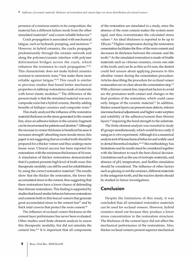
This study analyzed the influence of the restorative material thickness on the stress generated in the cement line, since an adhesive failure in the ceramic fragment can be inconvenient for patients and dentists.14 Although the increase in veneer thickness is beneficial because it increases strength6 absorbing more tensile stress, this paper is not suggesting that an eroded tooth should be prepared for a thicker veneer and thus undergo more tissue wear. Clinical success has been reported for restorations with the minimum thicknesses of 0.6 mm. A simulation of thicker restorations demonstrated that if a patient presents high level of tooth wear, this therapeutic modality can still be used for rehabilitation by using the correct restorative material.6 The results show that the thicker the restoration, the lower the accumulated stress in the cement, thus suggesting that these restorations have a lower chance of debonding than thinner restorations. This finding is supported by studies that found similar behavior between restorations and cements both in thin buccal veneers that generate great accumulated stress in the cement line37 and by thick total crowns that protect the resin cement.38
The influence of occlusal veneer thickness on the cement layer performance has never been evaluated. Other studies used finite element analysis to assess this therapeutic modality, but did not simulate the cement line.4,11 It is important that all components
of the restoration are simulated in a study, since the absence of the resin cement makes the system more rigid, and thus overestimates the calculated stress values.12 An ideal thickness is reported to be up to 120 μm.39 Higher compression during the restoration cementation facilitates the flow of the resin cement and decreases its thickness between the ceramic and the tooth.14 As the simulated restoration is made of friable materials such as vitreous ceramics, covers one side of the tooth, and can be as thin as 0.6 mm, the dentist could feel unsure about applying pressure to this ultrafine veneer during the cementation procedure. Articles describing the procedure for occlusal veneer restauration are no clear about the cementation step.2,5 With a thinner cement line, important factors to avoid are the premature teeth contact and changes in the final position of the restoration, which could cause early fatigue of the ceramic material.11 In addition, thicker cement layers can present more defects, inferior micromechanical adjustment, and higher water sorption and solubility of the adhesive/cement than thinner layers,40 impairing the bond strength to the substrate.
The finite element analysis was essential to study 45 groups simultaneously, which would be too costly if using an in vitro experiment. Although it is a numerical analysis tool of biomechanical behavior and widely used in dental theoretical studies,4,11,12 this methodology has limitations and its results must be considered together with the literature to reach the best clinical decision. Limitations such as the use of isotropic materials, and absence of pH, temperature, and biofilm simulation should be considered. The influence of other factors such as glazing or not the ceramics, different materials in the antagonist tooth, and the reactive dentin should be studied in future investigations.
Conclusion
Despite the limitations of this study, it was concluded that all simulated restorative materials can be used for occlusal veneers. However, hybrid ceramics stand out because they produce a lower stress concentration in the restoration structure. The thickness of the cement layer did not affect the mechanical performance of the restorations. Also, thicker occlusal veneers present superior mechanical
8 Braz. Oral Res. 2018;32:e118

Tribst JPM, Dal Piva AMO, Penteado MM, Borges ALS, Bottino MA
1. Abrahamsen TC. The worn dentition: pathognomonic patterns
of abrasion and erosion. Int Dent J. 2005;55(4 Suppl 1):268-
76. https://doi.org/10.1111/j.1875-595X.2005.tb00064.x
2. Schlichting LH, Resende TH, Reis KR, Magne P. Simplified
treatment of severe dental erosion with ultrathin CAD-
CAM composite occlusal veneers and anterior bilaminar
veneers. J Prosthet Dent. 2016 Oct;116(4):474-82.
https://doi.org/10.1016/j.prosdent.2016.02.013
3. Yazigi C, Kern M, Chaar MS. Influence of various bonding
techniques on the fracture strength of thin CAD/CAM-fabricated
occlusal glass-ceramic veneers. J Mech Behav Biomed Mater. 2017
Nov;75:504-11. https://doi.org/10.1016/j.jmbbm.2017.08.016
4. Magne P, Stanley K, Schlichting LH. Modeling of ultrathin
occlusal veneers. Dent Mater. 2012 Jul;28(7):777-82.
https://doi.org/10.1016/j.dental.2012.04.002
5. Magne P, Schlichting LH, Maia HP, Baratieri LN. In vitro fatigue
resistance of CAD/CAM composite resin and ceramic posterior
occlusal veneers. J Prosthet Dent. 2010 Sep;104(3):149-57.
https://doi.org/10.1016/S0022-3913(10)60111-4
6. Johnson AC, Versluis A, Tantbirojn D, Ahuja S. Fracture
strength of CAD/CAM composite and composite-ceramic
occlusal veneers. J Prosthodont Res. 2014 Apr;58(2):107-14.
https://doi.org/10.1016/j.jpor.2014.01.001
7. Carvalho AO, Bruzi G, Giannini M, Magne P. Fatigue
resistance of CAD/CAM complete crowns with a simplified
cementation process. J Prosthet Dent. 2014 Apr;111(4):310-7.
https://doi.org/10.1016/j.prosdent.2013.09.020
8. Lima JM, Souza AC, Anami LC, Bottino MA, Melo RM,
Souza RO. Effects of thickness, processing technique, and
cooling rate protocol on the flexural strength of a bilayer
ceramic system. Dent Mater. 2013 Oct;29(10):1063-72.
https://doi.org/10.1016/j.dental.2013.07.019
9. Ge C, Green CC, Sederstrom D, McLaren EA, White SN.
Effect of porcelain and enamel thickness on porcelain veneer
failure loads in vitro. J Prosthet Dent. 2014 May;111(5):380-7.
https://doi.org/10.1016/j.prosdent.2013.09.025
10. Abu-Izze FO, Ramos GF, Borges ALS, Anami LC, Bottino
MA. Fatigue behavior of ultrafine tabletop ceramic
restorations. Dent Mater. 2018 Sep;34(9):1401-9.
https://doi.org/10.1016/j.dental.2018.06.017
11. Magne P, Cheung R. Numeric simulation of occlusal interferences in
molars restored with ultrathin occlusal veneers. J Prosthet Dent. 2017
Jan;117(1):132-7. https://doi.org/10.1016/j.prosdent.2016.07.008
12. Tribst JP, Dal Piva AM, Borges AL. Biomechanical behavior of
indirect composite materials: a 3D-FEA study. Braz Dent Sci.
2017;20(3):52-7. https://doi.org/10.14295/bds.2017.v20i3.1444
13. Ausiello P, Ciaramella S, Martorelli M, Lanzotti A, Gloria A,
Watts DC. CAD-FE modeling and analysis of class II restorations
incorporating resin-composite, glass ionomer and glass
ceramic materials. Dent Mater. 2017 Dec;33(12):1456-65.
https://doi.org/10.1016/j.dental.2017.10.010
14. Prakki A, Cilli R, Costa AU, Gonçalves SE, Mondelli
RF, Pereira JC. Effect of resin luting film thickness
on fracture resistance of a ceramic cemented to
dentin. J Prosthodont. 2007 May-Jun;16(3):172-8.
https://doi.org/10.1111/j.1532-849X.2006.00168.x
15. Roscoe MG, Noritomi PY, Novais VR, Soares CJ. Influence
of alveolar bone loss, post type, and ferrule presence
on the biomechanical behavior of endodontically
treated maxillary canines: strain measurement and stress
distribution. J Prosthet Dent. 2013 Aug;110(2):116-26.
https://doi.org/10.1016/S0022-3913(13)60350-9
16. Joshi S, Mukherjee A, Kheur M, Mehta A.
Mechanical performance of endodontically treated
teeth. Finite Elem Anal Des. 2001;37(8):587-601.
https://doi.org/10.1016/S0168-874X(00)00059-7
17. Souza AC, Xavier TA, Platt JA, Borges AL. Effect of Base
and Inlay Restorative Material on the Stress Distribution and
Fracture Resistance of Weakened Premolars. Oper Dent. 2015
Jul-Aug;40(4):E158-66. https://doi.org/10.2341/14-229-L
18. Faria R, Bottino MA. High-translucent monolithic zirconia for
implant-supported rehabilitations. Prótese News. 2016;3:36-50.
19. Homaei E, Farhangdoost K, Tsoi JK, Matinlinna JP, Pow
EH. Static and fatigue mechanical behavior of three dental
CAD/CAM ceramics. J Mech Behav Biomed Mater. 2016
Jun;59:304-13. https://doi.org/10.1016/j.jmbbm.2016.01.023
20. Zimmermann M, Egli G, Zaruba M, Mehl A. Influence
of material thickness on fractural strength of CAD/
CAM fabricated ceramic crowns. Dent Mater J. 2017
Nov;36(6):778-83. https://doi.org/10.4012/dmj.2016-296
21. Ramos NC, Campos TM, Paz IS, Machado JP, Bottino MA,
Cesar PF, et al. Microstructure characterization and SCG
of newly engineered dental ceramics. Dent Mater. 2016
Jul;32(7):870-8. https://doi.org/10.1016/j.dental.2016.03.018
22. Jongsma LA, de Jager N, Kleverlaan CJ, Pallav P, Feilzer AJ. Shear
bond strength of three dual-cured resin cements to dentin analyzed
by finite element analysis. Dent Mater. 2012 Oct;28(10):1080-8.
https://doi.org/10.1016/j.dental.2012.07.002
23. Correia AMO, Tribst JPM, Matos FS, Platt JA, Caneppele
TMF, Borges ALS. Polymerization shrinkage stresses in different
restorative techniques for non-carious cervical lesions. J Dent.
2018 Sep;76:68-74. https://doi.org/10.1016/j.jdent.2018.06.010
References
performance than thinner restorations, but all three simulated conditions can withstand masticatory loads.
Conflict of interestThe authors declare no conflict of interest.
9Braz. Oral Res. 2018;32:e118

Inf luence of ceramic material, thickness of restoration and cement layer on stress distribution of occlusal veneers
24. Dal Piva AMO, Tribst JPM, Borges ALS, Souza ROAE, Bottino
MA. CAD-FEA modeling and analysis of different full crown
monolithic restorations. Dent Mater. 2018 Sep;34(9):1342-50.
https://doi.org/10.1016/j.dental.2018.06.024
25. Tribst JPM, Dal Piva AMO, Madruga CFL, Valera MC,
Borges ALS, Bresciani E et al. Endocrown restorations:
Influence of dental remnant and restorative material on
stress distribution. Dent Mater. 2018 Oct;34(10):1466-73.
https://doi.org/10.1016/j.dental.2018.06.012
26. Kok P, Pereira GK, Fraga S, Jager N, Venturini AB, Kleverlaan
CJ. The effect of internal roughness and bonding on the
fracture resistance and structural reliability of lithium
disilicate ceramic. Dent Mater. 2017 Dec;33(12):1416-25.
https://doi.org/10.1016/j.dental.2017.09.018
27. Marinis A, Aquilino SA, Lund PS, Gratton DG, Stanford
CM, Diaz-Arnold AM et al. Fracture toughness of
yttria-stabilized zirconia sintered in conventional and
microwave ovens. J Prosthet Dent. 2013 Mar;109(3):165-71.
https://doi.org/10.1016/S0022-3913(13)60037-2
28. Zhang Y, Mai Z, Barani A, Bush M, Lawn B. Fracture-resistant
monolithic dental crowns. Dent Mater. 2016 Mar;32(3):442-9.
https://doi.org/10.1016/j.dental.2015.12.010
29. Scherrer SS, Lohbauer U, Della Bona A, Vichi A, Tholey
MJ, Kelly JR et al. ADM guidance-ceramics: guidance
to the use of fractography in failure analysis of brittle
materials. Dent Mater. 2017 Jun;33(6):599-620.
https://doi.org/10.1016/j.dental.2017.03.004
30. Trindade FZ, Valandro LF, Jager N, Bottino MA,
Kleverlaan CJ. Elastic properties of lithium disilicate versus
feldspathic inlays: effect on the bonding by 3D finite
element analysis. J Prosthodont. 2018 Oct;27(8):741-7.
https://doi.org/10.1111/jopr.12550
31. Zhang Y, Lee JJ, Srikanth R, Lawn BR. Edge
chipping and flexural resistance of monolithic
ceramics. Dent Mater. 2013 Dec;29(12):1201-8.
https://doi.org/10.1016/j.dental.2013.09.004
32. Krüger S, Deubener J, Ritzberger C, Höland W. Nucleation
kinetics of lithium metasilicate in ZrO2-bearing lithium
disilicate glasses for dental application. Int J Appl Glass Sci.
2013;4(1):9-19. https://doi.org/10.1111/ijag.12011
33. Zhang Y, Sailer I, Lawn BR. Fatigue of dental
ceramics. J Dent. 2013 Dec;41(12):1135-47.
https://doi.org/10.1016/j.jdent.2013.10.007
34. Coldea A, Swain MV, Thiel N. Hertzian contact
response and damage tolerance of dental ceramics.
J Mech Behav Biomed Mater. 2014 Jun;34:124-33.
https://doi.org/10.1016/j.jmbbm.2014.02.002
35. El Zhawi H, Kaizer MR, Chughtai A, Moraes RR, Zhang Y.
Polymer infiltrated ceramic network structures for resistance to
fatigue fracture and wear. Dent Mater. 2016 Nov;32(11):1352-
61. https://doi.org/10.1016/j.dental.2016.08.216
36. Al-Akhali M, Chaar MS, Elsayed A, Samran
A, Kern M. Fracture resistance of ceramic and
polymer-based occlusal veneer restorations. J
Mech Behav Biomed Mater. 2017 Oct;74:245-50.
https://doi.org/10.1016/j.jmbbm.2017.06.013
37. Jankar AS, Kale Y, Kangane S, Ambekar A, Sinha M, Chaware
S. Comparative evaluation of fracture resistance of Ceramic
Veneer with three different incisal design preparations - An
In-vitro Study. J Int Oral Health. 2014 Feb;6(1):48-54.
38. Zhu J, Rong Q, Wang X, Gao X. Influence of remaining tooth
structure and restorative material type on stress distribution
in endodontically treated maxillary premolars: A finite
element analysis. J Prosthet Dent. 2017 May;117(5):646-55.
https://doi.org/10.1016/j.prosdent.2016.08.023
39. Almeida e Silva JS, Erdelt K, Edelhoff D, Araújo
É, Stimmelmayr M, Vieira LC et al. Marginal and
internal fit of four-unit zirconia fixed dental prostheses
based on digital and conventional impression
techniques. Clin Oral Investig. 2014;18(2):515-23.
https://doi.org/10.1007/s00784-013-0987-2
40. Silva NR, Souza GM, Coelho PG, Stappert CF, Clark EA,
Rekow ED et al. Effect of water storage time and composite
cement thickness on fatigue of a glass-ceramic trilayer system.
J Biomed Mater Res B Appl Biomater. 2008 Jan;84(1):117-23.
https://doi.org/10.1002/jbm.b.30851
10 Braz. Oral Res. 2018;32:e118