Influence of an electric field on the efficacy of Mitomycin C - DUCG
13
1 Influence of an electric i field on the efficacy of Mitomycin C treatment of bladder cancer. DaBlaCa study 3 Juan Luis Vsquez PhD Project description (Academically approved for ernrollment at The University of Copenhagen) August 2011
Transcript of Influence of an electric field on the efficacy of Mitomycin C - DUCG

1
Influence of an electrici field on the efficacy of Mitomycin C treatment of bladder cancer. DaBlaCa study 3
Juan Luis V�squezPhD Project description
(Academically approved for ernrollment at The University of Copenhagen)
August 2011
2
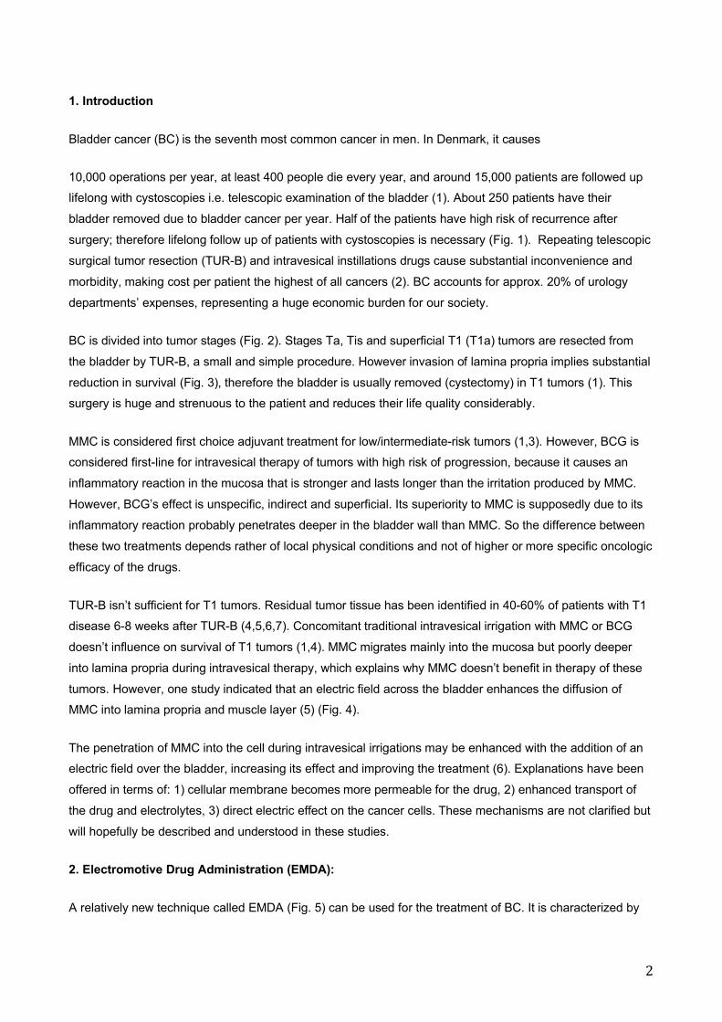
1. Introduction
Bladder cancer (BC) is the seventh most common cancer in men. In Denmark, it causes
10,000 operations per year, at least 400 people die every year, and around 15,000 patients are followed up
lifelong with cystoscopies i.e. telescopic examination of the bladder (1). About 250 patients have their
bladder removed due to bladder cancer per year. Half of the patients have high risk of recurrence after
surgery; therefore lifelong follow up of patients with cystoscopies is necessary (Fig. 1). Repeating telescopic
surgical tumor resection (TUR-B) and intravesical instillations drugs cause substantial inconvenience and
morbidity, making cost per patient the highest of all cancers (2). BC accounts for approx. 20% of urology
departments’ expenses, representing a huge economic burden for our society.
BC is divided into tumor stages (Fig. 2). Stages Ta, Tis and superficial T1 (T1a) tumors are resected from
the bladder by TUR-B, a small and simple procedure. However invasion of lamina propria implies substantial
reduction in survival (Fig. 3), therefore the bladder is usually removed (cystectomy) in T1 tumors (1). This
surgery is huge and strenuous to the patient and reduces their life quality considerably.
MMC is considered first choice adjuvant treatment for low/intermediate-risk tumors (1,3). However, BCG is
considered first-line for intravesical therapy of tumors with high risk of progression, because it causes an
inflammatory reaction in the mucosa that is stronger and lasts longer than the irritation produced by MMC.
However, BCG’s effect is unspecific, indirect and superficial. Its superiority to MMC is supposedly due to its
inflammatory reaction probably penetrates deeper in the bladder wall than MMC. So the difference between
these two treatments depends rather of local physical conditions and not of higher or more specific oncologic
efficacy of the drugs.
TUR-B isn’t sufficient for T1 tumors. Residual tumor tissue has been identified in 40-60% of patients with T1
disease 6-8 weeks after TUR-B (4,5,6,7). Concomitant traditional intravesical irrigation with MMC or BCG
doesn’t influence on survival of T1 tumors (1,4). MMC migrates mainly into the mucosa but poorly deeper
into lamina propria during intravesical therapy, which explains why MMC doesn’t benefit in therapy of these
tumors. However, one study indicated that an electric field across the bladder enhances the diffusion of
MMC into lamina propria and muscle layer (5) (Fig. 4).
The penetration of MMC into the cell during intravesical irrigations may be enhanced with the addition of an
electric field over the bladder, increasing its effect and improving the treatment (6). Explanations have been
offered in terms of: 1) cellular membrane becomes more permeable for the drug, 2) enhanced transport of
the drug and electrolytes, 3) direct electric effect on the cancer cells. These mechanisms are not clarified but
will hopefully be described and understood in these studies.
2. Electromotive Drug Administration (EMDA):
A relatively new technique called EMDA (Fig. 5) can be used for the treatment of BC. It is characterized by

3
combination of 3 electromolecular interactions: Iontophoresis increases transport of ions around target
molecules, dragging water along, which will entrain any non-ionised solutes present. This ‘‘solvent drag’’ is
called electroosmosis. Electroporation (EP) implies increasing biological membrane permeability under the
influence of the electric field (7).
EMDA uses a current generator (Physionizer�) (Fig. 5) that delivers pulsed electric current; passed between
the active intravesical electrode, which is integrated to a specifically designed catheter, and two ground
electrodes, placed on lower abdominal skin. MMC (40 mg) is instilled through the catheter. The current is set
to 20mA for 30 min.
Di Stasi estimated concentrations of MMC after EMDA and Passive Diffusion (PD) using HPLC in an in vitro
study. The concentrations were substantially higher in the 3 layers after EMDA than after PD (7) (Fig. 4).
A study compared MMC administered by PD and EMDA to BCG in patients with high-risk non-muscle
invasive BC (8). It suggests that EMDA/MMC is as effective as BCG and more effective than PD, in terms of
recurrence, progression and survival, with fewer side effects than BCG.
Juan Luis V�squez and I tested EMDA at our department in Frederiksberg hospital in 4 patients with high-
risk tumors. The procedure is easy to perform, well tolerated and patients don’t require major observation.
None of the patients have had recurrence of disease in one and a half year. Frequently reported side effects
are transient skin redness at the site of abdominal electrodes, tingling sensation on the abdomen during
treatment (7), and transient irritative urinary symptoms.
These data suggest that EMDA may become an alternative treatment for patients with high-risk non-invasive
BC. However, it is only used experimentally in 2 – 3 centers in Europe, because lack of knowledge about the
mechanisms involved.
Juan Luis V�squez is developing this project in order to reach a PhD degree. He has performed an in-vitro
study in which bladder cancer cell lines were electroporated with MMC. This study shows that EP it self
causes cell death of approx. 25%. Furthermore it shows that EP and MMC have an additive effect, and cell
death can be increased by 34% when compared to MMC alone at the same MMC concentrations (Fig. 6).
Publication of these data is being prepared.
3. Hypotheses:
(H0): EMDA improves MMC’s intravesical effect by electroporation, increasing influx of MMC into cells and
into deeper layers of the bladder.
A. In-vitro studies with BC cell lines:
1) Do electroporation improve MMC’s efficacy in in-vitro studies?

4
2) Does pH influence the efficacy of EMDA in-vitro?
B. In-vivo studies with bladder biopsies:
4) Does MMC induce more cellular changes and deeper in the bladder wall when associated to EMDA than
when given alone?
5) Is the concentration of MMC higher in biopsies after EMDA than after PD?
6) Is the penetration of MMC into the bladder wall improved when administered under the influence of an
electric field?
4. Objectives:
To investigate the mechanisms involved in the apparently enhanced penetration of MMC into bladder cancer
cells when combined with electricity.
A1: To demonstrate if MMC can be electroporated into BC cells and to compare its effect with non-
electroporated MMC.
A2: To investigate the influence of pH in the cytotoxicity of MMC when combined with electricity. Does pH of
MMC solution affect results of electroporation?
B4-6: To perform histopathological studies of bladder biopsies taken in relation to EMDA treatments in order
to determine its effect on cancer and stromal cells in-vivo.
5. Methodology:
A) Study 1:
MMC might have different charges according to the pH of the solution in which it is diluted. These charges
might play an important role in the penetration of MMC into the cell. We believe that MMC is charged in acid
environments like urine, which makes it difficult for the drug to penetrate through the membrane, unless
electroporation is present.
Juan Luis V�squez will perform experimental manipulation of the pH in order determine this. Cells will be
electroporated using the BTX T820 electroporator (9) to permeabilize T24 bladder cancer cell lines for MMC
at Center for Drug and Gene Electrotransfer, Oncology department, Herlev Hospital. MTT assay will be
performed; it makes a quantitative assessment of cytotoxicity.
If our in-vitro studies demonstrate increased cytotoxicity on cell lines when MMC is associated with
electroporation; and furthermore that an increased concentration of MMC can be detected intracellularly in

5
cell cultures, we will be able to suggest that EMDA could improve the oncologic efficacy of intravesical MMC.
B) Study 2:
MMC causes double strand breaks (DSB) in the DNA inducing apoptosis (10). Juan Luis V�squez will
perform immunohistochemical laboratory methods to detect apoptosis and tissue reaction, to assess MMC’s
effect on the bladder’s different layers. Paraffin sections from bladder biopsies will be analyzed for
proliferation, apoptosis and necrosis in the various components of the biopsy, tumor, stroma, and smooth
muscle using standard point counting morphometry (11).
Ten patients with high-risk superficial BC (including T1) unable or unwilling to undergo radical curative
treatment, who previously have received alternative treatment with BCG, will be included. In the first week, a
treatment with only electricity will be given. Then MMC without electricity in the 2nd week; and in the 3rd, 4th,
5th and 6th weeks, patients will be treated with electricity + MMC (EMDA) (Fig. 7). Thus, the effect on the
bladder wall of every modality can be evaluated on bladder biopsies. Crossover effects are possible from
modality to modality but this is expected to be minimal since the two weakest treatments are given first.
V�squez will take biopsies during flexible cystoscopy at Frederiksberg Hospital immediately after the first
four treatments and 3 days after treatment 2 and 3 (Fig. 7). They will be embedded in paraffin, and taken to
Rigshospitalet, where V�squez will analyze them. Paraffin sections can be stained with haematoxylin-eosin,
to determine necrosis.
Gamma-H2AX is considered marker of DSBs. Paraffin sections can be stained with gamma- H2AX antibody,
and finally the numbers of foci and level of gamma-H2AX expression per nucleus can be determined by
manual counting under a light microscope and by Automated Cellular Imaging System (12).
A way to determine cellular changes related to cell death is detecting proliferation. Sections can be
immunostained with the avidin-biotin complex (ABC) method.
Expected results are schematized in Fig 8.
This in vivo study is performed in order to demonstrate cellular changes in the deeper layers of the bladder.
This is an indirect way to show the presence of MMC and a direct way to show the effect of the drug on
tissue.
We expect to find that EMDA affects the deeper layers of the bladder wall. We expect that at least 9 of 10
biopsies from our patients will show this effect, which gives 95% confidence limits at 56% -100%, in order to
show that findings are correct.
The laboratory and clinical work will be performed and coordinated by Juan Luis V�squez. Specific days of
the week will be designated for specific activities at the place where they will be performed.

6
6. Statistics
Descriptive and non-parametric statistical methods will predominantly be used to analyze study results. Data
will be determined according to test numbers, data distribution, and data structure. Senior statistician
Susanne Rosth�j, Department of Biostatistics, University of Copenhagen, will be consulted in statistical
issues.
7. Clinical importance of this project:
Traditional intravesical chemotherapy supported by electric treatments could become an important adjunct to
the treatment of bladder cancer. We still lack of knowledge of how the treatment works on cells and tissue,
which electrical charges to be used, treatment durations, number of treatments, etc. These parameters are
important to be determined in order to perform larger studies involving patients, in which an optimized
method is being used and compared to standards.
EMDA may spare removal of bladders in Stage T1 bladder cancer and furthermore be a new treatment
modality to improve treatment and survival of patients, unfit or unwilling for cystectomy, but fit for a serie of
EMDA.
8. PhD:
The University of Copenhagen has found this project academically qualified for PhD enrollment at the PhD
school of The Faculty of Health Sciences. The next step is to raise the financial resources necessary for its
development over 3 years.
Juan Luis V�squez graduated MD from Universidad Central de Venezuela in November 2006. In August
2007 he immigrated to Denmark after he got a job offer in Copenhagen. In 1999-2000, he was exchange
student in Ribe and ever since he has cared very much about Denmark.
In 2007, he obtained the credentials that allowed him to work at danish hospitals. He wants to become an
urologist, and during the last 2 years he has been part of the bladder cancer team of the urology department
at Frederiksberg Hospital. When consultant urologist Gregers G Hermann offered him a scientific project
about EMDA it was obvious for him to accept it. This has inspired him to start a research career before he
goes further with his main surgical education. Gregers G Hermann has established from his network, an
advisory group of scientific clinicians with wide laboratory experience, with whom he works very well. He has
gotten very well into the bladder cancer issue and feels confident carrying out the experiments. He finds the
focus of these studies very exciting and feels committed to this PhD project, because of its clinical
significance, which opens greater research possibilities for the future. He has been on study visit at Dr. Di
Stasi’s clinic in Rome and got involved with the technique to be studied. He has become familiarized with
EMDA during this last 2 years.

7
In June 2011 Juan Luis V�squez received a grant of DKK 100.000 from The Danish Cancer Research
Foundation (Dansk Kr�ftforsknings Fond) for the development of in-vitro experiments involving
electroporation of bladder cancer cell lines and mitomycin C. These experiments were performed between
April and August 2011.
He will perform and coordinate the laboratory and clinical work. Specific days of the week will be designated
for specific activities at the place where they will be performed (Frederiksberg Hospital, Herlev Hospital,
Rigshospitalet).
9. Advisory group
DM Sc, Dr. Gregers G Hermann, (Frederiksberg Hospital, Urology, Dept.) is the main supervisor of this
project. As head of Urology Department’s Research Unit at Frederiksberg Hospital and former laboratory
research fellow, he will provide clinical guidance about bladder cancer patients, laboratory work and
implementation of all projects. Dr. Hermann guarantees that the studies can be performed at Frederiksberg
Hospital or equivalent place, in case the Urology Department is moved to Bispebjerg Hospital or
Rigshospitalet.
Clinical Associate Research Professor Julie Gehl (Herlev Hospital, Oncology Dept.), will be the contact to the
University of Copenhagen during PhD period. Prof. Gehl is chief at CEDGE, where the first in vitro study was
performed, and she has the necessary expertise and access to the equipment needed by Juan Luis V�squez
in order to conduct experiments with cell cultures, electroporation and chemotherapy.
DM Sc, Dr. Maxwell Sehested (Rigshospitalet, Pathology Dept.) will supervise the laboratory work during the
clinical study. Dr. Sehested guarantees that Juan Luis V�squez will be able to analyse the biopsies at the
Pathology Institute, Rigshospitalet.
Professor Mikael R�rth (Rigshospitalet, Oncology Dept.) and Consultant urologist Karin Mogensen
(Frederiksberg Hospital, Urology Dept.) will be advisors during all studies.
Dr. Hermann and Juan Luis Vasquez have been in ongoing communication with Dr. Savino Di Stasi, Italy
and Dr. Claus Riedl, Austria who use EMDA at their urology departments.
The Danish multidisciplinary Bladder Cancer Group (DaBlaCa) supports the study and has been accepted to
be DaBlaCa’s study number 3.
10. Publications
Results will be published in a peer-review journal with Juan Luis V�squez as first author, Gregers G
Hermann as last author; others by Vancouver rules.

8
11. Ethical Considerations
The in-vitro study doesn’t involve ethical considerations. Study 2 implies EMDA treatments of 10 patients
with T1 bladder cancer. Patients eligible for this study are those unable (due to age or co-morbidity) or
reluctant to undergo radical curative surgery, who don’t respond to alternative intravesical BCG treatment,
and therefore might benefit of EMDA.
During treatments patients undergo 6 flexible cystoscopies, and biopsies will be taken through telescopic instrument. The study is not significantly harmful and there are no risks associated, apart from a little risk of simple cystitis. The examination requires no anesthesia, and besides a little discomfort in the urethra during the cystoscopy and during the subsequent 10-12 hours, there are no major side effects.
Application for approval by The Scientific Ethics Committee is currently being written. However, we don’t
expect problems getting our protocol approved due to its clinical significance and the potential benefit for
these quite common patients.
9
12. References:
1) Hermann GG et al. Dansk Bl�reCancer Register DBCR. Year report 2010.
2)Botteman MF et al. The health economics of bladder cancer: A comprehensive review of the published
literature. Pharmacoeconomic2003; 21: 1315-30.
3) Solsona E, Iborra I, Ricos JV, Monros JL, Casanova J, Dumont R. Effectiveness of a single immediate
mitomycin C instillation in patients with low risk superficial bladder cancer: short and long-term followup. J
Urol 1999 Apr; 161(4): 1120-3.
4) Hermann GG, Mogensen K, Carlsson S, Marcussen N, Duun S. Fluorescence guided TUR-B reduces
bladder tumour recurrence due to less residual tumour tissue after TURB in Ta/T1 patients. In press, British
Journal of Urology.
5) Di Stasi SM et al. Electromotive versus Passive Difussion of Mitomycin C into human bladder wall:
Concentration-Depth Profiles Studies. Cancer Research 1999; 59: 4912-4918.
6) Di Stasi SM et al. Electromotive Delivery of Mitomycin C into Human Bladder Wall. Cancer Research
1997; 57: 875-880.
7) Riedl CR et al. Intravesical electromotive drug administration technique: Preliminary results and side
effects. J Urol 1998; 159: 1851 -1856.
8) Di Stasi et al. Intravesical electromotive mitomycin C versus passive transport mitomycin C for high-risk
superficial bladder cancer: A prospective randomized study. J Urol 2003; 170: 777 – 782.
9) Gehl J, Skovgaard T, and Muir L. Enhancement of citotoxicity by electropermeabilization: an improved
method for screening drugs. Anti-Cancer Drugs 1998; 9: 319-325.
10) Tomasz M. Mitomycin C: small, fast and deadly (but very selective). Chemistry & Biology 1999; 2: 575-
579.
11) El-Salhy M and Sitohy B. Triple therapy with octreotide, galanin and serotonin induces necrosis and
increases apoptosis of a rat colon carcinoma. Regulatory Peptides 2002; 55-62.
12) Krayewska Maryla et al. Image Analysis Algorithms for Immunohistochemical Assessment of Cell Death
Events and Fibrosis in Tissue Sections. J Histochem Cytochem 2009; 57:649–663.

10
13. Annex
1) Figure 1: This figure shows the follow-up model for patients with non-muscle invasive bladder cancer.
2) Figure 2: BC is divided into low-grade tumors and high-grade tumors and into clinical tumor stages (Ta-T4). Stage Ta and Tis are not invasive, stage T1 invades lamina propria, T2 invades the muscle and T3-T4 tumors invade the surrounding organs.

11
3) Figure 3: This figure shows patients’ survival after tumor invasion of the lamina propria, T1 tumors, (green line).
4) Figure 4: This figure shows the results of Di Stasi et al’s in-vitro study. The concentration of MMC was measured in the 3 layers of the bladder wall after EMDA and passive difussion.

12
5) Figure 5: A) Current generator, Physionizer�. B) Specifically designed catheter with the active electrode. C) EMDA treatment in course.
6) Figure 6: This graph shows the results of our preliminary results of our in vitro studies. Cell mortality in vitro can be increased by 34% by the Inhibitory Concentration 50% (IC50) when MMC is associated to electroporation. In this graph is also seen that when MMC is administered together with electroporation it is possible to reach the IC50 with a third of the concentration of MMC.
The most interesting thing is that MMC together with MMC increases cell mortality by 34%. We have tested EMDA at our outpatient clinic, and patients do not report major side effects. This might suggest that electroporation increases cytotoxicity without giving more side effects.

13
7) Figure 5: Scheme for treatments in study 3. The first procedure will be only electricity. One week after, one intravesical instillation of MMC (passive diffusion). The last treatments will 4 weekly EMDA treatments. Biopsies will be taken as marked with the red X.
8) Figure 6: This figure shows the expected results of the clinical study. Cellular changes related to apoptosis shall be observed in the deeper layers of the bladder wall after EMDA.