Inflammatory Bowel Diseases - Lancaster Universityeprints.lancs.ac.uk/71989/1/UC_paper_IBD.pdf ·...
33
For Peer Review Human gut dendritic cells drive aberrant, gut-specific T-cell responses in ulcerative colitis, characterised by increased IL-4 production and loss of IL-22 and IFNγ Journal: Inflammatory Bowel Diseases Manuscript ID: IBD-14-0541 Wiley - Manuscript type: Original Research Articles - Basic Science Date Submitted by the Author: 08-Jul-2014 Complete List of Authors: Mann, Elizabeth; Imperial College London, Antigen Presentation Research Group; Johns Hopkins University, School of Medicine Bernardo, David; Imperial College London, Antigen Presentation Research Group Chen, Siew Chien; Imperial College London, Antigen Presentation Research Group Rigby, Rachael; Lancaster University, Division of Biomedical and Life Sciences; Imperial College London, Antigen Presentation Research Group Al-Hassi, Hafid; Imperial College London, Antigen Presentation Research Group Landy, Jonathan; Imperial College London, Antigen Presentation Research Group; North West London Hospitals NHS Trust, Gastroenterology Peake, Simon; Imperial College London, Antigen Presentation Research Group; North West London Hospitals NHS Trust, Gastroenterology Spranger, Henning; North West London Hospitals NHS Trust, Gastroenterology English, Nicholas; Imperial College London, Antigen Presentation Research Group Thomas, Linda; Yakult UK Ltd., Science Stagg, Andrew; of Cell and Molecular Science, Centre for Infectious Disease Knight, Stella; Imperial College London, Antigen Presentation Research Group Hart, Ailsa; St. Mark's Hospital , Gastroenterology; Imperial College London, Antigen Presentation Research Group; North West London Hospitals NHS Trust, Gastroenterology Keywords: Dendritic Cells in IBD < Basic Science Areas, T Cells and Regulatory T Cells in IBD < Basic Science Areas, Adaptive Immune System in IBD < Basic Science Areas, Mucosal Immunology < Basic Science Areas Inflammatory Bowel Diseases
Transcript of Inflammatory Bowel Diseases - Lancaster Universityeprints.lancs.ac.uk/71989/1/UC_paper_IBD.pdf ·...
For Peer Review
Human gut dendritic cells drive aberrant, gut-specific T-cell
responses in ulcerative colitis, characterised by increased IL-4 production and loss of IL-22 and IFNγ
Journal: Inflammatory Bowel Diseases
Manuscript ID: IBD-14-0541
Wiley - Manuscript type: Original Research Articles - Basic Science
Date Submitted by the Author: 08-Jul-2014
Complete List of Authors: Mann, Elizabeth; Imperial College London, Antigen Presentation Research
Group; Johns Hopkins University, School of Medicine Bernardo, David; Imperial College London, Antigen Presentation Research Group Chen, Siew Chien; Imperial College London, Antigen Presentation Research Group Rigby, Rachael; Lancaster University, Division of Biomedical and Life Sciences; Imperial College London, Antigen Presentation Research Group Al-Hassi, Hafid; Imperial College London, Antigen Presentation Research Group Landy, Jonathan; Imperial College London, Antigen Presentation Research Group; North West London Hospitals NHS Trust, Gastroenterology Peake, Simon; Imperial College London, Antigen Presentation Research
Group; North West London Hospitals NHS Trust, Gastroenterology Spranger, Henning; North West London Hospitals NHS Trust, Gastroenterology English, Nicholas; Imperial College London, Antigen Presentation Research Group Thomas, Linda; Yakult UK Ltd., Science Stagg, Andrew; of Cell and Molecular Science, Centre for Infectious Disease Knight, Stella; Imperial College London, Antigen Presentation Research Group Hart, Ailsa; St. Mark's Hospital , Gastroenterology; Imperial College London, Antigen Presentation Research Group; North West London Hospitals NHS Trust, Gastroenterology
Keywords: Dendritic Cells in IBD < Basic Science Areas, T Cells and Regulatory T Cells in IBD < Basic Science Areas, Adaptive Immune System in IBD < Basic Science Areas, Mucosal Immunology < Basic Science Areas
Inflammatory Bowel Diseases

For Peer Review
Page 1 of 31 Inflammatory Bowel Diseases
123456789101112131415161718192021222324252627282930313233343536373839404142434445464748495051525354555657585960
For Peer Review
Human gut dendritic cells drive aberrant, gut-specific T-cell responses in ulcerative colitis, characterised by increased IL-4 production and loss of IL-22 and IFNγ. Elizabeth R. Mann1,2, David Bernardo1, Siew C. Ng3,1, Rachael J. Rigby4,1, Hafid O. Al-Hassi1, Jon Landy1,5, Simon T.C. Peake1,5, Henning Spranger5, Nicholas R. English1, Linda V. Thomas6, Andrew J. Stagg7, Stella C. Knight1 and Ailsa L. Hart1,5. 1Antigen Presentation Research Group, Imperial College London, Northwick Park and St. Mark’s Campus, Harrow, UK. 2Gastrointestinal Division, Johns Hopkins University School of Medicine, Baltimore, Marylands, US. 3Department of Medicine and Therapeutics, Institute of Digestive Disease, Li Ka Shing Institute of Health Science, Chinese University of Hong Kong, Hong Kong. 4Faculty of Health and Medicine, Division of Biomedical and Life Sciences, Lancaster, University, Lancaster, UK. 5Department of Gastroenterology, St. Mark’s Hospital, North West London Hospitals NHS Trust, Harrow, UK. 6Yakult UK Ltd., West End Road, South Ruislip, UK. 7Centre for Immunology and Infectious Disease, Blizard Institute of Cell and Molecular Science, Barts and the London School of Medicine and Dentistry, Queen Mary University of London, UK. Contact address: Ailsa L. Hart, IBD Unit, St. Mark’s Hospital, Harrow, HA1 3UJ, UK. Phone +44 (0) 20 8869 5805; Fax +44 (0) 20 8869 3532 Email [email protected] Grant support: Yakult Europe B.V., BBSRC. E.R.M. was involved in study concept and design, acquisition, analysis and interpretation of data, drafting, revision of manuscript and statistical analysis. D.B, H.O.A. and N.R.E. were involved in data acquisition/analysis, interpretation of data and revision of manuscript. S.C.N., R.J.R. and A.J.S. were involved in study concept and design, interpretation of data and revision of manuscript. J.L., S.T.C.P. and H.S. were involved in critical material and reagent support, data interpretation and revision of manuscript. L.V.T. was involved in initial project discussions, study concept and design, data interpretation and revision of manuscript. S.C.K. and A.L.H. were involved in study concept and design, interpretation of data, revision of manuscript and study supervision.
Page 2 of 31Inflammatory Bowel Diseases
123456789101112131415161718192021222324252627282930313233343536373839404142434445464748495051525354555657585960

For Peer Review
ABSTRACT The pathogenesis of inflammatory bowel disease (IBD) is incompletely understood but results from a dysregulated intestinal immune response to the luminal microbiota. CD4+ T-cells mediate tissue injury in the IBD-associated immune response. Dendritic cells (DC) generate primary T-cell responses and mediate intestinal immune tolerance to prevent overt inflammation in response to the gut microbiota. However, most information regarding function of intestinal DC has come from mouse models and information in humans is scarce. We show here that intestinal DC subsets are skewed in ulcerative colitis (UC) in humans, with a loss of CD103+ lymph-node homing DC; this intestinal DC subset preferentially generates regulatory T-cells in mice. We show infiltrates of DC negative for myeloid marker CD11c, with enhanced expression of Toll-like receptors (TLRs) for bacterial recognition. Following mixed leucocyte reaction, DC from the inflamed UC colon had an enhanced ability to generate gut-specific CD4+ T-cells with enhanced production of IL-4, but a loss of IFNγ and IL-22 production. Conditioning intestinal DC with probiotic strain Lactobacillus casei Shirota in UC partially restored their normal function indicated by reduced TLR2/4 expression and restoration of their ability to imprint homing molecules on T-cells and to generate IL-22 production by stimulated T-cells. This study suggests that T-cell dysfunction in UC is driven by DC. T-cell responses can be manipulated indirectly via effects of bacterial conditioning on gut DC, with implications for immunomodulatory effects of the commensal microbiota in vivo. Manipulation of DC to allow generation of DC-specific therapy may be beneficial in IBD. Keywords: Dendritic cells; ulcerative colitis; T-cells
Page 3 of 31 Inflammatory Bowel Diseases
123456789101112131415161718192021222324252627282930313233343536373839404142434445464748495051525354555657585960

For Peer Review
INTRODUCTION Inflammatory bowel disease (IBD), comprising Crohn’s disease (CD) and ulcerative colitis (UC), is characterized by chronic relapsing and remitting intestinal inflammation (1;26;30;59). The pathogenesis of IBD is incompletely understood but is thought to result from a dysregulated response of the intestinal immune system to components of the luminal microbiota, and breakdown of immune tolerance in individuals who are genetically predisposed to the disease. These processes lead to “inappropriate” activation of mucosal T-cells and production of inflammatory mediators (3;5;42;51). CD4+ T-cells are major players in the IBD-associated immune response; T-cell derived cytokines, either directly or indirectly, mediate intestinal tissue injury (26;52). Dendritic cells (DC) are specialized antigen-presenting cells (APC) that are unique in their ability to generate primary, antigen-specific T-cell responses (4;50). DC also determine whether T-cell responses generated are tolerogenic or immunogenic (4;19). In particular, intestinal DC maintain the delicate balance in the gut between immunogenicity against invading pathogens and tolerance of the commensal microbiota (reviewed in (12;33;45). Intestinal DC from the gut lamina propria (LP) recognize and respond to bacteria and bacterial products from the gut lumen, and transport bacterial antigens to mesenteric lymph nodes (MLN (31;57)), where they subsequently generate antigen-specific T-cell responses. Given their ability to drive primary T-cell responses, and bacterial sampling properties, intestinal DC play a critical role in the pathology of IBD, a T-cell dominated disease. Activated DC accumulate throughout the intestinal LP and MLN in mouse models of colitis (27;29;53), and murine models also provide strong evidence that interactions between the intestinal microbiota and intestinal DC are essential for IBD pathogenesis (24;40). Human studies have shown an increase in DC number within inflamed IBD tissue (25;55), and enhanced expression of bacterial recognition receptors on DC in IBD, providing a potential mechanism of inappropriate recognition of bacterial antigens from the gut lumen (15). However, there is currently little information available regarding mechanisms by which intestinal DC may cause T-cell pathology in IBD. Intestinal DC in the steady state are generally hyporesponsive to TLR ligation (11) and maintain immune tolerance in the gut by generation of regulatory T-cell responses towards food antigens and commensal bacteria, preventing unnecessary inflammation and hypersensitivity (10;11;58). In mice, CD103 (integrin αE)+ DC migrate to MLN in afferent lymph (44); in the steady state this constitutive migration generates tolerogenic T-cell responses against harmless luminal antigens and establishes oral tolerance (11;58). Furthermore, murine CD103+ DC specialize to imprint gut-homing markers on regulatory T-cell subsets (22). DC imprint specific homing and migratory properties on T-cells that they stimulate, to localize immune responses to specific tissues; the first studies on murine gut DC imprinting capacity demonstrated these DC generated T-cells expressing the gut-homing markers α4β7 and CCR9 (23;37;49). Dysregulated lymphocyte trafficking has been reported in both CD and UC, with enhanced lymphocyte expression of gut-homing molecule α4β7 (2;9;16;17). Mechanisms of how this occurs are unknown, but are likely to involve DC stimulation of T-cells.
Page 4 of 31Inflammatory Bowel Diseases
123456789101112131415161718192021222324252627282930313233343536373839404142434445464748495051525354555657585960
For Peer Review
Interactions between the host and the microbiota play a crucial role in maintaining mucosal immune homeostasis in the steady state (reviewed in (47)), as well as driving pathology in an inflammatory setting (51). Bacterial genera that can usually be found in the normal commensal microbiota include Lactobacillus and Bifidobacterium species; certain strains of these bacteria have been classed as probiotics because their consumption is associated with health benefits, usually mediated via the gut (13;14;28;36;43). The effects of intestinal commensal bacteria on gut DC which are so pivotal in early bacterial recognition and shaping T-cell responses are likely to be central to the maintenance of intestinal immune homeostasis. Indeed, reduced intestinal bacterial diversity and increased amounts of disease-associated bacterial strains have been reported in IBD (46). A critical barrier in the development of targeted immunomodulatory therapies in IBD stems from a lack of understanding how dysregulated T-cell responses directed against the gut microbiota are initiated in IBD. The aims of this study were to characterise human intestinal DC in healthy controls compared to UC patients, to determine whether alterations occur in DC ability to generate T-cell responses in IBD and to condition intestinal DC with probiotic strain Lactobacillus casei Shirota (LcS) to determine any knock on effects on T-cells. We hypothesised that DC from the UC colon drive dysregulated T-cell responses, and that bacterial conditioning of gut DC can shape their function and ability to prime specific T-cell responses, with implications in vivo being modulation of intestinal immunity via commensal and/or probiotic bacterial strains.
Page 5 of 31 Inflammatory Bowel Diseases
123456789101112131415161718192021222324252627282930313233343536373839404142434445464748495051525354555657585960
For Peer Review
MATERIALS AND METHODS Tissues and cells Colonic biopsies Biopsies were obtained at colonoscopy, following informed consent. Patient groups included active UC patients (biopsies taken from inflamed and non-inflamed areas for phenotyping experiments) and healthy controls. Healthy controls had macroscopically and histologically normal intestines, and were undergoing colonoscopy for rectal bleeding or because of a family history of colorectal cancer. Diagnosis for patients with active UC was made according to clinical parameters, radiographic studies, endoscopic and histological criteria. Disease activity for UC was assessed using the UC disease activity index (UCDAI) with active UC patients having a UCDAI score >4. Patients were treatment naive or on stable therapy with 5-aminosalicylic acid and/or azathioprine. No patients were on corticosteroids or anti-TNF therapies. Biopsies were collected in ice-chilled complete medium. Intestinal dendritic cells Total lamina propria cells were obtained from biopsy tissue by a cell migration/”walkout” method during 20 hour incubation of biopsies (37ºC, 5%CO2, high humidity) in complete medium (RPMI 1640 (Dutch modification; Sigma-Aldrich, Dorset, UK) supplemented with 10% FCS, 100µml-1 penicillin, 100µgml-1 streptomycin and 20mM L-glutamine), following removal of mucus and epithelial cells via DTT/EDTA incubation, as previously described4,i. DC were identified as HLA-DR+lineage cocktail- (HLA-DR+CD3-CD14-
CD16-CD19-CD34-) by flow cytometry and analysed for surface expression of molecules of interest or enriched for T-cell stimulation assays as described below. LcS conditioning of intestinal dendritic cells Gut biopsies were incubated for 20 hours for the walkout method following DTT/EDTA treatment, in complete medium with or without 1x10⁷CFU/ml heat-killed LcS (dose had been previous optimised using human blood DC (34)). DC were then identified as HLA-DR+lineage cocktail- by flow cytometry or enriched by centrifugation (600g, 15min at RT) over NycoprepTM and used for T-cell stimulation. These enriched DC have been characterised as functional colonic DC (7). Enrichment of T-cells for DC and T-cell co-culture Peripheral blood mononuclear cells obtained from a healthy donor were suspended in MiniMACS buffer (PBS containing 0.5% BSA and 2mM EDTA) and T-cells were enriched by depletion of CD14+, CD19+ and HLA-DR+ cells with immunomagnetic beads (Miltenyi Biotech, Bisley, UK) following manufacturer’s instructions.
Page 6 of 31Inflammatory Bowel Diseases
123456789101112131415161718192021222324252627282930313233343536373839404142434445464748495051525354555657585960

For Peer Review
T-cell proliferation assay Carboxyfluorescein diacetate succinimidyl ester (CFSE, Invitrogen Ltd, UK) labelled CD4+ naive T-cells (4x10⁵/well) were incubated for 5 days in U-bottomed 96-well microtitre plates with enriched intestinal allogeneic DC at 1%, 2% and 3%, in a mixed leucocyte reaction (MLR). Cells were recovered and CFSElo proliferating cells were identified and quantified by flow cytometry. T-cells were also cultured in the absence of DC, which did not proliferate and constituted an internal negative control. Antibody labelling Monoclonal antibodies with the following specificities and conjugations were used: CD40-FITC (LOB7/6), TLR2-FITC (TLR2.3), TLR4-FITC (HTA125), CD3-RPE-Cy5 (UCHT1) and CD19-RPE-Cy5 (SJ25-C1) were purchased from AbD Serotec (Oxford, UK); CD80-FITC (L307.4), CD83-PE (HB15e), CD86-FITC (24F), CD103-FITC (Ber-ACT8), β7-integrin-PE (FIB504), CLA-biotin (HECA-452), IL-10-APC (JES3-19F1), IL-4-PeCy7 (8D4-8), IFNγ-APC (25723.11), IL-17A-PE (SCPL1362), FoxP3-PE (259D/C7), HLA-DR-APC (G46-6), CD16-PeCy5 (3G8), CD14-PeCy5 (M5E2) and CD34-PeCy5 (581) were purchased from BD Biosciences (Oxford, UK); CCR7-PE (150503), CCR9-PE (112509), CCR4-APC (205410) and latent TGFβ1-PE (IC388P) were purchased from R&D systems (Abingdon, UK); CD11c-FITC (KB90) was purchased from Dako (Ely, UK); IL-22-PeCy7 (22URTI) was purchased from eBioscience (San Diego, USA). Appropriate isotype-matched control antibodies were purchased from the same manufacturers. After staining, cells were fixed with 1% paraformaldehyde in 0.85% saline and stored at 4°C prior to acquision on the flow cytometer, within 48 hours. Flow cytometry and data analysis Data were acquired on a FACSCanto II cytometer (BD Biosciences) and analysed using WinList 5.0 software (Verity, ME, US). Gating strategies to determine proportions of positive cells were determined by comparison to the appropriate isotype-matched control staining from the test histogram. Cytokine analysis The intracellular cytokine production by stimulated T-cells post-MLR was measured using superenhanced Dmax normalised subtraction upon data analysis following incubation +/- monensin, T-cell permeabilisation, antibody labelling and flow cytometry. Statistical analyses
Page 7 of 31 Inflammatory Bowel Diseases
123456789101112131415161718192021222324252627282930313233343536373839404142434445464748495051525354555657585960

For Peer Review
Data are presented as mean and standard errors (with representative histograms/dot plots). Two-way repeated measures ANOVA, and two-tailed paired t-tests were applied as stated in the figure legends. In the case of multiple comparisons, subsequent ad hoc Bonferroni correction was applied. P<0.05 was considered statistically significant.
Page 8 of 31Inflammatory Bowel Diseases
123456789101112131415161718192021222324252627282930313233343536373839404142434445464748495051525354555657585960

For Peer Review
RESULTS
Alterations in DC subsets in the UC colon
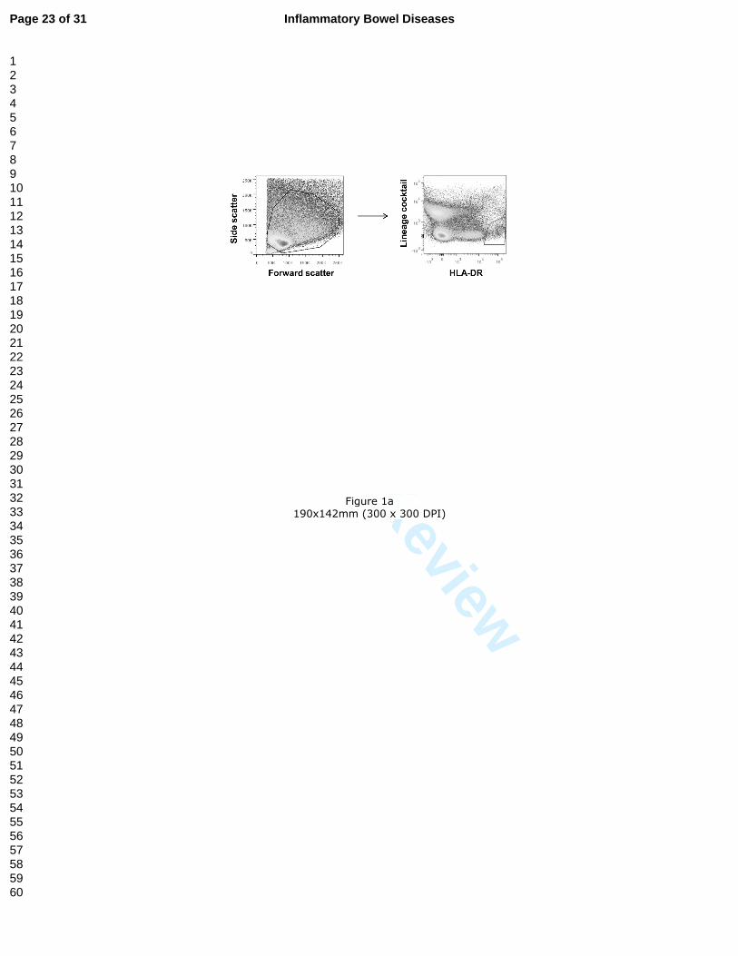
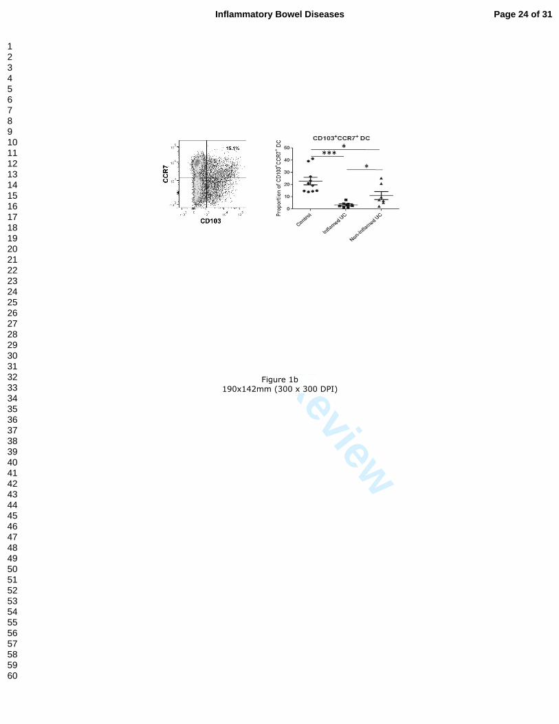
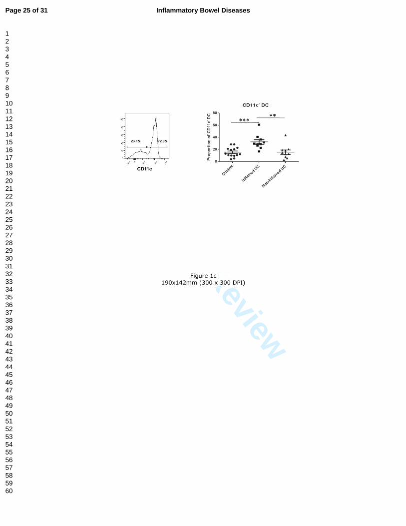
Loss of “regulatory” CD103+CCR7+ DC in UC Intestinal DC were isolated and characterised from healthy controls and from patients with active UC (biopsies were taken from both inflamed and non-inflamed colon in UC for phenotyping purposes). Human intestinal DC were identified as HLA-DR+/lineage cocktail (CD3/CD14/CD16/CD19/CD34)- cells by flow cytometry (figure 1a). In mice, CD103+ DC are migratory DC that travel to mesenteric lymph nodes (MLN) to prime gut-homing, regulatory T-cells; these cells are essential for maintenance of mucosal immune homeostasis (11). In this study, the UC colon contained a lower proportion of DC co-expressing CD103 with lymph node-homing marker CCR7 compared to the healthy colon; a greater loss was observed in the inflamed UC colon compared to the non-inflamed UC colon (figure 1b). There was a loss of total CD103+ DC from the inflamed UC colon but no differences in total CCR7+ DC between UC patients and healthy controls. Proportions of CD103+CCR7- DC were also comparable between healthy controls and UC patients (data not shown). Enhanced proportions of non-myeloid CD11c- DC in UC Although the majority of DC in the healthy human colon were CD11c+ (myeloid) cells, there was an enhanced proportion of CD11c- (non-myeloid) DC in the inflamed UC colon (figure 1c).
Alterations in DC phenotype in the UC colon
The acquisition of antigens by DC is enabled in part through their rich supply of pattern recognition receptors such as Toll-like receptors (TLRs). Bacterial recognition by TLRs on DC is one of the major pathways by which intestinal DC sample intestinal antigens. TLRs 2 and 4 were aberrantly expressed on intestinal DC in UC compared with healthy controls; in the latter, expression of both TLR2 and TLR4 was minimal. Expression of TLRs 2 and 4 was enhanced on DC from inflamed areas of the gut mucosa in UC, compared to healthy control DC (figure 2). These data support previous studies in which it has been proposed that enhanced expression of TLRs on intestinal DC in IBD may contribute to the dysregulated intestinal immune response to bacterial luminal components (15). LcS partially restored normal levels of TLR2/4 expression on gut DC from inflamed tissue in UC Colonic biopsies from healthy controls and patients with active UC (biopsies from both inflamed and non-inflamed areas for phenotyping purposes) were cultured in vitro for 20 hours in in the presence of 1x10⁷ CFU/ml heat-killed Lactobacillus casei Shirota (LcS; concentration previously optimised) or complete medium only. Conditioning gut DC from the inflamed UC colon with LcS partially restored the normal low levels of TLR2/4
Page 9 of 31 Inflammatory Bowel Diseases
123456789101112131415161718192021222324252627282930313233343536373839404142434445464748495051525354555657585960
For Peer Review
expression observed on healthy gut DC, by significantly reducing expression of both TLR2 and TLR4 (figure 2). LcS did not affect proportions of DC subsets and there were no significant effects of LcS conditioning of healthy gut DC on expression of TLRs 2 and 4 (data not shown); however this may be due to the low baseline level of expression of these receptors on human gut DC in healthy controls. LcS conditioning did not alter proportions of gut DC expressing CD103, CCR7 or CD11c in either UC patients or healthy controls (data not shown).
Reduced stimulatory capacity for T-cells by intestinal DC in UC
Intestinal DC were isolated and characterised from healthy controls and from patients with active UC (biopsies from inflamed colon only) and enriched for DC (7) prior to 5-day co-culture with CD4+ naive allogeneic T-cells (pre-sorted) from a separate, healthy blood donor in a mixed leucocyte reaction (MLR). Dividing T-cells were identified by flow cytometry post-culture as CFSElo T-cells (figure 3a). Following MLR, dose-dependent T-cell proliferative responses were generated by gut DC from both healthy controls and UC patients (figure 3b). Intestinal DC from UC patients exhibited a restricted stimulatory capacity for allogeneic T-cells, compared with their healthy control counterparts (figure 4b). LcS partially restored function of DC from inflamed tissue in UC: stimulation of T-cells For MLR experiments, DC (inflamed areas only in UC patients) were pre-conditioned with LcS, washed and then co-cultured with T-cells so that LcS cells were not in direct contact with T-cells; therefore effects of LcS observed on T-cells would be mediated via DC. Pre-conditioning UC-DC with LcS significantly enhanced their stimulatory capacity (figure 3b). Intestinal DC imprint aberrant homing properties on T-cells in UC
Following MLR, the homing profile of stimulated T-cells, imprinted by DC, was assessed via surface staining for a variety of gut- and skin-specific homing markers. Gut DC in UC induced enhanced expression of gut-homing marker CCR9 on stimulated T-cells, compared to their healthy control counterparts. Expression of gut-homing marker β7 integrin was expressed on the majority of dividing T-cells and proportions of β7+ T-cells were unchanged following stimulation with UC-DC compared to control DC (figure 4a). Levels of β7 expression measured via mean fluorescence intensity (MFI) were also unchanged between UC-DC and control DC (data not shown). Gut DC in UC induced reduced expression of skin-homing markers CLA and CCR4 on stimulated T-cells, compared to their healthy control counterparts (figure 4b).
Page 10 of 31Inflammatory Bowel Diseases
123456789101112131415161718192021222324252627282930313233343536373839404142434445464748495051525354555657585960

For Peer Review
LcS partially restored function of DC from inflamed tissue in UC: imprinting homing properties on T-cells
LcS treatment of gut DC in UC restored their ability to imprint skin-homing marker CLA on T-cells that they stimulated (figure 4b). However, there were no effects of LcS treatment on DC ability to imprint CCR4, CCR9 or β7 (figures 4a and 4b).
Intestinal DC skew the cytokine profile of stimulated T-cells in UC
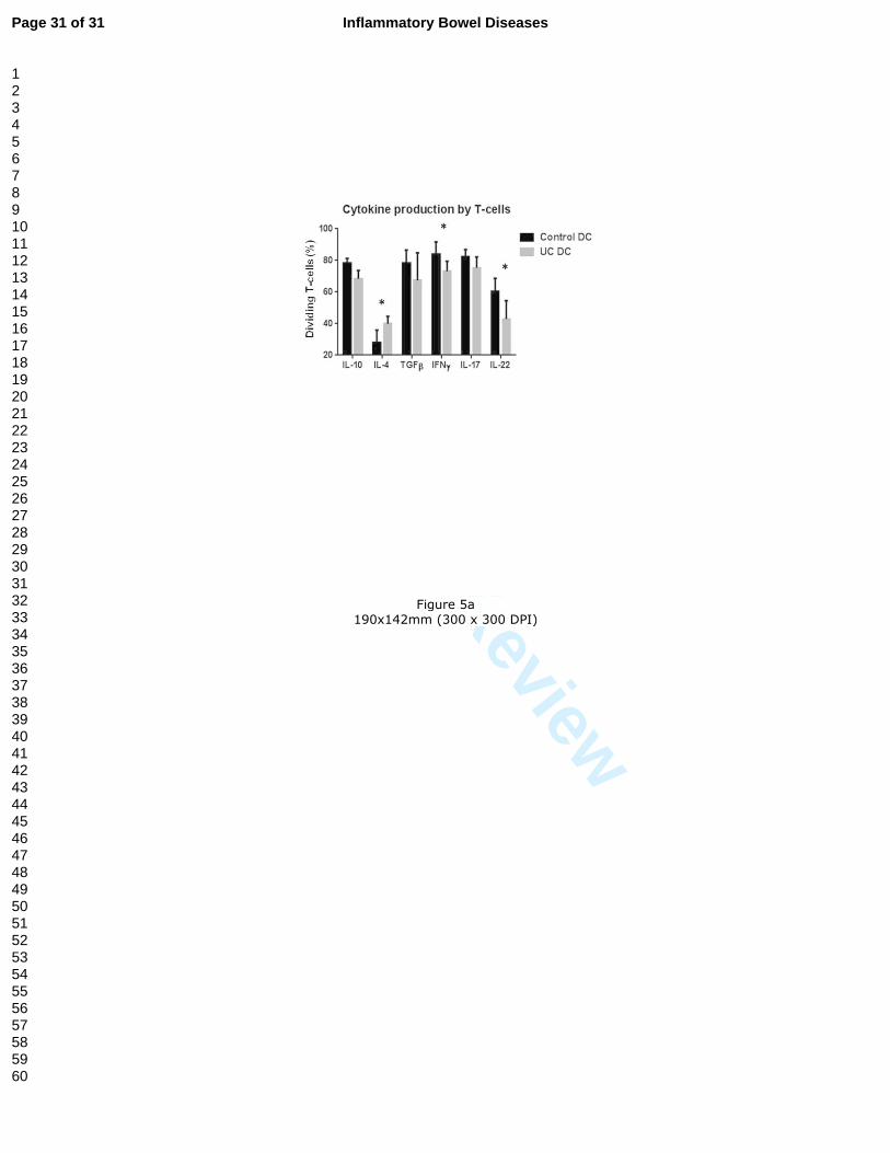
Gut DC in UC induced more IL-4 (Th2 cytokine) production, but less IFNγ (Th1 cytokine) and less IL-22 production from T-cells that they stimulated, compared to healthy control DC. Levels of IL-10, TGFβ and IL-17A produced by T-cells were unchanged regardless of type of DC stimulation (figure 5a).
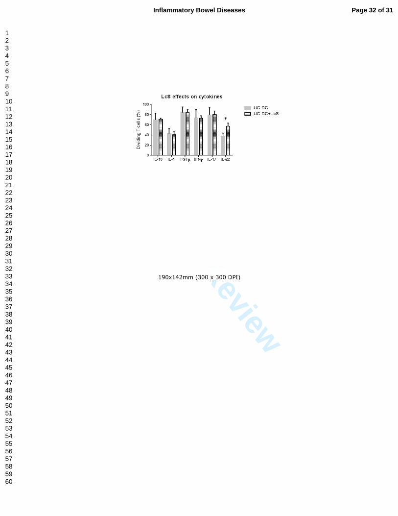
LcS partially restored function of DC from inflamed tissue in UC: cytokine production by T-cells
In a new series of paired experiments, LcS treatment of gut DC in UC restored their ability to induce IL-22 production by T-cells that they stimulated (figure 5b). However, there were no effects of LcS treatment on DC ability to induce IFNγ or IL-4 production by T-cells (or other cytokines measured; figure 5b).
Page 11 of 31 Inflammatory Bowel Diseases
123456789101112131415161718192021222324252627282930313233343536373839404142434445464748495051525354555657585960

For Peer Review
DISCUSSION We demonstrate in this study that in UC, human intestinal DC drive aberrant cytokine production by T-cells in vitro with an enhanced capacity to imprint gut-specific homing properties on T-cells, skewing gut-specific T-cell responses towards a Th2 type response. Inappropriate activation of mucosal CD4+ T-cells in IBD leads to production of T-cell cytokines, which either directly or indirectly, mediate intestinal tissue injury (26;52). A critical barrier in the development of targeted immunomodulatory therapies in IBD stems from a lack of understanding of how dysregulated T-cell responses directed against the gut microbiota are initiated in IBD. Intestinal DC recognize and respond to bacteria and bacterial products from the gut lumen and transport bacterial antigens to the MLN (31;57) to generate antigen-specific T-cell responses, strongly suggesting inappropriate T-cell responses to the gut microbiota in IBD are mediated by DC. However, constitutive migration of murine CD103+ DC to MLN generates tolerogenic T-cell responses against harmless luminal antigens and establishes oral tolerance (11;21;58). This study demonstrates a loss of CD103+ lymph-node homing DC in the UC gut, and suggests that skewing of DC subsets contributes to T-cell mediated pathogenesis in UC. In mice, this DC subset is responsible for generating regulatory T-cell responses and inducing B-cell class switching from IgM- to IgA-producing B-cells, a process critical for intestinal immune homeostasis (38). Furthermore, we demonstrate that probiotic strain LcS partially restores the normal function of intestinal DC in UC, suggesting that commensal and/or probiotic lactobacilli may directly modulate intestinal DC function to maintain intestinal immune homeostasis. These data also suggest that the reported beneficial effects of probiotic strains in IBD (13;14;28;36;43) may be mediated by gut DC due to their bacterial sampling function (18). Gut DC play a central role in immune homeostasis in the gut (10) and exhibit tolerogenic properties (15;45) that are likely to be due to the high antigenic load at intestinal sites, including food antigens and the commensal microbiota. However, these data provide a role for CD11c- non-myeloid DC in UC pathogenesis. The infiltration of CD11c- DC into inflamed tissue in the UC colon is likely to contribute to the reduced T-cell stimulatory capacity of the total intestinal DC population in UC, given the restricted stimulatory capacity of CD11c- DC compared with their CD11c+ (myeloid) counterparts (39). The CD11c- DC in the UC gut may represent plasmacytoid DC (pDC), which have been reported to be increased in human gut mucosa MLN in IBD (6) and which are a source of pro-inflammatory cytokines in the intestinal gut mucosa (20). Some CD11c-
DC may also represent CD56+CD11c- DC, which have previously been reported to infiltrate both inflamed and non-inflamed areas of the UC gut (39). Further studies will determine the subpopulations and functions of these CD11c- DC in UC. The enhanced TLR expression on total intestinal DC in UC have implications in enhanced ability to recognize and respond to bacterial antigens and potentially generating inappropriate T-cell responses to the gut microbiota. The enhanced ability of gut DC in UC to imprint gut-homing markers on T-cells, taken with the reduced capacity to imprint skin-homing markers reflects the dysregulated function of intestinal DC in UC, and may partially account for the increased infiltrates of
Page 12 of 31Inflammatory Bowel Diseases
123456789101112131415161718192021222324252627282930313233343536373839404142434445464748495051525354555657585960
For Peer Review
gut-homing T-cells at sites of inflammation in UC (16;17). Indeed, we have previously demonstrated fresh gut T-cells and those stimulated by gut DC in vitro express β7 by default, but that a gut-homing profile is conferred by loss of skin-homing markers (32). Our previous study demonstrated blood DC from UC patients have an enhanced ability to generate skin-homing T-cells (34). Taken together, these data demonstrate dysregulated DC function in UC occurs systemically and not just at intestinal sites. Furthermore, these data provide a potential explanation for the occurrence of infiltrating gut-homing T-cells at intestinal sites in IBD, yet the propensity of these patients to develop cutaneous manifestations of IBD (35). The enhanced ability of DC from the UC gut to generate IL-4 producing T-cells, with a loss of IFNγ, supports evidence of UC being a Th2-dominated disease (8) but implicates DC as the main drivers of dysregulated T-cell responses in UC. The loss of IL-22 production by T-cells stimulated by gut DC in UC has implications for reduced epithelial barrier maintenance and repair (48), a process which is dysfunctional in UC and can ultimately lead to bacterial translocation (41). This study suggest that luminal bacteria may directly or indirectly modulate intestinal DC function to contribute to intestinal immune homeostasis, and that the beneficial effects of probiotic strains in IBD (13;14;28;36;43) may be at least partially mediated by gut DC. In this case, immunomodulation of gut DC by probiotic LcS in vitro had immunoregulatory effects on T-cell responses generated. The restricted stimulatory capacity of DC for T-cells in UC was partially restored via LcS conditioning of gut DC. LcS also restored the ability of DC in UC to generate T-cells expressing skin-homing marker CLA and producing IL-22, with implications in vivo for reduced generation of pathogenic gut-specific T-cell responses, and epithelial barrier repair. These data support murine studies in which Lactobacillus ameliorated symptoms of colitis in mice in an IL-22 dependent fashion (54). In summary, our data demonstrate alterations in human intestinal DC in UC that drive aberrant T-cell responses skewed towards a Th2 profile, with an abnormal homing profile, and loss of cytokines involved in epithelial barrier maintenance. These data provide evidence that bacteria can modulate human intestinal DC function, generating a knock on effect on T-cell responses. Understanding mechanisms by which gut DC drive T-cell responses in IBD, and how these T-cell responses contribute to inflammation and pathology is of critical importance for determining how the intestinal immune system responds inappropriately to the commensal microbiota. Manipulation of the composition of the gut microbiota may be beneficial in IBD; however details of which bacterial strains confer health benefits are only slowly being defined. Furthermore, recent evidence suggests some probiotic strains may even be harmful for use in inflammatory conditions (56). DC, which are so pivotal in early bacterial recognition, tolerance induction, and shaping T-cell responses, are central to immunomodulation by probiotic bacteria and the commensal microbiota. Manipulation of DC to allow generation of DC-specific therapy may be beneficial in IBD and would provide novel therapeutic opportunities, minimising systemic side effects and immunosuppression.
Page 13 of 31 Inflammatory Bowel Diseases
123456789101112131415161718192021222324252627282930313233343536373839404142434445464748495051525354555657585960

For Peer Review
Page 14 of 31Inflammatory Bowel Diseases
123456789101112131415161718192021222324252627282930313233343536373839404142434445464748495051525354555657585960
For Peer Review
FIGURE LEGENDS Figure 1. Loss of CD103+CCR7+ DC from the UC colon and infiltrating CD11c- DC. a) FACS dot plots demonstrating identification of human intestinal DC according to forward scatter versus side scatter, and subsequent gating strategy to select HLA-DR+Lineage cocktail (CD3/CD14/CD16/CD19/CD34)- cells. b) FACS dot plots showing examples of CD103/CCR7 co-expression on gut DC (regions set based on isotype controls) and summary graph demonstrating proportions of intestinal DC co-expressing CD103 and CCR7 from biopsies obtained from healthy controls (n=10), inflamed UC colon (n=7) and non-inflamed UC colon (n=7). T-test (control versus UC) or paired T-test (inflamed UC versus non-inflamed UC) was carried out: *p<0.05, ***p<0.001. c) FACS histograms showing examples of CD11c expression (regions sets using isotype controls) and summary graph demonstrating proportions of CD11c- (non-myeloid) intestinal DC from biopsies obtained from healthy controls (n=14), inflamed UC colon (n=10) and non-inflamed UC colon (n=10). T-test (control versus UC) or paired T-test (inflamed UC versus non-inflamed UC) was carried out: **p<0.01, ***p<0.001. Figure 2. Enhanced TLR2 and TLR4 expression on intestinal DC in UC and reduction of TLR expression with LcS conditioning. FACS histograms showing examples of TLR2 and TLR4 expression (regions set using isotype controls) and summary graphs demonstrating proportions of intestinal DC expressing TLR2 and TLR4 from biopsies obtained from healthy controls (TLR2: n=11; TLR4: n=11), and inflamed UC +/- LcS (TLR2: n=9; TLR4: n=9). T-test (control versus UC) or paired T-test (inflamed UC +/- LcS) was carried out: *p<0.05. . Figure 3. Reduced T-cell stimulatory capacity of intestinal DC in UC and partial restoration with LcS conditioning. a) FACS plots showing identification of dividing T-cells post DC stimulation in MLR, via gating on blasting lymphocytes according to forward scatter versus side scatter plots and subsequent selection of CD3+CFSElo cells. T-cells were sorted as CD4+ naive (CD45RO-) T-cells, and CFSE labelled pre-culture. b) Summary graph demonstrating proportions of (the same pool of) dividing T-cells stimulated by either control DC (n=5) or DC from the inflamed UC colon +/-LcS (n=5). T-cells were stimulated at DC doses of 0%, 1%, 2% and 3% of the number of T-cells in both cases. Repeated measures two-way ANOVA was carried out: UC DC were significantly less stimulatory for T-cells than control DC, at 3% (**p<0.01), LcS conditioned UC DC were significantly more stimulatory than UC DC at 3% (*p<0.05).
Figure 4. Altered imprinting of homing molecules on T-cells by intestinal DC in UC and effects of LcS conditioning FACS histograms demonstrating examples of stimulated T-cells expressing tissue-specific homing markers and summary graphs demonstrating proportions of (the same pool of) dividing T-cells expressing a) gut-homing markers β7 and CCR9, and b) skin-homing markers CLA and CCR4, following stimulation by either control DC (n=5) or DC from the inflamed UC colon +/- LcS (n=5), at a DC concentration of 3% (of total T-cells). Paired T-test was carried out (*p<0.05, **p<0.01).
Page 15 of 31 Inflammatory Bowel Diseases
123456789101112131415161718192021222324252627282930313233343536373839404142434445464748495051525354555657585960
For Peer Review
Figure 5. Intestinal DC alter T-cell production of cytokines in UC and LcS restoration of DC ability to induce T-cell IL-22 production a) Summary graph demonstrating proportions of (the same pool of) dividing T-cells producing cytokines IL-10, IL-4, TGFβ, IFNγ, IL-17 and IL-22 following stimulation by either control DC (n=5) or DC from the inflamed UC colon (n=5), at a DC concentration of 3% (of total T-cells). Paired T-test was carried out (*p<0.05). b) Summary graph demonstrating proportions of (the same pool of) dividing T-cells producing cytokines IL-10, IL-4, TGFβ, IFNγ, IL-17 and IL-22 following stimulation by DC from the inflamed UC colon +/- LcS (n=5), at a DC concentration of 3% (of total T-cells). Paired T-test was carried out (*p<0.05).
Page 16 of 31Inflammatory Bowel Diseases
123456789101112131415161718192021222324252627282930313233343536373839404142434445464748495051525354555657585960

For Peer Review
ACKNOWLEDGEMENTS
This research was funded by Yakult Europe B.V. (Almere, The Netherlands). The Lactobacillus casei Shirota strain was provided by Yakult Honsha Co. Ltd (Tokyo, Japan). DB. SCK and NRE were funded by Biotechnology and Biological Research Council Institute Strategic Program for Gut Health and Food Safety BB/J004529/1
Page 17 of 31 Inflammatory Bowel Diseases
123456789101112131415161718192021222324252627282930313233343536373839404142434445464748495051525354555657585960
For Peer Review
Reference List
1. Abraham C, Cho JH. Inflammatory bowel disease. N Engl J Med. 2009;361:2066-2078.
2. Arihiro S, Ohtani H, Suzuki M, et al. Differential expression of mucosal addressin cell
adhesion molecule-1 (MAdCAM-1) in ulcerative colitis and Crohn's disease. Pathol Int.
2002;52:367-374.
3. Bamias G, Cominelli F. Immunopathogenesis of inflammatory bowel disease: current
concepts. Curr Opin Gastroenterol. 2007;23:365-369.
4. Banchereau J, Steinman RM. Dendritic cells and the control of immunity. Nature.
1998;392:245-252.
5. Baumgart DC, Carding SR. Inflammatory bowel disease: cause and immunobiology.
Lancet. 2007;369:1627-1640.
6. Baumgart DC, Metzke D, Guckelberger O, et al. Aberrant plasmacytoid dendritic cell
distribution and function in patients with Crohn's disease and ulcerative colitis. Clin Exp
Immunol. 2011;166:46-54.
7. Bell SJ, Rigby R, English N, et al. Migration and maturation of human colonic dendritic
cells. J Immunol. 2001;166:4958-4967.
8. Bouma G, Strober W. The immunological and genetic basis of inflammatory bowel
disease. Nat Rev Immunol. 2003;3:521-533.
9. Briskin M, Winsor-Hines D, Shyjan A, et al. Human mucosal addressin cell adhesion
molecule-1 is preferentially expressed in intestinal tract and associated lymphoid tissue.
Am J Pathol. 1997;151:97-110.
10. Chirdo FG, Millington OR, Beacock-Sharp H, et al. Immunomodulatory dendritic cells in
intestinal lamina propria. Eur J Immunol. 2005;35:1831-1840.
11. Coombes JL, Maloy KJ. Control of intestinal homeostasis by regulatory T cells and
dendritic cells. Semin Immunol. 2007;19:116-126.
12. Farache J, Zigmond E, Shakhar G, et al. Contributions of dendritic cells and macrophages
to intestinal homeostasis and immune defense. Immunol Cell Biol. 2013;91:232-239.
Page 18 of 31Inflammatory Bowel Diseases
123456789101112131415161718192021222324252627282930313233343536373839404142434445464748495051525354555657585960

For Peer Review
13. Gionchetti P, Rizzello F, Helwig U, et al. Prophylaxis of pouchitis onset with probiotic
therapy: a double-blind, placebo-controlled trial. Gastroenterology. 2003;124:1202-1209.
14. Gionchetti P, Rizzello F, Venturi A, et al. Oral bacteriotherapy as maintenance treatment
in patients with chronic pouchitis: a double-blind, placebo-controlled trial.
Gastroenterology. 2000;119:305-309.
15. Hart AL, Al-Hassi HO, Rigby RJ, et al. Characteristics of intestinal dendritic cells in
inflammatory bowel diseases. Gastroenterology. 2005;129:50-65.
16. Hart AL, Kamm MA, Knight SC, et al. Prospective evaluation of intestinal homing
memory T cells in ulcerative colitis. Inflamm Bowel Dis. 2004;10:496-503.
17. Hart AL, Kamm MA, Knight SC, et al. Quantitative and functional characteristics of
intestinal-homing memory T cells: analysis of Crohn's disease patients and healthy
controls. Clin Exp Immunol. 2004;135:137-145.
18. Hart AL, Lammers K, Brigidi P, et al. Modulation of human dendritic cell phenotype and
function by probiotic bacteria. Gut. 2004;53:1602-1609.
19. Hart DN. Dendritic cells: unique leukocyte populations which control the primary
immune response. Blood. 1997;90:3245-3287.
20. Hostmann A, Kapp K, Beutner M, et al. Dendritic cells from human mesenteric lymph
nodes in inflammatory and non-inflammatory bowel diseases: subsets and function of
plasmacytoid dendritic cells. Immunology. 2013;139:100-108.
21. Jaensson E, Uronen-Hansson H, Pabst O, et al. Small intestinal CD103+ dendritic cells
display unique functional properties that are conserved between mice and humans. J Exp
Med. 2008;205:2139-2149.
22. Johansson-Lindbom B, Svensson M, Pabst O, et al. Functional specialization of gut
CD103+ dendritic cells in the regulation of tissue-selective T cell homing. J Exp Med.
2005;202:1063-1073.
23. Johansson-Lindbom B, Svensson M, Wurbel MA, et al. Selective generation of gut tropic
T cells in gut-associated lymphoid tissue (GALT): requirement for GALT dendritic cells
and adjuvant. J Exp Med. 2003;198:963-969.
24. Karlis J, Penttila I, Tran TB, et al. Characterization of colonic and mesenteric lymph node
dendritic cell subpopulations in a murine adoptive transfer model of inflammatory bowel
disease. Inflamm Bowel Dis. 2004;10:834-847.
25. Kaser A, Ludwiczek O, Holzmann S, et al. Increased expression of CCL20 in human
inflammatory bowel disease. J Clin Immunol. 2004;24:74-85.
26. Kaser A, Zeissig S, Blumberg RS. Inflammatory bowel disease. Annu Rev Immunol.
2010;28:573-621.
Page 19 of 31 Inflammatory Bowel Diseases
123456789101112131415161718192021222324252627282930313233343536373839404142434445464748495051525354555657585960

For Peer Review
27. Krajina T, Leithauser F, Moller P, et al. Colonic lamina propria dendritic cells in mice
with CD4+ T cell-induced colitis. Eur J Immunol. 2003;33:1073-1083.
28. Kruis W, Fric P, Pokrotnieks J, et al. Maintaining remission of ulcerative colitis with the
probiotic Escherichia coli Nissle 1917 is as effective as with standard mesalazine. Gut.
2004;53:1617-1623.
29. Leach MW, Bean AG, Mauze S, et al. Inflammatory bowel disease in C.B-17 scid mice
reconstituted with the CD45RBhigh subset of CD4+ T cells. Am J Pathol.
1996;148:1503-1515.
30. Levine A, Griffiths A, Markowitz J, et al. Pediatric modification of the Montreal
classification for inflammatory bowel disease: the Paris classification. Inflamm Bowel
Dis. 2011;17:1314-1321.
31. Macpherson AJ, Uhr T. Induction of protective IgA by intestinal dendritic cells carrying
commensal bacteria. Science. 2004;303:1662-1665.
32. Mann ER, Bernardo D, Al-Hassi HO, et al. Human gut-specific homeostatic dendritic
cells are generated from blood precursors by the gut microenvironment. Inflamm Bowel
Dis. 2012;18:1275-1286.
33. Mann ER, Landy JD, Bernardo D, et al. Intestinal dendritic cells: their role in intestinal
inflammation, manipulation by the gut microbiota and differences between mice and
men. Immunol Lett. 2013;150:30-40.
34. Mann ER, You J, Horneffer-van der Sluis V, et al. Dysregulated circulating dendritic cell
function in ulcerative colitis is partially restored by probiotic strain Lactobacillus casei
Shirota. Mediators Inflamm. 2013;2013:573576.
35. Marzano AV, Borghi A, Stadnicki A, et al. Cutaneous Manifestations in Patients With
Inflammatory Bowel Diseases: Pathophysiology, Clinical Features, and Therapy.
Inflamm Bowel Dis. 2013.
36. Mimura T, Rizzello F, Helwig U, et al. Once daily high dose probiotic therapy (VSL#3)
for maintaining remission in recurrent or refractory pouchitis. Gut. 2004;53:108-114.
37. Mora JR, Bono MR, Manjunath N, et al. Selective imprinting of gut-homing T cells by
Peyer's patch dendritic cells. Nature. 2003;424:88-93.
38. Mora JR, Iwata M, Eksteen B, et al. Generation of gut-homing IgA-secreting B cells by
intestinal dendritic cells. Science. 2006;314:1157-1160.
39. Ng SC, Plamondon S, Al-Hassi HO, et al. A novel population of human CD56+ human
leucocyte antigen D-related (HLA-DR+) colonic lamina propria cells is associated with
inflammation in ulcerative colitis. Clin Exp Immunol. 2009;158:205-218.
Page 20 of 31Inflammatory Bowel Diseases
123456789101112131415161718192021222324252627282930313233343536373839404142434445464748495051525354555657585960

For Peer Review
40. Niess JH. Role of mucosal dendritic cells in inflammatory bowel disease. World J
Gastroenterol. 2008;14:5138-5148.
41. Porras M, Martin MT, Yang PC, et al. Correlation between cyclical epithelial barrier
dysfunction and bacterial translocation in the relapses of intestinal inflammation.
Inflamm Bowel Dis. 2006;12:843-852.
42. Sartor RB. Cytokine regulation of experimental intestinal inflammation in genetically
engineered and T-lymphocyte reconstituted rodents. Aliment Pharmacol Ther. 1996;10
Suppl 2:36-42.
43. Sartor RB. Therapeutic manipulation of the enteric microflora in inflammatory bowel
diseases: antibiotics, probiotics, and prebiotics. Gastroenterology. 2004;126:1620-1633.
44. Schulz O, Jaensson E, Persson EK, et al. Intestinal CD103+, but not CX3CR1+, antigen
sampling cells migrate in lymph and serve classical dendritic cell functions. J Exp Med.
2009;206:3101-3114.
45. Scott CL, Aumeunier AM, Mowat AM. Intestinal CD103+ dendritic cells: master
regulators of tolerance? Trends Immunol. 2011;32:412-419.
46. Sokol H, Seksik P. The intestinal microbiota in inflammatory bowel diseases: time to
connect with the host. Curr Opin Gastroenterol. 2010;26:327-331.
47. Sommer F, Backhed F. The gut microbiota--masters of host development and physiology.
Nat Rev Microbiol. 2013;11:227-238.
48. Sonnenberg GF, Fouser LA, Artis D. Functional biology of the IL-22-IL-22R pathway in
regulating immunity and inflammation at barrier surfaces. Adv Immunol. 2010;107:1-29.
49. Stagg AJ, Kamm MA, Knight SC. Intestinal dendritic cells increase T cell expression of
alpha4beta7 integrin. Eur J Immunol. 2002;32:1445-1454.
50. Steinman RM. Dendritic cells: understanding immunogenicity. Eur J Immunol. 2007;37
Suppl 1:S53-S60.
51. Strober W, Fuss I, Mannon P. The fundamental basis of inflammatory bowel disease. J
Clin Invest. 2007;117:514-521.
52. Strober W, Fuss IJ. Proinflammatory cytokines in the pathogenesis of inflammatory
bowel diseases. Gastroenterology. 2011;140:1756-1767.
53. Strober W, Fuss IJ, Blumberg RS. The immunology of mucosal models of inflammation.
Annu Rev Immunol. 2002;20:495-549.
54. Takamura T, Harama D, Fukumoto S, et al. Lactobacillus bulgaricus OLL1181 activates
the aryl hydrocarbon receptor pathway and inhibits colitis. Immunol Cell Biol.
2011;89:817-822.
Page 21 of 31 Inflammatory Bowel Diseases
123456789101112131415161718192021222324252627282930313233343536373839404142434445464748495051525354555657585960
For Peer Review
55. te Velde AA, van KY, Braat H, et al. Increased expression of DC-SIGN+IL-12+IL-18+
and CD83+IL-12-IL-18- dendritic cell populations in the colonic mucosa of patients with
Crohn's disease. Eur J Immunol. 2003;33:143-151.
56. Tsilingiri K, Barbosa T, Penna G, et al. Probiotic and postbiotic activity in health and
disease: comparison on a novel polarised ex-vivo organ culture model. Gut.
2012;61:1007-1015.
57. Voedisch S, Koenecke C, David S, et al. Mesenteric lymph nodes confine dendritic cell-
mediated dissemination of Salmonella enterica serovar Typhimurium and limit systemic
disease in mice. Infect Immun. 2009;77:3170-3180.
58. Worbs T, Bode U, Yan S, et al. Oral tolerance originates in the intestinal immune system
and relies on antigen carriage by dendritic cells. J Exp Med. 2006;203:519-527.
59. Xavier RJ, Podolsky DK. Unravelling the pathogenesis of inflammatory bowel disease.
Nature. 2007;448:427-434.
Page 22 of 31Inflammatory Bowel Diseases
123456789101112131415161718192021222324252627282930313233343536373839404142434445464748495051525354555657585960
For Peer Review
Figure 1a
190x142mm (300 x 300 DPI)
Page 23 of 31 Inflammatory Bowel Diseases
123456789101112131415161718192021222324252627282930313233343536373839404142434445464748495051525354555657585960
For Peer Review
Figure 1b
190x142mm (300 x 300 DPI)
Page 24 of 31Inflammatory Bowel Diseases
123456789101112131415161718192021222324252627282930313233343536373839404142434445464748495051525354555657585960
For Peer Review
Figure 1c
190x142mm (300 x 300 DPI)
Page 25 of 31 Inflammatory Bowel Diseases
123456789101112131415161718192021222324252627282930313233343536373839404142434445464748495051525354555657585960

For Peer Review
Figure 2
190x142mm (300 x 300 DPI)
Page 26 of 31Inflammatory Bowel Diseases
123456789101112131415161718192021222324252627282930313233343536373839404142434445464748495051525354555657585960

For Peer Review
Figure 3a
190x142mm (300 x 300 DPI)
Page 27 of 31 Inflammatory Bowel Diseases
123456789101112131415161718192021222324252627282930313233343536373839404142434445464748495051525354555657585960

For Peer Review
Figure 3b
190x142mm (300 x 300 DPI)
Page 28 of 31Inflammatory Bowel Diseases
123456789101112131415161718192021222324252627282930313233343536373839404142434445464748495051525354555657585960

For Peer Review
Figure 4a
190x142mm (300 x 300 DPI)
Page 29 of 31 Inflammatory Bowel Diseases
123456789101112131415161718192021222324252627282930313233343536373839404142434445464748495051525354555657585960

For Peer Review
Figure 4b
190x142mm (300 x 300 DPI)
Page 30 of 31Inflammatory Bowel Diseases
123456789101112131415161718192021222324252627282930313233343536373839404142434445464748495051525354555657585960
For Peer Review
Figure 5a
190x142mm (300 x 300 DPI)
Page 31 of 31 Inflammatory Bowel Diseases
123456789101112131415161718192021222324252627282930313233343536373839404142434445464748495051525354555657585960
For Peer Review
190x142mm (300 x 300 DPI)
Page 32 of 31Inflammatory Bowel Diseases
123456789101112131415161718192021222324252627282930313233343536373839404142434445464748495051525354555657585960