Inflammatory bowel disease
32
Inflammatory Bowel Diseases Alka Ingnam
-
Upload
alka-ingnam -
Category
Health & Medicine
-
view
152 -
download
0
Transcript of Inflammatory bowel disease

Inflammatory Bowel Diseases
Alka Ingnam

Inflammatory bowel disease (IBD)
Refers to two chronic inflammatory GI disorders: regional enteritis (i.e. crohn's disease)
&ulcerative colitis.
Both have striking similarities but as the same time several differences.

Incidence In the US, has increased in the past
century: over 30,000 new cases occur annually.
15 and 30 years of age at the greatest risk, followed by people between 50 and 70.
Women and men are equally affected.

Aetiology• Unknown• Believed is caused by a
combination of these factors: Immune system problemsGeneticsEnvironmental factors
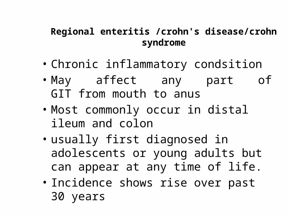
Regional enteritis /crohn's disease/crohn syndrome
• Chronic inflammatory condsition • May affect any part of GIT from mouth to
anus• Most commonly occur in distal ileum and
colon• usually first diagnosed in adolescents or
young adults but can appear at any time of life.
• Incidence shows rise over past 30 years

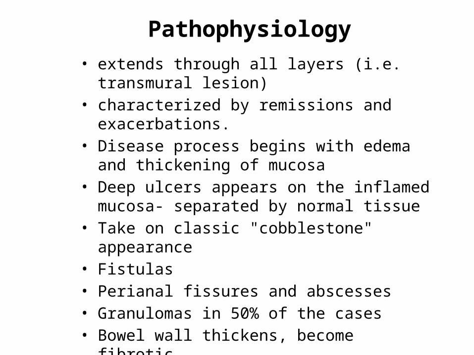
Pathophysiology• extends through all layers (i.e. transmural lesion)• characterized by remissions and exacerbations.• Disease process begins with edema and thickening
of mucosa• Deep ulcers appears on the inflamed mucosa-
separated by normal tissue• Take on classic "cobblestone" appearance • Fistulas• Perianal fissures and abscesses • Granulomas in 50% of the cases• Bowel wall thickens, become fibrotic• Intestinal lumen narrows
Cobblestone mucosa

Clinical features
• Unpredictable exacerbations and remissions.
• Quality of life is diminished • Overall mortality is 2x of general populationFeatures depend on site and extent of bowel
affected:• Abdominal pain
Most frequently in Rt lower quadrant due to involvement of terminal ileum and Rt side of colon
Crampy abdominal pain, usually after food
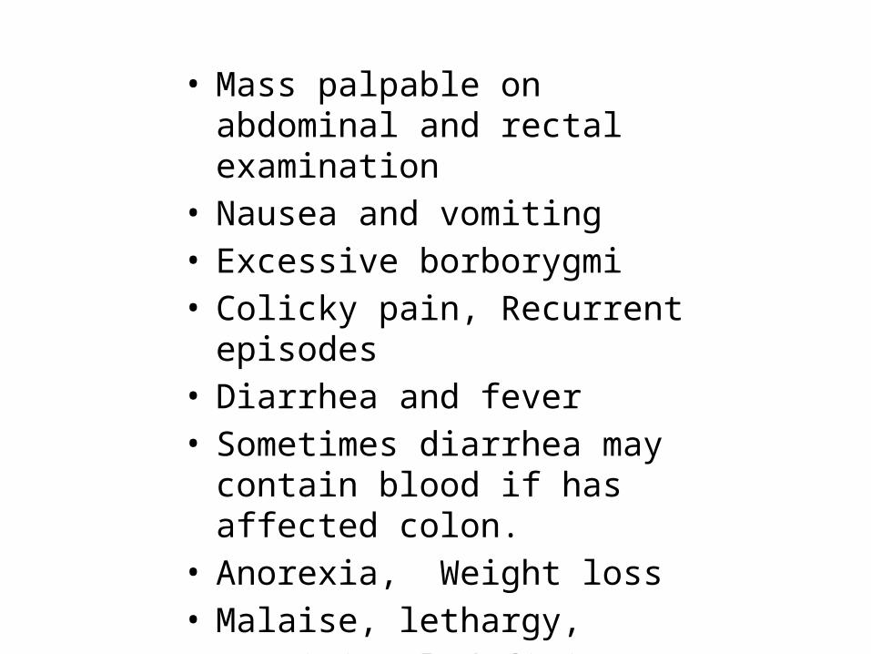
• Mass palpable on abdominal and rectal examination
• Nausea and vomiting • Excessive borborygmi• Colicky pain, Recurrent episodes• Diarrhea and fever • Sometimes diarrhea may contain
blood if has affected colon.• Anorexia, Weight loss• Malaise, lethargy, • Nutritional deficiency , anemia • Psychosocial issues
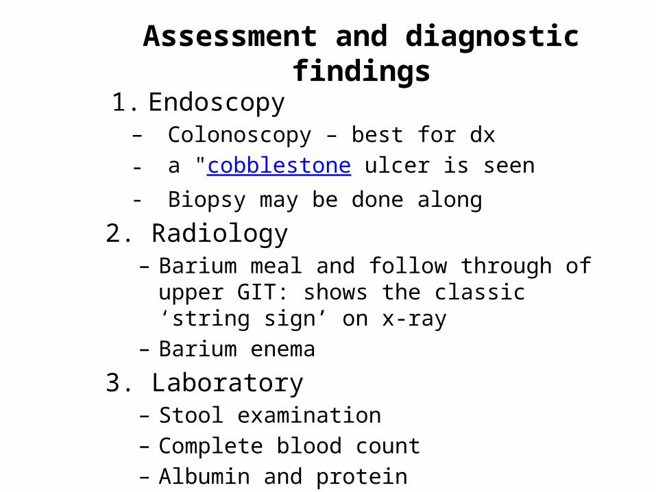
Assessment and diagnostic findings
1. Endoscopy– Colonoscopy – best for dx- a "cobblestone ulcer is seen- Biopsy may be done along
2. Radiology– Barium meal and follow through of upper GIT: shows
the classic ‘string sign’ on x-ray– Barium enema
3. Laboratory– Stool examination– Complete blood count– Albumin and protein

String sign on barium study

Complications
• Intestinal obstruction/ stricture formation
• Perianal disease• Fluid and electrolyte imbalance• Malnutrition• Fistula and abscess. Commonly
enterocutaneous fistula• Colon cancer: increased risk

Ulcerative colitis• Recurrent ulcerative and inflammatory
disease of the mucosal and submucosal layers of the colon and rectum.
• Peak incidence is between 30 and 50 years of age
• Serious disease accompanied by systemic complications and a high mortality rate.
• 10 to 15 % of these patients develop colon carcinoma
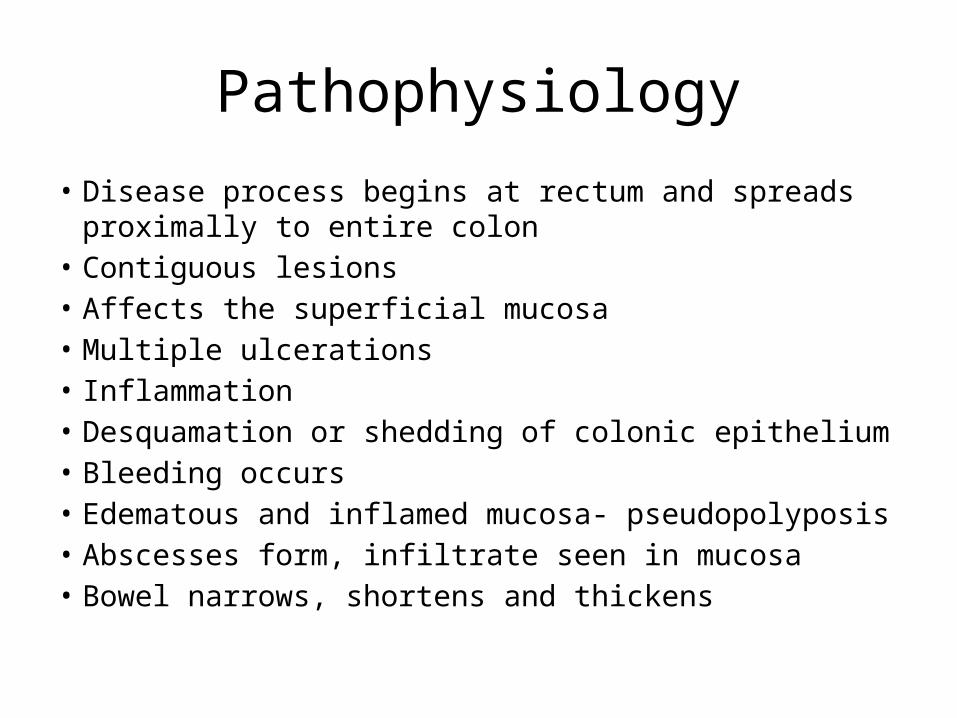
Pathophysiology
• Disease process begins at rectum and spreads proximally to entire colon
• Contiguous lesions • Affects the superficial mucosa• Multiple ulcerations• Inflammation• Desquamation or shedding of colonic epithelium• Bleeding occurs• Edematous and inflamed mucosa- pseudopolyposis• Abscesses form, infiltrate seen in mucosa• Bowel narrows, shortens and thickens

Clinical features
• Bloody diarrhea is the hallmark• Lower left quadrant abdominal pain• Tenesmus • Rectal bleeding• Anorexia, weight loss, fever, vomiting and dehydration• Cramping• Fecal urgency• Passage of 10-20 liquid stools each day.• Hypocalcemia and anemia • Rebound tenderness in right lower quadrant• Extraintestinal symptoms such as skin lesion (erythema
nodosum), eye lesions(uveitis), joint abnormalities(eg. arthritis) and liver disease
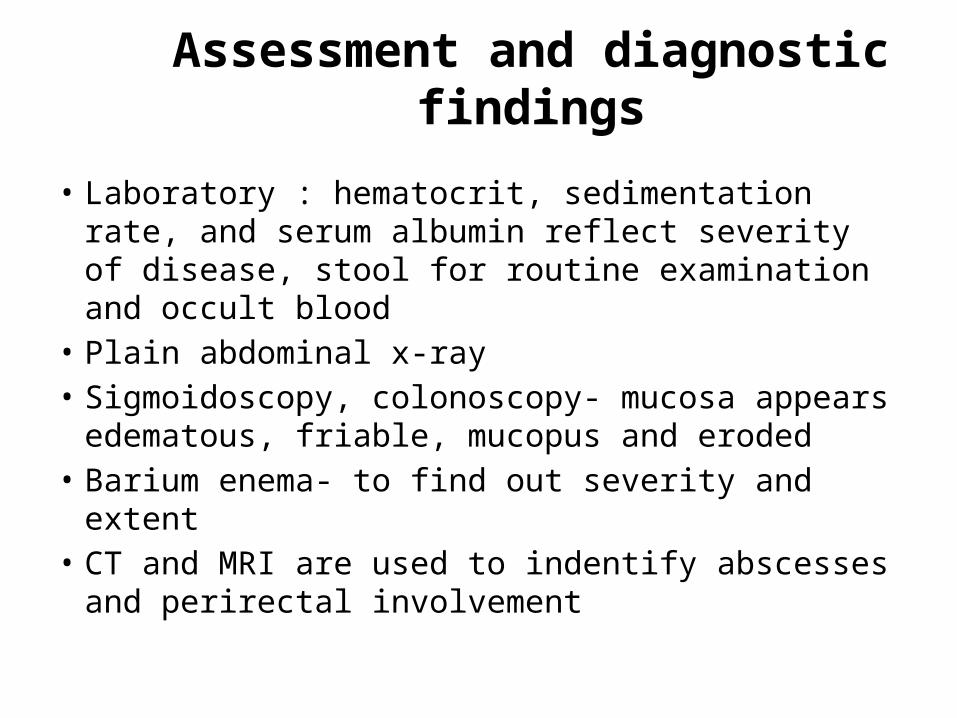
Assessment and diagnostic findings
• Laboratory : hematocrit, sedimentation rate, and serum albumin reflect severity of disease, stool for routine examination and occult blood
• Plain abdominal x-ray • Sigmoidoscopy, colonoscopy- mucosa appears
edematous, friable, mucopus and eroded• Barium enema- to find out severity and extent• CT and MRI are used to indentify abscesses and
perirectal involvement

Complications
• Toxic megacolon• Perforation and hemorrhage• Peritonitis• Nutritional deficiency• Colon cancer• Osteoporotic fractures- due to
decreased bone mineral density
Medical management of chronic IBD
Focuses on:• reducing inflammation• suppressing inappropriate immune
responses• providing rest for the diseased bowel• improving quality of life• preventing or minimizing complications

Nutritional therapy
• Oral fluids• Low residue, high protein and high calorie
diet• Supplemental vitamin• Iron replacement• IV therapy in fluid and electrolyte
imbalances• Avoid milk in those with lactose intolerance• Avoid cold food and smoking • Parenteral nutrition

Pharmacologic therapy
1. Aminosalicylate formulations; sulfasalazine (azulfidine)
- treat inflammation and prevent recurrences
2. Antibiotics; metronidazole & ciprofloxacin- In secondary infections
3. Corticosteroids- suppress the acute clinical symptoms
4. Immunomodulators; (eg. Azathioprene [Imuran], 6mercaptopurine, methotrexate)
- Alter immune response

Surgical management
• Total colectomy and ileostomy- procedure of choice for crohn’s disease.
• Intestinal transplant- not a cure, to improve quality of life for some who are terminally ill.
• Almost 25% of ulcerative colitis patients require surgery. Surgical excision usually improves life.
• Proctocolectomy with ileostomy• Ileoanal anastomosis• Strictureplasty- blocked and narrowed section of
bowel is widened, leaving the bowel intact

Nursing Management
Nursing ProcessAssessment
Health history regarding ◦Abdominal pain: onset,
duration and characteristics◦presence of diarrhea, ◦fecal urgency, tenesmus, ◦nausea and vomiting, ◦anorexia, weight loss, ◦family history of IBD.

• Dietary patterns• Patterns of bowel elimination: – character, frequency– presence of blood, pus, fat or mucus,
• Allergies and food intolerance especially milk.• Sleep disturbances if diarrhea and pain occurs
at night• Assess abdomen: – bowel sound– tenderness or pain, – distension, – evidence of fistula, – signs of dehydration
• Assess stool, rectal bleeding

Nursing Diagnoses
1. Acute pain related to increased peristalsis and GI inflammation
2. Diarrhea related to the inflammatory process3. Fluid volume deficit related to anorexia, nausea
and diarrhea4. Anxiety related to impending surgery 5. Imbalanced nutrition, less than body
requirements related to dietary restrictions, nausea and malabsorption

Nursing Interventions

Relieving pain
• Identifying type of pain, onset before/after food, pattern, if relieved with medication or not?
• Position changes, heat application, diversional activities
• Prevention of fatigue to decrease pain• Administer antichonilergics as prescribed 30
min prior to food to decrease intestinal motility • Give analgesics as prescribed

Maintaining normal elimination patterns
• Identifying precipitating factors such as food, activity, stress, etc. if any and avoiding those.
• Provide ready access to bathroom, commode or bedpan
• Keep the environment clean and odor free• ‘administer anti diarrheal agents as prescribed• Record frequency and consistency of stool
before, during and after therapy• Encourage bed rest to decrease peristalsis

Maintaining fluid intake
• Record oral and IV fluids intake and outputs• Assess daily weight for fluid gains or losses• Look for signs of fluid deficit; decreased skin
turgor, oliguria, dry skin and mucosa, increased hematocrit, hypotension
• Encourage oral fluid intake and maintain IV intake• Measures to decrease diarrhea; dietary
restriction, stress reduction, anti diarrheal agents

Maintaining optimal nutrition
• Parenteral nutrition when symptoms are severe; monitors weight, fluid intake and output. The patient should gain 0.5kg daily during PN therapy
• Monitor Feedings high in protein and low in fat and residue• Note for intolerance; signs are nausea, vomiting, diarrhea
or abdominal distention• Restrict activity to conserve energy, reduce peristalsis and
reduce calorie requirements• If oral foods are tolerated; small, frequent, low residue
feedings to avoid over distending the stomach and stimulating peristalsis

ANY QUESTIONS?

Thank you