Inflammation in Age-Related Macular Degeneration ... · Inflammation in Age-Related Macular...
24
6 Inflammation in Age-Related Macular Degeneration – Implications for Therapy Mei Chen and Heping Xu Centre for Vision and Vascular Science, School of Medicine, Dentistry and Biomedical Sciences, Queen’s University Belfast, UK 1. Introduction The macula or macula lutea (originally from Latin macula, “spot” and lutea, “yellow”) is an oval-shaped spot, five mm in diameter, located temporal to the optical nerve and near the centre of the human retina. Although it comprises only a small part of the retina, it is responsible for the sharp central vision and colour vision. Age-related macular degeneration (AMD) is a disease in which the neuroretina and retinal pigment epithelia (RPE) of the macula degenerate with age resulting in profound loss of visual function. At the early stages, so called age-related maculopathy, patients present with “drusen”, the “biological waste materials” deposition, between RPE cells and the choroid especially in the macular region (Coleman et al. 2008, Jager et al. 2008). Visual acuity is normally not affected at this stage. As disease progresses to the advanced stages, patients may lose their central vision. There are two forms of advanced AMD: dry and wet. Dry-AMD also called central geographic atrophy. Apoptosis of RPE cells and subsequent death of photoreceptors in the macula underlie the pathology of dry-AMD (Coleman et al. 2008). “Wet” AMD refers to the neovascular or exudative form of the disease and is associated with rapid vision loss caused by the infiltration of abnormal blood vessels from the choroid into the subretinal space leading to haemorrhage, leakage of fluid and eventual scar tissue formation (Chopdar et al. 2003, Coleman et al. 2008). Dry-AMD is more common than wet-AMD accounting for nearly 85~90% of AMD cases; however, wet-AMD contributes to 90% of severe vision loss resulting from AMD (Chopdar et al. 2003). In addition, AMD is generally thought to progress along a continuum from atrophic or dry-AMD to neovascular (wet) AMD with approximately 10- 15% of all AMD patients eventually developing the wet form (Sunness et al. 1999). Occasionally, patients can also present with exudative (wet) AMD as the first manifestation of the condition without prior signs of dry-AMD. 1.1 The social and economic burden of age-related macular degeneration AMD is not painful. However, it affects the central vision and patients with AMD may have a distorted, or blurred vision (early AMD), or even a total loss of central vision (advanced AMD), and they cannot see things in details. It is therefore, very difficult for them to cope with daily life on their own. AMD is the largest cause of blindness in most developed countries. More than half a million people in the UK suffer from AMD with over half of all registrations of severe visual impairment attributed to the disease (Bunce www.intechopen.com
Transcript of Inflammation in Age-Related Macular Degeneration ... · Inflammation in Age-Related Macular...

6
Inflammation in Age-Related Macular Degeneration – Implications for Therapy
Mei Chen and Heping Xu Centre for Vision and Vascular Science, School of Medicine,
Dentistry and Biomedical Sciences, Queen’s University Belfast, UK
1. Introduction
The macula or macula lutea (originally from Latin macula, “spot” and lutea, “yellow”) is an oval-shaped spot, five mm in diameter, located temporal to the optical nerve and near the centre of the human retina. Although it comprises only a small part of the retina, it is responsible for the sharp central vision and colour vision. Age-related macular degeneration (AMD) is a disease in which the neuroretina and retinal pigment epithelia (RPE) of the macula degenerate with age resulting in profound loss of visual function. At the early stages, so called age-related maculopathy, patients present with “drusen”, the “biological waste materials” deposition, between RPE cells and the choroid especially in the macular region (Coleman et al. 2008, Jager et al. 2008). Visual acuity is normally not affected at this stage. As disease progresses to the advanced stages, patients may lose their central vision. There are two forms of advanced AMD: dry and wet. Dry-AMD also called central geographic atrophy. Apoptosis of RPE cells and subsequent death of photoreceptors in the macula underlie the pathology of dry-AMD (Coleman et al. 2008). “Wet” AMD refers to the neovascular or exudative form of the disease and is associated with rapid vision loss caused by the infiltration of abnormal blood vessels from the choroid into the subretinal space leading to haemorrhage, leakage of fluid and eventual scar tissue formation (Chopdar et al. 2003, Coleman et al. 2008). Dry-AMD is more common than wet-AMD accounting for nearly 85~90% of AMD cases; however, wet-AMD contributes to 90% of severe vision loss resulting from AMD (Chopdar et al. 2003). In addition, AMD is generally thought to progress along a continuum from atrophic or dry-AMD to neovascular (wet) AMD with approximately 10-15% of all AMD patients eventually developing the wet form (Sunness et al. 1999). Occasionally, patients can also present with exudative (wet) AMD as the first manifestation of the condition without prior signs of dry-AMD.
1.1 The social and economic burden of age-related macular degeneration AMD is not painful. However, it affects the central vision and patients with AMD may have a distorted, or blurred vision (early AMD), or even a total loss of central vision (advanced AMD), and they cannot see things in details. It is therefore, very difficult for them to cope with daily life on their own. AMD is the largest cause of blindness in most developed countries. More than half a million people in the UK suffer from AMD with over half of all registrations of severe visual impairment attributed to the disease (Bunce
www.intechopen.com

Inflammatory Diseases – Immunopathology, Clinical and Pharmacological Bases
130
and Wormald. 2006). The incidence of AMD is predicated to increase as the proportion of the elderly increases and this will have a major impact on morbidity with implications for economic and social cost. For example, a recent analysis of AMD in Australia shows that current treatment in AMD costs society $2.6 billion per year; this figure is predicted to rise to $6.5 billion by 2025 and a total of $59 billion over the next 20 years will be needed for AMD management. (Taylor H, Guymer R, Keeffe J. 2006). In the UK, the anti-vascular endothelial growth factor A (VEGF-A) antibody, Ranibizumab (Lucentis) is approved by the National Health Service (NHS) for neovascular AMD therapy. Based on the information published in 2008, there are about 26,000 new cases of neovascular AMD each year in England (http://www.nice.org.uk/nicemedia/pdf /TA155guidance.pdf). The estimated cost to NHS for Lucentis alone is about £1.3 billion/year in England. The social and economic cost for caring and treating AMD is huge. A search for effective ways to prevent or treat AMD is urgent.
2. Inflammation in age-related macular degeneration
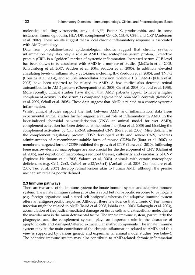
2.1 Para-inflammation and retinal aging To develop an effective and safe therapy for AMD, it is important that we understand the cause and the pathogenesis of the disease. Although a lot of risk factors of AMD have been identified, the precise mechanism on how these risk factors lead to macular damage remains ill-defined. Inflammation is believed to play an important role in AMD. How is inflammation initiated in the aging retina? According to Harman’s “free radical theory of aging” (Harman. 1956), aging is the accumulation of free-radical induced damage in cells and tissues. We have shown previously that a number of oxidized materials, including nitrotyrosine, oxidized low-density lipoprotein (LDL), and oxidized protein (identified by dinitrophenyl (DNP) staining) accumulate in the aging retina (Xu et al. 2009) (Figures 1A-1E). These oxidized (or damaged) molecules represent an endogenous threat to normal tissue physiology. However, under normal aging conditions, overt pathology does not occur in the retina. We now understand that the immune system has evolved to clear senescent, damaged cells and maintain normal tissue functions. Cells of the innate immune system, such as tissue resident macrophages sense signals from damaged cells/molecules and mount a para-inflammatory response, a concept proposed by Medzhitov (Medzhitov. 2008) as a “physiological” inflammatory response. Para-inflammation describes a “physiological” inflammatory process that lies between the overt destructive inflammation and normal quiescent state, and is required for tissue homeostatic processes in clearing damaged cells and molecules (Medzhitov. 2008). In the retina, RPE and photoreceptors encounter age-related increases in oxidative or metabolic stress. We have shown previously that age-related retinal “para-inflammation” comprises (1) microglial activation and subretinal migration (Figure 1F); and (2) complement activation (Figures 1G, 1H) (Chen et al. 2010, Xu et al. 2009). This para-inflammatory response may protect the retina from age-related free radical mediated damage as overt retinal pathology does not occur under physiological aging conditions (Chen et al. 2010, Xu et al. 2009). The protective effect of the para-inflammatory response is, however, limited. During aging, the noxious stimuli persist for many decades, which will inevitably result in loss of functional cells and molecules; the host has evolved to adjust the thresholds to maintain tissue function as well as to avoid an overt inflammation (Medzhitov. 2008).
www.intechopen.com
Inflammation in Age-Related Macular Degeneration – Implications for Therapy
131
Fig. 1. Oxidative stress and para-inflammation in the aging retina (Xu et al. 2009). (A-E), Cryosections of mouse eyes were stained for dinitrophenyl (DNP, red) and DAPI (blue). A weak positivity of DNP was observed in the retina (A) and RPE/choroid (C) of 3-month old mice. DNP was strongly positive in the retina (B) and RPE/choroid (D) of 24 month old mice. (E), Isotype control staining did not reveal any positivity. (F), Subretinal macrophages in a 20 months old mouse. RPE/choroidal flatmounts were stained for F4/80 (red) and Iba-1 (green) and observed by confocal microscopy. The majority of subretinal macrophages are F4/80+Iba-1+. A small number of cells were F4/80+Iba-1-. (G, H), Complement C3d deposition in mouse retina. Cryosections of mouse eye were stained for C3d (green) and propidium iodide (PI, red) and observed by confocal microscopy. C3d was not detected in the retina of a 3-month old mouse (G), but detected at the retina/choroidal interface in a 24-month old mouse (H). Ch, choroid; INL, inner nuclear layer; ONL, outer nuclear layer; RPE, retinal pigment epithelium.
2.2 Evidence of the role of inflammation in AMD Evidence supporting the association between chronic inflammation and AMD emerged over 25 years ago. In donated human AMD eyes, Penfold et al detected inflammatory cells (macrophages, lymphocytes and mast cells) in the choroid (Penfold et al. 1985, Penfold et al. 1997). Drusen, the hallmark of early AMD, also contains a variety of inflammatory
www.intechopen.com
Inflammatory Diseases – Immunopathology, Clinical and Pharmacological Bases
132
molecules including vitronectin, amyloid A/P, Factor X, prothrombin, and in some instances, immunoglobulin, HLA-DR, complement C3, C5, C5b-9, CFH, and CRP (Anderson et al. 2002). These results suggest that a local chronic inflammatory response is associated with AMD pathology. Data from population-based epidemiological studies suggest that chronic systemic
inflammation may also play a role in AMD. The acute-phase serum protein, C-reactive
protein (CRP) is a “golden” marker of systemic inflammation. Increased serum CRP level
has been shown to be associated with AMD in a number of studies (McGwin et al. 2005,
Schaumberg et al. 2007, Seddon et al. 2004, Seddon et al. 2005). In addition, elevated
circulating levels of inflammatory cytokines, including IL-6 (Seddon et al. 2005), and TNF-
(Cousins et al. 2004), and soluble intercellular adhesion molecule 1 (sICAM-1) (Klein et al.
2005) have been reported to be related to AMD. A few studies also detected retinal
autoantibodies in AMD patients (Cherepanoff et al. 2006, Gu et al. 2003, Penfold et al. 1990).
More recently, clinical studies have shown that AMD patients appear to have a higher
complement activity in the serum as compared age-matched non-AMD controls (Reynolds
et al. 2009, Scholl et al. 2008). These data suggest that AMD is related to a chronic systemic
inflammation.
Whilst clinical studies support the link between AMD and inflammation, data from
experimental animal studies further suggest a causal role of inflammation in AMD. In the
laser-induced choroidal neovascularisation (CNV, an animal model for wet AMD),
complement activation has been detected at the lesion site (Bora et al. 2005) and blocking the
complement activation by CFB siRNA attenuated CNV (Bora et al. 2006). Mice deficient in
the complement regulatory protein CD59 developed early and severe CNV, whereas
administration of a recombinant soluble form of mouse CD59a-Fc (Bora et al. 2007) or
membrane-targeted form of CD59 inhibited the growth of CNV (Bora et al. 2010). Infiltrating
bone marrow-derived macrophages are also crucial for the development of CNV (Galimi et
al. 2005), and depletion of macrophages reduced the size, cullularity and vascularity of CNV
(Espinosa-Heidmann et al. 2003, Sakurai et al. 2003). Animals with certain macrophage
deficiencies (e.g. Ccl2, Ccr2, Cx3cr1 or ccl2/cx3cr1) (Ambati et al. 2003, Combadiere et al.
2007, Tuo et al. 2007) develop retinal lesions akin to human AMD, although the precise
mechanism remains poorly defined.
2.3 Immune pathways There are two arms of the immune system: the innate immune system and adaptive immune system. The innate immune system provides a rapid but non-specific response to pathogens (e.g. foreign organisms and altered self antigens); whereas the adaptive immune system offers an antigen-specific response. Although there is evidence that chronic C. Pneumoniae infection might be related to AMD (Baird et al. 2008, Ishida et al. 2003, Kalayoglu et al. 2003), accumulation of free radical-mediated damage on tissue cells and extracellular molecules at the macular area is the main detrimental factor. The innate immune system, particularly the phagocytes and the complement system, plays an important role in the clearance of apoptotic cells and damaged/altered extracellular matrix components. The innate immune system may be the main contributor of the chronic inflammation related to AMD, and this view is supported by various genetic and experimental animal model studies (see below). The adaptive immune system may also contribute to AMD-related chronic inflammation
www.intechopen.com
Inflammation in Age-Related Macular Degeneration – Implications for Therapy
133
albeit to a less extent. Retinal autoantibodies have been detected in AMD patients (Cherepanoff et al. 2006, Gu et al. 2003, Penfold et al. 1990), and AMD-like lesions can be modelled in animals with DHL-immunization (Hollyfield et al. 2008).
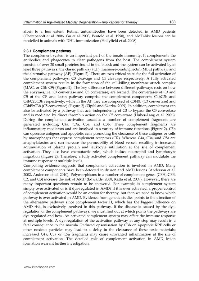
2.3.1 Complement pathway The complement system is an important part of the innate immunity. It complements the antibodies and phagocytes to clear pathogens from the host. The complement system consists of over 20 small proteins found in the blood; and the system can be activated by at least three pathways: the classic pathway (CP), mannose-binding lectin (MBL) pathway, and the alternative pathway (AP) (Figure 2). There are two critical steps for the full activation of the complement pathways: C3 cleavage and C5 cleavage respectively. A fully activated complement system results in the formation of the cell-killing membrane attack complex (MAC, or C5b-C9) (Figure 2). The key difference between different pathways rests on how the enzymes, i.e. C3 convertase and C5 convertase, are formed. The convertases of C3 and C5 of the CP and lectin pathway comprise the complement components C4bC2b and C4bC2bC3b respectively, while in the AP they are composed of C3bBb (C3 convertase) and C3bBbC3b (C5 convertase) (Figure 2) (Zipfel and Skerka. 2009). In addition, complement can also be activated by a pathway that acts independently of C3 to bypass the C3 convertase and is mediated by direct thrombin action on the C5 convertase (Huber-Lang et al. 2006). During the complement activation cascades a number of complement fragments are generated including C4a, C3a, C5a, and C3b. These complement fragments are inflammatory mediators and are involved in a variety of immune functions (Figure 2). C3b can opsonise antigens and apoptotic cells promoting the clearance of these antigens or cells by macrophages that express complement receptors (CR). Whereas C4a, C3a, and C5a are anaphylatoxins and can increase the permeability of blood vessels resulting in increased accumulation of plasma protein and leukocyte infiltration at the site of complement activation. They also have chemotactic roles, which induce neutrophil and lymphocyte migration (Figure 2). Therefore, a fully activated complement pathway can modulate the immune response at multiple levels. Compelling evidence suggests that complement activation is involved in AMD. Many complement components have been detected in drusen and AMD lesions (Anderson et al. 2002, Anderson et al. 2010). Polymorphisms in a number of complement genes (CFH, CFB, C2, and C3) increase the risk of AMD (Edwards. 2008, Katta et al. 2009). However, there are many important questions remain to be answered. For example, is complement system simply over activated or is it dys-regulated in AMD? If it is over activated, a proper control of complement activation would be an option for therapy, but then we need to know which pathway is over activated in AMD. Evidence from genetic studies points to the direction of the alternative pathway since complement factor H, which has the biggest influence on AMD risk, is exclusively involved in this pathway. If the disease is caused by the dys-regulation of the complement pathways, we must find out at which points the pathways are dys-regulated and how. An activated complement system may affect the immune response at multiple levels. A dys-regulation of the activation pathway at any step may result in a fatal consequence to the macula. Reduced opsonisation by C3b on apoptotic RPE cells or other noxious particles may lead to a delay in the clearance of these toxic materials; increased C4a, C3a or C5a fragments may cause unwanted inflammation at the site of complement activation. The detailed role of complement activation in AMD lesion formation warrant further investigation.
www.intechopen.com

Inflammatory Diseases – Immunopathology, Clinical and Pharmacological Bases
134
Fig. 2. Complement activation and immune regulation. Complement system can be activated by three pathways: the classical pathway (CP), mannose-binding lectin (MBL) pathway and the alternative pathway (AP); all lead to the cleavage of C3 and C5 and the formation of C5b-C9. Activation of the complement system generates C4a, C3a, C3b and C5a fragments that are actively involved in immune responses. C3b opsonises foreign antigens and apoptotic cells promoting phagocytosis. C4a, C3a and C5a are anaphylatoxins, which may cause increased vascular permeability enhancing inflammation. They are also potent leukocytes chemoattractants and can induce the migration of neutrophils and lymphocytes to the sites of complement activation. C5b-C9 may directly kill antibody coated particles.
2.3.2 Monocytes and macrophages Monocytes are a subset of circulating white blood cells that are originated from bone marrow haematopoietic stem cells. Monocytes migrate from the bloodstream to peripheral tissues and then differentiate into tissue resident macrophages or dendritic cells. In the central nervous system, they differentiate into brain (or retinal) resident macrophages, and these cells are traditionally called microglial cells. Monocytes and macrophages, dendritic cells are important components of the innate immune system and play a crucial role in detecting antigens (including non-self foreign antigens and altered self antigens) and the removal of the antigens as well as apoptotic cells in pathophysiological conditions. Pathologies in AMD are restricted to the retina-choroid interface. Choroidal macrophages and retinal microglial cells are major immune cells involved in the pathological process. Under normal physiological conditions, subretinal space (the interface between the retina and RPE/choroid) is devoid of any immune cells. However, in the aging eye, microglia and macrophages accumulate not only in the subretinal space, but also in the choroid (Xu et al. 2009). Presumably, this form of para-inflammation is a protective response to RPE or
www.intechopen.com

Inflammation in Age-Related Macular Degeneration – Implications for Therapy
135
photoreceptor damage in the aging eye. Subretinal microglial cells may remove apoptotic RPE cells or damaged photoreceptors, whereas choroidal macrophages scavenge waste materials produced by RPE cells preventing drusen formation. These cells are, therefore, crucial for retinal homeostasis, and dys-function or mal-function of these cells may result in macular pathology at the retina-choroid interface (AMD lesions). Compelling evidence suggests that macrophages may play a detrimental role in AMD. Polymorphisms in chemokine receptor cx3cr1 gene, a gene that is widely expressed by myeloid-derived cells, increase the risk of AMD (Tuo et al. 2004, Yang et al. 2010). Animals with monocyte dysfunction due to lack of certain chemokine (e.g. CCL2) (Ambati et al. 2003) or chemokine receptors (CCR2, or CX3CR1) (Ambati et al. 2003, Combadiere et al. 2007, Tuo et al. 2007) develop AMD-like lesions. Furthermore, in the laser-induced wet AMD animal model, retinal lesion can be attenuated by macrophage depletion (Espinosa-Heidmann et al. 2003, Sakurai et al. 2003). The precise mechanism underlying the detrimental effect of macrophages to AMD remains to be elucidated.
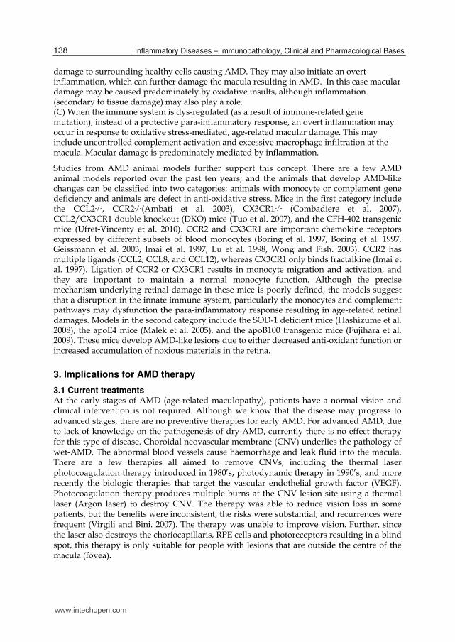
2.4 Age-related macular degeneration - an imbalance between macular damage and para-inflammation? Under normal physiological aging conditions, the para-inflammatory response, which is characterized by complement activation and microglia/macrophage activation and subretinal migration (Figure 1) (Xu et al. 2009), protects the macula against the age-related free radical-mediated damage. This suggests that microglia and complement activation are beneficial to retinal aging. However, under pathological conditions, these two arms of innate immune pathways are harmful and their activation leads to macular damage, such as in AMD. How is the outcome of their activation determined? In other worlds, activation of microglia and the complement system at the macular site occurs in all aged people, why some people develop AMD, some others do not? In order to maintain a healthy functional macula, the level of age-related free radical-mediated tissue/cell damage and the capacity of the immune system to cope with accelerating damage (via para-inflammatory response) needs to be balanced (Figure 3). In persons who have a relatively healthy lifestyle (hence the age-related oxidative damage accumulates at the average level during the process of aging), and have no genetic predispositions to AMD (hence be able to maintain a good defence system), their immune system can maintain macular homeostasis throughout their life and AMD will not occur (Figure 3A). In persons who do not have a healthy lifestyle (e.g. heavy smokers, high-fat diet, and extensive light exposure throughout their life), even if they do not have any genetic predispositions to AMD, the age-related free radical-mediated macular damage may exceed the repair capacity of the para-inflammatory response, macular function may decline and overt pathology (AMD) may ensure (Figure 3B). On the other hand, if the person has a healthy lifestyle and the age-related free radical-mediated damage accumulates at a normal level in the macula, but he/she has genetic predispositions to AMD, and the immune system is unable to initiate a functional para-inflammatory response, macular pathology (AMD) may also occur (Figure 3C). Needless to say, if a person has an unhealthy lifestyle, and he/she is unfortunate enough to have genetic risk factors, his/her risk to develop AMD is significantly higher than others. In terms of the immune response, during the disease initiation stage, the para-inflammatory responses are protective (although they may not be able to fully protect the macula). The inducer of para-inflammation is a low-grade chronic macular damage, and pathways involved
www.intechopen.com

Inflammatory Diseases – Immunopathology, Clinical and Pharmacological Bases
136
are monocytes, macrophages and the complement system (Xu et al. 2009). Once the disease begins, an overt inflammation, instead of para-inflammation, may occur. At this stage, the inducers are toxic molecules released by dead cells and altered extracellular molecules. In addition to monocytes, macrophages (Penfold et al. 1985, Penfold et al. 1997) and the complement system, other immune components such as T and B lymphocytes, and autoantibodies (Penfold et al. 1985, Penfold et al. 1990) may also be involved in inflammation at this stage. The physiological purpose of the inflammation is to remove dead cells, tissue debris and other toxic molecules and to promote tissue repair and remodelling. However, like in many other disease conditions, inflammation is a double-edged sword. Activated immune cells may release inflammatory cytokines and chemokines such as TNF-and IL-1, and further tissue damage (so called collateral damage) (Nathan. 2002) is unavoidable. The concept that AMD is related to an imbalance between the level of age-related macular damage and the protective capacity of the immune system is supported by evidence from numerous epidemiological and genetic studies. Over the last decades epidemiological studies have identified a number of environmental factors that may increase the risk of AMD, including smoking (Baird et al. 2008, Chakravarthy et al. 2007, Cong et al. 2008, DeBlack. 2003, Hughes et al. 2007, Khan et al. 2006, Thornton et al. 2005), high-fat diet (Evans. 2001), sunlight exposure (Hirakawa et al. 2008, Plestina-Borjan and Klinger-Lasic. 2007) and alcohol consumption (Chong et al. 2008). Cigarette smoking is the single most important environmental risk factor for AMD. Current smokers have 45% greater probability of developing AMD and exhibit enhanced disease progression when compared to non-smokers (Klein et al. 2008). Although the mechanism underlying the environmental factors mediated increased risk of AMD is not known, it is believed that they may increase macular damage and/or alter the immune function. Taken cigarette smoking as an example, the blood borne products of tobacco combustion damage RPE cells, alter Bruch’s membrane and exacerbate sub-PRE deposition (Bertram et al. 2009, Espinosa-Heidmann et al. 2006, Jia et al. 2007, Wang et al. 2009). Such lesions can be experimentally induced in vivo and in vitro after exposure to cigarette smoke (Espinosa-Heidmann et al. 2006, Wang et al. 2009), or defined extracts such as hydroquinone (HQ) (Espinosa-Heidmann et al. 2006, Wang et al. 2009), polycyclic aromatic hydrocarbons (PHA) (Espinosa-Heidmann et al. 2006, Wang et al. 2009) and acrolein (Jia et al. 2007). Cigarette smoking also affects the immune system (Arnson et al. 2010, Klareskog et al. 2007), including the ocular immune responses. Cigarette smoking increases the incidence of uveitis (Lin et al. 2010, Lois et al. 2008, Thorne et al. 2008)and scleritis (Boonman et al. 2005), and enhances the risk of developing cystoid macular oedema in uveitis patients (Lin et al. 2010, Thorne et al. 2008). It appears, therefore, that cigarette smoking can cause macular damage, and alter the immune system resulting in a declined para-inflammatory response. In addition to environmental factors, clinical studies have found that the risk of AMD is also affected by genetic factors. Genes that are involved in AMD susceptibility fall into three categories: immune-related genes (CFH, CFB, C2, C3, C5, Cx3cr1, TLRs, IL-8, HLAs), mitochondrial and oxidative stress-related genes (ARMS2 and HTRA1) and extracellular matrix related genes (PRELP, LAMC1, LAMC2, LAMB3, FIBULIN2, and ITGB4) (Katta et al. 2009). Importantly, the majority of the immune-related genes are related to the innate immunity, including the complement system (CFH, CFB, C2, C3 and C5) (Lotery and Trump. 2007, Montezuma et al. 2007), and monocyte/macrophage functions (Cx3cr1 (Chan et al. 2005, Combadiere et al. 2007, Tuo et al. 2004, Yang et al. 2010) and TLRs (Kaarniranta and Salminen. 2009)). These data suggest that genetic factors may predispose individuals to the risk of AMD by (1) decreasing the anti-oxidative ability, and/or (2) altering the immune function.
www.intechopen.com

Inflammation in Age-Related Macular Degeneration – Implications for Therapy
137
Fig. 3. The balance between oxidative damage and para-inflammation in retinal aging. (A) Under normal physiological conditions, oxidative damage accumulates at the macula with age. A healthy immune system can mount a para-inflammatory response to remove damaged molecules/cells and maintain macular function. The level of damage is within the capacity of the immune system and pathology (AMD) will not occur. (B) When the level of stress-mediated macular damage exceeds the capacity of the immune system (as a result of unhealthy life style, anti-stress related gene mutations, etc), the damaged molecules and cells may not be cleared away through the para-inflammatory response. The damaged cells and oxidised molecules are toxic and may cause further
www.intechopen.com
Inflammatory Diseases – Immunopathology, Clinical and Pharmacological Bases
138
damage to surrounding healthy cells causing AMD. They may also initiate an overt inflammation, which can further damage the macula resulting in AMD. In this case macular damage may be caused predominately by oxidative insults, although inflammation (secondary to tissue damage) may also play a role. (C) When the immune system is dys-regulated (as a result of immune-related gene mutation), instead of a protective para-inflammatory response, an overt inflammation may occur in response to oxidative stress-mediated, age-related macular damage. This may include uncontrolled complement activation and excessive macrophage infiltration at the macula. Macular damage is predominately mediated by inflammation.
Studies from AMD animal models further support this concept. There are a few AMD animal models reported over the past ten years; and the animals that develop AMD-like changes can be classified into two categories: animals with monocyte or complement gene deficiency and animals are defect in anti-oxidative stress. Mice in the first category include the CCL2-/-, CCR2-/-(Ambati et al. 2003), CX3CR1-/- (Combadiere et al. 2007), CCL2/CX3CR1 double knockout (DKO) mice (Tuo et al. 2007), and the CFH-402 transgenic mice (Ufret-Vincenty et al. 2010). CCR2 and CX3CR1 are important chemokine receptors expressed by different subsets of blood monocytes (Boring et al. 1997, Boring et al. 1997, Geissmann et al. 2003, Imai et al. 1997, Lu et al. 1998, Wong and Fish. 2003). CCR2 has multiple ligands (CCL2, CCL8, and CCL12), whereas CX3CR1 only binds fractalkine (Imai et al. 1997). Ligation of CCR2 or CX3CR1 results in monocyte migration and activation, and they are important to maintain a normal monocyte function. Although the precise mechanism underlying retinal damage in these mice is poorly defined, the models suggest that a disruption in the innate immune system, particularly the monocytes and complement pathways may dysfunction the para-inflammatory response resulting in age-related retinal damages. Models in the second category include the SOD-1 deficient mice (Hashizume et al. 2008), the apoE4 mice (Malek et al. 2005), and the apoB100 transgenic mice (Fujihara et al. 2009). These mice develop AMD-like lesions due to either decreased anti-oxidant function or increased accumulation of noxious materials in the retina.
3. Implications for AMD therapy
3.1 Current treatments At the early stages of AMD (age-related maculopathy), patients have a normal vision and clinical intervention is not required. Although we know that the disease may progress to advanced stages, there are no preventive therapies for early AMD. For advanced AMD, due to lack of knowledge on the pathogenesis of dry-AMD, currently there is no effect therapy for this type of disease. Choroidal neovascular membrane (CNV) underlies the pathology of wet-AMD. The abnormal blood vessels cause haemorrhage and leak fluid into the macula. There are a few therapies all aimed to remove CNVs, including the thermal laser photocoagulation therapy introduced in 1980’s, photodynamic therapy in 1990’s, and more recently the biologic therapies that target the vascular endothelial growth factor (VEGF). Photocoagulation therapy produces multiple burns at the CNV lesion site using a thermal laser (Argon laser) to destroy CNV. The therapy was able to reduce vision loss in some patients, but the benefits were inconsistent, the risks were substantial, and recurrences were frequent (Virgili and Bini. 2007). The therapy was unable to improve vision. Further, since the laser also destroys the choriocapillaris, RPE cells and photoreceptors resulting in a blind spot, this therapy is only suitable for people with lesions that are outside the centre of the macula (fovea).
www.intechopen.com
Inflammation in Age-Related Macular Degeneration – Implications for Therapy
139
Photodynamic therapy (PDT) was introduced in 2000. PDT involves an intravenous injection of a photosensitizing drug called Verteporfin. The drug is carried out by blood lipoproteins and reaches to the site of CNV in the macula. A non-thermal laser (blue laser at 689 nm) is then used to sensitize the drug, and this photochemical reaction produces cytotoxic free radicals resulting in direct cellular injury to vascular endothelial cells and subsequent regression of CNV. Clinical studies have shown that PDT is much less destructive and achieves better results for vision than photocoagulation (Spaide et al. 2003). Blood vessels that have been eradicated in this way do now grow back, however other vessels may still be formed if angiogenic stimuli persist. Clinical studies have shown that PDT is safe and effective for treating a range of lesions, including predominant classic lesions, CNV secondary to pathological myopia and occult with no classic subfoveal lesion, but it has no effect on minimally classic lesions (Spaide et al. 2003). It should be noted that PDT does not improve vision (Bressler et al. 2009). The most recent treatment to be developed is called Anti Vascular Endothelial Growth Factor (Anti –VEGF) drug therapy (Campa and Harding. 2011). Anti-VEGF therapy involves blocking the VEGF-A, an important and essential growth factor that is involved in the breakdown of blood-retinal barrier (BRB) (thus the leakage of blood components and macular oedema) and the growth of new blood vessels (CNV) (Bressler. 2009). Therefore, this therapy will make its effect by stabilising tight-junctions of the BRB and inducing regression of the neovascularisation once formed. There are three main drugs used in this category of treatment: Macugen, Lucentis and Avastin. The drugs are administered intravitreally, and the therapy needs to be repeated a number of times. Macugen was the first drug that was approved by the US Food & Drug Administration (FDA) in 2004 and by the European Medicines Evaluation Agency (EMEA) in 2006. Clinical studies have shown that Macugen is more effective than PDT at slowing vision loss, but it does not improve visual acuity (Edwards et al. 2008, Gragoudas et al. 2004, VEGF Inhibition Study in Ocular Neovascularization (V.I.S.I.O.N.) Clinical Trial Group et al. 2006). The reason for this is probably because it only targets the VEGF-165 isoform. Lucentis is another anti-VEGF antibody that targets all isoforms of VEGF, and it was released one year after Macugen. Clinical evidence suggests that Lucentis can substantially improve visual acuity in wet-AMD patients (Bressler et al. 2009, Brown et al. 2006, Rosenfeld et al. 2006). Avastin is another anti-VEGF drug and is approved for the treatment of advanced colorectal disease, and it has similar properties to Lucentis. However, it is a full-length antibody and is much less expensive than Lucentis. A recent study has shown that Avastin has the same effect as Lucentis in treating wet-AMD, although it may have a slightly higher cardiovascular side effect (Arevalo et al. 2010, Campa and Harding. 2011, Giansanti et al. 2007). Although the drugs that inhibit VEGF prevent vision loss, and even improve visual acuity in some cases of wet AMD, their effect on improving vision depends greatly on the time at which they are administered and there is a huge variability in functional outcomes. A recent study on Avastin treatment for wet AMD (Arevalo et al. 2010) reported functional outcomes and showed that after 2 years treatment, 43.5% of cases had improved vision, 43% remained stable and 13.5% had decreased vision (even with >10 injections within 24 months) (Arevalo et al. 2010). The anti-VEGF therapy, although is better than PDT and other treatments for wet AMD, has its limitations and side effects. More target-specific, safe and effective therapies are urgently needed for both dry and wet AMD.
3.2 Immune therapy - a future for Age-related macular degeneration? The importance of inflammation in AMD pathology offers an opportunity for therapy. Based on the “oxidative stress/para-inflammation balance” theory of AMD, the disease can
www.intechopen.com
Inflammatory Diseases – Immunopathology, Clinical and Pharmacological Bases
140
be theoretically prevented or at least delayed if we know where the unbalance is. By living in a healthy lifestyle (avoid the environmental risk factors), we can minimise the age-related oxidative stress. In reality, however, we can chose healthy food, but we may have a very limited choice on the environment that we live. With advanced gene therapy technology, in the future, we might be able to reduce the genetic risk factors of AMD. Once the disease begins, we must treat the disease. We know that inflammation may have dual roles in disease progression stages. How can we modulate the immune system to treat AMD?
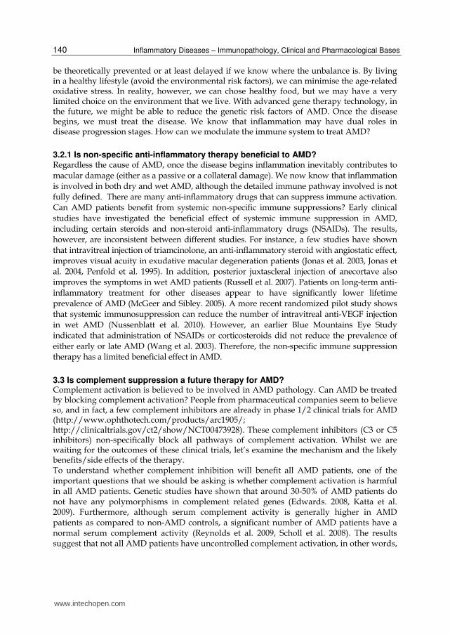
3.2.1 Is non-specific anti-inflammatory therapy beneficial to AMD? Regardless the cause of AMD, once the disease begins inflammation inevitably contributes to macular damage (either as a passive or a collateral damage). We now know that inflammation is involved in both dry and wet AMD, although the detailed immune pathway involved is not fully defined. There are many anti-inflammatory drugs that can suppress immune activation. Can AMD patients benefit from systemic non-specific immune suppressions? Early clinical studies have investigated the beneficial effect of systemic immune suppression in AMD, including certain steroids and non-steroid anti-inflammatory drugs (NSAIDs). The results, however, are inconsistent between different studies. For instance, a few studies have shown that intravitreal injection of triamcinolone, an anti-inflammatory steroid with angiostatic effect, improves visual acuity in exudative macular degeneration patients (Jonas et al. 2003, Jonas et al. 2004, Penfold et al. 1995). In addition, posterior juxtascleral injection of anecortave also improves the symptoms in wet AMD patients (Russell et al. 2007). Patients on long-term anti-inflammatory treatment for other diseases appear to have significantly lower lifetime prevalence of AMD (McGeer and Sibley. 2005). A more recent randomized pilot study shows that systemic immunosuppression can reduce the number of intravitreal anti-VEGF injection in wet AMD (Nussenblatt et al. 2010). However, an earlier Blue Mountains Eye Study indicated that administration of NSAIDs or corticosteroids did not reduce the prevalence of either early or late AMD (Wang et al. 2003). Therefore, the non-specific immune suppression therapy has a limited beneficial effect in AMD.
3.3 Is complement suppression a future therapy for AMD? Complement activation is believed to be involved in AMD pathology. Can AMD be treated by blocking complement activation? People from pharmaceutical companies seem to believe so, and in fact, a few complement inhibitors are already in phase 1/2 clinical trials for AMD (http://www.ophthotech.com/products/arc1905/; http://clinicaltrials.gov/ct2/show/NCT00473928). These complement inhibitors (C3 or C5 inhibitors) non-specifically block all pathways of complement activation. Whilst we are waiting for the outcomes of these clinical trials, let’s examine the mechanism and the likely benefits/side effects of the therapy. To understand whether complement inhibition will benefit all AMD patients, one of the important questions that we should be asking is whether complement activation is harmful in all AMD patients. Genetic studies have shown that around 30-50% of AMD patients do not have any polymorphisms in complement related genes (Edwards. 2008, Katta et al. 2009). Furthermore, although serum complement activity is generally higher in AMD patients as compared to non-AMD controls, a significant number of AMD patients have a normal serum complement activity (Reynolds et al. 2009, Scholl et al. 2008). The results suggest that not all AMD patients have uncontrolled complement activation, in other words,
www.intechopen.com

Inflammation in Age-Related Macular Degeneration – Implications for Therapy
141
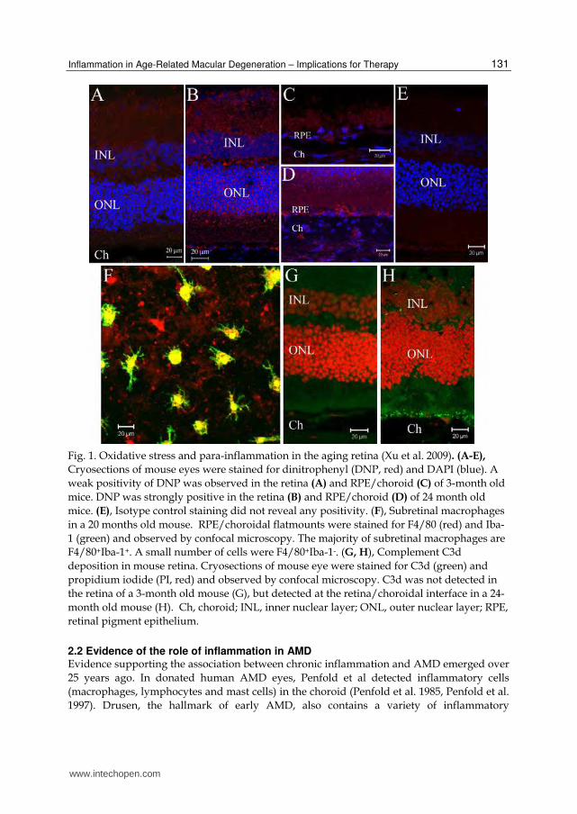
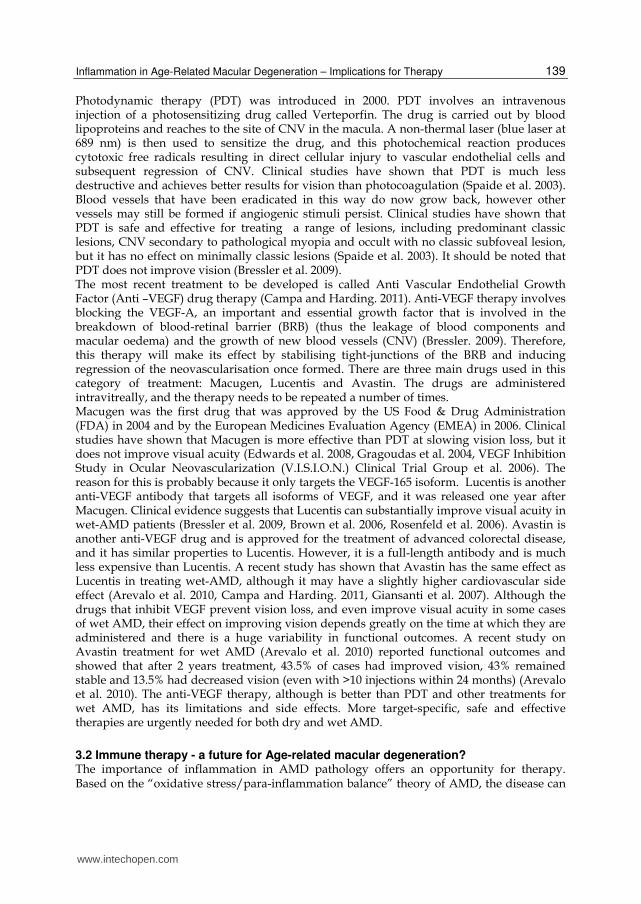
AMD pathology may occur in the absence of abnormal uncontrolled complement activation. To further test this hypothesis, we have examined the complement activation in the CCL2 KO and CCR2 KO mice, the mouse models of AMD (Ambati et al. 2003). We found that 40% of CCL2 KO mice and 28% of CCR2 KO mice (>18 months old) develop retinal atrophies (Figure 4A) (Chen et al. 2011). Further mechanic study shows that there is no significant increase in the serum complement activity (Figure 4C). The expression levels of complement genes in the liver (figure 4D), retinal and RPE/choroidal tissue (data not shown) in the KO do not significantly differ from those in age-matched control mice. Furthermore, the complement system is only partially activated resulting in complement C3b but not C5b-C9 deposition at the lesion site (Figure 4E, 4F) (Chen et al. 2011). The results suggest that retinal lesion in these mice is not caused by dys-regulated complement activation. Since C3b/C3d plays an important role in opsonising apoptotic cells, we believe this partial complement activation is beneficial. Complement activation in these mice is a consequence of retinal damage and the physiological purpose may be to promote the removal of apoptotic cells from the lesion site.
Fig. 4. Complement activation in aged CCL2 KO or CCR2 KO mice. A, a fundus image of a 24-m old C57BL/6 mouse showing multiple small white dots (correlated to subretinal microglia (Xu et al. 2008). B, a fundus image of a 24-m old CCL2 KO mouse showing patches of white lesions akin to human geographic atrophy (arrows). C, Serum complement activity determined by haemolytic assay. D, real-time RT-PCR analysis of complement gene expression in the liver tissue in different strains of mice. E & F, complement C3d (red) and C5b-9 (green) expression in retinal lesion in a 24-m old CCL2 KO mouse (E) and an experimental autoimmune uveoretinitis mouse (F, as a positive control). C5b-9 (green) was detected in uveoretinitis lesion but not in CCL2 KO mouse lesion (arrowheads). RPE, retinal pigment epithelium; Ch, choroid; ONL, outer nuclear layer (Chen et al. 2011).
www.intechopen.com
Inflammatory Diseases – Immunopathology, Clinical and Pharmacological Bases
142
So it appears that not all AMD has a complement component in its pathogenesis, and complement inhibition may not benefit every AMD patient. In patients whose macular pathology is not caused by uncontrolled complement activation, the therapy may even worsen the disease, as a partially activated complement system may help the clearance of dead cells and debris from the lesion site and promote tissue repair/remodelling. The efficacy and safety of complement inhibitors in AMD therapy warrant further investigation.
3.4 Can we modify monocyte function to treat AMD? Genetic studies have shown that cx3cr1 gene polymorphism is a risk factor of AMD (Tuo et al. 2004, Yang et al. 2010), and this risk is independent of any complement gene polymorphisms (Yang et al. 2010). Our studies in the aged CCL2 KO and CCR2 KO mice show that AMD-like lesion can develop in the absence of any complement dys-regulation (Figure 4) (Chen, et al. 2011). The results suggest that under aging conditions, monocyte malfunction may result in macular damage in the absence of complement dys-regulation. Modulating the monocyte function may, therefore, offer an opportunity for therapy under this situation.
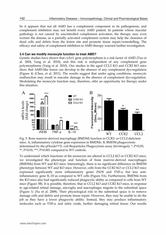
Fig. 5. Bone marrow-derived macrophage (BMDM) function in CCR2- or CCL2-deficient mice. A, inflammatory cytokine gene expression in BMDMs. B, BMDM phagocytosis determined by the pHrodoTM E. coli Bioparticles Phagocytosis assay (Invitrogen). *, P<0.05; **, P<0.01; ***, P<0.001 compared to WT controls.
To understand which functions of the monocyte are altered in CCL2 KO or CCR2 KO mice, we investigated the phenotype and function of bone marrow-derived macrophages (BMDMs) from WT and KO mice. Interestingly, there is no significant difference on BMDM phenotype between WT and KO mice. However, cells from the CCR2 KO or CCL2 KO mice
expressed significantly more inflammatory genes iNOS and TNF- but less anti-inflammatory gene IL-10 as compared to WT cells (Figure 5A). Furthermore, BMDMs from the KO mice also had significantly reduced phagocytic ability as compared to cells from WT mice (Figure 5B). It is possible, therefore, that in CCL2 KO and CCR2 KO mice, in response to age-related retinal damage, microglia and macrophages migrate to the subretinal space (Figure 1) (Xu et al. 2008). Their physiological role in the subretinal space is to remove damage cells and debris and promote tissue repair. However, they may be unable to do the job as they have a lower phagocytic ability. Instead, they may produce inflammatory
molecules such as TNF- and nitric oxide, further damaging retinal tissue. Our results
www.intechopen.com

Inflammation in Age-Related Macular Degeneration – Implications for Therapy
143
suggest that a normal monocyte function is important to maintain retinal homeostasis. Genetic study did not reveal any link between AMD risk and CCL2 or CCR2 gene polymorphism (Despriet et al. 2008). However, monocyte function can be affected by many factors, it is entirely possible that similar monocyte functional alterations may exist in AMD patients (although they may not be caused by CCL2 or CCR2 deficiency), and these functional changes may be responsible for inflammation mediated retinal pathology (with or without complement dys-regulation). Further studies on how monocyte function might be changed in AMD patients will be essential to develop monocyte-specific immune therapy.
4. Conclusions
AMD is a multifactorial disease. Old age, environmental risk factors, genetic predispositions all work together leading to macular damage. The immune system plays an important role in the initiation and progression of the disease. A healthy immune system can prevent overt pathology during aging by initiating a para-inflammatory response. Whereas an altered immune system either in the complement pathways or the monocyte functions, may fail to induce a protective para-inflammatory response. Instead, it may respond aggressively (overt inflammation) causing further damage to the aging retina. Complement over activation and monocyte malfunction may work together enhancing age-related macular damage. They may also work independently contributing to AMD pathology. Due to the complexity of the immunomechanism of the disease, there will be no universal immune therapy for AMD. Blocking complement activation may benefit patients who have uncontrolled complement activity (presumably as a result of complement gene polymorphism), it may make the disease worse in patients who do not have a dys-regulated complement system. We suggest that complement gene polymorphisms should be used as a guide for complement inhibitors therapy in AMD. Modulating monocyte function may be beneficial for patients who have monocyte malfunction (may be related CX3CR1 gene polymorphism) (Tuo et al. 2004, Yang et al. 2010). Further studies are required to understand the pathways related to monocyte-mediated macular damage to identify specific target for therapy.
5. References
Ambati J., Anand A., Fernandez S., Sakurai E., Lynn B.C., Kuziel W.A., Rollins B.J., Ambati B.K., 2003. An animal model of age-related macular degeneration in senescent Ccl-2- or Ccr-2-deficient mice. Nat. Med. 9, 1390-1397.
Anderson D.H., Mullins R.F., Hageman G.S., Johnson L.V., 2002. A role for local inflammation in the formation of drusen in the aging eye. Am. J. Ophthalmol. 134, 411-431.
Anderson D.H., Radeke M.J., Gallo N.B., Chapin E.A., Johnson P.T., Curletti C.R., Hancox L.S., Hu J., Ebright J.N., Malek G., Hauser M.A., Rickman C.B., Bok D., Hageman G.S., Johnson L.V., 2010. The pivotal role of the complement system in aging and age-related macular degeneration: hypothesis re-visited. Prog. Retin. Eye Res. 29, 95-112.
Arevalo J.F., Sanchez J.G., Wu L., Berrocal M.H., Alezzandrini A.A., Restrepo N., Maia M., Farah M.E., Brito M., Diaz-Llopis M., Rodriguez F.J., Reategui G., Iturralde-Iraola J., Udaondo-Mirete P., Pan-American Collaborative Retina Study Group, 2010.
www.intechopen.com

Inflammatory Diseases – Immunopathology, Clinical and Pharmacological Bases
144
Intravitreal bevacizumab for subfoveal choroidal neovascularization in age-related macular degeneration at twenty-four months: the Pan-American Collaborative Retina Study. Ophthalmology 117, 1974-81, 1981.e1.
Arnson Y., Shoenfeld Y., Amital H., 2010. Effects of tobacco smoke on immunity, inflammation and autoimmunity. J. Autoimmun. 34, J258-65.
Baird P.N., Robman L.D., Richardson A.J., Dimitrov P.N., Tikellis G., McCarty C.A., Guymer R.H., 2008. Gene-environment interaction in progression of AMD - the CFH gene, smoking and exposure to chronic infection. Hum. Mol. Genet. 17(9), 1299-1305.
Bertram K.M., Baglole C.J., Phipps R.P., Libby R.T., 2009. Molecular regulation of cigarette smoke induced-oxidative stress in human retinal pigment epithelial cells: implications for age-related macular degeneration. Am. J. Physiol. Cell. Physiol. 297, C1200-10.
Boonman Z.F., de Keizer R.J., Watson P.G., 2005. Smoking delays the response to treatment in episcleritis and scleritis. Eye (Lond) 19, 949-955.
Bora N.S., Jha P., Lyzogubov V.V., Kaliappan S., Liu J., Tytarenko R.G., Fraser D.A., Morgan B.P., Bora P.S., 2010. Recombinant membrane-targeted form of CD59 inhibits the growth of choroidal neovascular complex in mice. J. Biol. Chem. 285, 33826-33833.
Bora N.S., Kaliappan S., Jha P., Xu Q., Sivasankar B., Harris C.L., Morgan B.P., Bora P.S., 2007. CD59, a complement regulatory protein, controls choroidal neovascularization in a mouse model of wet-type age-related macular degeneration. J. Immunol. 178, 1783-1790.
Bora N.S., Kaliappan S., Jha P., Xu Q., Sohn J.H., Dhaulakhandi D.B., Kaplan H.J., Bora P.S., 2006. Complement activation via alternative pathway is critical in the development of laser-induced choroidal neovascularization: role of factor B and factor H. J. Immunol. 177, 1872-1878.
Bora P.S., Sohn J.H., Cruz J.M., Jha P., Nishihori H., Wang Y., Kaliappan S., Kaplan H.J., Bora N.S., 2005. Role of complement and complement membrane attack complex in laser-induced choroidal neovascularization. J. Immunol. 174, 491-497.
Boring L., Gosling J., Chensue S.W., Kunkel S.L., Farese R.V.,Jr, Broxmeyer H.E., Charo I.F., 1997. Impaired monocyte migration and reduced type 1 (Th1) cytokine responses in C-C chemokine receptor 2 knockout mice. J. Clin. Invest. 100, 2552-2561.
Bressler N.M., 2009. Antiangiogenic approaches to age-related macular degeneration today. Ophthalmology 116, S15-23.
Bressler N.M., Chang T.S., Fine J.T., Dolan C.M., Ward J., Anti-VEGF Antibody for the Treatment of Predominantly Classic Choroidal Neovascularization in Age-Related Macular Degeneration (ANCHOR) Research Group, 2009. Improved vision-related function after ranibizumab vs photodynamic therapy: a randomized clinical trial. Arch. Ophthalmol. 127, 13-21.
Brown D.M., Kaiser P.K., Michels M., Soubrane G., Heier J.S., Kim R.Y., Sy J.P., Schneider S., ANCHOR Study Group, 2006. Ranibizumab versus verteporfin for neovascular age-related macular degeneration. N. Engl. J. Med. 355, 1432-1444.
Bunce C., Wormald R., 2006. Leading causes of certification for blindness and partial sight in England & Wales. BMC Public Health, 6, 58.
Campa C., Harding S.P., 2011. Anti-VEGF compounds in the treatment of neovascular age related macular degeneration. Curr. Drug Targets, 12, 173-181.
Chakravarthy U., Augood C., Bentham G.C., de Jong P.T., Rahu M., Seland J., Soubrane G., Tomazzoli L., Topouzis F., Vingerling J.R., Vioque J., Young I.S., Fletcher A.E., 2007.
www.intechopen.com

Inflammation in Age-Related Macular Degeneration – Implications for Therapy
145
Cigarette smoking and age-related macular degeneration in the EUREYE Study. Ophthalmology, 114, 1157-1163.
Chan C.C., Tuo J., Bojanowski C.M., Csaky K.G., Green W.R., 2005. Detection of CX3CR1 single nucleotide polymorphism and expression on archived eyes with age-related macular degeneration. Histol. Histopathol. 20, 857-863.
Chen M., Muckersie E., Forrester J.V., Xu H., 2010. Immune activation in Retinal Aging: A Gene Expression Study. Invest. Ophthalmol. Vis. Sci. 51, 5888-5896.
Chen M., Forrester J., Xu H., 2011. Dysregulation in retinal para-inflammation and age-related retinal degeneration in CCL2 or CCR2 deficient mice. PLoS ONE. 6(8):e22818.
Chen Y., Bedell M., Zhang K., 2010. Age-related macular degeneration: genetic and environmental factors of disease. Mol. Interv. 10, 271-281.
Cherepanoff S., Mitchell P., Wang J.J., Gillies M.C., 2006. Retinal autoantibody profile in early age-related macular degeneration: preliminary findings from the Blue Mountains Eye Study. Clin. Experiment. Ophthalmol. 34, 590-595.
Chong E.W., Kreis A.J., Wong T.Y., Simpson J.A., Guymer R.H., 2008. Alcohol consumption and the risk of age-related macular degeneration: a systematic review and meta-analysis. Am. J. Ophthalmol. 145, 707-715.
Chopdar A., Chakravarthy U., Verma D., 2003. Age related macular degeneration. BMJ, 326, 485-488.
Coleman H.R., Chan C.C., Ferris F.L.,3rd, Chew E.Y., 2008. Age-related macular degeneration. Lancet, 372, 1835-1845.
Combadiere C., Feumi C., Raoul W., Keller N., Rodero M., Pezard A., Lavalette S., Houssier M., Jonet L., Picard E., Debre P., Sirinyan M., Deterre P., Ferroukhi T., Cohen S.Y., Chauvaud D., Jeanny J.C., Chemtob S., Behar-Cohen F., Sennlaub F., 2007. CX3CR1-dependent subretinal microglia cell accumulation is associated with cardinal features of age-related macular degeneration. J. Clin. Invest. 117, 2920-2928.
Cong R., Zhou B., Sun Q., Gu H., Tang N., Wang B., 2008. Smoking and the risk of age-related macular degeneration: a meta-analysis. Ann. Epidemiol. 18, 647-656.
Cousins S.W., Espinosa-Heidmann D.G., Csaky K.G., 2004. Monocyte activation in patients with age-related macular degeneration: a biomarker of risk for choroidal neovascularization? Arch. Ophthalmol. 122, 1013-1018.
DeBlack S.S., 2003. Cigarette smoking as a risk factor for cataract and age-related macular degeneration: a review of the literature. Optometry, 74, 99-110.
Despriet D.D., Bergen A.A., Merriam J.E., Zernant J., Barile G.R., Smith R.T., Barbazetto I.A., van Soest S., Bakker A., de Jong P.T., Allikmets R., Klaver C.C., 2008. Comprehensive analysis of the candidate genes CCL2, CCR2, and TLR4 in age-related macular degeneration. Invest. Ophthalmol. Vis. Sci. 49, 364-371.
Edwards A.O., 2008. Genetics of age-related macular degeneration. Adv. Exp. Med. Biol. 613, 211-219.
Edwards A.O., Fridley B.L., James K.M., Sharma A.K., Cunningham J.M., Tosakulwong N., 2008. Evaluation of clustering and genotype distribution for replication in genome wide association studies: the age-related eye disease study. PLoS One, 3, e3813.
Espinosa-Heidmann D.G., Suner I.J., Catanuto P., Hernandez E.P., Marin-Castano M.E., Cousins S.W., 2006. Cigarette smoke-related oxidants and the development of sub-RPE deposits in an experimental animal model of dry AMD. Invest. Ophthalmol. Vis. Sci. 47, 729-737.
www.intechopen.com

Inflammatory Diseases – Immunopathology, Clinical and Pharmacological Bases
146
Espinosa-Heidmann D.G., Suner I.J., Hernandez E.P., Monroy D., Csaky K.G., Cousins S.W., 2003. Macrophage depletion diminishes lesion size and severity in experimental choroidal neovascularization. Invest. Ophthalmol. Vis. Sci. 44, 3586-3592.
Evans J.R., 2001. Risk factors for age-related macular degeneration. Prog. Retin. Eye Res. 20, 227-253.
Fujihara M., Bartels E., Nielsen L.B., Handa J.T., 2009. A human apoB100 transgenic mouse expresses human apoB100 in the RPE and develops features of early AMD. Exp. Eye Res. 88, 1115-1123.
Galimi F., Summers R.G., van Praag H., Verma I.M., Gage F.H., 2005. A role for bone marrow-derived cells in the vasculature of noninjured CNS. Blood, 105, 2400-2402.
Geissmann F., Jung S., Littman D.R., 2003. Blood monocytes consist of two principal subsets with distinct migratory properties. Immunity, 19, 71-82.
Giansanti F., Virgili G., Bini A., Rapizzi E., Giacomelli G., Donati M.C., Verdina T., Menchini U., 2007. Intravitreal bevacizumab therapy for choroidal neovascularization secondary to age-related macular degeneration: 6-month results of an open-label uncontrolled clinical study. Eur. J. Ophthalmol. 17, 230-237.
Gragoudas E.S., Adamis A.P., Cunningham E.T.,Jr, Feinsod M., Guyer D.R., VEGF Inhibition Study in Ocular Neovascularization Clinical Trial Group, 2004. Pegaptanib for neovascular age-related macular degeneration. N. Engl. J. Med. 351, 2805-2816.
Gu X., Meer S.G., Miyagi M., Rayborn M.E., Hollyfield J.G., Crabb J.W., Salomon R.G., 2003. Carboxyethylpyrrole protein adducts and autoantibodies, biomarkers for age-related macular degeneration. J. Biol. Chem. 278, 42027-42035.
Harman D., 1956. Aging: a theory based on free radical and radiation chemistry. J. Gerontol. 11, 298-300.
Hashizume K., Hirasawa M., Imamura Y., Noda S., Shimizu T., Shinoda K., Kurihara T., Noda K., Ozawa Y., Ishida S., Miyake Y., Shirasawa T., Tsubota K., 2008. Retinal dysfunction and progressive retinal cell death in SOD1-deficient mice. Am. J. Pathol. 172, 1325-1331.
Hirakawa M., Tanaka M., Tanaka Y., Okubo A., Koriyama C., Tsuji M., Akiba S., Miyamoto K., Hillebrand G., Yamashita T., Sakamoto T., 2008. Age-related maculopathy and sunlight exposure evaluated by objective measurement. Br. J. Ophthalmol. 92, 630-634.
Hollyfield J.G., Bonilha V.L., Rayborn M.E., Yang X., Shadrach K.G., Lu L., Ufret R.L., Salomon R.G., Perez V.L., 2008. Oxidative damage-induced inflammation initiates age-related macular degeneration. Nat. Med. 14, 194-198.
Huber-Lang M., Sarma J.V., Zetoune F.S., Rittirsch D., Neff T.A., McGuire S.R., Lambris J.D., Warner R.L., Flierl M.A., Hoesel L.M., Gebhard F., Younger J.G., Drouin S.M., Wetsel R.A., Ward P.A., 2006. Generation of C5a in the absence of C3: a new complement activation pathway. Nat. Med. 12, 682-687.
Hughes A.E., Orr N., Patterson C., Esfandiary H., Hogg R., McConnell V., Silvestri G., Chakravarthy U., 2007. Neovascular age-related macular degeneration risk based on CFH, LOC387715/HTRA1, and smoking. PLoS Med. 4, e355.
Imai T., Hieshima K., Haskell C., Baba M., Nagira M., Nishimura M., Kakizaki M., Takagi S., Nomiyama H., Schall T.J., Yoshie O., 1997. Identification and molecular characterization of fractalkine receptor CX3CR1, which mediates both leukocyte migration and adhesion. Cell, 91, 521-530.
www.intechopen.com

Inflammation in Age-Related Macular Degeneration – Implications for Therapy
147
Ishida O., Oku H., Ikeda T., Nishimura M., Kawagoe K., Nakamura K., 2003. Is Chlamydia pneumoniae infection a risk factor for age related macular degeneration? Br. J. Ophthalmol. 87, 523-524.
Jager R.D., Mieler W.F., Miller J.W., 2008. Age-related macular degeneration. N. Engl. J. Med. 358, 2606-2617.
Jia L., Liu Z., Sun L., Miller S.S., Ames B.N., Cotman C.W., Liu J., 2007. Acrolein, a toxicant in cigarette smoke, causes oxidative damage and mitochondrial dysfunction in RPE cells: protection by (R)-alpha-lipoic acid. Invest. Ophthalmol. Vis. Sci. 48, 339-348.
Jonas J.B., Kreissig I., Degenring R.F., 2004. Factors influencing visual acuity after intravitreal triamcinolone acetonide as treatment of exudative age related macular degeneration. Br. J. Ophthalmol. 88, 1557-1562.
Jonas J.B., Kreissig I., Hugger P., Sauder G., Panda-Jonas S., Degenring R., 2003. Intravitreal triamcinolone acetonide for exudative age related macular degeneration. Br. J. Ophthalmol. 87, 462-468.
Kaarniranta K., Salminen A., 2009. Age-related macular degeneration: activation of innate immunity system via pattern recognition receptors. J. Mol. Med. 87, 117-123.
Kalayoglu M.V., Galvan C., Mahdi O.S., Byrne G.I., Mansour S., 2003. Serological association between Chlamydia pneumoniae infection and age-related macular degeneration. Arch. Ophthalmol. 121, 478-482.
Katta S., Kaur I., Chakrabarti S., 2009. The molecular genetic basis of age-related macular degeneration: an overview. J. Genet. 88, 425-449.
Khan J.C., Thurlby D.A., Shahid H., Clayton D.G., Yates J.R., Bradley M., Moore A.T., Bird A.C., Genetic Factors in AMD Study, 2006. Smoking and age related macular degeneration: the number of pack years of cigarette smoking is a major determinant of risk for both geographic atrophy and choroidal neovascularisation. Br. J. Ophthalmol. 90, 75-80.
Klareskog L., Padyukov L., Alfredsson L., 2007. Smoking as a trigger for inflammatory rheumatic diseases. Curr. Opin. Rheumatol. 19, 49-54.
Klein R., Klein B.E., Knudtson M.D., Wong T.Y., Shankar A., Tsai M.Y., 2005. Systemic markers of inflammation, endothelial dysfunction, and age-related maculopathy. Am. J. Ophthalmol. 140, 35-44.
Klein R., Knudtson M.D., Cruickshanks K.J., Klein B.E., 2008. Further observations on the association between smoking and the long-term incidence and progression of age-related macular degeneration: the Beaver Dam Eye Study. Arch. Ophthalmol. 126, 115-121.
Lin P., Loh A.R., Margolis T.P., Acharya N.R., 2010. Cigarette smoking as a risk factor for uveitis. Ophthalmology, 117, 585-590.
Lois N., Abdelkader E., Reglitz K., Garden C., Ayres J.G., 2008. Environmental tobacco smoke exposure and eye disease. Br. J. Ophthalmol. 92, 1304-1310.
Lotery A., Trump D., 2007. Progress in defining the molecular biology of age related macular degeneration. Hum. Genet. 122, 219-236.
Lu B., Rutledge B.J., Gu L., Fiorillo J., Lukacs N.W., Kunkel S.L., North R., Gerard C., Rollins B.J., 1998. Abnormalities in monocyte recruitment and cytokine expression in monocyte chemoattractant protein 1-deficient mice. J. Exp. Med. 187, 601-608.
Malek G., Johnson L.V., Mace B.E., Saloupis P., Schmechel D.E., Rickman D.W., Toth C.A., Sullivan P.M., Bowes Rickman C., 2005. Apolipoprotein E allele-dependent
www.intechopen.com

Inflammatory Diseases – Immunopathology, Clinical and Pharmacological Bases
148
pathogenesis: a model for age-related retinal degeneration. Proc. Natl. Acad. Sci. U.
S. A. 102, 11900-11905. McGeer P.L., Sibley J., 2005. Sparing of age-related macular degeneration in rheumatoid
arthritis. Neurobiol. Aging, 26, 1199-1203. McGwin G., Hall T.A., Xie A., Owsley C., 2005. The relation between C reactive protein and
age related macular degeneration in the Cardiovascular Health Study. Br. J. Ophthalmol. 89, 1166-1170.
Medzhitov R., 2008. Origin and physiological roles of inflammation. Nature, 454, 428-435. Montezuma S.R., Sobrin L., Seddon J.M., 2007. Review of genetics in age related macular
degeneration. Semin. Ophthalmol. 22, 229-240. Nathan C., 2002. Points of control in inflammation. Nature, 420, 846-852. Nussenblatt R.B., Byrnes G., Nida H., Yeh S., Faia L., Meyerle C., Wroblewski K., Li Z., Liu
B., Chew E., Sherry P.R., Friedman P., Gill F., Ferris F.,3rd, 2010. A Randomized Pilot Study of Systemic Immunosuppression in the Treatment of Age-Related Macular Degeneration with Choroidal Neovascularization. Retina, 30(10),1579-1587.
Penfold P.L., Gyory J.F., Hunyor A.B., Billson F.A., 1995. Exudative macular degeneration and intravitreal triamcinolone. A pilot study. Aust. N. Z. J. Ophthalmol. 23, 293-298.
Penfold P.L., Killingsworth M.C., Sarks S.H., 1985. Senile macular degeneration: the involvement of immunocompetent cells. Graefes Arch. Clin. Exp. Ophthalmol. 223, 69-76.
Penfold P.L., Liew S.C., Madigan M.C., Provis J.M., 1997. Modulation of major histocompatibility complex class II expression in retinas with age-related macular degeneration. Invest. Ophthalmol. Vis. Sci. 38, 2125-2133.
Penfold P.L., Provis J.M., Furby J.H., Gatenby P.A., Billson F.A., 1990. Autoantibodies to retinal astrocytes associated with age-related macular degeneration. Graefes Arch. Clin. Exp. Ophthalmol. 228, 270-274.
Plestina-Borjan I., Klinger-Lasic M., 2007. Long-term exposure to solar ultraviolet radiation as a risk factor for age-related macular degeneration. Coll. Antropol. 31 Suppl 1, 33-38.
Reynolds R., Hartnett M.E., Atkinson J.P., Giclas P.C., Rosner B., Seddon J.M., 2009. Plasma complement components and activation fragments: associations with age-related macular degeneration genotypes and phenotypes. Invest. Ophthalmol. Vis. Sci. 50, 5818-5827.
Rosenfeld P.J., Brown D.M., Heier J.S., Boyer D.S., Kaiser P.K., Chung C.Y., Kim R.Y., MARINA Study Group, 2006. Ranibizumab for neovascular age-related macular degeneration. N. Engl. J. Med. 355, 1419-1431.
Russell S.R., Hudson H.L., Jerdan J.A., Anecortave Acetate Clinical Study Group, 2007. Anecortave acetate for the treatment of exudative age-related macular degeneration--a review of clinical outcomes. Surv. Ophthalmol. 52 Suppl 1, S79-90.
Sakurai E., Anand A., Ambati B.K., van Rooijen N., Ambati J., 2003. Macrophage depletion inhibits experimental choroidal neovascularization. Invest. Ophthalmol. Vis. Sci. 44, 3578-3585.
Schaumberg D.A., Christen W.G., Buring J.E., Glynn R.J., Rifai N., Ridker P.M., 2007. High-sensitivity C-reactive protein, other markers of inflammation, and the incidence of macular degeneration in women. Arch. Ophthalmol. 125, 300-305.
Scholl H.P., Charbel Issa P., Walier M., Janzer S., Pollok-Kopp B., Borncke F., Fritsche L.G., Chong N.V., Fimmers R., Wienker T., Holz F.G., Weber B.H., Oppermann M., 2008.
www.intechopen.com

Inflammation in Age-Related Macular Degeneration – Implications for Therapy
149
Systemic complement activation in age-related macular degeneration. PLoS ONE 3, e2593.
Seddon J.M., Gensler G., Milton R.C., Klein M.L., Rifai N., 2004. Association between C-reactive protein and age-related macular degeneration. JAMA, 291, 704-710.
Seddon J.M., George S., Rosner B., Rifai N., 2005. Progression of age-related macular degeneration: prospective assessment of C-reactive protein, interleukin 6, and other cardiovascular biomarkers. Arch. Ophthalmol. 123, 774-782.
Spaide R.F., Sorenson J., Maranan L., 2003. Combined photodynamic therapy with verteporfin and intravitreal triamcinolone acetonide for choroidal neovascularization. Ophthalmology, 110, 1517-1525.
Sunness J.S., Gonzalez-Baron J., Bressler N.M., Hawkins B., Applegate C.A., 1999. The development of choroidal neovascularization in eyes with the geographic atrophy form of age-related macular degeneration. Ophthalmology, 106, 910-919.
Taylor H, Guymer R, Keeffe J, 2006. The Impact of Age-Related Macular Degeneration, Access Economics Pty Limited. Melbourne: University of Melbourne, p. 1-72.
Thorne J.E., Daniel E., Jabs D.A., Kedhar S.R., Peters G.B., Dunn J.P., 2008. Smoking as a risk factor for cystoid macular edema complicating intermediate uveitis. Am. J. Ophthalmol. 145, 841-846.
Thornton J., Edwards R., Mitchell P., Harrison R.A., Buchan I., Kelly S.P., 2005. Smoking and age-related macular degeneration: a review of association. Eye, 19, 935-944.
Tuo J., Bojanowski C.M., Zhou M., Shen D., Ross R.J., Rosenberg K.I., Cameron D.J., Yin C., Kowalak J.A., Zhuang Z., Zhang K., Chan C.C., 2007. Murine ccl2/cx3cr1 deficiency results in retinal lesions mimicking human age-related macular degeneration. Invest. Ophthalmol. Vis. Sci. 48, 3827-3836.
Tuo J., Smith B.C., Bojanowski C.M., Meleth A.D., Gery I., Csaky K.G., Chew E.Y., Chan C.C., 2004. The involvement of sequence variation and expression of CX3CR1 in the pathogenesis of age-related macular degeneration. FASEB J. 18, 1297-1299.
Ufret-Vincenty R.L., Aredo B., Liu X., McMahon A., Chen P.W., Sun H., Niederkorn J.Y., Kedzierski W., 2010. Transgenic mice expressing variants of complement factor H develop AMD-like retinal findings. Invest. Ophthalmol. Vis. Sci. 51, 5878-5887.
VEGF Inhibition Study in Ocular Neovascularization (V.I.S.I.O.N.) Clinical Trial Group, Chakravarthy U., Adamis A.P., Cunningham E.T.,Jr, Goldbaum M., Guyer D.R., Katz B., Patel M., 2006. Year 2 efficacy results of 2 randomized controlled clinical trials of pegaptanib for neovascular age-related macular degeneration. Ophthalmology, 113, 1508.e1-1508.25.
Virgili G., Bini A., 2007. Laser photocoagulation for neovascular age-related macular degeneration. Cochrane Database Syst. Rev. (3), CD004763.
Wang A.L., Lukas T.J., Yuan M., Du N., Handa J.T., Neufeld A.H., 2009. Changes in retinal pigment epithelium related to cigarette smoke: possible relevance to smoking as a risk factor for age-related macular degeneration. PLoS One, 4, e5304.
Wang J.J., Mitchell P., Smith W., Gillies M., Billson F., Blue Mountains Eye Study, 2003. Systemic use of anti-inflammatory medications and age-related maculopathy: the Blue Mountains Eye Study. Ophthalmic Epidemiol. 10, 37-48.
Wong M.M., Fish E.N., 2003. Chemokines: attractive mediators of the immune response. Semin. Immunol. 15, 5-14.
Xu H., Chen M., Forrester J.V., 2009. Para-inflammation in the aging retina. Prog. Retin. Eye Res. 28, 348-368.
www.intechopen.com

Inflammatory Diseases – Immunopathology, Clinical and Pharmacological Bases
150
Xu H., Chen M., Manivannan A., Lois N., Forrester J.V., 2008. Age-dependent accumulation of lipofuscin in perivascular and subretinal microglia in experimental mice. Aging Cell. 7, 58-68.
Yang X., Hu J., Zhang J., Guan H., 2010. Polymorphisms in CFH, HTRA1 and CX3CR1 confer risk to exudative age-related macular degeneration in Han Chinese. Br. J. Ophthalmol. 94(9),1211-1214.
Zipfel P.F., Skerka C., 2009. Complement regulators and inhibitory proteins. Nat. Rev. Immunol. 9, 729-740.
www.intechopen.com

Inflammatory Diseases - Immunopathology, Clinical andPharmacological BasesEdited by Dr Mahin Khatami
ISBN 978-953-307-911-0Hard cover, 396 pagesPublisher InTechPublished online 10, February, 2012Published in print edition February, 2012
InTech EuropeUniversity Campus STeP Ri Slavka Krautzeka 83/A 51000 Rijeka, Croatia Phone: +385 (51) 770 447 Fax: +385 (51) 686 166www.intechopen.com
InTech ChinaUnit 405, Office Block, Hotel Equatorial Shanghai No.65, Yan An Road (West), Shanghai, 200040, China
Phone: +86-21-62489820 Fax: +86-21-62489821
This book is a collection of comprehensive reviews contributed by experts in the diverse fields of acute andchronic inflammatory diseases, with emphasis on current pharmacological and diagnostic options. Interestedprofessionals are also encouraged to review the contributions made by experts in a second related bookentitled "Inflammation, Chronic Diseases and Cancer"; it deals with immunobiology, clinical reviews, andperspectives of the mechanisms of immune inflammatory responses that are involved in alterations of immunedynamics during the genesis, progression and manifestation of a number of inflammatory diseases andcancers, as well as perspectives for diagnosis, and treatment or prevention of these disabling and potentiallypreventable diseases, particularly for the growing population of older adults around the globe.
How to referenceIn order to correctly reference this scholarly work, feel free to copy and paste the following:
Mei Chen and Heping Xu (2012). Inflammation in Age-Related Macular Degeneration – Implications forTherapy, Inflammatory Diseases - Immunopathology, Clinical and Pharmacological Bases, Dr Mahin Khatami(Ed.), ISBN: 978-953-307-911-0, InTech, Available from: http://www.intechopen.com/books/inflammatory-diseases-immunopathology-clinical-and-pharmacological-bases/inflammation-in-age-related-macular-degeneration-implications-for-therapy

© 2012 The Author(s). Licensee IntechOpen. This is an open access articledistributed under the terms of the Creative Commons Attribution 3.0License, which permits unrestricted use, distribution, and reproduction inany medium, provided the original work is properly cited.