Inflammation and Acute Phase Proteins in...
24
Chapter 2 Inflammation and Acute Phase Proteins in Haemostasis Simon J. Davidson Additional information is available at the end of the chapter http://dx.doi.org/10.5772/55998 1. Introduction Inflammation is a very complex reaction to infection or injury the endeavour being to contain the infection and harm to a limited area. The process is associated with the activation of the coagulation system. To think that inflammation occurs and then leads to activation of the coagulation system is not quite true as there is much cross-talk between the two systems. This is a natural host response to cellular damage or infection however an overshooting of this cross- talk between coagulation and inflammation can lead to an exaggerated prothrombotic state and exacerbate the disease process. A wide range of inflammatory conditions such as infec‐ tions, acute respiratory distress syndrome and SIRS (systemic inflammatory response syn‐ drome) following major surgery e.g.cardiac surgery can lead to an uncontrolled inflammatory response and to profound disturbance of the coagulation system leading to an imbalance in the normal anticoagulated state of blood to that of a procoagulant state. When coagulation is compromised it can contribute to the pathogenesis of the inflammatory condition with deposition of fibrin within the microvasculature directly enhancing the inflammatory reaction. This in turn leads to a modulation of protein manufacture mainly via Liver hepatocytes in the upregulation and downregulation of at least twenty factors directly involved in blood coagulation. This process is controlled via cytokines and leads to the imbalance in what are called the haemostatic acute phase proteins. All of which puts the haemostatic system at an increased thrombotic potential [1, 2, 3, 4]. The haemostatic system maintains blood in a fluid phase under normal physiological condi‐ tions and provides a mechanism to prevent exsanguination upon vascular damage. Morawitz had created the ‘classic’ theory of blood coagulation in 1905 but it was Macfarlane who first reported the coagulation cascade as a biochemical amplification pathway of pro-enzyme- enzyme transformations in 1964 [5]. Davie and Ratnoff later the same year referred to it as a waterfall stepwise sequence of activation [6]. Macfarlane’s idea that amplification of the cascade and acceleration of earlier stages of the pathway culminating in the conversion of © 2013 Davidson; licensee InTech. This is an open access article distributed under the terms of the Creative Commons Attribution License (http://creativecommons.org/licenses/by/3.0), which permits unrestricted use, distribution, and reproduction in any medium, provided the original work is properly cited.
-
Upload
phungkhanh -
Category
Documents
-
view
216 -
download
1
Transcript of Inflammation and Acute Phase Proteins in...
Chapter 2
Inflammation and Acute Phase Proteins in Haemostasis
Simon J. Davidson
Additional information is available at the end of the chapter
http://dx.doi.org/10.5772/55998
1. Introduction
Inflammation is a very complex reaction to infection or injury the endeavour being to containthe infection and harm to a limited area. The process is associated with the activation of thecoagulation system. To think that inflammation occurs and then leads to activation of thecoagulation system is not quite true as there is much cross-talk between the two systems. Thisis a natural host response to cellular damage or infection however an overshooting of this cross-talk between coagulation and inflammation can lead to an exaggerated prothrombotic stateand exacerbate the disease process. A wide range of inflammatory conditions such as infec‐tions, acute respiratory distress syndrome and SIRS (systemic inflammatory response syn‐drome) following major surgery e.g.cardiac surgery can lead to an uncontrolled inflammatoryresponse and to profound disturbance of the coagulation system leading to an imbalance inthe normal anticoagulated state of blood to that of a procoagulant state. When coagulation iscompromised it can contribute to the pathogenesis of the inflammatory condition withdeposition of fibrin within the microvasculature directly enhancing the inflammatory reaction.This in turn leads to a modulation of protein manufacture mainly via Liver hepatocytes in theupregulation and downregulation of at least twenty factors directly involved in bloodcoagulation. This process is controlled via cytokines and leads to the imbalance in what arecalled the haemostatic acute phase proteins. All of which puts the haemostatic system at anincreased thrombotic potential [1, 2, 3, 4].
The haemostatic system maintains blood in a fluid phase under normal physiological condi‐tions and provides a mechanism to prevent exsanguination upon vascular damage. Morawitzhad created the ‘classic’ theory of blood coagulation in 1905 but it was Macfarlane who firstreported the coagulation cascade as a biochemical amplification pathway of pro-enzyme-enzyme transformations in 1964 [5]. Davie and Ratnoff later the same year referred to it as awaterfall stepwise sequence of activation [6]. Macfarlane’s idea that amplification of thecascade and acceleration of earlier stages of the pathway culminating in the conversion of
© 2013 Davidson; licensee InTech. This is an open access article distributed under the terms of the CreativeCommons Attribution License (http://creativecommons.org/licenses/by/3.0), which permits unrestricted use,distribution, and reproduction in any medium, provided the original work is properly cited.

fibrinogen to fibrin was a major breakthrough in the understanding of how the coagulationfactors interacted and forms the basis of what we understand today.
Platelets are the primary haemostatic plug when damage first occurs to a vessel. The multi‐meric protein von Willebrand factor mediates the adhesion of platelets to the site of vasculardamage. The platelets bind to the matrix proteins exposed by the damage to the vessel wall,particularly collagen. These platelets are activated by small amounts of thrombin (~1nM)produced by tissue factor exposure at the site of vascular damage. The tissue factor binds factorVII that is rapidly activated. The tissue factor-VIIa complex subsequently activates factor X.The activated platelet provides binding sites for the coagulation enzymes, localising coagula‐tion to the site of injury and prolonging activation of coagulation by protecting the enzymesfrom inhibition and inactivation [7]. Factor X is essential to this ‘propagation’ phase ofcoagulation. When bound to the platelet and activated via the VIIIa-IXa complex Xa isprotected from inhibition by tissue factor pathway inhibitor and antithrombin.
Figure 1. Cell based model coagulation. Reproduced with kind permission of Professor M.Hoffman
Tissue factor pathway inhibitor (TFPI) rapidly blunts this driving force of tissue factor-VIIacomplex that initiates coagulation and generates the sudden burst of thrombin [8]. Once traceamounts of thrombin have been formed this is then able to activate factors V, VIII, IX and XI.This positive feedback mechanism ensures prolonged activation of the system with sufficientquantities of thrombin being produced to activate platelets, white cells, endothelial cells, andthe protein C anticoagulant pathway and to continue producing thrombin.
Acute Phase Proteins32

Coagulation activation yields proteases that not only interact with coagulation proteinzymogens but also with specific cell receptors to induce signaling pathways that mediateinflammatory responses [9]. Many in vitro observations point to a role of coagulation proteasesin upregulating the expression of proinflammatory mediators [10]. The most importantmechanism by which coagulation proteases influence inflammation is by binding to proteaseactivated receptors (PARs), of which four types (PAR 1 to 4) have been identified, all belongingto the family of transmembrane domain, G-protein–coupled receptors [11]. Tissue factor is alsoa potential mediator of intracellular signaling of established inflammatory pathways, func‐tioning as an intermediate for factor VIIa–induced activation of mitogen-activated proteinkinases and calcium signalling [12]. It is tissue factor that binds to factor VII and drivesthrombin generation leading to fibrin formation. Tissue factor is an integral membrane proteinnormally separated from blood by the vascular endothelium. Tissue factor is expressed in thevascular adventitia in astroglial cells. It also appears in tumour cells where it appears relatedto their metastatic potential. All of this activation of coagulation increases in some procoagu‐lant factors (fibrinogen and fator VIII) with reduced fibrinolytic response and dampening ofthe natural anticoagulant potential have a profound effect on mortality.
Thrombin plays many parts (Figure 2) and with the anticoagulant protein, activated proteinC, can activate specific cell receptors on mononuclear cells and endothelial cells which canaffect cytokine production and inflammatory cell apoptosis.
Vascular endothelium
Thrombin
VIII V
XIII
XI
Leukocyte
Platelet
Fibrinogen
Activated protein C
TM
TAFI
t-PAICAM-1 P-selectin
Figure 2. Thrombins role in activating some of the components of coagulation and inflammation. Coagulation factors V, VIII,XI and XIII, TM –
thrombomodulin, TAFI – thrombin activatable fibrinolytic inhibitor, t-PA – tissue plasminogen activator
2. Initiation of the pro-inflammatory response
Pathogen recognition receptors, Toll receptors (TLR), are essential in the host defence against pathogens. Platelets express these
pattern recognition receptors involved in innate immunity. TLR’s recognise microbial structures that are conserved among species.
TLR1,2,4 6,8 & 9 all appear on platelets and form one of the many bridges between inflammation and coagulation. TLR’s are
responsible for LPS induced thrombocytopenia[13]. Platelets express TLR – pattern recognition receptors involved in innate
immunity. TLR’s are able to recognise danger associated molecular patterns (DAMPs). For example fibrinogen that is released
during inflammation is a DAMP and further enhances the proinflammatory response through TLR4. It is via TLR and DAMPs that
induction of caspase-1 activation which causes the processing of the proinflammatory response through various cytokines[14].
Figure 3
Figure 2. Thrombins role in activating some of the components of coagulation and inflammation. Coagulation factorsV, VIII,XI and XIII, TM – thrombomodulin, TAFI – thrombin activatable fibrinolytic inhibitor, t-PA – tissue plasminogenactivator, ICAM-1 intracellular adhesion molecule 1
Inflammation and Acute Phase Proteins in Haemostasishttp://dx.doi.org/10.5772/55998
33
2. Initiation of the pro-inflammatory response
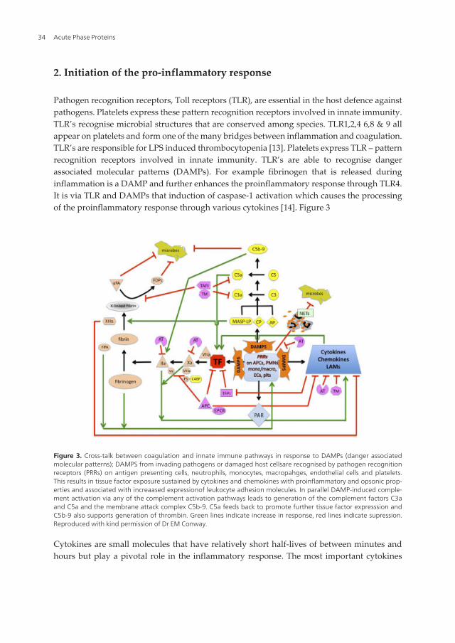
Pathogen recognition receptors, Toll receptors (TLR), are essential in the host defence againstpathogens. Platelets express these pattern recognition receptors involved in innate immunity.TLR’s recognise microbial structures that are conserved among species. TLR1,2,4 6,8 & 9 allappear on platelets and form one of the many bridges between inflammation and coagulation.TLR’s are responsible for LPS induced thrombocytopenia [13]. Platelets express TLR – patternrecognition receptors involved in innate immunity. TLR’s are able to recognise dangerassociated molecular patterns (DAMPs). For example fibrinogen that is released duringinflammation is a DAMP and further enhances the proinflammatory response through TLR4.It is via TLR and DAMPs that induction of caspase-1 activation which causes the processingof the proinflammatory response through various cytokines [14]. Figure 3
Figure 3. Cross-talk between coagulation and innate immune pathways in response to DAMPs (danger associatedmolecular patterns); DAMPS from invading pathogens or damaged host cellsare recognised by pathogen recognitionreceptors (PRRs) on antigen presenting cells, neutrophils, monocytes, macropahges, endothelial cells and platelets.This results in tissue factor exposure sustained by cytokines and chemokines with proinflammatory and opsonic prop‐erties and associated with increaased expressionof leukocyte adhesion molecules. In parallel DAMP-induced comple‐ment activation via any of the complement activation pathways leads to generation of the complement factors C3aand C5a and the membrane attack complex C5b-9. C5a feeds back to promote further tissue factor expresssion andC5b-9 also supports generation of thrombin. Green lines indicate increase in response, red lines indicate supression.Reproduced with kind permission of Dr EM Conway.
Cytokines are small molecules that have relatively short half-lives of between minutes andhours but play a pivotal role in the inflammatory response. The most important cytokines
Acute Phase Proteins34

involved in regulating the coagulation response during inflammation are IL-6, TNF-alpha,IL-8, MCP-1 and IL-1 [15].
Acute phase proteins are released as mediators of the inflammatory cascade as a chemical andcellular response to injury. They increase rapidly in plasma in response to a inflammatoryinsult. Some acute phase proteins increase transiently (C-reactive protein) while others havea more sustained elevation (Haptoglobin) [16].
The inflammatory response to surgery, atherosclerosis, infection and cardiovascular diseasehas a profound effect upon the haemostatic system, including fibrinolysis. The cytokinesinterleukin 1β (IL-1β) and interleukin 6 (IL-6) modulate the production and suppression ofmany of the coagulation enzymes formed by the Liver.
The effect is to make the endothelium and whole coagulation system more procoagulant.Figure 4
Inflammation
AnticoagulantAnti-inflammatoryProfibrinolytic
ProcoagulantAntifibrinolyticPro-inflammatory
TMEPCRProstoglandint-PA Tissue factor
vWfPAI-1PAFP-selectin
Figure 4. Inflammatory drive to a procoagulant state following endothelial stimulation.
3. Coagulation acute phase proteins
An acute-phase protein has been defined as one whose plasma concentration increases(positive acute-phase proteins) or decreases (negative acute-phase proteins) by at least 25percent during inflammatory disorders [16]. The changes in the concentrations of acute-phase proteins are due largely to changes in their production by hepatocytes. Although
Inflammation and Acute Phase Proteins in Haemostasishttp://dx.doi.org/10.5772/55998
35

the mechanism by which the liver processes the stimulation to increase and decreaseprotein production may be different in different forms of inflammatory insult e.g. sepsisand chronic inflammation [17].
Proteins whose plasma concentrations increase with
inflammation
Proteins whose plasma concentrations decrease with
inflammation
Fibrinogen
Factor VIII
Protein S
Plasminogen activator inhibitor PAI-1 C4b-binding
protein
Urokinase
α 1 Antitrypsin
α 2 Macrogloulin
von Willebrand factor
C1-esterase inhibitor
C-reactive protein
Thrombopoietin
Thrombin activatable fibrinolytic inhibitor (TAFI)?
Factor XII
Antithrombin
Histidine rich glycoprotein
Thrombomodulin
Endothelial protein C receptor
Thrombin activatable fibrinolytic inhibitor TAFI?
Protein C (? No change/decrease)
Table 1. Acute phase proteins that directly affect haemostasis
4. Coagulation proteins whose plasma concentrations increase duringinflammation
4.1. Fibrinogen
Fibrinogen is a soluble glycoprotein synthesised in the Liver with a normal plasma concen‐tration of 2 – 4 g/L and half life of 4 days. When the coagulation cascade is activated fibrinogenis the final substrate in the formation of a clot being converted to its insoluble fibrin form.Thrombin cleaves fibrinogen releasing fibrinopeptide A and B forming fibrin monomers whichhave exposed polymerisation sites on the fibrin molecule. Thrombin activates factor XIII whichcross-links these fibrin fibrils increasing clot strength and rendering them more resistant toproteolysis [18]. Fibrin creation is required with platelets (stimulating platelet aggregation bybinding to the glycoprotein IIb/IIIa platelet membrane receptor) to repair any breach in thevascular integrity and prevent haemorrhage. This process is not left unchecked and isregulated via the fibrinolytic system (plasmin production) to prevent excess fibrin accumula‐tion at the site of damage despite the procoagulant signalling drive. The local production ofplasmin is regulated via two plasminogen activators, tissue plasminogen activator (t-PA) andurokinase plasminogen activator (u-PA) which under normal physiological conditions keepsthis fibrin matrix production and its lysis tightly controlled [19].
Acute Phase Proteins36

During an inflammatory reaction fibrinogen can increase in the order of 2-3 fold and thiswill significantly increase blood viscosity and cause some degree of red cell aggregationas well as transform vascular pathologies such as atherosclerotic plaques. Fibrinogen bymeans of increasing the production of endothelin-1, is also capable of directly inducingvasoconstriction [18].
Figure 5. Fibrinogen and fibrin modulation of inflammation. Fibrin(ogen) modulates the inflammatory response byaffecting leukocyte migration but also by induction of cytokine/chemokine expression mostly via Mac-1 signalling.NO-nitric oxide, Mac-1 Macrophage antigen 1, PI3K Phosphoinositide-3-kiniase, TNF tumor necrosis factor. Repro‐duced with kind permission of Prof. K Zacharowski
There is good evidence that fibrinogen has a function in regulation of the inflammatoryresponse as is seen in the increased concentration of fibrinogen associated with atherosclerosisand cardiovascular risk. Fibrin(ogen) also participates in activation of vascular cells andregulation of the inflammatory response via the ability to bind to and activate a number ofimmune cells. The cellular ligand binding of fibrinogen is totally distinct to any coagulation
Inflammation and Acute Phase Proteins in Haemostasishttp://dx.doi.org/10.5772/55998
37
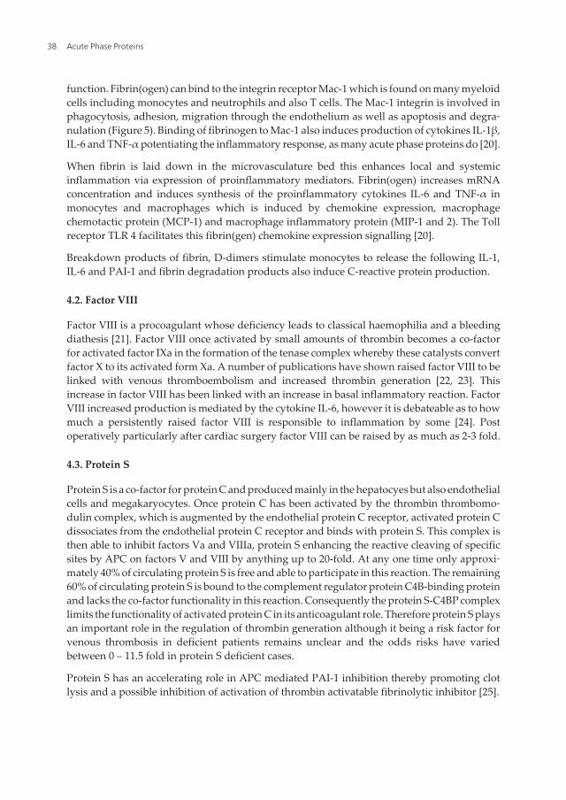
function. Fibrin(ogen) can bind to the integrin receptor Mac-1 which is found on many myeloidcells including monocytes and neutrophils and also T cells. The Mac-1 integrin is involved inphagocytosis, adhesion, migration through the endothelium as well as apoptosis and degra‐nulation (Figure 5). Binding of fibrinogen to Mac-1 also induces production of cytokines IL-1β,IL-6 and TNF-α potentiating the inflammatory response, as many acute phase proteins do [20].
When fibrin is laid down in the microvasculature bed this enhances local and systemicinflammation via expression of proinflammatory mediators. Fibrin(ogen) increases mRNAconcentration and induces synthesis of the proinflammatory cytokines IL-6 and TNF-α inmonocytes and macrophages which is induced by chemokine expression, macrophagechemotactic protein (MCP-1) and macrophage inflammatory protein (MIP-1 and 2). The Tollreceptor TLR 4 facilitates this fibrin(gen) chemokine expression signalling [20].
Breakdown products of fibrin, D-dimers stimulate monocytes to release the following IL-1,IL-6 and PAI-1 and fibrin degradation products also induce C-reactive protein production.
4.2. Factor VIII
Factor VIII is a procoagulant whose deficiency leads to classical haemophilia and a bleedingdiathesis [21]. Factor VIII once activated by small amounts of thrombin becomes a co-factorfor activated factor IXa in the formation of the tenase complex whereby these catalysts convertfactor X to its activated form Xa. A number of publications have shown raised factor VIII to belinked with venous thromboembolism and increased thrombin generation [22, 23]. Thisincrease in factor VIII has been linked with an increase in basal inflammatory reaction. FactorVIII increased production is mediated by the cytokine IL-6, however it is debateable as to howmuch a persistently raised factor VIII is responsible to inflammation by some [24]. Postoperatively particularly after cardiac surgery factor VIII can be raised by as much as 2-3 fold.
4.3. Protein S
Protein S is a co-factor for protein C and produced mainly in the hepatocyes but also endothelialcells and megakaryocytes. Once protein C has been activated by the thrombin thrombomo‐dulin complex, which is augmented by the endothelial protein C receptor, activated protein Cdissociates from the endothelial protein C receptor and binds with protein S. This complex isthen able to inhibit factors Va and VIIIa, protein S enhancing the reactive cleaving of specificsites by APC on factors V and VIII by anything up to 20-fold. At any one time only approxi‐mately 40% of circulating protein S is free and able to participate in this reaction. The remaining60% of circulating protein S is bound to the complement regulator protein C4B-binding proteinand lacks the co-factor functionality in this reaction. Consequently the protein S-C4BP complexlimits the functionality of activated protein C in its anticoagulant role. Therefore protein S playsan important role in the regulation of thrombin generation although it being a risk factor forvenous thrombosis in deficient patients remains unclear and the odds risks have variedbetween 0 – 11.5 fold in protein S deficient cases.
Protein S has an accelerating role in APC mediated PAI-1 inhibition thereby promoting clotlysis and a possible inhibition of activation of thrombin activatable fibrinolytic inhibitor [25].
Acute Phase Proteins38
Protein S has been shown to increase during inflammation. This may in part be due tocounterbalancing the procoagulant drive of the coagulation system in these circumstances byproviding more anticoagulant effect via the protein C pathway. It may also have to do withother non-anticoagulant actions it has via its binding with C4BP (see below). Certainlyinhibition of protein S in in-vivo models of bacteraemia have shown to provoke the cytokineresponse and what was a non-lethal injected dose of E.coli in baboons resulted in a lethalreaction [26].
4.4. C4b-binding protein
The complement binding protein C4BP is a co-factor to a serine protease in the degredation ofC4 in the classical complement pathway and providing it has been suggested protection frominflammation at a cellular level. The C4BP molecule has a central region with seven α chainsand one β chain emanating from it each with distinct ligand binding regions. It is on the βchain side arm that the protein S binding region resides and in normal human plasma ~80%of C4BP is found as this β chain form. C4BP is an acute phase protein and its levels can increaseto 400% of normal [25]. However this is mainly of the C4BPα form so the Protein S-C4BPcomplex concentration is not generally affected. As so much protein S is normally bound toC4BP and the concentration of the free anticoagulant active co-factor protein S level quitetightly controlled is there a physiological role for the PS-C4BP complex? It has been suggestedthat protein S forms one of the bridges between coagulation and inflammation. As protein Shas a high affinity to bind to negatively charged phospholipids it can bring C4BP into closeproximity to these sites allowing controlled complement activation at areas of vascular damagewhere coagulation is activated. This has been linked to apoptosis and the complement systemallowing rapid clearance of apoptotic cells from the site of damage by macrophages but moreprobably limiting or inhibiting further complement activation via necrotic cells [27, 28].
4.5. Plasminogen activator inhibitor PAI-1
The fibrinolytic system is the opposite of the coagulation system. It limits clot formation to thesite of injury and breaks down existing clot by an enzyme cascade. It is also, like haemostasis,linked to inflammation with acute phase proteins that increase and others that are decreased.Plasminogen provides the fibrinolytic potential and when converted to its active form plasminis able to bind to fibrin and begin its degradation. Plasminogen is activated by two activators,tissue plasminogen activator (t-PA) and urokinase tissue plasmingen activator (u-PA). Thesein turn are inhibited by plasminogen activator inhibitor 1 and 2 (PAI-1, PAI-2) and plasmin isinhibited by α-2-antiplasmin. Of these inhibitors PAI-1 is a positive acute phase protein andincreases during inflammation. The cytokine tissue necrosis factor
(TNF-α) suppresses finbrinolysis by down regulating t-PA expression in endothelial cellswhile production PAI-1 then in turn PAI-1 inhibits TNF-α is inhibited by PAI-1. It has alsobeen shown that by inhibiting TNF-α plasma levels of PAI-1 decrease [19].
Inflammation and Acute Phase Proteins in Haemostasishttp://dx.doi.org/10.5772/55998
39

The increase in PAI-1 levels is again a driver of the haemostatic system towards a moreprothrombotic state. PAI-2 will not be discussed here but for interested readers they aredirected to the following publication [29].
4.6. Urokinase
Urokinase plasminogen activator (u-PA) is a multifunctional serine protease. It has beenshown to act as both a signalling ligand and a proteolytic enzyme. As an acute phase proteinits increase has been found in a number of metastatic carcinomas and this has led to a link inits role of tumor growth and cellular expansion in conditions such as cardiac fibrosis andatherosclerosis [30]. As well as displaying involvement in tissue remodelling u-PA alsoappears to regulate macrophage activation and function. The macrophages not only synthesisand release u-PA but also have receptors for the u-PA protease on their membranes. Theexcreted u-PA induces TNF-α synthesis and secretion. The u-PA molecule has also been shownto initiate inflammation via the release of IL-6 and IL-1β from monocytes and lymphocytes[31]. The u-PA and its receptor uPAR also mediate immune complex induced inflammationin the lung. This is achieved via generation of u-PA at the site of cellular inflammation withsubsequent activation of its receptor uPAR. This then sets off cellular signalling with C5a/C5aRon the alveolar macrophages, recruitment of polymorphonuclear leukocytes and adequateTNF-α production [32].
4.7. α 1 Antitrypsin
The α-1-antitrypsin glycoprotein is a proteinase inhibitor synthesised and secreted in thehepatocytes of the Liver. It is one of two physiological inhibtors of activated protein C. It alsohas a profound anti-inflammatory effect in the lungs with concentrations during a inflamma‐tory response reaching levels found in the plasma. The range of antiprotease activity seen inthe lungs includes neutrophil elastase and plasminogen acitvators. If there is a deficiency ofα-1-antitrypsin (as with the genetically abnormal SERPINA1 gene mutations) then neutrophilelastase provoked by infection and inflammation will unchecked breakdown elastin anddestroy alveolar walls leading to emphysema [33].
4.8. Alpha-2-macroglobulin
Alpha-2-Macroglobulin (α-2M) is an ancient serine protease binding host or foreign peptidesand particles, thereby serving as humoral defense barrier against pathogens in the plasma andtissues. In humans α-2M, interacts and captures virtually any proteinase whether self orforeign, suggesting a function as a unique "panproteinase inhibitor." In adult humans itprovides somewhere between 10-25% of the overall anti-thrombin activity in plasma. It is alsothe primary inhibitor of thrombin in neonates and infants under 1 year of age until the Livermatures and begins producing sufficient Antithrombin to take over the role. At times ofinflammation when antithrombin levels are low α-2M can become a ‘back-up’ thrombininhibitor [34].
Acute Phase Proteins40
4.9. C1-esterase inhibitor
C1-inhibitor (C1INH) is a serpin and major inhibitor of the contact coagulation system thatinvolves factor XII, kallikrein and kininogen. It is also an important regulator of complementactivation inhibiting the first component of complement C1. Another important biological roleof C1INH is vascular permeability regulation [35]. This is well illustrated in patients who sufferfrom hereditary angioedema where there is a deficiency of the C1INH activity. As it now seemsfactor XII plays a substantial part in the formation of thrombin during sepsis and inflammationwhere neutrophil extracellular traps are present that release polyphosphate that in turnactivate factor XII it would appear normal for C1INH to act as a positive acute phase proteinin limiting this activation as well as its complement inhibitory role. A rapid appearance ofC1INH-factor XIIa complexes is reported during sepsis with a sharp fall in the C1INH activitywherein the inhibitory function of α-2Macroglobin becomes more important in its kallikreininhibiting role [36].
4.10. von Willebrand factor
The von Willebrand factor (VWF) is a multimeric protein that mediates the adhesion ofplatelets to the exposed subendothelium at the site of vascular injury. It also serves as a carrierprotein for the labile coagulation factor VIII. VWF is synthesised in and stored in the Weibal-Palade bodies of endothelial cells and megakaryocytes/platelets. Endothelial cells release VWFwhen activated or stimulated. These VWF molecules are ultralarge and hyperactive, proficientin binding to platelets via the glycoprotein Ib-X-V complex without any external activation.Normally the ultralarge VWF multimers are cleaved into smaller less active multimers beforebeing released into plasma. Cleavage is performed by ADAMTS-13 (a disintegrin and metal‐loprotease with thrombospondin motif). A deficiency of ADAMTS-13 leads to a thromboticthrombocytopenic purpura a thrombotic microangiopathy. Increased plasma levels of VWFhave been reported in a number of disease states including coronary artery disease, autoim‐mune disease, trauma and infections. A common underlying process in all of these is inflam‐mation. There appears to be a complex interaction between the common inflammatorycytokines IL-6, IL-1β and TNF-α the release and cleavage of the VWF multimers byADAMTS-13.
Release of VWF multimers from endothelial cells occurs in a dose dependent fashion whenstimulated with IL-8 and TNF-α and IL-6 inhibits cleavage of the ultrlarge multimers. Thisdemonstrates that inflammatory cytokines interfere with the equilibrium of release and rateof cleavage of the ultralarge VWF multimers.
This increases prothrombotic risk in this setting as most of the positive acute phase proteinshave a propensity to do creating a procoagulant environment. It is of interest to note that IL-8resides in the Weibel-Palade bodies of the platelet the same place as VWF and is involved inthe platelet leukocyte aggregation and it is TNF-α that activates endothelial cells releasing IL-8.The inhibition by IL-6 of ultralarge multimer cleavage by ADAMTS-13 may be by way ofsynthetic and secretion inhibition. In overwhelming sepsis there is also probably an elementof exhaustion of metalloprotease activity [37].
Inflammation and Acute Phase Proteins in Haemostasishttp://dx.doi.org/10.5772/55998
41
4.11. C-reactive protein
C-reactive protein (CRP) is a pentameric molecule that increases several hundred foldfollowing an inflammatory stimulatory response this being primarily due to IL-6 stimulationof production of CRP in the hepatocytes of the Liver. CRP amplifies the host defence mecha‐nism by activation of complement via C1 and stimulation of macrophages. CRP also upregu‐lates tissue factor expression on monocytes [38] and induces release of PAI-1 therebydownregulating fibrinolysis [39] promoting a procoagulant state. The pentameric CRP isthought to be directly proinflammatory at high concentration like those of sepsis or majorsurgery but more subtle inflammatory reactions take place when monomeric CRP is releasedfrom the pentameric form. This appears to be driven by activated platelets revealing new lipidmessenger sites; lysophosphatidycholine (LPC) that bind and dissociate the pentameric formof CRP to the monmeric form [40].
During sepsis when disseminated intravascular coagulation (DIC) occurs this has been foundexperimentally to co-inside with formation of a calcium-dependent complex between CRP andvery low density lipoprotein (VLDL). The VLDL molecular makeup is different in septicpatients to normal controls due to a deficiency of phosphatidylethanolamine. This CRP-VLDLlipid raft increases the procoagulant effect through an increase in prothrombinase activity. TheCRP-VLDL complex exists in vivo and it has been postulated that is has a pathogenic role indisseminating the intravascular coagulation [41].
5. Coagulation proteins whose plasma concentrations decrease duringinflammation
5.1. Antithrombin
Antithrombin (AT) is one of the major natural anticoagulats inhibiting thrombin, factor Xa,IXa and factor VIIa bound to tissue factor. Inhibition of factors Xa, IXa and the VIIa-tissue factorare accelerated via the endothelial cell heparin like proteoglycans. The importance of thisanticoagulant pathway is highlighted when AT levels are low, either congenitally or acquired,the risk of thrombosis is significantly increased.
AT has been shown in-vitro using HUVEC cells and IL-6 to act as a negative acute phase protein[42]. Other mechanisms that can reduce AT function during an inflammatory response includeincreased consumption from activation of haemostasis and increased degradation by proteo‐lytic enzymes (elastase released from activated neutrophils). Furthermore inflammatorycytokines can induce a reduction in the production of glycosaminoglycans such as heparinand chondroitin sulphate on the endothelial cell which may contribute to the impaired functionof AT due to GAGs acting as physiological heparin-like cofactors promoting the anticoagulantanti-thrombin activity of AT [43, 44].
AT can indirectly act as an anti-inflammatory molecule by directly inhibiting thrombinreducing its inflammatory properties of haemostatic and complement activation, leukocyte,
Acute Phase Proteins42

endothelial and platelet activation. AT also appears to directly act as an anti-inflammatoryagent by binding with leukocytes receptors blocking their communication with endothelialcells and limiting their adhesion and migration [45]. In animaI models it has also been shownthat AT induces the release of prostacyclin from endothelial cells. The prostacyclin is a plateletinhibitor and abrogates neutrophils adhering to endothelial cells both of which contribute tothe inflammatory response [46].
In addition AT can also modulate cellular receptor expression by downregulating the expres‐sion of CD11b/CD18 on leucocytes which bind factor X aiding its activation [47].
AT is also able to decrease expression of tissue factor and IL-6 expression on monocytes andendothelial cells reducing the inflammatory drive [48].
5.2. Histidine rich glycoprotein
Histindine rich glycoprotein (HRGP) is a single polypeptide chain protein that is synthesised inthe Liver found in plasma and on the surface of leukocytes, monocytes and the α-granules ofplatelets. HRGP has been shown to bind to a number of molecules including, haem, Zn2+,plasminogen, fibrinogen, IgG, complement and factor XIIa (not the inactive zymogen) [49].HRGP exerts some degree of innate immune response by exhibiting antimicrobial activity tosome organisms and removal of necrotic cells by binding to free decondensed DNA (polyphos‐phate).
HRGP behaves as a negative acute protein during inflammation [50]. This is of interest asfibrinogen one of its primary ligands is increased. Possibly more importantly is the fact thatHRGP strongly inhibits polyphosphate induced factor XII autoactivation via increased Zn2+
levels in response to local platelet activation, showing it to be a significant modulator of factorXII activation during sepsis and inflammation [51]. Because of its negative acute phase naturethis will allow increased activation of factor XII in these circumstances potentiating the pro-inflammatory and pro-coagulant condition.
5.3. Thrombomodulin
Thrombomodulin (TM) is present on endothelial cells of the entire vasculature and is atransmembrane protein with epidermal growth factor (EGF) repeating molecules that stick outof the plasma membrane and provide binding and activation sites for a number of molecules.There is a high affinity binding site for thrombin on EGF domain 5-6 from which protein C isactivated at EGF domains 4-6. Thrombin activatable fibrinolytic inhibitor is activated bythrombin at EGF sites 3-6 [52]. When thrombin binds and complexes with thrombomodulinon the cell membrane its ability to activate protein C increases by greater than 1000-fold.Activated protein C anti-inflammatory effects are discussed later in this chapter, see below.Once thrombin has bound to TM it becomes quickly neutralised by its natural serine proteasesinhibitors antithrombin, heparin co-factor II and protein C inhibitor and therefore activationof protein C also ceases.
The effect of inflammation on TM is to decrease its presence on the endothelial cell surface byits cellular internalisation by endocytosis via TNF-α. This creates a site where coagulation can
Inflammation and Acute Phase Proteins in Haemostasishttp://dx.doi.org/10.5772/55998
43

take place as the anticoagulant barrier has been removed and it may also stimulated furtherinflammatory response [43]. CRP has also been shown in experimental conditions usinghuman coronary artery endothelial cells treated with CRP in a dose and time dependentmanner to reduce messenger RNA levels of TM [53]. TM also provides anti-inflammatoryprotection from complement activation by enhancing inactivation of C3b and by promotingactivation of thrombin-activatable fibrinolysis inhibitor that inactivates complement anaphy‐latoxins C5a and C3a [54]. Others have shown that thrombin-activatable fibrinolysis inhibitoractivation via TM is attenuated by platelet factor 4 released from activated platelets [55].
5.4. Endothelial protein C receptor
The thrombomodulin-thrombin conversion of protein C to its activated form is facilitated bythe endothelial protein C receptor (EPCR). The EPCR is another endothelial cell transmem‐brane protein located in close proximity to the thrombomodulin molecule and with highaffinity binding for protein C. EPCR as well as binding protein C also binds its activated formand via membrane lipid rafts they complex with PAR-1 [56]. The EPCR-activated protein Cmolecule activates PAR-1 in a different way to thrombin which allows it to signal through apotent Gi protein pathway by which anti-inflammatory pathways are stimulated within theendothelial cell [57]. C-reactive protein has also been shown, as in the case of thrombomodulin,under experimental cell culture conditions to down regulate EPCR.
5.5. Thrombin activatable fibrinolytic inhibitor
Thrombin activatable fibrinolytic inhibitor (TAFI) is a basic carboxypeptidase identical to theplasma procarboxypeptidases B, U and R. TAFI is activated by thrombin and plasmin althoughthe most efficient activator is the thrombomodulin-thrombin cellular membrane boundcomplex. TAFI inhibits fibrinolysis by cleaving lysine residues from fibrin which restricts thebinding of tissue plasminogen activator to these sites and enhancing the plasminogen con‐version to plasmin potentiating further fibrin breakdown [58]. TAFI certainly modulates thebalance between coagulation and fibrinolysis but it appears its linking role with inflammationstill needs to be fully elucidated. For instance TAFI has been shown to be a positive acute phaseprotein in mice [59] but in the same year Boffa et al interestingly showed that IL-6 administeredto cultured HepG2 cells resulted in a 60% decrease in TAFI mRNA [58]. Subsequently TAFIhas been shown to be raised in experimental endotoxemia [60]. Its anti-inflammatory proper‐ties are also downregulated by platelet factor 4 which is release from activated platelets andendothelial cells during activating stimuli and inflammatory insults which prevents TAFIactivation by binding to the thrombin-thrombomodulin complex thereby preventing TAFI’sinactivation of the complement anaphylatoxins C5a and C3a [55].
Acute Phase Proteins44

6. Other major components of the acute phase response that affecthaemostasis
6.1. Protein C
The protein C pathway is known to be an important anticoagulant system with patientsdeficient in protein C being at risk of thrombosis or in its homozygous form purpura fulminans.Activated protein C inhibits factors V and VIII this being supported by the activation of proteinC by thrombin bound to thrombomodulin on the endothelial cell surface. As well as acting asan anticoagulant activated protein C is also able to inhibit PAI-1. The anticoagulant andantifibrinolytic aspects of the protein C pathway have been elucidated and well describedalthough there still appears much to learn from the interaction of protein C during theinflammatory response.
It is unclear from studies of the acute phase proteins whether protein C acts as a positive ornegative acute phase protein. Most studies show it to have no change in concentration duringan inflammatory response or its plasma concentration to decrease. A decrease in protein Ccould be attributed to consumption as well as a cytokines limiting the natural anticoagulantresponse. Activated protein C also confers a cytoprotective, anti-inflammatory, anti-apoptosisand endothelial barrier stabilisation effect when active [61].
Activated protein C signals its anti-inflammatory effects mainly via PAR-1 pathways wherebyfollowing Gi signalling and sphingosine-1-phosphate production there is improvement inendothelial cell barrier function [62].
Transcriptional profiling studies using cell cultures of human umbilical vein endothelial cells(HUVECs) have demonstrated that recombinant human activated protein C can regulateendothelial cell gene expression linked to inflammation and cell survival [63]. The activatedprotein C suppresses NFκB a cell nuclear transcription factor, and by reducing its expressionand function this in turn causes inhibition of cytokine signaling [64].
6.2. Cytokines
Cyotkines are a superfamily of molecules involved in cell signalling many of which areincreased greater than 1000-fold during an inflammatory insult. Cytokines such as interleukin(IL)-6, but also platelet-derived growth factor and monocyte chemoattractant protein (MCP)-1are capable of stimulating tissue factor expression in mononuclear cells. Tissue factor beingthe initiator of thrombin generation and fibrin formation.
IL-6 is a multifunctional cytokine that is induced in many disease states such as sepsis,endotoxaemia and in-vitro after administration of tumor necrosis factor (TNF). Several studieshave suggested IL-6 to be a potential mediator of endotoxin induced coagulation activation.This has been validated by treatment of chimpanzees with a monoclonal anti-IL6 antibody thatablated the activation of coagulation [65]. Cytokines are pivotal in providing a means of crosstalk between inflammation and coagulation [66].
Inflammation and Acute Phase Proteins in Haemostasishttp://dx.doi.org/10.5772/55998
45
Inflammation and coagulation can not be considered as two separate processes because thereare several interlocking points making them a unique defensive host reaction. The endotheli‐um is one of the major links between the two since damaged endothelium during inflammationrepresents a surface where proteins involved in both coagulation and fibrinolysis and thedevelopment of inflammation are expressed. Cytokines down regulate the surface receptorthrombomodulin and the activation of protein C but at the same time increase the expressionof tissue factor. Platelets adhere to these sites of vascular damage and when activated alsorelease several cytokine mediators of inflammation, adhesion molecules and growth factorsincluding IL-1β, CD40 ligand, vitronectin and RANTES [13], [67].
In addition to the inflammatory cytokines like interleukin 1 and TNF, infection per se cantrigger the release of neutrophil extracellular traps (NETs). Which in turn release cytokinesand microparticles exacerbating the acute phase haemostatic response [68].
Cytokines also upregulate the complement system activated C5b9 complexes can assembleclot promoting membrane phospholipids, TNF-α downregulates thrombomodulin andvascular heparin sulphates promoting a procoagulant environment. The net effect of this is tofurther lessen the inhibitory mechanisms that control thrombin generation.
6.3. Platelets
Platelet numbers increase following surgery, trauma and sepsis following a inflammatoryresponse. Both IL-6 and thrombopoetin stimulate the production of platelets [69]. This reactivethrombocytosis can last up to13 days post surgery and may responsible for PAF adverse eventsin liver reperfusion and thrombotic complications post cardiac surgery. It appears thatthrombopoietin is an acute phase reactant but not uniquely responsible for the rise in theplatelet count during a reactive thrombocytosis but is probably aided and abetted by IL-6 [70].
Platelets release CD40 ligand which induces tissue factor expression and increases inflamma‐tory cytokines IL-6 and IL-8. (Esmon CT). The CD40 ligand is a transmembrane protein relatedto TNF-α which was originally identified on stimulated CD4+ T cells. The interaction of CD40on T and B cells is integral to the development and function of the humoral immune response.It is now known that CD40 ligand is found on many cells including macrophages, endothelialcells and platelets. Upon activation platelets express CD40L within seconds. As wtih TNF-αand IL-1 the CD40L on platelets induces endothelial cells to exude cytokines and up regulatethe expression of adhesion molecules. This all increases the general recruitment of leukocytesto the site of injury. Platelets therefore directly initiate the inflammatory response at the vesselwall [14]. Figure 6
The concept that platelets play a key role in the host defence and inflammatory response hastaken longer to realise mainly due to their role in primary haemostasis. This new role ofplatelets as immune effector cells is enhanced by the finding that platelets directly interact withmicrobes and bacteria.
Platelet activation along with procoagulant events and fibrin formation seem crucial for thecontainment and killing of bacteria. Platelets have recently been shown to induce the formationof neutrophil extracellulat traps (NETs). NETs are lattice arrangements of decondensed nuclear
Acute Phase Proteins46

chromatin which are laced with histones that have antimicrobial properties. It also appearsthat this may be an ‘overshooting’ of the hosts defence mechanism with further plateletactivation, thrombosis and endothelial cell injury [71], [72], [73].
Figure 6. Inflammation and platelet activation. Platelet derived growth factor – PDGF, platetlet factor 4 – PF4.
The concept that platelets play a key role in the host defence and inflammatory response has taken longer to realise mainly due to
their role in primary haemostasis. This new role of platelets as immune effector cells is enhanced by the finding that platelets
directly interact with microbes and bacteria.
Platelet activation along with procoagulant events and fibrin formation seem crucial for the containment and killing of bacteria.
Platelets have recently been shown to induce the formation of neutrophil extracellulat traps (NETs). NETs are lattice arrangements
of decondensed nuclear chromatin which are laced with histones that have antimicrobial properties. It also appears that this may
be an ‘overshooting’ of the hosts defence mechanism with further platelet activation, thrombosis and endothelial cell injury[71],[ 72],
[73].
6.4. Endothelium
Endothelial cells in the vasculature are actively involed in haemostasis providing an anticoagulant surface preventing activation of
the coagulation system. Endothelial cells produce elements with proinflammatory, procoagulant and antifibrinolytic properties as
well as those with the opposite anti-inflammatory, anticoagulant and profibrinolytic properties. When endothelial cells are
activated or damaged they release into the local surroundings procoagulant components such as von Willebrand factor ( a platelet
binding factor and a carrier protein for factor VIII) and thromboxane A2 (a platelet activator) and plasminogen activator inhibitor
(PAI-1 a potent inhibitor of tissue plasmingen activator). The opposite is true of components that provide an anticoagulant surface
in the milieu of the blood vascular barrier. Thrombomodulin, a thrombin binding transmembrane protein that switches thrombin
from a procoagulant enzyme to one that activates protein C, a nautral anticoagulant that inhibits the activity of factors V and VIII,
in internalised within the endothelial cell. Thrombomodulin is also cleaved from the endothelium by activated Neutrophils. Tissue
factor is expressed on the cell surface along with adhesion molecules that mediate the interaction of neutrophils and platelets these
included vascular cell adhesion molecule (VCAM-1), P and E selectin and intracellular
cell adhesion molecule (ICAM-1) all of which promote the inflammatory response. So the endothelium serves as an interface for the
inflammatory response leading to local activation of the coagulation system, vasodilatation and pro-inflammatory state[3], [57].
6.5. Complement
Complement is part of the innate immune system and the effector of antibody mediated immunity. The biological functions of
complement include the defence against infections and the clearance of immune complexes and apoptotic cells. The complement
cascade is made up of approximately 30 proteins circulating in plasma and expressed on cellular surfaces. The complement cascade
is activated via three pathways: classical, lectin and alternative. The classical pathway is initiated by the binding of C1q to antigen–
antibody complexes. The lectin pathway is initiated via the binding of mannose-binding lectin or ficolins to sugars found at the
bacterial cell wall. Both of these pathways lead to the formation of a C3 convertase. The alternative pathway is stimulated by
spontaneous hydrolysis of internal thioester bonds within C3. C3a and C5a are anaphylatoxins and inflammatory mediators which
are inhibited by TAFI.
Figure 6. Inflammation and platelet activation. Platelet derived growth factor – PDGF, platetlet factor 4 – PF4.
6.4. Endothelium
Endothelial cells in the vasculature are actively involed in haemostasis providing an anticoa‐gulant surface preventing activation of the coagulation system. Endothelial cells produceelements with proinflammatory, procoagulant and antifibrinolytic properties as well as thosewith the opposite anti-inflammatory, anticoagulant and profibrinolytic properties. Whenendothelial cells are activated or damaged they release into the local surroundings procoagu‐lant components such as von Willebrand factor ( a platelet binding factor and a carrier proteinfor factor VIII) and thromboxane A2 (a platelet activator) and plasminogen activator inhibitor(PAI-1 a potent inhibitor of tissue plasmingen activator). The opposite is true of componentsthat provide an anticoagulant surface in the milieu of the blood vascular barrier. Thrombo‐modulin, a thrombin binding transmembrane protein that switches thrombin from a procoa‐gulant enzyme to one that activates protein C, a nautral anticoagulant that inhibits the activityof factors V and VIII, in internalised within the endothelial cell. Thrombomodulin is alsocleaved from the endothelium by activated Neutrophils. Tissue factor is expressed on the cellsurface along with adhesion molecules that mediate the interaction of neutrophils and plateletsthese included vascular cell adhesion molecule (VCAM-1), P and E selectin and intracellular
Inflammation and Acute Phase Proteins in Haemostasishttp://dx.doi.org/10.5772/55998
47
cell adhesion molecule (ICAM-1) all of which promote the inflammatory response. So theendothelium serves as an interface for the inflammatory response leading to local activationof the coagulation system, vasodilatation and pro-inflammatory state [3], [57].
6.5. Complement
Complement is part of the innate immune system and the effector of antibody mediatedimmunity. The biological functions of complement include the defence against infections andthe clearance of immune complexes and apoptotic cells. The complement cascade is made upof approximately 30 proteins circulating in plasma and expressed on cellular surfaces. Thecomplement cascade is activated via three pathways: classical, lectin and alternative. Theclassical pathway is initiated by the binding of C1q to antigen–antibody complexes. The lectinpathway is initiated via the binding of mannose-binding lectin or ficolins to sugars found atthe bacterial cell wall. Both of these pathways lead to the formation of a C3 convertase. Thealternative pathway is stimulated by spontaneous hydrolysis of internal thioester bonds withinC3. C3a and C5a are anaphylatoxins and inflammatory mediators which are inhibited by TAFI.
Complement and coagulation are again two systems that cannot be viewed separately duringan inflammatory response. Complement contributes significantly to the prothrombotic stateduring inflammation. The direct procoagulant activities of complement involve the activationof platelets via C3a and the C5b-9 membrane attack complex and the upregulation of tissue factorand PAI-1 expression on various cell types by C5b. Thrombin has also recently been identifiedas an activator of C5 linked to the coexistence of a C5 convertase enzyme [74], [75], [76].
It is of interest to note the complement factor C4B-binding protein is a positive acute phase proteinthat can increase greater than 400% during inflammatory states. The C4B-bp in normal circum‐stances binds the natural anticoagulant protein S. Protein S acts as a co-factor with activatedprotein C in the inactivation of factors V and VIII. It was thought that an increase in C4B-bp mayincrease the binding of circulating protein S, approximately 60% of protein S is normally boundleaving the other 45% free protein S to aid in the V and VIII inactivation. However this in‐crease is restricted to the C4BPα+ form, which does not bind to PS. Therefore, the blood levelsof the active free form of PS remain stable even during an acute phase response. The normalbinding of protein S to C4B-bp probably allows this complex to bind via protein S high affinityfor negatively charged phospholipids depositing it at area of cellular damage and limiting furtherapoptosis through complement activation as it will block C4b (see Protein S).
Author details
Simon J. Davidson
Department of Haematology, Royal Brompton Hospital, London, UK
Acute Phase Proteins48

References
[1] Lipinski S, Bremer L, Lammers T, Thieme F, Schreiber S, Rosenstiel P.Coagulationand inflammation. Molecular insights and diagnostic implications.Hamostaseologie.2011;31:94-104.
[2] Levi M, van der Poll T, Büller HR.Bidirectional relation between inflammation andcoagulation Circulation. 2004;109:2698-2704.
[3] Margetic S. Inflammation and haemostasis. Biochemica Medica. 2012;22:49-62.
[4] Esmon CT. Inflammtion and thrombosis. J Thromb Haemost 2003;1:1343-1348.
[5] MacFarlane RG. An enzyme cascade in the blood clotting mechanism and its func‐tion as a biochemical amplifier. Nature. 1964;202:498-499.
[6] Davie EW, Ratnoff OD. Waterfall sequence for intrinsic blood clotting. Science.1964;145:1310-1312.
[7] Munroe DM, Hoffman M, Roberts HR. Platelets and thrombin generation. ATVB.2002;22:1381-1389.
[8] Price GC, Thompson SA, Kam PCA. Tissue factor and tissue factor pathway inhibi‐tor. Anaesthesia2004;59: 483–492
[9] Esmon CT, Xu J, Lupu F. Innate immunity and coagulation. J Thromb Haemost.2011;9 (suppl.1):182-188.
[10] Levi M The coagulant response in sepsis and inflammation. Hamostaseologie.2010;30:10-16
[11] Ma L, Dorling A. The roles of thrombin and protease-activated receptors in inflam‐mation.Semin Immunopathol. 2012 ;34:63-72.
[12] Butenas S, Orefo T, Mann KG. Tissue factor in coagulation. Which? Where? When?Arterioscler Thromb Vasc Biol 2009;29:1989-1996.
[13] Vieira-de-Abreu A, Campbell RA, Weyrich AS, Zimmerman GA. Platelets: versatileeffector cells in hemostasis, inflammation, and the immune continuum. Semin Immu‐nopathol. 2012 ;34:5-30.
[14] Delvaeye M, Conway EM. Coagulation and innate immune responses: can we viewthem separately? Blood 2009;114:2367-2374
[15] Ceciliani F, Giordano A, Spagnolo V. The systemic reaction during inflammation: theacute-phase proteins.Protein Pept Lett. 2002;9:211-223
[16] Gabay C, Kushner I. Acute phase proteins and other systemic responses to inflamma‐tion. NEJM. 1999;340:448-454.
Inflammation and Acute Phase Proteins in Haemostasishttp://dx.doi.org/10.5772/55998
49
[17] Vary TC, Kimball SR. Regulation of hepatic protein synthesis in chronic inflamma‐tion and sepsis. Am J Physiol. 1992;262:445-452.
[18] Davalos D, Akassoglou K.Fibrinogen as a key regulator of inflammation in disease.Semin Immunopathol. 2012;34:43-62
[19] Medcalf RL. Fibrinolysis, inflammation and regulation of the plasminogen activatingsystem. J Thromb Haemost. 2007;5 (suppl 1):132-142.
[20] Jennewein C, Tran N, Paulus P, Ellinghaus P, Eble JA, Zacharowski K. Novel aspectsof fibrin(ogen) fragments during inflammation. Mol Med. 2011;17:568-573.
[21] van den Berg HM, De Groot PH, Fischer K. Phenotypic heterogeneity in severe he‐mophilia. J Thromb Haemost. 2007;5 Suppl 1:151-156.
[22] O'Donnell J, Mumford AD, Manning RA, Laffan MA. Marked elevation of thrombingeneration in patients with elevated FVIII:C and venous thromboembolism. Br J Hae‐matol. 2001;115:687-691.
[23] Rosendaal FR. High levels of factor VIII and venous thrombosis. Thromb Haemost.2000 ;83:1-2
[24] Tichelaar V, Mulder A, Kluin-Nelemans H, Meijer K. The acute phase reaction ex‐plains only a part of initially elevated factor VIII:C levels: a prospective cohort studyin patients with venous thrombosis. Thromb Res 2012;129:183-186.
[25] Rezende SM, Simmonds RE, Lane DA. Coagulation, inflammation, and apoptosis:different roles for protein S and the protein S-C4b binding protein complex. Blood.2004;103:1192-1201.
[26] Esmon CT. Role of coagulation inhibitors in inflammation.Thromb Haemost.2001;86:51-56
[27] Kask L, Trouw LA, Dahlbäck B, Blom AM. The C4b-binding protein-protein S com‐plex inhibits the phagocytosis of apoptotic cells.J Biol Chem. 2004;279:23869-23873.
[28] Trouw LA, Nilsson SC, Gonçalves I, Landberg G, Blom AM. C4b-binding proteinbinds to necrotic cells and DNA, limiting DNA release and inhibiting complementactivation. J Exp Med. 2005;201:1937-1948.
[29] Lee JA, Cochran BJ, Labov S, Ransom M. Forty Years Later and the Role of Plasmino‐gen Activator Inhibitor Type 2/SERPINB2 Is Still an Enigma. Semin Thromb Hemost.2011;37:395-407
[30] Cozen AE, Moriwaki H, Kremen M, DeYoung MB, Dichek HL, Slezicki KI, YoungSG, Véniant M, Dichek DA. Macrophage-targeted overexpression of urokinase caus‐es accelerated atherosclerosis, coronary artery occlusions, and premature death.Cir‐culation. 2004;109:2129-2135.
Acute Phase Proteins50

[31] Shushakova N, Eden G, Dangers M, Menne J, Gueler F, Luft FC, Haller H, Dumler I.The urokinase/urokinase receptor system mediates the IgG immune complex-in‐duced inflammation in lung. J Immunol. 2005 Sep 15;175(6):4060-8.
[32] Sitrin RG, Shollenberger SB, Strieter RM, Gyetko MR. Endogenously produced uroki‐nase amplifies tumor necrosis factor-alpha secretion by THP-1 mononuclear phago‐cytes. J Leukoc Biol. 1996;59:302-311.
[33] Brantly M. Α1-antitrypsin: not just an antiprotease. Am J Respir Cell Mol Biol.2002;27:652-654.
[34] Borth W. Alpha 2-macroglobulin, a multifunctional binding protein with targetingcharacteristics. FASEB J. 1992;6:3345-3453.
[35] Zeerleder S. C1-inhibitor: more than a serine protease inhibitor. Semin Thromb He‐most. 2011;37:362-374.
[36] Davis AE 3rd, Lu F, Mejia P. C1 inhibitor, a multi-functional serine protease inhibi‐tor. Thromb Haemost. 2010;104:886-893
[37] Bernardo A, Ball C, Nolasco L, Moake JF, Dong JF. Effects of inflammatory cytokineson the release and cleavage of the endothelial cell-derived ultralarge von Willebrandfactor multimers under flow. Blood. 2004;104:100-106.
[38] Cermak J, Key NS, Bach RR, Balla J, Jacob HS, Vercellotti GM. C-reactive protein in‐duces human peripheral blood monocytes to synthesize tissue factor.Blood.1999;82:513-520.
[39] Boudjeltia KZ, Piagnerelli M, Brohee D, Guillaume M, Cauchie P, Vincent JL, Rema‐cle C, Bouckaert Y, Vanhaeverbeek M. Relationship between CRP and hypofibrinoly‐sis: Is this a possible mechanism to explain the association between CRP andoutcome in critically ill patients? Thrombosis Journal. 2004;2:1-5.
[40] Filep JG. Platelets affect the structure and function of C-reactive protein. Circ Res.2009;105:109-111.
[41] Toh CH, Samis J, Downey C, Walker J, Becker L, Brufatto N, Tejidor L, Jones G, Hou‐dijk W, Giles A, Koschinsky M, Ticknor LO, Paton R, Wenstone R, Nesheim M. Bi‐phasic transmittance waveform in the APTT coagulation assay is due to theformation of a Ca(++)-dependent complex of C-reactive protein with very-low-densi‐ty lipoprotein and is a novel marker of impending disseminated intravascular coagu‐lation. Blood. 2002;100:2522-2529.
[42] Niessen RW, Lamping RJ, Jansen PM, Prins MH, Peters M, Taylor FB Jr, de Vijlder JJ,ten Cate JW, Hack CE, Sturk A.Antithrombin acts as a negative acute phase proteinas established with studies on HepG2 cells and in baboons. Thromb Haemost.1999;78:1088-1092.
Inflammation and Acute Phase Proteins in Haemostasishttp://dx.doi.org/10.5772/55998
51

[43] Bourin J, Lindahl U. Glycosaminoglycans and the regulation of blood coagulation.Biochem J. 1993;289:313-330.
[44] Klein, N.J., Shennan, G.I., Heyderman, R.S. & Levin, M. Alteration in glycosaminogly‐can metabolism and surface charge on human umbilical vein endothelial cells inducedby cytokines, endotoxin and neutrophils. Journal of Cell Science, 1992;102, 821–832.
[45] Ostrovsky L, Woodman RC, Payne D, Teoh D, Kubes P.Antithrombin III preventsand rapidly reverses leukocyte recruitment in ischemia/reperfusion.Circulation.1997;96:2302-2310.
[46] Mizutani A, Okajima K, Uchiba M, Isobe H, Harada N, Mizutani S, Noguchi T.An‐tithrombin reduces ischemia/reperfusion-induced renal injury in rats by inhibitingleukocyte activation through promotion of prostacyclin production. Blood.2003;101:3029-3036.
[47] Altieri, D.C., Morrissey, J.H. &Edgington, T.S. (1988) Adhesive receptor Mac-1 coor‐dinates the activation of factor X on stimulated cells of monocytic and myeloid differ‐entiation: An alternativeinitiation of the coagulation protease cascade. Proc NatlAcad Sci U S A. 1988; 85:7462-7466
[48] Souter PJ, Thomas S, Hubbard AR, Poole S, Römisch J, Gray EAntithrombin inhibits lip‐opolysaccharide-induced tissue factor and interleukin-6 production by mononuclearcells, human umbilical vein endothelial cells, and whole blood. Crit Care Med.2001;29:134-139.
[49] Poon IK, Patel KK, Davis DS, Parish CR, Hulett MD. Histidine-rich glycoprotein: theSwiss Army knife of mammalian plasma. Blood. 2011;117:2093-2101.
[50] Morrissey JH. Taking the brakes off? Blood. 2011;117:3939-3940
[51] MacQuarrie JL, Stafford AR, Yau JW, Leslie BA, Vu TT, Fredenburgh JC, Weitz JI.Histidine-rich glycoprotein binds factor XIIa with high affinity and inhibits contact-initiated coagulation. Blood 2011 ;117:4134-4141.
[52] Conway EM. Thrombomodulin and its role in inflammation.Semin Immunopathol.2012;34:107-125.
[53] Nan B, Yang H, Yan S, Lin PH, Lumsden AB, Yao Q, Chen C. C-reactive protein de‐creases expression of thrombomodulin and endothelial protein C receptor in humanendothelial cell. Surgery. 2005;138:212-222.
[54] Ito T, Maruyama I. Thrombomodulin: protectorate God of the vasculature in throm‐bosis and inflammation. J Thromb Haemost. 2011; 9 (Suppl. 1): 168–173.
[55] Mosnier LO. Platelet factor 4 inhibits thrombomodulin-dependent activation ofthrombin-activatable fibrinolysis inhibitor (TAFI) by thrombin.J Biol Chem.2011;286:502-510.
Acute Phase Proteins52

[56] Bae JS, Yang L, Rezaie AR. Receptors of the protein C activation and activated pro‐tein C signaling pathways are colocalized in lipid rafts of endothelial cells. Proc NatlAcad Sci U S A. 2007;104:2867-2872.
[57] van Hinsbergh VWM. Endothelium – role in the regulation of coagulation and in‐flammation. Semin Immunopathol. 2012;34:93-106.
[58] Boffa MB, Hamill JD, Maret D, Brown D, Scott ML, Nesheim ME, Koschinsky ML.Acute phase mediators modulate thrombin-activable fibrinolysis inhibitor (TAFI)gene expression in HepG2 cells. J Biol Chem. 2003;278:9250-9257.
[59] Myles T, Nishimura T, Yun TH, Nagashima M, Morser J, Patterson AJ, Pearl RG,Leung LL. Thrombin activatable fibrinolysis inhibitor, a potential regulator of vascu‐lar inflammation. J Biol Chem. 2003;278:51059-51067
[60] Skeppholm M, Wallén NH, Mobarrez F, Sollevi A, Soop A, Antovic JP. Inflammationand thrombin generation cause increased thrombin activatable fibrinolysis inhibitorlevels in experimental human endotoxemia. Blood Coagul Fibrinolysis.2009;20:611-613.
[61] Esmon CT. Protein C anticoagulant system – anti-inflammatory effects. Semin Immu‐nopathol. 2012;34:127-132.
[62] Danese S, Vetrano S, Zhang L, Poplis VA, Castellino FJ.The protein C pathway in tis‐sue inflammation and injury: pathogenic role and therapeutic implications Blood.2010;115:1121-1130.
[63] Joyce DE, Gelbert L, Ciaccia A, DeHoff B, Grinnell BW. Gene expression profile ofantithrombotic protein c defines new mechanisms modulating inflammation andapoptosis. J Biol Chem. 2001;276:11199-11203.
[64] Mosnier LO, Zlokovic BV, Griffin JH. The cytoprotective protein C pathway. Blood.2007;109:3161-3172.
[65] Levi M, van Der POLL T, ten CATE H, Kuipers B, Biemond BJ, Jansen HM, ten CATEJW. Differential effects of anti-cytokine treatment on bronchoalveolar hemostasis inendotoxemic chimpanzees. Am J Respir Crit Care Med. 1998;158:92-98.
[66] de Jong HK, van der Poll T, Wiersinga WJ. The systemic pro-inflammatory responsein sepsis. J Innate Immun. 2010;2:422-430.
[67] Henn V, Slupsky JR, Gräfe M, Anagnostopoulos I, Förster R, Müller-Berghaus G,Kroczek RA.CD40 ligand on activated platelets triggers an inflammatory reaction ofendothelial cells. Nature. 1998;391:591-594.
[68] Borissoff JI, ten Cate H. From neutrophil extracellular traps release to thrombosis: anovershooting host-defense mechanism?J Thromb Haemost. 2011;9:1791-1794.
[69] Kaser A, Brandacher G, Steurer W, Kaser S, Offner FA, Zoller H, Theurl I, Widder W,Molnar C, Ludwiczek O, Atkins MB, Mier JW, Tilg H. Interleukin-6 stimulates
Inflammation and Acute Phase Proteins in Haemostasishttp://dx.doi.org/10.5772/55998
53

thrombopoiesis through thrombopoietin: role in inflammatory thrombocytosis.Blood. 2001;98:2720-2725.
[70] Klinger MH, Jelkmann W. Role of blood platelets in infection and inflammation. J In‐terferon Cytokine Res. 2002;22:913-922.
[71] Müller F, Mutch NJ, Schenk WA, Smith SA, Esterl L, Spronk HM, Schmidbauer S,Gahl WA, Morrissey JH, Renné T. Platelet polyphosphates are proinflammatory andprocoagulant mediators in vivo. Cell. 2009;139:1143-1156
[72] Morrissey JH, Choi SH, Smith SA. Polyphosphate: an ancient molecule that links pla‐telets, coagulation, and inflammation. Blood. 2012;119:5972-5979
[73] Caudrillier A, Kessenbrock K, Gilliss BM, Nguyen JX, Marques MB, Monestier M,Toy P, Werb Z, Looney MR. Platelets induce neutrophil extracellular traps in transfu‐sion-related acute lung injury. J Clin Invest. 2012;122:2661-2671
[74] Oikonomopoulou K, Ricklin D, Ward PA, Lambris JD. Interactions between coagula‐tion and complement--their role in inflammation.Semin Immunopathol.2012;34:151-165.
[75] Markiewski MM, Nilsson B, Ekdahl KN, Mollnes TE, Lambris JD. Complement andcoagulation: strangers or partners in crime?Trends Immunol. 2007;28:184-192.
[76] Krisinger MJ, Goebeler V, Lu Z, Meixner SC, Myles T, Pryzdial EL, ConwayEM.Thrombin generates previously unidentified C5 products that support the termi‐nal complement activation pathway. Blood. 2012;120:1717-1725.
Acute Phase Proteins54