15. paleolatitude inferred from cretaceous sedimentary and igneous

lable at ScienceDirect
Cretaceous Research 50 (2014) 304e317
Contents lists avai
Cretaceous Research
journal homepage: www.elsevier .com/locate/CretRes
Inferred bite marks on a Late Cretaceous (Santonian) bothremydidturtle and a hylaeochampsid crocodilian from Hungary
G�abor Botfalvai a, b, *, Edina Prondvai b, Attila }Osi b
a E€otv€os Lor�and University, Department of Applied and Physical Geology, P�azm�any P�eter s�et�any 1/c, Budapest 1117, Hungaryb MTA-ELTE, Lendület Dinosaur Research Group, P�azm�any P�eter s�et�any 1/c, Budapest 1117, Hungary
a r t i c l e i n f o
Article history:Received 17 March 2014Accepted in revised form 7 May 2014Available online
Keywords:Bone pathologyLate CretaceousIharkútBite marksBone histologyPredatoreprey interactionTurtlesCrocodilians
* Corresponding author. P�azm�any P�eter s�et�any 1Tel.: þ36 1 3722 500/8722; fax: þ36 1 381 2130.
E-mail addresses: [email protected] (G.gmail.com (E. Prondvai), [email protected] (A. }Os
http://dx.doi.org/10.1016/j.cretres.2014.05.0060195-6671/Published by Elsevier Ltd.
a b s t r a c t
The Iharkút locality in the Bakony Mountains of western Hungary has provided a rich and diverseassemblage of Late Cretaceous vertebrates. Here we present two specimens of this assemblage, a platefragment of the bothremydid turtle Foxemys trabanti, and a partial skull roof of the hylaeochampsidcrocodilian, Iharkutosuchus makadii, that exhibit pathological traits, such as shallow and deep pits,bisected pits, and scores on their surface, and in the case of the skull roof, also a hole piercing through theentire bone thickness. Morphological and bone histological features of these pathological traits implythat they probably represent bite marks rather than deformations due to pre-mortem shell diseases,infections or post-mortem invertebrate bioerosion, and microbial activity. Morphological similaritieswith experimentally investigated crocodilian tooth marks suggest that both elements bear the bite marksof a crocodilian predator with typical conical teeth, possibly the Allodaposuchus-like crocodile, alsoknown from the locality. The inferred tooth marks on the dorsal surface of the Iharkutosuchus skull roofindicate a rarely documented predatoreprey interaction between two different crocodilian taxa ratherthan antagonistic behaviour over common resources. Nevertheless, more comparative studies are neededon different traumatic as well as non-traumatic bone pathologies that may eventuate in bite-mark-likeabnormalities.
Published by Elsevier Ltd.
1. Introduction
The study of bite marks represents a significant research field inpaleontology because such traces on the fossil bone surface indicatea factual interaction between two animals (either antagonistic orpredatoreprey interaction). As such, it may provide direct evidenceon the feeding behaviour of extinct carnivores and information onthe trophic structure of the palaeocommunity. Crushing the bonesof the prey to access the nutritious marrow is a common behaviouramong mammalian carnivores and related traces are frequentlyfound in modern ecosystems as well as in fossil assemblages (e.g.,Haynes, 1983; Weigelt, 1989; Fiorillo, 1991; Domínguez-Rodrigo,1999; Hu et al., 2005; Faith and Behrensmeyer, 2006; Faith et al.,2007). However, direct evidence of bones showing such mammal-like bone-crushing activity is quite rare among sauropsid groupsdue to their usually different dentition and feeding behaviour
/c, Budapest 1117, Hungary.
Botfalvai), edina.prondvai@i).
(Fiorillo, 1991; Farlow and Holtz, 2012; Hone and Rauhut, 2009;D'Amore and Blumenschine, 2009). The number of studies focus-sing on fossil bones with sauropsid bite marks has increased lately(Fiorillo, 1991; Carpenter, 1998; Jacobsen, 1998; Farlow and Holtz,2012; Avilla et al., 2004; Buffetaut et al., 2004; Hone and Rauhut,2009; Fiorelli, 2010; Longrich et al., 2010; Mil�an et al., 2010;Schwimmer, 2010; Bell et al., 2012; Noto et al., 2012; Boyd et al.,2013), and some experiments have been conducted on thefeeding traces of extant sauropsids as well (Njau and Blumenschine,2006; D'Amore and Blumenschine, 2009, 2012; Vasconcellos andCarvalho, 2010). In most investigations of sauropsid feedingbehaviour, the study objects were restricted to dinosaurs (e.g.,Fiorillo, 1991; Erickson and Olson, 1996; Carpenter, 1998; Jacobsen,1998; Farlow and Holtz, 2012; Rogers et al., 2003; Fowler andSulivan, 2006; Hone and Rauhut, 2009; Peterson et al., 2009;Hone et al., 2010; Paik et al., 2011) while feeding traces of othersauropsids, such as crocodilians, have only recently receivedattention (e.g., Karl and Tichy, 2004; Njau and Blumenschine, 2006,2012; Fiorelli, 2010; Schwimmer, 2010; Vasconcellos and Carvalho,2010; Boyd et al., 2013; Martin, 2013). The predatoreprey interac-tion between crocodyliforms and turtles has long been recognized

G. Botfalvai et al. / Cretaceous Research 50 (2014) 304e317 305
in modern and ancient ecosystems. However, the number of sys-tematic descriptions of this interaction in the fossil assemblages islimited (Carpenter and Lindsey, 1980; Hutchison and Frye, 2001;Schwimmer, 2002, 2010; McCoy et al., 2012; Noto et al., 2012)because most studies focus on different taxonomic and/or moregeneral palaeoecological aspects, and less on these specific in-teractions (Antunes and de Broin, 1988; Mead et al., 2006; Mikul�asand Dvor�ak, 2010).
The aim of the present study is to explore and investigate po-tential bitemarks on fossil bones of the Late Cretaceous (Santonian)vertebrate assemblage from Iharkút, Hungary, and to interpret thecharacteristics of these traces in order to assess possible preda-toreprey interactions in the palaeocommunity. We provide bothmorphological and histological descriptions and comparisons ofthe pathological features to assess the probability that they indeedrepresent bite marks. After arguing for a tooth mark origin, weattempt to infer the identity of the putative predator responsiblefor the bite marks on the fossil bones, and discuss their significancefor possible trophic interactions in this ancient palaeocommunity.
2. Stratigraphy, geological setting and faunal composition
The Iharkút vertebrate locality is situated in a rehabilitatedopen-pit bauxite mine in the northern part of the Bakony Moun-tains (Transdanubian Central Range), western Hungary, near thevillages of N�emetb�anya and Bakonyj�ak�o (Fig. 1A).
The oldest rocks in the Iharkút open-pit mine are Late Triassicshallowmarine dolomites (Main Dolomite Formation) the irregularkarstic surface of which was filled by bauxite (Nagyt�ark�any BauxiteFormation) during the Late Cretaceous (pre-Santonian) subaerialexposure phase (B�ardossy and Mindszenty, 2013). The dolomiteand the bauxite deposits are overlain by the Upper CretaceousCsehb�anya Formation, rich in both plant and vertebrate fossils. TheCsehb�anya Formation represents a typical alluvial, flood-plain de-posit consisting mainly of fine-grained silty-clayey overbank sedi-ments with several palaeosol horizons and are crosscut by shallowchannel-filling sandstones (}Osi and Mindszenty, 2009; Tuba et al.,2006; Botfalvai et al., 2012). The deposition of the Csehb�anya For-mation started in the Santonian (OculopolliseComplexiopollis Zone,Siegl-Farkas, 1991) which is also confirmed by palaeomagneticstudies (Szalai, 2005); however, there is no data on the upper agelimit of the formation. Absence of desiccation cracks and subordi-nate pedogenic carbonate accumulation in the paleosol horizonsindicate humid climate in agreement with the reconstructed sub-tropical floodplain forest vegetation (Bodor et al., 2012). At someplaces in the quarry, higher up in the sequence, middle Eocene(Lutetian) conglomerates and limestones unconformably cover theCsehb�anya Formation (B�ardossy and Mindszenty, 2013). Theyoungest deposit exposed in themine is Pleistocene loess forming adiscontinuous blanket over most of the area (Fig. 1B).
Most of the vertebrate fossils were discovered in the basalbreccia of the site SZ-6. Site SZ-6 is situated in the east wall of themine and represents a fluviatile sequence in the Csehb�anya For-mation. This layer is 10e50 cm thick, composed of grey sand, silt-stone, clay clasts, pebbles and plant debris, and occasionallycontains complete, but more frequently fragmentary bones(Fig. 1C). Normal gradation of this unit suggests that energy con-ditions changed during the deposition of the bone bed complex.The basal breccia layer of site SZ-6 is covered by a less fossiliferoussandstone bed. The uppermost bed of this sequence is 30e50 cmthick, laminated, greyish siltstone which contains fewer bones andsometimes incomplete skeletons of Hungarosaurus. Vertebratefossils are common in the coarse-grained poorly sorted sedimentsof the lower segment of the sequence at site SZ-6, while they areonly rarely encountered in the upper laminated deposit. The age of
the bone beds at site SZ-6 was examined by palynological methodswhich resulted in an age corresponding to the Santonian Oculopolliszaklinskaiae-Tetracolporopollenites (Brecolpites) globosus Zone(Bodor and Baranyi, 2012). The fossils presented in this study wererecovered from the basal breccia of site SZ-6 and represent part ofthe attritional isolated bone assemblage of the Iharkút vertebratematerial (Botfalvai et al., submitted for publication).
The Late Cretaceous vertebrate locality of Iharkút yielded a veryrich and diverse fauna of terrestrial and freshwater animals, thecomposition of which is similar to other Late Cretaceous conti-nental vertebrate faunas of Europe (see e.g., Buffetaut and Le Loeuff,1991; Allain and Pereda Suberbiola, 2003;Weishampel et al., 2010).The fish fauna recovered from Iharkút includes one pycnodonti-form and one lepisosteiform taxa (}Osi et al., 2012a). Amphibianswere a diverse group in this palaeoenvironment and are repre-sented by both allocaudatans (albanerpetontid) and anurans (e.g.,Szentesi and Vencel, 2010; Szentesi et al., 2013). Turtle fossils arethe most frequent remains in the Iharkút bone assemblage andrepresent at least four different taxa. Among these, isolated post-cranial elements and skulls of the bothremydid Foxemys trabantiRabi, Tong and Botfalvai, 2012 are the most abundant, whereasremains of dortokid turtles and Kallokibotion sp. are less common(Rabi et al., 2012; }Osi et al., 2012a). Squamates show a high di-versity, including at least seven, small to medium-sized taxa oflizards and the freshwater mosasaur Pannoniasaurus inexpectatusMak�adi, Caldwell and }Osi, 2012 (Mak�adi et al., 2012; Mak�adi,2013a,b). The crocodyliform assemblage is relatively diverse beingrepresented by two terrestrial (Doratodon sp. and a Theriosuchus-like neosuchian) and two semi-aquatic taxa (Iharkutosuchusmakadii }Osi, Clark and Weishampel, 2007 and an Allodaposuchus-related form; }Osi, 2008; }Osi et al., 2012a). One of the characteristicsof the Iharkút palaeocommunity is the surprisingly high individualnumber of pterosaurs including members of the family Azhdarch-idae and indeterminate pterodactyloids (}Osi et al., 2011: Prondvaiet al., 2014). At least ten different taxa of dinosaurs can be distin-guished: the theropods are represented by five different taxa(Tetanurae indet, Abelisauridae indet, Pneumatoraptor fodori }Osi,Apesteguía and Kowalewski, 2010 Paraves indet., Bauxitornismindszentyae Dyke and }Osi, 2010, Enantiornithes indet.), whereasthe known herbivorous dinosaur fauna includes two nodosauridankylosaurs (Hungarosaurus tormai }Osi, 2005, cf. Struthiosaurus sp.),a small-bodied rhabdodontid (Mochlodon vorosi }Osi et al., 2012) anda ceratopsian dinosaur (Ajkaceratops kozmai }Osi, Butler andWeishampel, 2010), the latter of which is the first undisputableevidence for the European occurrence of the clade Ceratopsia (e.g.,}Osi and Buffetaut, 2011; }Osi et al., 2010, 2012a,b; }Osi and Prondvai,2013).
3. Material and methods
Among the vertebrate remains, only two specimens, a carapacefragment (MTM PAL 2013.93.1) of the turtle Foxemys trabanti and apartial skull roof (MTM PAL 2013.94.1) of the crocodilian Iharku-tosuchus makadii showed clear pathological traits resembling toothmarks, and hence were suitable for this study (Figs. 2 and 3). Thefossils are housed in the vertebrate palaeontological collection ofthe Hungarian Natural History Museum (MTM) in Budapest. Mor-phologies of the inferred tooth marks were described followingNjau and Blumenschine (2006).
Both elements showing bite-mark-like deformities were cutthrough the pathologic regions for histological investigation. Tocompare the histological characteristics of a healthy and a patho-logical region, two samples were taken from the anterior margin ofthe carapace fragment (MTM PAL 2013.93.1, Fig. 2A): one rightthrough the largest presumed tooth mark on the left peripheral 1,

N
8
Bakonyj ká ó N metb nyaé á
V roslődá
Kislőd
Farkasgyepű
Ajkarendek Herend
Iharkút
2 km
A
Gley patches
Root traces
Ripped up clay clasts
Vertebrate fossils
Legend to C
Depression marks
High organic content
C
2 m
SandstoneSiltstone
SandSand with clay clasts
Clay
Gray fine sandy channel deposit withripped-up clay clasts
(bonebed)
Grey fine sandy channel depositwith charcoal
Chocolate brownsiltstone with
depression marks
Grey fine sandy channel deposit
with yellowhydromorphic stains
Green clay with red gley(rate of pedogenesis
decreases towards top of section)
Site Sz-6 section
Bauxite
Dolomite
Hydromorphicpaleosol
Well-drainedpaleosol
~ ~~ ~
Sandstone ribbon
Legend to B
Fig. 1. Map and stratigraphicesedimentological sections of the Iharkút locality (see colour version online). A, Location map of the Iharkút vertebrate locality. B, Schematic section ofthe Iharkút open-pit mine after }Osi and Mindszenty (2009). C, Schematic stratigraphic section of the site SZ-6 showing the main palaeoenvironment and lithofacies associations.
G. Botfalvai et al. / Cretaceous Research 50 (2014) 304e317306

Fig. 2. Carapace fragment (MTM PAL 2013.93.1) of a Foxemys turtle with pathological traits. A, Arrangement of scutes in a reconstructed intact carapace with the red outline markingthe position of MTM PAL 2013.93.1 in dorsal view. B, Actual specimen in dorsal view with the indication of pathologies (white arrows) and the locations of histological sampling(purple squares). Black square frames the area of the magnified region shown in D. C, Line drawing of the specimen in dorsal view outlining the pathological traits (solid grey lines),the bony sutures (blue solid lines) between the first peripherals (per1) and the nuchal (nu), the sulci (green dotted lines) between the four marginals (m1, m2) and the vertebral(ver1) scales, and the plane of histological sectioning (dashed purple lines). D, Close-up image of some prominent pathologic pits found on the dorsal surface of the carapacefragment. Dotted white lines indicate the outline of the pits. E, Actual specimen and F, its line drawing in ventral view with the indication of the same structures as in B and C. G,Magnification of the largest confluent pathologic depression and part of a smaller pit on the ventral surface of the carapace fragment with indication of their outlines (dotted whitelines). See colour version online. Abbreviations: hs1 and hs2, location of histological sampling of the intact and pathologic regions, respectively; hsp1 and hsp2, planes of histologicalsectioning through the largest pit mark and the intact region, respectively; m1-2, marginals 1 and 2; nu, nuchal; per1, peripheral 1; ver1, vertebral 1.
G. Botfalvai et al. / Cretaceous Research 50 (2014) 304e317 307
whereas the other from an intact area on the right peripheral 1. Thetransverse (i.e., vertical) section plane was directed parallel to thenatural free margin of the peripheral (Fig. 2B). By contrast, theentire skull roof fragment was cut in half through the parietal andthe frontal (Fig. 3B) slicing vertically through areas that lookedintact as well as through the most distinct, tooth mark lookingdepressions, including a hole piercing the entire bone thickness.Transverse thin sections of these samples were prepared followingstandard methods (Wells, 1989) and examined under Nicon LV 100polarized light microscope. Pictures of the histological slides wereacquired with QImaging MP5.0 digital microscope camera and
processed with Image Pro Insight software. Histological de-scriptions follow the nomenclature of Marotti (2010) and Stein andProndvai (2014) and partially that of Scheyer and Sander (2007).
4. Results
4.1. Description of pathologies on the turtle plate fragment (MTMPAL 2013.93.1)
Material and description: The turtle shell fragment exhibiting thepathological marks (MTM PAL 2013.93.1) is 13.9 cm in length and

Fig. 3. Skull roof fragment of the Iharkutosuchus (MTM PAL 2013.94.1) with pathological traits. A, Pathological pits (white arrow) in dorsal view. B, Sketch of the same skull fragmentwith pathological marks indicated by grey line in dorsal view. Dotted lines mark sutures; hatched area indicates the hole piercing the skull roof. Dashed line denotes the direction ofcut of the histological sample. C, Reconstruction of the skull of Iharkutosuchus with indication of the position of MTM PAL 2013.94.1 (red line) in dorsal view (see colour versiononline). Abbreviations: fr, frontal; pa, parietal; po, postorbital; sq, squamosum.
G. Botfalvai et al. / Cretaceous Research 50 (2014) 304e317308
6.8 cm in width and represents the anterior edge of the carapace,including the nuchal and both peripherals 1 (Fig. 2). Of the scutes,the anterior fourth of the first vertebral and four marginals can beobserved. The left and right first two marginals are complete,whereas the second marginals on both sides preserve only theirmedial portion. The specimen is assigned to Foxemys trabanti on thebasis of its size, the emarginated nuchal, the absence of charac-teristic surface decoration, and the absence of a cervical scale (Rabiet al., 2012, 2013; }Osi et al., 2012a,b).
Taphonomical features: The dorsal surface of the specimen ismoderately well preserved, with no significant abrasion orweathering, whereas the ventral surface is more worn due tophysical or chemical effects. The margin of the carapace fragment isinterrupted by two pathological depressions, but there are anumber of other pits distributed on the dorsal and ventral surfaceof the element (Fig. 2). The edges of some of these marks arerounded and polished, which indicates that the bone surfaceexperienced some abrasion after the depressions were formed. Therounded margins and the abraded ventral side of the plate frag-ment indicate that it was exposed to the physical impacts oftransportation before the burial, such as the polishing effect ofminute particles in the wind or water current. As in the case of thisspecimen, the dorsal sides of fossil turtle plates often show betterpreservation than the ventral side due to the protective hornyscales covering the dorsal but missing from the ventral surfacemaking the dorsal side more resistant to physical impacts (e.g.,Brand et al., 2003). The carapace was deformed and bent along thescale sulci probably due to mechanical impact.
Morphology of the pathological traits: Several pathological de-pressions can be detected on the ventral, dorsal and lateral surfacesof the plate showing different morphologies (Fig. 2). The pit marksappear as irregular or bowl-shaped depressions on the plate sur-face, vary in diameter from 1 to 13.5 mm and in depth from 1 to7mm, showU-shaped cross section, and a circular to oval outline indorsal view. Most of the pit marks are arranged in rows of differentorientations. Some of them look bisected via a slight lineardepression, but this feature was observed only in a few pits. Thereare two large circular marks situated in the marginal region of thespecimen, which completely pass through the carapace. Scoremarks also appear on the more proximal part of the shell, ranging13e38 mm in length and 1e4 mm in width. They are shallow andU-shaped in cross section, and in many cases they originate from
the pit marks. The scores are mostly perpendicular to the rows ofpits and diagonal to the long axis of the carapace. On the ventralsurface of the carapace there is a pathological region with de-formations of complex morphology including one shallow andthree deeper pits which coalesce into a deep groove (Fig. 2D,E).
Bone histology: Although the staining effect of the pyrite ob-scures details of fibre orientation and osteocyte lacuna features inmost areas, the microstructural preservation of the turtle shellfragment is sufficient for comparing the healthy and pathologicbone tissue (Figs. 4 and 5). Histology of the intact area of the shellreveals a diploe structure common in terrestrial and semi-aquaticturtles (Scheyer, 2007a; Scheyer and Sander, 2007) with cancel-lous bone sandwiched between the well-developed external andinternal compact cortices (Fig. 4A). Apart from being slightlythinner, the external cortex (Fig. 4B) appears to have the samemicrostructural features as the internal cortex (Fig. 4F) in thissection. In this context, however, it is noteworthy that the periph-eral was sampled at its free, rounded margin, and therefore theexternal and internal cortices in this area correspond to the dorsaland ventral compact bone of the plate fragment, respectively.Hence, the ventral compact bone must be considered externalcortex, as well (Scheyer, 2007a) which may explain the unexpectedsymmetrical diploe structure in the peripheral of this turtle incontrast to the reduced internal cortex in the shell of otherbothremydid turtles that is considered a synapomorphic trait(Scheyer, 2007a). Variably spaced growth marks mostly eventu-ating in lines of arrested growth (LAGs) are visible, locally even inthe highly porous cancellous bone, although not in the innermostmiddle layer. Primary vascular canals run radially, longitudinally orirregularly in both the dorsal and ventral cortices, and towards thecancellous layer the canals have progressively wider lumen andscalloped outlines due to secondary resorption (Fig. 4B,D,F). In thecancellous areas (Fig. 4D), most of the large cavities are also theresult of extensive secondary resorption, although deposition ofsecondary bone tissue on these irregular resorption surfaces is alsoevident mainly at the transitional region between the cancellousand dorsal compact bone. Well-compacted secondary osteons,however, are not present in any area of this section, and most of thebony material in the cancellous layer is also primary. The entireprimary cortex is invaded by extrinsic structural fibres mostlyrunning parallel to the surface of the plate fragment (Fig. 4C,D).There are extensive, apparently acellular cortical areas, although

Fig. 4. Transverse thin section of the intact region of the turtle plate fragment MTM PAL 2013.93.1. A, Complete section revealing the diploe microanatomy of the shell with dorsaland ventral compact bone (dcb, vcb) and the cancellous bone (cb) sandwiched in between. Labelled squares indicate corresponding magnified areas showing finer details of thetripartite structure in B, D, and F. C, Close-up of the dorsal compact bone with short irregular and circular primary vascular canals (pvc) and abundant bundles of extrinsic structuralfibres (esf) appearing as dark stripes. Areas of apparently acellular bone (acb?) are also visible. E, Higher magnification of some preserved osteocyte lacunae probably derived fromdynamic osteogenesis (DO-l?), and the extrinsic structural fibres running between them. Further abbreviations: ds, dorsal surface; ec, erosion cavity; so, secondary osteon; vs,ventral surface.
G. Botfalvai et al. / Cretaceous Research 50 (2014) 304e317 309
the lack of osteocyte lacunae in these regions may be a preserva-tional artefact (Fig. 4C).
The appearance of the bone tissue in the pathologic region(Fig. 5) suggests mechanical abrasion of the dorsal compact bone inthe U-shaped pit that exposed the inner cancellous layer(Fig. 5AeD,G). This exposed cancellous layer contains cavities ofdiverse sizes and shapes most of which have smooth rims formedby a thin layer of secondary bone (Fig. 5C). It is observable by nakedeye as well that the loss of the dorsal cortex is restricted to the areaof the U-shaped depression. Unequivocal microstructural charac-teristics of osteomyelitis such as irregular lesion or necrosis of thebony tissue caused by different shell diseases (Lovich et al., 1996;Garner et al., 1997; Homer et al., 1998; Hernandez-Divers et al.,2009; Aleksi�c-Kova�cevi�c et al., 2013; Rothschild et al., 2013 andreferences therein) cannot be detected. No callus-like tissue orother pathological secondary bone tissues disfiguring shell diseaseor referring to wound healing can be observed. Apart from the
erosion of the dorsal cortex in the U-shaped pit, the microstructureof this region of the plate corresponds with that of the intact region(compare Figs. 4 and 5), and there is no other evidently pathologicalcondition observed in this thin section.
4.2. Description of pathologies on the Iharkutosuchus skull roof(MTM PAL 2013.94.1)
Material and description: The skull roof fragment of Iharkuto-suchus makadii (MTM PAL 2013.94.1) exhibits at least elevenpathological marks on its dorsal surface (Fig. 3). The ante-roposterior length of this specimen is 4 cm and its mediolateralwidth is 3.1 cm. It represents the anterior part of the skull tablepreserving the posterior part of the frontal, the anterior and centralportions of the parietal, the medial sides of both postorbitals, andthe anteromedial corner of the right squamosal. As it is character-istic for Iharkutosuchus, this specimen has also closed
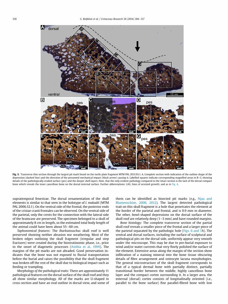
Fig. 5. Transverse thin section through the largest pit mark found on the turtle plate fragment MTM PAL 2013.93.1. A, Complete section with indication of the outline-shape of thedepression (dashed line) and the direction of the presumed mechanical impact (black arrow) causing it. Labelled squares indicate corresponding magnified areas in BeG showingdetails of the pathologically eroded surface (pes) and the deeper shell layers. Note, that the only evident pathology compared to the intact section is the lack of the dorsal compactbone which reveals the inner cancellous bone on the dorsal external surface. Further abbreviations: LAG, lines of arrested growth; and as in Fig. 4.
G. Botfalvai et al. / Cretaceous Research 50 (2014) 304e317310
supratemporal fenestrae. The dorsal ornamentation of the skullelements is similar to that seen in the holotype of I. makadii (MTMPAL 2006.52.1). On the ventral side of the frontal, the posterior endsof the cristae cranii frontales can be observed. On the ventral side ofthe parietal, only the crests for the connection with the lateral sideof the braincase are preserved. The specimen belonged to a skull ofapproximately 8 cm in length, so the estimated total body length ofthe animal could have been about 55e60 cm.
Taphonomical features: The Iharkutosuchus skull roof is wellpreserved showing neither abrasion nor weathering. Most of thebroken edges outlining the skull fragment (irregular and stepfractures) were created during the biostratinomic phase, i.e., priorto the onset of diagenetic processes (Astibia et al., 1999). Themargins of the pit marks are not abraded. Good preservation in-dicates that the bone was not exposed to fluvial transportationbefore the burial and raises the possibility that the skull fragmentwas broken off the rest of the skull due to biological impact such asfeeding or trampling.
Morphology of the pathological traits: There are approximately 11pathological features on the dorsal surface of the skull roof and theyall show similar morphology. All of the marks are U-shaped incross-section and have an oval outline in dorsal view, and some of
them can be identified as bisected pit marks (e.g., Njau andBlumenschine, 2006, 2012). The largest detected pathologicaltrait on this skull fragment is a hole that penetrates the element atthe border of the parietal and frontal, and is 0.9 mm in diameter.The other, bowl-shaped depressions on the dorsal surface of theskull roof are relatively deep (1e3 mm) and have rounded margins.
Bone histology: The complete transverse section of the partialskull roof reveals a smaller piece of the frontal and a larger piece ofthe parietal separated by the pathologic hole (Figs. 6 and 7A). Theventral and dorsal surfaces, including the surface of sculptural andpathological pits on the dorsal side, uniformly appear very smoothunder the microscope. This may be due to pre-burial exposure towind and/or water currents that very finely polished the surface ofthe element. Extensive areas along the margin of the section showinfiltration of a staining mineral into the bone tissue obscuringdetails of fibre arrangement and osteocyte lacuna morphologies.The general microstructure of the skull fragment corresponds tothat of a typical dermal bone with partially distinct, partiallytransitional border between the middle, highly cancellous bonelayer and the compact cortex surrounding it. In a larger area, theexternal (dorsal) cortex consists of longitudinally oriented (i.e.,parallel to the bone surface) fine parallel-fibred bone with low

Fig. 6. Transverse thin section of the Iharkutosuchus (MTM PAL 2013.94.1) skull fragment. A, Complete section under plane polarized light showing the general tripartite micro-structure of the skull roof (delineated by dotted lines) including intact as well as pathologic regions. Black arrows indicate pathologic depressions, whereas dashed lines the outlineof the eroded surfaces of the depressions. Large black arrow marks the pathological hole where the element was pierced through. Letter labels refer to the magnified areas shown inBeD. B, Histological details of the margin of a pathological pit at the broken edge of the fragment under single plane polarizers and C, under cross polarized light. Note the abrupttermination of the bone fibres running parallel to the ventral bone surface and the complete loss of the dorsal compact bone (dcb) in the deeper part of the pit revealing thecancellous layer on the eroded surface. D, Close-up of the intact sculptural pit (scp) and the distinct structural change of the primary parallel-fibred bone (pfb) at the border betweenthe dorsal compact bone (dcb) and the middle cancellous bone (cb). Further abbreviations as in Figs. 4 and 5.
G. Botfalvai et al. / Cretaceous Research 50 (2014) 304e317 311
birefringence (Fig. 6D) and abundant Sharpey's fibres that runroughly perpendicular or oblique to the dorsal surface of the bone.In other areas, the orientation of the parallel-fibred bone is variablepartially following the orientation of the vascular canals. Osteocytelacunae are sparse in the majority of the dorsal cortex with areasthat seem to be void of lacunae; however, the latter may be a pure
Fig. 7. Counterpart of the transverse thin section of the Iharkutosuchus (MTM PAL 2013.94under cross polarized light. Black arrows and dashed lines indicate pathologic pits and the ohole where the element was pierced through. Letter labels refer to the magnified areas shownpolarizers revealing the cut-off nature of the bone fibres and vascular canals at the edge of thpit at the edge of the broken fragment. Note the apparent erosion cutting off the fibres of
preservational artefact. No evident plywood-like arrangement canbe observed. Some indistinct incremental lines can be detected butusually cannot be followed all along the length of the section.Vascular canals are sparse but of relatively wide lumen, and theyrun mostly radially and parallel to the dorsal surface of the bone.Secondary remodelling can also be observed around some vascular.
.1) skull fragment. A, Complete section showing the general tripartite microstructureutline of their eroded surfaces, respectively. Large black arrow marks the pathologicalin BeD. B and C, magnified areas of the margins of pathologic pits under crossed plane
e pits and the loss of the dorsal cortex. D, Close-up of the shallowmargin of a pathologicsecondary osteons close to the surface. Abbreviations as in Figs. 4e6.

G. Botfalvai et al. / Cretaceous Research 50 (2014) 304e317312
A distinct structural change characterizes the border between theexternal cortex and the middle cancellous bone layer. Here, the fineparallel-fibred bone of the dorsal cortex with low lacunar densitiesand low birefringence abruptly changes into the strongly birefrin-gent parallel-fibred bone of the middle cancellous layer whichshows much higher lacunar densities (Fig. 6D). The majority of theparallel-fibred bone in the middle layer is primary and orientedparallel or subparallel to the external and internal cortical surfaces(Fig. 7A), but in small areas interwoven structural fibres charac-teristic of dermal bones (e.g., Scheyer, 2007a,b; Scheyer and Sander,2007; Witzmann, 2009) also occur. External structural fibres arealso present in this middle layer. Secondary remodelling bylamellar parallel-fibred bone is restricted to the margin of somemedium-sized secondary osteons and the large erosion cavities.There is no distinct border between themiddle cancellous layer andthe internal (ventral) cortex; the parallel-fibred bone of the middlelayer continues in the ventral compacta without any structuralinterruption (Figs. 6 and 7C). In the ventral cortex, parallel-fibredbone is oriented mainly parallel to the internal bone surface, andlocally it shows lamellation (Figs. 6C and 7A,C). As in the dorsalcortex, Sharpey's fibres abundantly cross the internal compactaperpendicular or oblique to its surface. Vascularity is much lowerthan in the external cortex with a few radially oriented canals andlarge, entirely avascular areas. Numerous growth marks are presentwhich, in contrast to e.g., the frontal bone of the Eocene Crocodyluscf. affinis and the recent C. niloticus (Buffrenil and Buffetaut, 1981),are much more distinct than in the dorsal cortex.
The complete section reveals the pathologic as well as thepresumably intact areas of the skull fragment, including a sup-posedly intact sculptural pit of the external surface. The mostimportant microstructural difference between the ornamental andthe pathological pits lies in the apparently pathological loss of alarger amount of bone in the bite-mark-like pits and around thehole piercing through the entire element. Although the surface ofthe element is uniformly smoothened, in contrast to the sculpturalpit, where the external cortex is still thick and the fibres seem tofollow the undulation of the dorsal surface, in all pathological pitsthe external cortex is lost or reduced to a thin layer and there is anabrupt termination of the longitudinally oriented parallel-fibredbone at the margin of the pits and the hole (Figs. 6C and 7A,C).The preserved bone layers do not exhibit any other histologicaldifference compared to the intact region of the bone which showsno apparent deviations from the characteristic microstructure ofdermal bones, either (e.g., Buffrenil and Buffetaut, 1981; Scheyer,2007a,b; Scheyer and Sander, 2007; Witzmann, 2009). Hence, asin the turtle plate fragment MTM PAL 2013.94.1, no histologicalfeatures indicate that pre-mortem pathogens were responsible forthe formation of the bite-mark-like pits. The microstructure of thisskull roof fragment rather suggests an external, strong mechanicalimpact that removed the dorsal cortex in the pits and brokethrough the entire bone thickness in the thinner part of theelement.
5. Discussion
Some morphological features (spatial distribution and shapes)of the diverse pits, bisected marks and scores detected on thesurface of the carapace fragment and the skull roof imply that theyprobably represent feeding traces. Microanatomical and histologi-cal comparison of the intact and pathologic regions of both ele-ments shows that, apart from the large amount of missing bone inthe pathologic regions, the preserved bone tissue does not exhibitsuch deviations from the normal microstructure that would indi-cate pre-mortem bone diseases or infections (Lovich et al., 1996;Garner et al., 1997; Homer et al., 1998; Knotkova et al., 2005;
Hernandez-Divers et al., 2009; Rothschild et al., 2013). Hence,these histological observations are consistent with the hypothesisthat the investigated pits have resulted from the massive intrusionof conical objects, most probably teeth, into the bones. The abun-dant fractures and deformations present in both elements may alsobe the result of powerful mechanical impacts to which the speci-mens were exposed prior to fossilization (e.g., Noto et al., 2012).Nevertheless, post-mortem invertebrate and small vertebratefeeding traces (Hutchison and Frye, 2001; Farinati and Zavala,2002; Bader et al., 2009; Kirkland and Bader, 2010; Saneyoshiet al., 2011; Holden et al., 2013) and microbial (fungal and bacte-rial) activity (Pereda Suberbiola et al., 2000; Hutchinson and Frye,2001; Slater et al., 2011) must also be considered as possible cau-ses of the pathologies detected on the fossil specimens. The overallmorphology and spatial distribution of pits, notches, borings andchannels discovered on Mongolian dinosaur skeletons and attrib-uted to the scavenging activity of insect larvae (Kirkland and Bader,2010; Saneyoshi et al., 2011) differ from the pathologies describedin this study. First, our fossils show neither borings nor channels;deformations that are usually associated with insect larval feedingand pupation and generally cluster together with the pits. Second,the pits on the fossils studied here have smooth surface as opposedto the rough and irregular walls of insect feeding traces with groovemarks of no preferred orientation (Saneyoshi et al., 2011). Althoughthe polished surface may not have preserved such details on theIharkút fossils, known differences in taphonomical conditions alsoindicate different trace makers in these two cases. The occurrenceof the insect feeding traces described in the Mongolian dinosaursare believed to be related to the arid environment and hence theutilization of the dried carcasses by invertebrate scavengers; anessentially different condition from that expected in two aquaticanimals (a turtle and a crocodile) living in a subtropical floodplainecosystem reconstructed for the Iharkút locality (Bodor et al., 2012).In addition, the lack of such pathologies on other specimens amongthe Iharkút vertebrate fossils also contrasts the frequent occurrenceof insect feeding traces on the Mongolian dinosaurs (Kirkland andBader, 2010; Saneyoshi et al., 2011). Feeding traces created bysmall mammals are larger borings and usually affect the articularsurfaces where thick cartilage pads covered the bone surface (Fejfarand Kaiser, 2005; Saneyoshi et al., 2011); unlike the pathologiesseen on the turtle carapace and crocodilian skull fragments.Moreover, no mammals of any kind have been found in the Iharkútvertebrate assemblage so far (e.g., }Osi et al., 2012a). Finally, traces ofneither pre-mortem shell diseases and infections, nor post-mortembioturbation by microbes, invertebrates and small vertebrates areexpected to be spatially restricted to evenly distributed, coherentrows of pits, as is the case in our fossils. Thus, no unequivocalmorphological and histological features associated with the abovealternative causes are detected on the Iharkút fossils renderingthem less likely sources of the pathologies described in this study.However, it must be noted that, at present, inadequate morpho-logical and histological descriptions and illustrations of pathologiesoccurring in dermal bones (such as skull bones or turtle shells) withknown causes (including trauma, infection and metabolic diseases)prevent precise comparison (Rothschild et al., 2013) and henceinferences on the unknown agent of pathologies in fossils. As aconsequence, most reports on inferred bite marks (Antunes and deBroin, 1988; Schwimmer, 2002, 2010; Steadman et al., 2007; Mil�anet al., 2011; Noto et al., 2012; Valais et al., 2012; Karl, 2012,; McCoyet al., 2012; Morgan and Albury, 2013) do not take non-traumaticorigin of the detected pathologies into account which are other-wise very common in both extant and fossil turtle shells (Hutchisonand Frye, 2001; Rothschild, 2010; Rothschild et al., 2013 and ref-erences therein). Non-traumatic skeletal pathologies resulting inholes and grooves near the articular surfaces have also been

G. Botfalvai et al. / Cretaceous Research 50 (2014) 304e317 313
documented in crocodilians (Rothschild, 2010); however not intheir skull bones. Bone pathological evaluation of fossil specimensis even more problematic because post-mortem alterations of thebone surface due to diagenetic processes, weathering, and differentmicrobial and invertebrate feeding activities sometimes eventu-ating in bite-mark-like pathologies (Hutchison and Frye, 2001;Fejfar and Kaiser, 2005; Fern�andez-Jalvo et al., 2010; Holdenet al., 2013) cannot be assessed with high confidence. Neverthe-less, based on the comparison of morphological and histologicalfeatures of the pathologies detected in our specimens with thosereported in other studies, the bite mark origin is still consistent inboth elements studied here.
The bowl shaped deep depressions and bisected pits detected onthe fossils closely resemble the experimentally investigated toothmarks of Crocodylus niloticus which possesses conical teeth withsharp mesial and distal carinae (e.g., Njau and Blumenschine, 2006,2012). These morphological similarities suggest that both thecarapace and skull roof fragments studied here bear the bite marksof a crocodilian with similar tooth morphologies. This hypothesis isfurther supported by: (1) the U-shaped cross section and the cir-cular outline of tooth marks in dorsal view lacking extensivepunctures or furrowswhich have been associated with mammalianstyle of chewing (Noto et al., 2012); (2) the high concentration offeeding traces in a small area (Boyd et al., 2013); and (3) the lack ofdiagnostic marks from serrated ziphodont and unserrated pseu-doziphodont teeth, such as parallel clusters of elongate and narrowmarks or striations and deeper notches and pits with a more ovaloutline in dorsal view and a V-shaped cross section, which arecharacteristic of most theropod dinosaurs and some crocodilians(Fiorillo, 1991; Farlow and Holtz, 2012; Rogers et al., 2003; D'Amoreand Blumenschine, 2009; Hone and Rauhut, 2009; Paik et al., 2011;Noto et al., 2012). The freshwater mosasaur Pannoniasaurus, a po-tential top predator known from the locality, has slender, pointedand slightly distally curved teeth (Mak�adi et al., 2012); a toothmorphology that, in contrast to conical teeth, is considered to beinadequate for crushing hard food items, such as bony elements(e.g., Massare, 1987). Therefore it is also highly unlikely that Pan-noniasaurus was responsible for the feeding traces detected on theinvestigated carapace and the skull roof fragments.
If the feeding trace hypothesis is true, the bite marks detectedon the Iharkutosuchus skull roof fragment most probably representtraces of a predatoreprey interaction between two crocodilianspecies rather than scavenging, because the skull is an undesirablefood item for a scavenger (Dodson, 1971; Weigelt, 1989). Antago-nistic behaviour due to competition over common resources is alsovery unlikely, since Iharkutosuchus was a small-bodied crocodile(estimated body length 50e100 cm) with spatulate anterior andflat, molariform posterior teeth referring to oral food processingand a specialized omnivorous/herbivorous diet (}Osi andWeishampel, 2009), whereas its attacker was apparently a largerspecies with tooth morphologies typical for generalist crocodilianpredators (e.g., Buffetaut, 1983). Hence, it is more likely that thesetooth marks were created when the Iharkutosuchus specimen wascaught by another, larger-bodied crocodile species that tried to killits prey by perforating the skull roof, which injury may have causedthe death of this Iharkutosuchus individual.
Having restricted the circle of possible predators to a croco-dilian, the most probable identity of the attacker can be assessed.Four different taxa of Mesoeucrocodylia are documented from theIharkút vertebrate assemblage (}Osi et al., 2007, 2012a). Doratodonsp. is represented by several serrated, labiolingually compressed(i.e., ziphodont) teeth, an incomplete dentary and a maxilla (Martinet al., 2010; }Osi et al., 2012a). The occurrence of a second meso-eucrocodylian taxon is inferred from the presence of labiolinguallycompressed teeth lacking serration (i.e., pseudoziphodont teeth).
This tooth morphology is similar to that found in the genus Ther-iosuchus (}Osi et al., 2012a). The remains of the other two, semi-aquatic mesoeucrocodylians, an indeterminate neosuchian and thehylaeochampsid eusuchian Iharkutosuchus, yield the richest diag-nostic crocodilian material of the Iharkút vertebrate assemblage.Iharkutosuchus is known on the basis of nearly complete skulls andskull fragments, dentaries, and teeth (}Osi et al., 2007, 2012a). Theindeterminate neosuchian taxon is represented by conical teethwith sharp mesial and distal carinae, dentaries, and different skullelements which are reminiscent of those of Allodaposuchus (Rabi,2006; }Osi et al., 2012a; Rabi and Delfino, 2012); a taxon reportedfrom numerous European Late Cretaceous vertebrate localities (e.g.,Buscalioni et al., 2001; Delfino et al., 2008; Martin, 2010; Pu�ertolas-Pascual et al., 2013). Among the abundant remains of Iharkuto-suchus, the most unmistakable elements are its unique molariform,multicuspid teeth (}Osi et al., 2007; }Osi, 2008) which suggestspecialized feeding involving elaborate chewing mechanism (}Osiand Weishampel, 2009). Differences in tooth morphology andpresumed lifestyle of these four crocodilian taxa suggest distinctfeeding strategies.
The tooth morphology and enamel microstructure of Iharkuto-suchus makadii suggest that its diet could have included fibrousplants, fruits, arthropods, and possibly small-bodied vertebrates(}Osi and Weishampel, 2009). These features, combined with itsrelatively small body size shows that Iharkutosuchus certainly didnot belong to the top predators of the Iharkút paleocommunity.Instead, it may have represented an important food source for thetop predators of the area. The other crocodilians were probablymore generalist carnivores (Fig. 8C). The largest of them, the Allo-daposuchus-like neosuchian, may have been among the top pred-ators at least in the aquatic environment along with the mosasaurPannoniasaurus.
The ziphodont and pseudoziphodont teeth of Doratodon and theTheriosuchus-like crocodilian suggest different feeding strategiesfrom the Allodaposuchus-like neosuchian with conical toothmorphology. Whereas ziphodont and pseudoziphodont teeth aremore suitable for cutting and slicing, conical teeth have more po-tential for crushing hard elements, such as bones (e.g., Massare,1987; Fiorillo, 1991; Farlow and Holtz, 2012; D'Amore andBlumenschine, 2009, 2012). Hence, it is most likely that the bowl-shaped pits and bisected bite marks detected on the Iharkuto-suchus skull roof fragment (MTM PAL 2013.94.1) originated fromthis Allodaposuchus-like predator with tooth morphologies similarto those of Crocodylus niloticus (Njau and Blumenschine, 2006).
Studies focussing on predatoreprey interaction or cannibalismamong extant crocodilians based on stomach content in-vestigations in modern ecosystems are rare (e.g., Delany andAbercrombie, 1986; Gabrey, 2010). Reports on croc-odilianecrocodilian interaction in the fossil record are also scarce,and most of them are interpreted as intraspecific antagonisticbehaviour rather than predation or scavenging (Buffetaut, 1983;Williamson, 1996; Avilla et al., 2004; Vasconcellos and Carvalho,2010; Martin, 2013). Interspecific predatoreprey interactionsamong different crocodilian taxa are also poorly documented(Fiorelli, 2010). Therefore, the inferred tooth marks on the dorsalsurface of the Iharkutosuchus skull roof (MTM PAL 2013.94.1) indi-cating a predatoreprey interaction between two different croco-dilian taxa are of great importance. Considering its abundance inthe locality, it is possible that, besides turtles, the small-bodied,semiaquatic Iharkutosuchus was also a potential prey for larger-bodied carnivores in the palaeoenvironment of Iharkút, includingthe Allodaposuchus-like crocodilians.
Turtles are the most common and most important sauropsidfood source for the wild populations of larger-bodied (>3 m) extantalligators (Alligator mississippiensis) in Florida and Louisiana where,
170
cm A
B
C
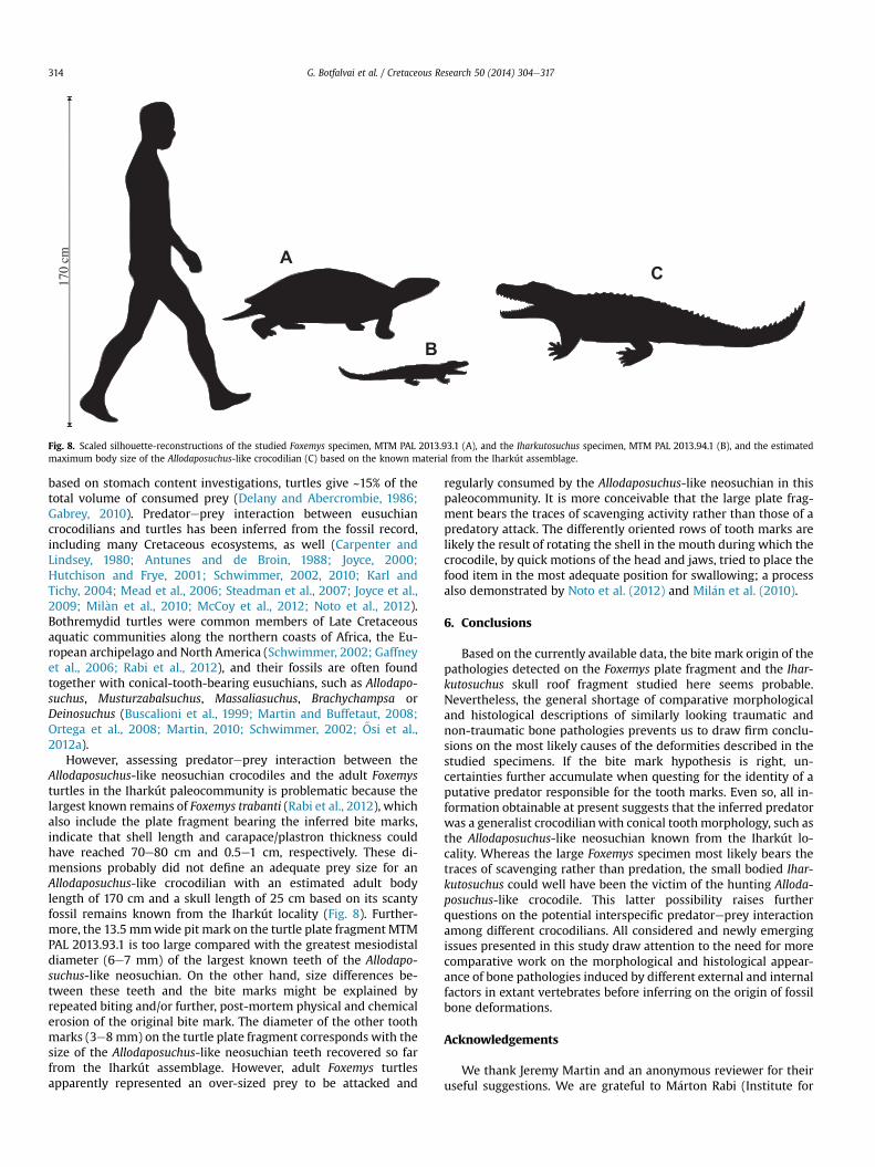
Fig. 8. Scaled silhouette-reconstructions of the studied Foxemys specimen, MTM PAL 2013.93.1 (A), and the Iharkutosuchus specimen, MTM PAL 2013.94.1 (B), and the estimatedmaximum body size of the Allodaposuchus-like crocodilian (C) based on the known material from the Iharkút assemblage.
G. Botfalvai et al. / Cretaceous Research 50 (2014) 304e317314
based on stomach content investigations, turtles give ~15% of thetotal volume of consumed prey (Delany and Abercrombie, 1986;Gabrey, 2010). Predatoreprey interaction between eusuchiancrocodilians and turtles has been inferred from the fossil record,including many Cretaceous ecosystems, as well (Carpenter andLindsey, 1980; Antunes and de Broin, 1988; Joyce, 2000;Hutchison and Frye, 2001; Schwimmer, 2002, 2010; Karl andTichy, 2004; Mead et al., 2006; Steadman et al., 2007; Joyce et al.,2009; Mil�an et al., 2010; McCoy et al., 2012; Noto et al., 2012).Bothremydid turtles were common members of Late Cretaceousaquatic communities along the northern coasts of Africa, the Eu-ropean archipelago and North America (Schwimmer, 2002; Gaffneyet al., 2006; Rabi et al., 2012), and their fossils are often foundtogether with conical-tooth-bearing eusuchians, such as Allodapo-suchus, Musturzabalsuchus, Massaliasuchus, Brachychampsa orDeinosuchus (Buscalioni et al., 1999; Martin and Buffetaut, 2008;Ortega et al., 2008; Martin, 2010; Schwimmer, 2002; }Osi et al.,2012a).
However, assessing predatoreprey interaction between theAllodaposuchus-like neosuchian crocodiles and the adult Foxemysturtles in the Iharkút paleocommunity is problematic because thelargest known remains of Foxemys trabanti (Rabi et al., 2012), whichalso include the plate fragment bearing the inferred bite marks,indicate that shell length and carapace/plastron thickness couldhave reached 70e80 cm and 0.5e1 cm, respectively. These di-mensions probably did not define an adequate prey size for anAllodaposuchus-like crocodilian with an estimated adult bodylength of 170 cm and a skull length of 25 cm based on its scantyfossil remains known from the Iharkút locality (Fig. 8). Further-more, the 13.5 mmwide pit mark on the turtle plate fragment MTMPAL 2013.93.1 is too large compared with the greatest mesiodistaldiameter (6e7 mm) of the largest known teeth of the Allodapo-suchus-like neosuchian. On the other hand, size differences be-tween these teeth and the bite marks might be explained byrepeated biting and/or further, post-mortem physical and chemicalerosion of the original bite mark. The diameter of the other toothmarks (3e8mm) on the turtle plate fragment corresponds with thesize of the Allodaposuchus-like neosuchian teeth recovered so farfrom the Iharkút assemblage. However, adult Foxemys turtlesapparently represented an over-sized prey to be attacked and
regularly consumed by the Allodaposuchus-like neosuchian in thispaleocommunity. It is more conceivable that the large plate frag-ment bears the traces of scavenging activity rather than those of apredatory attack. The differently oriented rows of tooth marks arelikely the result of rotating the shell in the mouth during which thecrocodile, by quick motions of the head and jaws, tried to place thefood item in the most adequate position for swallowing; a processalso demonstrated by Noto et al. (2012) and Mil�an et al. (2010).
6. Conclusions
Based on the currently available data, the bite mark origin of thepathologies detected on the Foxemys plate fragment and the Ihar-kutosuchus skull roof fragment studied here seems probable.Nevertheless, the general shortage of comparative morphologicaland histological descriptions of similarly looking traumatic andnon-traumatic bone pathologies prevents us to draw firm conclu-sions on the most likely causes of the deformities described in thestudied specimens. If the bite mark hypothesis is right, un-certainties further accumulate when questing for the identity of aputative predator responsible for the tooth marks. Even so, all in-formation obtainable at present suggests that the inferred predatorwas a generalist crocodilianwith conical toothmorphology, such asthe Allodaposuchus-like neosuchian known from the Iharkút lo-cality. Whereas the large Foxemys specimen most likely bears thetraces of scavenging rather than predation, the small bodied Ihar-kutosuchus could well have been the victim of the hunting Alloda-posuchus-like crocodile. This latter possibility raises furtherquestions on the potential interspecific predatoreprey interactionamong different crocodilians. All considered and newly emergingissues presented in this study draw attention to the need for morecomparative work on the morphological and histological appear-ance of bone pathologies induced by different external and internalfactors in extant vertebrates before inferring on the origin of fossilbone deformations.
Acknowledgements
We thank Jeremy Martin and an anonymous reviewer for theiruseful suggestions. We are grateful to M�arton Rabi (Institute for
G. Botfalvai et al. / Cretaceous Research 50 (2014) 304e317 315
Geosciences, University of Tübingen, Germany) for his valuablecomments on the manuscript and for sharing literature. We thankthe 2000e2013 field crews for their assistance in the fieldwork. Weare especially grateful to the Bakony BauxiteMines and to Geovol�anZrt. for their logistic help. We are grateful to Pavel Skutschas (SaintPetersburg State University, Russia) for helpful discussions andcomments to an earlier version of the manuscript. R�eka Kalm�ar isthanked for skillful preparation of histological slices. The fieldworkwas supported by the Hungarian Natural History Museum, theNational Geographic Society (Grant Nos. 7228e02, 7508e03), theHungarian Scientific Research Fund (OTKA Te38045, PD 73021, NF84193), MTA-ELTE Lendület Program (Project number: 95102) andthe Hungarian Oil and Gas Company (MOL). M�aty�as Vremir isthanked for providing literature. This project was also supported bythe Jurassic Foundation, the Hantken Miksa Foundation, and theBolyai Fellowship (A.}O). We thank the staff of the Department ofApplied and Physical Geology and the Department of Palae-ontology, Budapest for their support and help provided through theyears.
References
Aleksi�c-Kova�cevi�c, S., €Ozvegy, J., Krsti�c, N., Rusvai, M., Jakab, Cs, Stanimirovi�c, Z.,Becskei, Zs, 2013. Skin and skeletal system lesions of European pond turtles(Emys orbicularis) from natural habitats. Acta Veterinaria Hungarica. http://dx.doi.org/10.1556/AVet.2013.060.
Allain, R., Pereda Suberbiola, X., 2003. Dinosaurs of France. Comptes Rendus Palevol2, 27e44.
Antunes, M.T., de Broin, F., 1988. Le Cr�etac�e terminal de Beira Litoral, Portugal:remarques stratigraphiques et �ecologiques, etude compl�ementaire de Rosasiasoutoi (Chelonii, Bothremydidae). Ciencias da Terra 9, 153e200.
Astibia, H., Murelaga, X., Pereda Suberbiola, X., Elorza, J.J., Gomez-Alday, J.J., 1999.Taphonomy and palaeoecology of the Upper Cretaceous continental vertebrate-bearing beds of the La~no Quarry (Iberian Peninsula). Estudios Museo de CienciaNaturales de Alava 14, 43e104.
Avilla, L., Fernandes, R., Ramos, D.F.B., 2004. Bite marks on a crocodylomorph fromthe Upper Cretaceous of Brazil. Evidence of Social Behaviour 24 (9), 971e973.
Bader, K.S., Hasiotis, S.T., Martin, L.D., 2009. Application of forensic science tech-niques to trace fossils on dinosaur bones from a quarry in the Upper JurassicMorrison Formation, Northeaster Wyoming. Palaios 24, 140e158.
B�ardossy, Gy, Mindszenty, A., 2013. In: Piros, O. (Ed.), The Iharkút Bauxite. Geologicaand Geophysical Institute of Hungary, Budapest, pp. 1e64.
Bell, P.R., Currie, P.J., Lee, Y.N., 2012. Tyrannosaur feeding traces on Deinocheirus(Theropoda:? Ornithomimosauria) remains from Nemegt Formation (LateCretaceous), Mongolia. Cretaceous Research 37, 186e190.
Bodor, E.R., Baranyi, V., 2012. The Normapolles complex and related mesofossilsfrom the Iharkút vertebrate site, Bakony Mountains (Hungary). Central Euro-pean Journal of Geology 55 (3), 259e292.
Bodor, E.R., Baranyi, V., Hermanov�a, Z., 2012. The earliest Sabiaceae fruit remains ofHungary. Hantkeniana 7, 11e18.
Botfalvai, G., Mindszenty, A., }Osi, A., 2012. Sedimentology of the bonebeds of theLate Cretaceous (Santonian) Iharkút Dinosaur locality (Csehb�anya Formation,Bakony Mts, western Hungary). In: Royo-Torres, R., Gasc�o, F., Alcal�a, L. (Eds.),10th Annual Meeting of the European Association of Vertebrate Paleontologists.Fundamental, Abstract Book, pp. 25e26.
Botfalvai, G., }Osi, A., Mindszenty, A., 2014. Taphonomical and Palaeoecologicalinvestigation of the Late Cretaceous Iharkút vertebrate assemblage, submittedfor publication.
Boyd, C.A., Drumheller, S.K., Gates, T.A., 2013. Crocodyliform feeding traces on ju-venile Ornithischian dinosaur from the Upper Cretaceous (Campanian) Kai-parowits Formation, Utah. PLoS One 8 (2), e57605. http://dx.doi.org/10.1371/journal.pone.0057605.
Brand, L.R., Hussey, M., Taylor, J., 2003. Taphonomy of freshwater turtles: Decay anddisarticulation in controlled experiments. Journal of Taphonomy 1, 233e245.
Buffetaut, E.B., 1983. Wounds on the jaw of an Eocene mesosuchian crocodilian aspossible evidence for the antiquity of crocodilian interspecific fighting behav-iour. Pal€aontologische Zeitschrift 57, 143e145.
Buffetaut, E., Le Loeuff, J., 1991. Late Cretaceous dinosaur faunas of Europe: somecorrelation problems. Cretaceous Research 12, 159e176.
Buffetaut, E., Martill, D., Escuillie, F., 2004. Pterosaurs as part of a spinosaur diet.Nature 430, 33.
Buffrenil, V., Buffetaut, E., 1981. Skeletal growth lines in an Eocene crocodilian skullfrom Wyoming as an indicator of ontogenic age and paleoclimatic conditions.Journal of Vertebrate Paleontology 1 (1), 57e66.
Buscalioni, �A.D., Ortega, F., Vasse, D., 1999. The Upper Cretaceous crocodilianassemblage from Lano (Northcentral Spain): Implications in the knowledge ofthe finicretaceous European faunas. Estudios Museo de Ciencia Naturales deAlava 14, 213e233.
Buscalioni, �A.D., Ortega, F., Weishampel, D.B., Jianu, C.M., 2001. A revision of thecrocodyliform Allodaposuchus precedens from the Upper Cretaceous of theHateg Basin, Romania. Its relevance in the phylogeny of eusuchia. Journal ofVertebrate Paleontology 21 (1), 74e86.
Carpenter, K., 1998. Evidence of predatory behaviour by carnivorous dinosaurs. Gaia15, 135e144.
Carpenter, K., Lindsey, D., 1980. The dentary of Brachychampsa montana Gilmore(Alligatorinae; Crocodylidae), a Late Cretaceous turtle-eating alligator. Journalof Paleontology 54, 1213e1217.
D'Amore, D.C., Blumenschine, R.J., 2009. Komodo monitor (Varanus komodoensis)feeding behaviour and dental function reflected through tooth marks on bonesurfaces, and the application to ziphodont Paleobiology. Paleobiology 35 (4),525e552.
D'Amore, D.C., Blumenschine, R.J., 2012. Using striated tooth marks on bone topredict body size in theropods dinosaurs: a model based on feeding obser-vations of Varanus komodoensis, the Komodo monitor. Paleobiology 38,79e100.
Delany, M.F., Abercrombie, C.L., 1986. American alligator food habits in NorthcentralFlorida. Journal of Wildlife Management 50 (2), 348e353.
Delfino, M., Codrea, V., Folie, A., Dica, P., Godefroit, P., Smith, T., 2008. A completeskull of Allodaposuchus precedens Nopcsa, 1928 (Eusuchia) and a reassessmentof the morphology of the taxon based on the Romanian remains. Journal ofVertebrate Paleontology 28 (1), 111e122.
Dodson, P., 1971. Sedimentology and Taphonomy of the Oldman Formation (Cam-panian), Dinosaur Provincial Park, Alberta (Canada). Palaeogeography, Palae-oclimatology, Palaeoecology 10, 21e74.
Domínguez-Rodrigo, M., 1999. Flesh availability and bone modifications in carcassesconsumed by lions: palaeoecological relevance in hominid foraging patterns.Palaeogeography, Palaeoclimatology, Palaeoecology 149, 373e388.
Erickson, G.M., Olson, K.H., 1996. Bite marks attributable to Tyrannosaurus rex:Preliminary description and implications. Journal of Vertebrate Paleontology 16(1), 175e178.
Faith, J.T., Behrensmeyer, A.K., 2006. Changing patterns of carnivore modification ina landscape bone assemblage, Amboseli Park, Kenya. Journal of ArchaeologicalScience 33, 1718e1733.
Faith, J.T., Marean, C.W., Behrensmeyer, A.K., 2007. Carnivore competition, bonedestruction, and bone density. Journal of Archaeological Science 34,2025e2034.
Farinati, E., Zavala, C., 2002. Trace fossils on shelly substrate. An example from theMiocene of Patagonia, Argentina. Acta Geologica Hispanica 34, 29e36.
Farlow, J.O., Holtz Jr., T.R., 2012. The fossil record of predation in dinosaurs. Pale-ontological Society Papers 8, 251e266.
Fejfar, O., Kaiser, T.M., 2005. Insect bone-modification and paleoecology of Oligo-cene mammal-bearings sites in the Doupov Mountains, northwestern Bohemia.Palaeontologica Electronica 8. http://palaeo-electronica.org/paleo/2005_1/fejfar8/issue1_05.htm.
Fern�andez-Jalvo, Y., Andrews, P., Pesquero, D., Smith, C., Marín-Monfort, D.,S�anchez, B., Geigl, E.-M., Alonso, A., 2010. Early bone diagenesis in temperateenvironments Part I: Surface features and histology. Palaeogeography, Palae-oclimatology, Palaeoecology 288, 62e81.
Fiorelli, L.E., 2010. Predation bite-marks on a peirosaurid crocodyliform from theUpper Cretaceous of Neuqu�en Province, Argentina. Ameghiniana 47 (3),387e400.
Fiorillo, A.R., 1991. Prey bone utilization by predatory dinosaurs. Palaeogeography,Palaeoclimatology, Palaeoecology 88, 157e166.
Fowler, D.W., Sullivan, R.M., 2006. A ceratopsid pelvis with tooth marks from theUpper Cretaceous Kirtland Formation, New Mexico: Evidences of Late Campa-nian tyrannosaurid feeding behaviour. New Mexico Museum of Natural Historyand Science, Bulletin 35, 127e130.
Gabrey, S.W., 2010. Demographic and geographic variation in food habits ofAmerican alligators (Alligator mississippiensis) in Lousiana. HerpetologicalConservation and Biology 5 (2), 241e250.
Gaffney, E.S., Tong, H., Meylan, P.A., 2006. Evolution of the side-necked turtles: thefamilies Bothremydidae, Euraxemydidae, and Araripemydae. Bulletin of theAmerican Museum of Natural History 300, 1e698.
Garner, M.M., Herrington, R., Howerth, E.W., Homer, B.L., Nettles, V.F., Isaza, R.,Shotta, E.B., Jacobs, E.R., 1997. Shell disease in river cooters (Pseudemys con-cinna) and yellow-bellied turtles (Trachemys scripta) in a Georgia (USA) Lake.Journal of Wildlife Disease 33 (1), 78e86.
Haynes, G., 1983. Frequencies of spiral and green-bone fractures on ungulate limbbones in modern surface assemblages. American Antiquity 48, 102e114.
Hernandez-Divers, S.J., Hensel, P., Gladden, J., Hernandez-Divers, S.M.,Buhlmann, K.A., Hagen, C., Sanchez, S., Latimer, K.S., Ard, M., Camus, A.C., 2009.Investigation of shell disease in map turtles. Journal of Wildlife Disease 45 (3),637e652.
Holden, A.R., Harris, J.M., Timm, R.M., 2013. Paleoecological and taphonomic im-plications of insect-damaged Pleistocene vertebrate remains from Rancho LaBrea, Southern California. PLoS One 8 (7), e67119. http://dx.doi.org/10.1371/journal.pone.0067119.
Homer, B.L., Berry, K.H., Brown, M.B., Ellis, G., Jacobson, E.R., 1998. Pathology ofdiseases in wild desert tortoises from California. Journal of Wildlife Disease 34(3), 508e523.
Hone, D.W.E., Rauhut, O.W.M., 2009. Feeding behaviour and bone utilization bytheropod dinosaurs. Lethaia. http://dx.doi.org/10.1111/j.1502e3931.2009.00187.x.
G. Botfalvai et al. / Cretaceous Research 50 (2014) 304e317316
Hone, D., Choiniere, J., Sullivan, C., Xu, X., Pittman, M., Tan, Q., 2010. New evidencefor a trophic relationship between the dinosaurs Velociraptor and Protoceratops.Palaeogeography, Palaeoclimatology, Palaeoecology 291, 488e492.
Hu, Y., Meng, J., Wang, Y., Li, C., 2005. Large Mesozoic mammals fed on young di-nosaurs. Nature 433, 149e152.
Hutchison, J.H., Frye, F.L., 2001. Evidence of pathology in early Cenozoic turtles.PaleoBios 21, 12e19.
Jacobsen, A.R., 1998. Feeding behaviour of carnivorous dinosaurs as determined bytooth marks on dinosaur bones. Historical Biology 13, 17e26.
Joyce, W.G., 2000. The first complete skeleton of Solnhofia parsonsi (Cryptodira,Eurysternidae) from the Upper Jurassic of Germany and its taxonomic impli-cations. Journal of Paleontology 74, 684e700.
Joyce, W.G., Revan, A., Lyson, T.R., Danilov, I.G., 2009. Two new Plastomenine soft-shell turtles from the Paleocene of Montana and Wyoming. Bulletin of thePeabody Museum of Natural History 50 (2), 307e325.
Karl, H.V., 2012. Bite traces in a turtle shell fragment from the Kimmeridgian (UpperJurassic) of Northern Germany. Studia Palaeocheloniologica 4, 25e30.
Karl, H.V., Tichy, G., 2004. The structure of fossils teeth of chelonophagous croco-diles (Diapsida: Crocodylia). Studia Geologica Salmanticensia 40, 116e124.
Kirkland, J.I., Bader, K., 2010. Insect trace fossils associated with Protoceratops car-casses in the Djadokhta Formation (Upper Cretaceous), Mongolia. In: Ryan, M.J.,Chinnery-Allgeier, B.J., Eberth, D.A. (Eds.), New Perspectives on Horned Di-nosaurs. Indiana University Press, Bloomington, pp. 509e519.
Knotkova, Z., Mazanek, S., Hovorka, M., Sloboda, M., Knotek, Z., 2005. Haematologyand plasma chemistry of Bornean river turtles suffering from shell necrosis andhaemogregarin parasites. Veterin�arní Medicína 55 (9), 421e426.
Longrich, N.R., Horner, J.R., Erickson, G.M., Currie, P.J., 2010. Cannibalism in Tyran-nosaurus rex. PLoS One 5 (10), e13419. http://dx.doi.org/10.1371/journal.pone.0013419.
Lovich, J.E., Gotte, S.W., Ernst, C.H., Harshbarger, J.C., Laemmerzahl, A.F.,Gibbons, J.W., 1996. Prevalence and histopathology of shell disease in turtlesfrom Lake Blackshear, Georgia. Journal of Wildlife Disease 32 (2), 259e265.
Mak�adi, L., Caldwell, M.W., }Osi, A., 2012. The first freshwater mosasauroid (UpperCretaceous, Hungary) and a new clade of basal mosasauroids. PLoS ONE 7 (12),e51781. http://dx.doi.org/10.1371/journal.pone.0051781.
Mak�adi, L., 2013a. A new polyglyphanodontine lizard (Squamata: Borioteiioidea)from the Late Cretaceous Iharkút locality (Santonian, Hungary). CretaceousResearch 46, 166e176.
Mak�adi, L., 2013b. The first known champsiid lizard (Squamata) from the UpperCretaceous of Europe (Csehb�anya Formation; Hungary, Bakony Mts). Annales dePal�eontologie 99, 261e274.
Marotti, G., 2010. Static and dynamic osteogenesis. Italian Journal of Anatomy andEmbryology 115, 123e126.
Martin, J.E., 2010. Allodaposuchus Nopcsa, 1928 (Crocodylia, Eusuchia), from the LateCretaceous of Southern France and its relationships to Alligatoroidea. Journal ofVertebrate Paleontology 30 (3), 756e767.
Martin, J.E., 2013. Surviving a potentially lethal injury? Bite mark and associatedtrauma in the vertebrae of a dyrosaurid crocodilian. Palaios 28, 6e8.
Martin, J.E., Buffetaut, E., 2008. Crocodilus affuvelensis Matheron, 1869 from the LateCretaceous of Southern France: a reassessment. Zoologica Journal of theLinnean Society 152, 567e580.
Martin, J.E., Rabi, M., Csiki, Z., 2010. Survival of Theriosuchus (Mesoeucrocodylia:Atoposauridae) in a Late Cretaceous archipelago: a new species from theMaastrichtian of Romania. Naturwissenschaften 97, 845e854.
Massare, J.A., 1987. Tooth morphology and prey preference of mesozoic marinereptiles. Journal of Vertebrate Paleontology 7 (2), 121e137.
McCoy, M.R., Karl, H.V., Tichy, G., Steinbacher, J., Aigner, G., Cemper-Kisslich, J., 2012.Radiological evaluation of a fossil turtle trauma from the Upper Jurassic ofEichst€att (Testudines: Cryptodira). Studia Geologica Salmanticensia 48 (1),37e44.
Mead, J.I., Cubero, R., Zamora, A.L.V.Z., Swift, S.L., Lauritio, C., G�omez, L.D., 2006.Plio-Pleistocene Crocodylus (Crocodylia) from southwestern Costa Rica. Studieson Neotropical Fauna and Environment 41 (1), 1e7.
Mikul�as, R., Dvor�ak, Z., 2010. Possible crocodylian bite traces, Miocene of the MostBasin (Czech Republic). New Mexico Museum of Natural History and Science,Bulletin 51, 191e194.
Mil�an, J., Kofoed, J., Bromley, R.G., 2010. Crocodylian-Chelonian carnivory: Bitetraces of dwarf caiman, Paleosuchus palpebrosus, in red-eared slinder, Trachemysscripta, carapaces. NewMexico Museum of Natural History and Science, Bulletin51, 195e200.
Mil�an, J., Lindow, B.E.K., Lauridsen, B.W., 2011. Bite traces in a turtle carapacefragment from the middle Danian (Lower Paleocene) bryozoan limestone, Fexe,Denmark. Bulletin of the Geological Society of Denmark 55, 61e67.
Morgan, G.S., Albury, N.A., 2013. The Cuban crocodile (Crocodylus rhombifer) fromlate Quaternary fossil deposits in the Bahamas and Cayman Islands. Bulletin ofthe Florida Museum of Natural History 52, 161e236.
Njau, J.K., Blumenschine, R.J., 2006. A diagnosis of crocodile feeding traces on largemammal bone, with fossil examples from the Plio-Pleistocene Olduvai Basin,Tanzania. Journal of Human Evolution 50, 142e162.
Njau, J.K., Blumenschine, R.J., 2012. Crocodylian and mammalian carnivore feedingtraces on hominid fossils from FLK 22 and FLK NN 3, Plio-Pleistocene, OlduvaiGorge. Journal of Human Evolution 63, 408e417.
Noto, C.R., Main, D.J., Drumheller, S.K., 2012. Feeding traces and paleobiology of aCretaceous (Cenomanian) crocodyliform: example from the Woodbine Forma-tion of Texas. Palaios 27, 105e115.
Ortega, F., Sanz, J.L., Barroso-Barcenilla, F., Cambra-Moo, O., Escaso, F., García-Oliva, M., Fern�andez, F.M., 2008. El yacimiento de macrovertebrados f�osiles delCret�acico superior de “lo hueco” (Fuentes, Cuenca). Palaeontologica Nova 8,119e131.
}Osi, A., 2008. Cranial osteology of Iharkutosuchus makadii, a Late Cretaceous basaleusuchian crocodyliform from Hungary. Neues Jahrbuch für Geologie undPal€aontologie Abhandlungen 248 (3), 279e299.
}Osi, A., Mindszenty, A., 2009. Iharkút, dinosaur-bearing alluvial complex ofthe Csehb�anya Formation. In: Babinszky, E. (Ed.), Cretaceous sediments ofthe Transdanubian Range. Hungarian Geological Society, Budapest, pp. 51e63.
}Osi, A., Weishampel, D.B., 2009. Jaw mechanism and dental function in the LateCretaceous basal eusuchian Iharkutosuchus. Journal of Morphology 270,903e920.
}Osi, A., Buffetaut, E., 2011. Additional non-avian theropod and bird remains from theearly Late Cretaceous (Santonian) of Hungary and a review of the Europeanabelisauroid record. Annales de Paleontologie 97, 35e49.
}Osi, A., Prondvai, E., 2013. Sympatry of two ankylosaurs (Hungarosaurus andCf. Strutiosaurus) in the Santonian of Hungary. Cretaceous Research 44,58e63.
}Osi, A., Clark, J.M., Weishampel, D.B., 2007. First report on a new basal eusuchiancrocodyliform with multicusped teeth from the Upper Cretaceous (Santonian)of Hungary. Neues Jahrbuch für Geologie und Pal€aontologie Abhandlungen 243/2, 169e177.
}Osi, A., Butler, R.J., Weishampel, D.B., 2010. A Late Cretaceous ceratopsians dinosaurfrom Europe with Asian affinities. Nature 465, 466e468.
}Osi, A., Buffetaut, E., Prondvai, E., 2011. New pterosaurian remains from the LateCretaceous (Santonian) of Hungary (Iharkút, Csehb�anya Formation). CretaceousResearch 32, 456e463.
}Osi, A., Rabi, M., Mak�adi, L., Szentesi, Z., Botfalvai, G., Guly�as, P., 2012a. The LateCretaceous continental vertebrate fauna from Iharkút (western Hungary, Cen-tral Europe): a review. In: Godefroit, P. (Ed.), Bernissart dinosaurs and EarlyCretaceous Terrestrial Ecosystems. Indiana University Press, Bloomington,pp. 532e569.
}Osi, A., Prondvai, E., Butler, R., Weishampel, D.B., 2012b. Phylogeny, histology andinferred body size evolution in a new Rhabdodontid dinosaur from the LateCretaceous of Hungary. PLoS One 7 (9), e44318. http://dx.doi.org/10.1371/journal.pone.0044318.
Paik, I.S., Kim, H.J., Lim, J.D., Huh, M., Lee, H.I., 2011. Diverse tooth marks on an adultsauropod bone from Early Cretaceous, Korea: Implications in feeding behaviourof theropods dinosaurs. Palaeogeography, Palaeoclimatology, Palaeoecology309, 342e346.
Pereda-Suberbiola, X., Astibia, H., Murelaga, X., Elorza, J.J., G�omez-Alday, J.J., 2000.Taphonomy of the Late Cretaceous dinosaur-bearing beds of the La~no Quarry(Iberian Peninsula). Palaeogeography, Palaeoclimatology, Palaeoecology 157,247e275.
Peterson, J.E., Henderson, M.D., Scherer, R.P., Vittore, C.P., 2009. Face biting on ajuvenile tyrannosaurid and behavioral implications. Palaios 24, 780e784.
Prondvai, E., Bodor, E.R., }Osi, A., 2014. Does morphology reflect osteohistology-based ontogeny? A case study of Late Cretaceous pterosaur jaw symphysesfrom Hungary reveals hidden taxonomic diversity. Paleobiology 40 (2),288e321.
Pu�ertolas-Pascual, E., Candudo, J.I., Moreno-Azanza, M., 2013. The eusuchiancrocodylomorp Allodaposuchus subjuniperus sp. nov., a new species fromthe latest Cretaceous (upper Maastrichtian) of Spain. Historical Biology 26,91e109.
Rabi, M., 2006. Do alligatoroids really derive from North America? In: Pazonyi, P.(Ed.), 4th Annual Meeting of the European Association of Vertebrate Paleon-tologists. Hantkeniana, Abstract Book, p. 102.
Rabi, M., Delfino, M., 2012. A Reassessment of the “Alligatoroid” Eusuchian from theLate Cretaceous of Hungary and its Taxonomic Implications. In: Royo-Torres, R.,Gasc�o, F., Alcal�a, L. (Eds.), 10th Annual Meeting of the European Association ofVertebrate Paleontologists. Fundamental, Abstract Book, pp. 203e206.
Rabi, M., Tong, H., Botfalvai, G., 2012. A new species of the side-necked turtleFoxemys (Pelomedusoides: Bothremydidae) from the Late Cretaceous ofHungary and the historical biogeography of the Bothremydini. GeologicalMagazine 149, 662e674.
Rabi, M., Vremir, M., Tong, H., 2013. Preliminary overview of Late Cretaceous turtlediversity in eastern central Europe (Austria, Hungary, and Romania). In:Brinkman, P.A. (Ed.), Morphology and Evolution of Turtles; Origin and EarlyDiversification. Springer, Dordrecht, pp. 307e336.
Rogers, R.R., Krause, D.W., Rogers, K.C., 2003. Cannibalism in the Madagascandinosaur Majungatholus atopus. Nature 422, 515e518.
Rothschild, B., 2010. Macroscopic recognition of nontraumatic osseous pathology inthe postcranial skeletons of crocodilians and lizards. Journal of Herpetology 44(1), 13e20.
Rothschild, B.M., Schultze, H.-P., Pellegrini, R., 2013. Osseous and other hard tissuepathologies in turtles and abnormalities of mineral deposition. In:Brinkman, D.B., Holroyd, P.A., Gradner, J.D. (Eds.), Morphology and Evolution ofTurtles. Vertebrate Paleobiology and Paleoanthropology. Springer Scienceþ-Business Media, Dordrecht, pp. 501e534. http://dx.doi.org/10.1007/978-94-007-4309-0_27, 2013.
Saneyoshi, M., Watabe, M., Suzuki, S., Tsogtbaatar, K., 2011. Trace fossils on dinosaurbones from Upper Cretaceous eolian deposits in Mongolia: Taphonomic inter-pretation of palaeoecosystem desert environments. Palaeogeography, Palae-oclimatology, Palaeoecology 311, 38e47.

G. Botfalvai et al. / Cretaceous Research 50 (2014) 304e317 317
Scheyer, T.M., 2007a. Comparative bone histology of the turtle shell (carapace andplastron): implications for turtle systematics, functional morphology and turtleorigins. PhD Thesis, published on-line: http://hss.ulb.uni-bonn.de/diss_online.
Scheyer, T.M., 2007b. Skeletal histology of the dermal armor of Placodontia: theoccurrence of ‘postcranial fibro-cartilaginous bone’ and its developmental im-plications. Journal of Anatomy 211, 737e753.
Scheyer, T.M., Sander, P.M., 2007. Shell bone histology indicates terrestrial palae-oecology of basal turtles. Proceedings of the Royal Society B 274, 1885e1893.
Schwimmer, D.R., 2002. King of the crocodylians: The paleobiology of Deinosuchus.Indiana University Press, Bloomington, p. 221.
Schwimmer, D.R., 2010. Bite marks of the giant crocodilian Deinosuchus on LateCretaceous (Campanian) bones. New Mexico Museum of Natural History andScience, Bulletin 51, 183e190.
Siegl-Farkas, �A., 1991. Bauxite deposits and Senonian Formations in Hungary(Palynological analysis). Acta Geologica Hungarica 34/4, 345e350.
Slater, B.J., Reolid, M., Schouten, R., Benton, M.J., 2011. A new Late Jurassic turtlefrom Spain: Phylogenetic implication, taphonomy and paleoecology. Palae-ontology 54, 1393e1414.
Steadman, D.W., Franz, R., Morgan, G.S., Albury, N.A., Kakuk, B., Broad, K., Franz, S.E.,Tinker, K., Pateman, M.P., Lott, T.A., Jarzen, D.M., Dilcher, D.L., 2007. Excep-tionally well preserved late Quaternary plant and vertebrate fossils from a bluehole on Abaco, the Bahamas. PNAS 104. www.pnas.org_cgi_doi_10.1073_pnas.0709572104.
Stein, K., Prondvai, E., 2014. Rethinking the nature of fibrolamellar bone: an inte-grative biological revision of sauropod plexiform bone formation. BiologicalReviews 89, 24e47.
Szalai, E., 2005. Paleomagnetic studies in Iharkút. Manuscript. E€otv€os Lor�andUniversity.
Szentesi, Z., Venczel, M., 2010. An advanced anuran from the Late Cretaceous(Santonian) of Hungary. Neues Jahrbuch für Geologie und Pal€aontologieAbhandlungen 256 (3), 21e302.
Szentesi, Z., Gardner, J.D., Venczel, M., 2013. Albanerpetontid amphibians from theLate Cretaceous (Santonian) of Iharkút, Hungary, with remarks on regionaldifferences in Late Cretaceous Laurasian amphibian assemblages. CanadianJournal of Earth Sciences 50, 268e281.
Tuba, Gy, Kiss, P., P�osafai, M., Mindeszenty, A., 2006. Preliminary data on thediagenesis of Cretaceous bones from the Bakony Mts, Hungary. F€oldtaniK€ozl€ony 136, 1e24.
Valais, S., Apesteguía, S., Garrido, A.C., 2012. Cretaceous small scavengers: Feedingtrace in tetrapod bones from Patagonia, Argentina. PLoS One 7 (1), e29841.http://dx.doi.org/10.1371/journal.pone.0029841.
Vasconcellos, F.M., Carvalho, I.D., 2010. Paleoichnological assemblage associatedwith Baurusuchus salgadoensis remains, a Barusuchidae Mesoeucrocodylia fromthe Bauru Basin, Brazil (Late Cretaceous). New Mexico Museum of NaturalHistory and Science, Bulletin 51, 227e238.
Weigelt, J., 1989. Recent Vertebrate Carcasses and Their Palaeobiological Implica-tions (translated by J. Schaefer). University of Chicago Press, Chicago, pp. 1e166.
Weishampel, D.B., Csiki, Z., Benton, M., Grigorescu, D., Codrea, V., 2010. Palae-obiogeographic relationships of the Hateg biota d Between isolation andinnovation. Palaeogeography, Palaeoclimatology, Palaeoecology 293, 419e437.
Wells, N.A., 1989. Making thin sections. In: Feldmann, R.M., Chapman, R.E.,Hannibal, J.T. (Eds.), Paleotechniques. University of Tennessee, Knoxville,pp. 120e129.
Williamson, T.E., 1996. ?Brachychapsa sealeyi, sp. nov., (Crocodylia, Alligatoroidea)from the Upper Cretaceous (Lower Campanian) Menefee Formation, north-western New Mexico. Journal of Vertebrate Paleontology 16 (3), 421e431.
Witzmann, F., 2009. Comparative histology of sculptured dermal bones in basaltetrapods, and the implications for the soft tissue dermis. Palaeodiversity 2,233e270.