Infective Factors Exacerbations of Bronchitis and Asthma
5
BRITISH MEDICAL JOURNAL 5 AUGUST 1972 323 possible case of diabetes for the first time plasma glucose values ought to be reported. If anticoagulants interfere with the pre- cipitating second antibody of double antibody insulin assay systems then separation of insulin ought to be achieved by other methods-for example, by adsorption to charcoal. They are equally efficient and simple to use. Glucose and insulin responses are being studied under widely varying circumstances-relation between mother and fetus, hypoglycaemic disturbances in the neonate, and the effects of new oral hypoglycaemic agents. Precise comparisons of data in these and other fields cannot be made unless glucose and insulin concentrations are determined on the same basis. An agreement on basic methodology will help to save a great deal of confusion in the future. Requests for reprints should be addressed to Dr. T. Lind, M.R.C. Reproduction and Growth Unit, Princess Mary Maternity Hospital, Great North Road, Newcastle upon Tyne NE2 3BD. References Fitzgerald, M. G., and Keen, H. (1964). British Medical J7ournal, 1, 1568 Foster, G. L. (1923).3Journal of Biological Chemistry, 55, 291. Grant, D. B. (1972). Lancet, 1, 207. Henderson, J. R. (1970). Lancet, 2, 545. Hunter, W. M. (1969). In Protein and Polypeptide Hormones, ed. M. Mar- goulies, I.C.S. No. 161. Amsterdam, Excerpta Medica. Hunter, W. M., and Ganguli, P. C. (1971). In Radioimmunoassay Methods, ed. K. E. Kirkham and W. M. Hunter, p. 243. Edinburgh, Livingstone. Lind, T., Cheyne, G. A., Billewicz, W. Z., and Fairweather, D. V. I. (1968). Journal of Obstetrics and Gynaecology of the British Commonwealth, 75, 540. Middleton, J. E., and Griffiths, W. J. (1957). British Medical J'ournal, 2, 1525. Orosz, L., Michael, R., and Ziegler, M. (1971). Lancet, 2, 1149. W.H.O. Expert Committee on Diabetes Mellitus (1965). World Health Organization. Technical Report Series, No. 310. Infective Factors in Exacerbations of Bronchitis and Asthma H. P. LAMBERT, H. STERN British Medical journal, 1972, 3, 323-327 Summary Infections of the respiratory tract were studied in a group of families each containing a patient with chronic bron- chitis or with asthma. A wide variety of infective agents may be associated with exacerbations in susceptible subjects, but the types of organism to which patients are most at risk differ according to the family structure. Exacerbations in the susceptible subject are more likely to be related to viral infections when the family contains children than when it does not. Two patients with asthma experienced frequent respir- atory infections, many of which provoked attacks of asthma. Introduction Attempts to assess the role of infective agents in promoting exacerbations of chronic bronchitis and other chronic respiratory diseases have given rise to widely discrepant results. Many agents have been found in association with exacerbations, but their relative importance has varied greatly in different studies. In particular, viruses have been identified with very variable frequency in several careful surveys (Somerville, 1963; Carilli et al., 1964; Eadie et al., 1966; Ross et al., 1966; Stenhouse, 1967; McNamara et al., 1969; Fisher et al., 1969). The paucity of viruses in some surveys is hard to reconcile with the common observation made in general practice that patients often have exacerbations of bronchitis when other members of the family are suffering mild, and presumably mainly viral, infection of the respiratory tract. One reason for this discrepancy may be that few studies have been made in the patient's home, and as soon St. George's Hospital, London S.W.17 H. P. LAMBERT, M.D., F.R.C.P., Consultant Physician St. George's Hospital Medical School, London S.W.1 H. STERN, M.B., F.R.C.PATH, Professor of Virology as possible after the onset of symptoms; viral pathogens pro- voking exacerbations of chronic respiratory disease might well be more difficult to identify by the time the patient has reached hospital. The family structure too, particularly the presence and age of children, might affect the chances of virus-induced exacerbations in a susceptible subject. In the present study an attempt was made to identify infective agents implicated in provoking exacerbations of bronchitis or of asthma within the patients' households, and to study the distri- bution and spread of such agents in the families of these sus- ceptible subjects. In this way it was hoped to make some esti- mate of the types of infective risk which "chesty" people may experience in their own homes. Methods Eight families, each containing at least one member with chronic bronchitis or asthma, were studied in considerable detail over an average period of two years. Each family .-s visited every two weeks by a nurse specially trained in the taking of specimens, and additional visits were made if the susceptible patient developed a respiratory illness. At each visit a record card was completed for every member of the family, giving information about the following symptoms; cold, sore throat, earache, cough, sputum, wheezing, general aching, fever, diarr- hoea, and vomiting. In the case of persistent symptoms, such as cough and sputum, a simple code was recorded denoting "better," "worse," or "the same." An exacerbation was defined as an increase in any two of the three symptoms of cough, sputum, and wheezing. Visits to the family doctor, or by the family doctor to the house, were also noted, together with the doctor's diagnosis. At every visit the following specimens were taken from each member of the family: a throat swab into Stuart's transport medium for Streptococcus pyogenes, throat and nasal swabs placed together in viral transport medium, and a further throat swab which was inoculated directly into diphasic mycoplasma medium (Grayston et al., 1965). Sputum from the susceptible *subjects was similarly examined for viral, mycoplasmal, and bacterial pathogens. A stool specimen for enterovirus isolation was often obtained by using containers left on the previous visit. on 19 November 2021 by guest. Protected by copyright. http://www.bmj.com/ Br Med J: first published as 10.1136/bmj.3.5822.323 on 5 August 1972. Downloaded from
Transcript of Infective Factors Exacerbations of Bronchitis and Asthma

BRITISH MEDICAL JOURNAL 5 AUGUST 1972 323
possible case of diabetes for the first time plasma glucose valuesought to be reported. If anticoagulants interfere with the pre-cipitating second antibody of double antibody insulin assaysystems then separation of insulin ought to be achieved by othermethods-for example, by adsorption to charcoal. They areequally efficient and simple to use.
Glucose and insulin responses are being studied under widelyvarying circumstances-relation between mother and fetus,hypoglycaemic disturbances in the neonate, and the effects ofnew oral hypoglycaemic agents. Precise comparisons of data inthese and other fields cannot be made unless glucose and insulinconcentrations are determined on the same basis. An agreementon basic methodology will help to save a great deal of confusionin the future.
Requests for reprints should be addressed to Dr. T. Lind, M.R.C.
Reproduction and Growth Unit, Princess Mary Maternity Hospital,Great North Road, Newcastle upon Tyne NE2 3BD.
ReferencesFitzgerald, M. G., and Keen, H. (1964). British Medical J7ournal, 1, 1568Foster, G. L. (1923).3Journal of Biological Chemistry, 55, 291.Grant, D. B. (1972). Lancet, 1, 207.Henderson, J. R. (1970). Lancet, 2, 545.Hunter, W. M. (1969). In Protein and Polypeptide Hormones, ed. M. Mar-
goulies, I.C.S. No. 161. Amsterdam, Excerpta Medica.Hunter, W. M., and Ganguli, P. C. (1971). In Radioimmunoassay Methods,
ed. K. E. Kirkham and W. M. Hunter, p. 243. Edinburgh, Livingstone.Lind, T., Cheyne, G. A., Billewicz, W. Z., and Fairweather, D. V. I. (1968).
Journal of Obstetrics and Gynaecology of the British Commonwealth, 75,540.
Middleton, J. E., and Griffiths, W. J. (1957). British Medical J'ournal, 2,1525.
Orosz, L., Michael, R., and Ziegler, M. (1971). Lancet, 2, 1149.W.H.O. Expert Committee on Diabetes Mellitus (1965). World Health
Organization. Technical Report Series, No. 310.
Infective Factors in Exacerbations of Bronchitis and Asthma
H. P. LAMBERT, H. STERN
British Medical journal, 1972, 3, 323-327
Summary
Infections ofthe respiratory tract were studied in a groupof families each containing a patient with chronic bron-chitis or with asthma. A wide variety of infective agentsmay be associated with exacerbations in susceptiblesubjects, but the types of organism to which patients aremost at risk differ according to the family structure.Exacerbations in the susceptible subject are more likelyto be related to viral infections when the family containschildren than when it does not.Two patients with asthma experienced frequent respir-
atory infections, many of which provoked attacks ofasthma.
Introduction
Attempts to assess the role of infective agents in promotingexacerbations of chronic bronchitis and other chronic respiratorydiseases have given rise to widely discrepant results. Many agentshave been found in association with exacerbations, but theirrelative importance has varied greatly in different studies. Inparticular, viruses have been identified with very variablefrequency in several careful surveys (Somerville, 1963; Carilliet al., 1964; Eadie et al., 1966; Ross et al., 1966; Stenhouse,1967; McNamara et al., 1969; Fisher et al., 1969). The paucityof viruses in some surveys is hard to reconcile with the commonobservation made in general practice that patients often haveexacerbations of bronchitis when other members of the familyare suffering mild, and presumably mainly viral, infection of therespiratory tract. One reason for this discrepancy may be thatfew studies have been made in the patient's home, and as soon
St. George's Hospital, London S.W.17H. P. LAMBERT, M.D., F.R.C.P., Consultant Physician
St. George's Hospital Medical School, London S.W.1H. STERN, M.B., F.R.C.PATH, Professor of Virology
as possible after the onset of symptoms; viral pathogens pro-voking exacerbations of chronic respiratory disease might wellbe more difficult to identify by the time the patient has reachedhospital. The family structure too, particularly the presence andage of children, might affect the chances of virus-inducedexacerbations in a susceptible subject.
In the present study an attempt was made to identify infectiveagents implicated in provoking exacerbations of bronchitis or ofasthma within the patients' households, and to study the distri-bution and spread of such agents in the families of these sus-ceptible subjects. In this way it was hoped to make some esti-mate of the types of infective risk which "chesty" people mayexperience in their own homes.
Methods
Eight families, each containing at least one member withchronic bronchitis or asthma, were studied in considerable detailover an average period of two years. Each family .-s visitedevery two weeks by a nurse specially trained in the taking ofspecimens, and additional visits were made if the susceptiblepatient developed a respiratory illness. At each visit a recordcard was completed for every member of the family, givinginformation about the following symptoms; cold, sore throat,earache, cough, sputum, wheezing, general aching, fever, diarr-hoea, and vomiting. In the case of persistent symptoms, such ascough and sputum, a simple code was recorded denoting"better," "worse," or "the same." An exacerbation was definedas an increase in any two of the three symptoms of cough,sputum, and wheezing. Visits to the family doctor, or by thefamily doctor to the house, were also noted, together with thedoctor's diagnosis.At every visit the following specimens were taken from each
member of the family: a throat swab into Stuart's transportmedium for Streptococcus pyogenes, throat and nasal swabsplaced together in viral transport medium, and a further throatswab which was inoculated directly into diphasic mycoplasmamedium (Grayston et al., 1965). Sputum from the susceptible*subjects was similarly examined for viral, mycoplasmal, andbacterial pathogens. A stool specimen for enterovirus isolationwas often obtained by using containers left on the previous visit.
on 19 Novem
ber 2021 by guest. Protected by copyright.
http://ww
w.bm
j.com/
Br M
ed J: first published as 10.1136/bmj.3.5822.323 on 5 A
ugust 1972. Dow
nloaded from

324
Every two months, and again one to two weeks after an exacer-bation in the patients, a specimen of blood was taken from theadults and older children in the family, but few blood sampleswere taken from the small children.
Specimens for virus isolation were inoculated within one hourof collection into primary human kidney, primary monkeykidney, and diploid human embryonic fibroblast tissue cultures.Serum specimens were examined for complement-fixing anti-bodies against Mycoplasma pneumoniae, Rickettsia burneti, psit-tacosis, herpes simplex, influenza virus types A, B, and C, para-influenza virus types 1-3, mumps virus, respiratory syncytialvirus, and adenovirus by the microtitre method; an eightfold orgreater rise in antibody level was accepted as evidence of recentinfection.The sex and age distribution of the families is shown in Table
I, together with the diagnosis of the chronic respiratory illness inthe susceptible subject. The Medical Research Council (1960,1965a) shortened questionary on bronchitis was completed for
BRITISH MEDICAL JOURNAL 5 AUGUST 1972
throats, etc., which did not progress to exacerbations. Bycontrast, infective agents were found on only 13 out of 332(4%) routine visits.The variety of bacteria and viruses associated with an exacer-
bation of bronchitis, or with the development of acute asthma,is shown in Table III. The illnesses were characteristic exacer-
bations of bronchitis or acute attacks of asthma, and infectionscaused by different agents were generally indistinguishablefrom one another on clinical grounds. The single exacerbationassociated with infection by R. burneti (Q fever) followed a farmholiday in Scotland.
TABLE III-Incidence and Nature of Infective Agents Recognized duringExacerbations in Six Patients with Chronic Bronchitis and Two Patients withAsthma
ExacerbationsFamilyCode Total Agent
No. Recognized
Agents Identified
TABLE I.-Age and Sex Composition of Eight Families, Each having a Patientwith Chronic Bronchitis or Asthma
Sex and Age ofFamily -- Diagnosis of Index CaseCode Index Other Members of
Case Family
A .. M. 52 F. 49, F. 14* Chronic mucopurulent obstructivebronchitis
D M. 74 F. 78, F. 40, F. 34 ,.F M. 62 F.M64J .. M. 54 F. 44, F. 76, M. 15*e .. F. 56 M. 62, F. 19 Chronic simple bronchitis, Grade 2G . F. 46 M. 49, F. 17, F. 16 1B M. 7* M. 34, F. 30, F. 3 Bronchial asthmaC .. F. 25 M. 27, F. 6,*, F. 2/12
* At school.
each of the patients and measurements were made of vitalcapacity and peak flow. Two additional families were studiedby serial record cards and serum antibody profile, butavailable facilities did not allow regular attempts at viralisolation from them. Results which include data from theseadditional families are indicated in the text. The families wererecruited at short intervals and each remained under detailedstudy for two years. For some of the families serial specimensof serum were obtained during a third year.
Results
The infective agents recognized in eight patients with chronicbronchitis or asthma during periods of illness and duringroutine two-weekly visits is shown in Table II. An infective
TABLE iI-Incidence of Infective Agents Recognized in Eight Patients withChronic Bronchitis or Asthma during Routine Home Visits and during Episodesof Respiratory Illness
Six Patients with Two Patients TotalChronic Bronchitis with Asthma Percentage
of Visits inReason for which
Visit to No. of No. of No. of InfectivePatients No. of Infectve No. of Infective No. of Infectve Agents
Visits Agents Visits Agents Visits Agents wereDetected Detected Detected
Routine 234 6 98 7 332 13 4Acute exac-
erbation ofbronchitisor asthma 30 13 11 6 41 19 46
Other respir-atory illness 10 1 22 9 32 10 31
i
agent was found in 19 out of 41 (46%) exacerbations and in 10out of 32 (31 %) other respiratory illnesses-namely, colds, sore
AD
EFGJ
B
C
65
6643
5
6
Patients with Chronic Bronchitis3 Parainfluenza virus 1 (twice). R. burneti4 H. influenzae. Staph. pyogenes. Str. pneumoniae
Parainfluenza virus 32 Str. pneunoniae. Parainfluenza virus 32 R.S. virus. Parainfluenza virus 11 Psittacosis1 Rhinovirus H
Patients with Asthma3 Rhinovirus M + parainfluenza virus 1. Rhinovirus
H. Adenovirus type 1 >3 H. influenzae. Paramfluenza virus 3. Rhinovirus
H + Str. pneumoniae
Infective agents identified in 10 out of 32 respiratory illne.- sunassociated with exacerbations of chronic bronchitis or asthmaare listed in Table IV. Agents were also identified on 13 oc..casions without associated illness. These included isolationsof influenza virus B, herpesvirus hominis, Str. pneumoniae, andpoliovirus (vaccine strain), and serological evidence of infection
TABLE Iv-Incidence and Nature of Infective Agents Recognized in EightPatients with Chronic Bronchitis or Asthma during Episodes of Mild RespiratoryIllness
No. inwhich
Patients No. of an Agent Isolation or Isolation + SerologyIllnesses was Serology
Recog-nized
6 patients withchronic bronchitis 10 1 - Adenovirus
0
Str. pyogenes (twice) R.S. virusInfluenza virus A and Adenovrus
2 patients with 22 9 Str. pneumoniae M. pneu-asthma 2 Paramnfluenza virus 4a and moniac
H. influenzae Mumps virusCoxsackie virus type B3
TABLE v-Methods by which Viral Infections were Detected in the EightSusceptible Patients
No. of Isolations from No. DiagnosedAgent by Serology
Sputum Nose and Stool OnlyRhinovirus H .. 3 0Rhinovirus M .. 1 0Parainfluenza virus 1 2 1 2Parainfluenza virus 2 1Parainfluenza virus 3 0 0 5Parainfluenza virus 4a 1 0Influenza virus A .. 1 1Influenza virus B .. 1 0Influenza virus C .. 0 0 2R.S. virus .. .. 0 0 3Psittacosis .. .. 1R. burneti .. .. 1M. pneumoniae .. 1Adenovirus . 1 3Coxsackie B3 .. 1
Total . 9 2 2 19
on 19 Novem
ber 2021 by guest. Protected by copyright.
http://ww
w.bm
j.com/
Br M
ed J: first published as 10.1136/bmj.3.5822.323 on 5 A
ugust 1972. Dow
nloaded from

BRITISH MEDICAL JOURNAL 5 AUGUST 1972
with influenza viruses B and C, parainfluenza virus 2 and 3,respiratory syncytial virus, and adenovirus; some of them on twooccasions. Again, a wide variety of agents was found both onroutine visits and in association with minor respiratory illness.Double infections with a pathogenic bacterium and a virus wereidentified three times and on another occasion two respiratoryviruses were isolated at the same time.The methods contributing to diagnosis of the respiratory viral
infections in the susceptible subjects are shown in Table V. Thevalue of sputum as a source for virus isolation is strikingly!vident.
FAMILY STRUCTURE AND VIRAL INFECTION
Respiratory infection is more common in children than in adults,and among adults those who are in contact with children suffermore infections than those who are not. This relation of familystructure to the clinical syndromes of respiratory infection wasclearly shown in the Cleveland Survey (Badger et al., 1953) andwas also apparent in the present study. Thus, excluding the sus-ceptible index members of the families, viruses and pathogenicbacteria were isolated on 14 occasions from five school or pre-school children (including rhinovirus, herpes simplex, para-influenza virus 1 twice, adenovirus twice, Coxsackie B3 virustwice, vaccine poliomyelitis virus, and Str. pyogenes twice) ascompared with only seven isolations (parainfluenza virus 1 twiceand Str. pyogenes five times) from 14 adults. In Table VI viralinfections in adult members of the families which included
TABLE VI-Frequency of Virus Infection in Adult Members of Families with andwithout Schoolchildren
No. of Illnesses No. of SubclinicalDiagnosed by Infections
Families No. of Diagnosed by Total No. ofAdults Infections
Isolation Serology Isolation SerologyOnly Only
With school and/orpreschool children 9 7 7 *1 14 28Without school orpreschool children 13 0 7 *1 4 11
* Herpes simplex virus; not included in total.
children are compared with infections in the adult-only families.Evidence of clinical or subclinical viral infection was obtainedon 28 occasions in nine adults belonging to families which in-cluded children but on only 11 occasions in the 13 members ofadult-only families.The effect of family structure on the impact of respiratory
illness is also shown by noting the spread of infection in familieswith and without schoolchildren. Illnesses with family spreadwere defined as those in which a respiratory infection wasrecorded in more than one member of the family on the same oron a proximate visit, following a record of normal health on thepreceding visit. Two families containing asthmatic patients wereexcluded from this comparison, since both susceptible subjectswere young, 7 and 25 years respectively, and their inclusionwould have invalidated this comparison. The two additionalfamilies not listed in Table I, who were studied by serial recordcards and serum profiles only, were, however, included. Wewere able in this way to compare four families with and fourfamilies without schoolchildren. Transmission within thefamily was noted in 31% of the respiratory illnesses (17 out of55) in the families with schoolchildren, but in only 18% (12 outof 68) of the respiratory illnesses in the families without school-children. This difference does not, however, achieve the con-ventional level of significance (005 > P > 001).
325
RESPIRATORY PATHOGENS AND SUSCEPTIBILITY
Several bacterial, mycoplasmal, and viral infections wereidentified in the healthy family members, some associated withrespiratory illnesses and others causing subclinical infection.Infections in the healthy contacts are not analysed fully in thispaper, but they do provide vivid illustrations both of the generalfrequency of respiratory pathogens and of the varied effects ofthe same pathogen, acting at the same time, on subjects ofdifferent susceptibility. Three examples may be cited.
A girl aged 14 develoved a runny nose and sore throat andparainfluenza virus 1 was isolated from the throat swab. Her serumantibody titre to this virus rose from 8 to 256. Two weeks laterher father, a patient with chronic bronchitis, developed a severeexacerbation. Parainfluenza virus 1 was isolated from his sputum.The mother remained entirely well but her serum antibody titreagainst parainfluenza virus 1 rose from <8 to 32.An asthmatic child aged 7 developed a typical severe attack of
asthma. Parainfluenza virus 1 and a rhinovirus M were isolatedfrom his sputum. The parinfluenza virus 1 was also isolated fromhis father and from his 3-year-old sister who both remained well.The same child developed an attack of asthma. Adenovirus type
1 was isolated from his stool and his serum antibody level againstthis virus rose from 8 to 128. His 3-year-old sister was found tohave the virus in her throat and faeces and her antibody level rosefrom <8 to >512; she remained perfectly well.
ANTIBODY TO STR. PNEUMONIAE AND TO H. INFLUENZAE
All -era collected in the study were examined for antibody tothese bacteria by Professor J. R. May at the Institute of Diseasesofthe Chest. The sera were tested by the double-diffusionmethodagainst both antigens. Sera showing reactions to the haemo-philus extract, which contains both specific and cross-reactingantigens, were re-examined by immunoelectrophoresis in orderto detect specific antibody against H. influenzae (Burns and May,1967; Burns, 1968). Some patients' sera regularly containedantibody to one or other or both antigens, and there was noevident correlation between this finding and the subjects'clinical state or liability to respiratory infections. On six oc-casions individuals previously without antibody developedspecific antibody to one or other of the bacteria. These reactionsare summarized in Table VII together with a comment on theclinical events associated in time with the antibody rise.
TABLE VIi-Illnesses Associated with Development of Serum Antibody toStr. pneumoniae and H. influenzae in Two Patients with Chronic Bronchitisand Four Healthy Subjects
Family Sex Age Development of Associated IllnessCode Antibody against
Susceptible subjectsD M. 74 Str. pneumoniae Exacerbation of bronchitis
Normal SubjectsA .. F. 49 Str. pneumoniae Cold 12 days previouslyH .. F. 14 ,, ,, Sore throat 5 days previouslyG .. M. 49 H. influenzae Cold and bronchitisG .. F. 16 ,, ,, Cold I week previously
Discussion
The role of viruses in provoking exacerbation of bronchitis hasproved difficult to evaluate, partly because reliable techniquesfor isolation of respiratory viruses have only in recent yearsbecome available, and partly because patients have often beeninvestigated after admission to hospital, many days after theirinitial symptoms, when it might no longer be possible to recovera virus provocative of the initial illness.
Nevertheless, clear evidence has emerged that a number ofviruses, some of them of low pathogenicity for the healthy adult,may cause exacerbations of bronchitis, and that in chronic
on 19 Novem
ber 2021 by guest. Protected by copyright.
http://ww
w.bm
j.com/
Br M
ed J: first published as 10.1136/bmj.3.5822.323 on 5 A
ugust 1972. Dow
nloaded from
326 BRITISH MEDICAL JOURNAL 5 AUGUST 1972
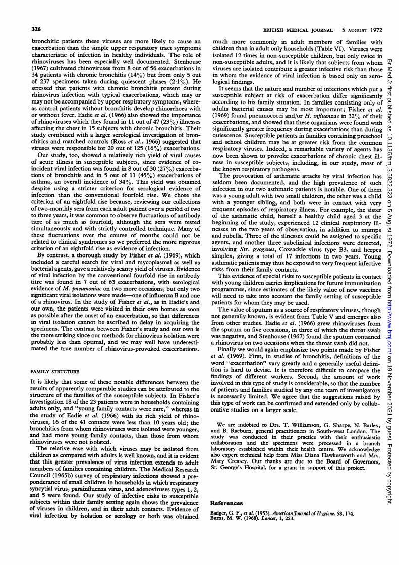
bronchitic patients these viruses are more likely to cause anexacerbation than the simple upper respiratory tract symptomscharacteristic of infection in healthy individuals. The role ofrhinoviruses has been especially well documented. Stenhouse(1967) cultivated rhinoviruses from 8 out of 56 exacerbations in34 patients with chronic bronchitis (14%) but from only 5 outof 237 specimens taken during quiescent phases (2-1%). Hestressed that patients with chronic bronchitis present duringrhinovirus infection with typical exacerbations, which may ormay not be accompanied by upper respiratory symptoms, where-as control patients without bronchitis develop rhinorrhoea withor without fever. Eadie et al. (1966) also showed the importanceof rhinoviruses which they found in 11 out of 47 (23%) illnessesaffecting the chest in 15 subjects with chronic bronchitis. Theirstudy combined with a larger serological investigation of bron-chitics and matched controls (Ross et al., 1966) suggested thatviruses were responsible for 20 out of 125 (16%) exacerbations.Our study, too, showed a relatively rich yield of viral causes
of acute illness in susceptible subjects, since evidence of co-incident viral infection was found in 8 out of 30 (27%) exacerba-tions of bronchitis and in 5 out of 11 (45%) exacerbations ofasthma, an overall incidence of 34%. This yield was obtaineddespite using a stricter criterion for serological evidence ofinfection than the conventional fourfold rise. We chose thecriterion of an eightfold rise because, reviewing our collectionsof two-monthly sera from each adult patient over a period of twoto three years, it was common to observe fluctuations of antibodytitre of as much as fourfold, although the sera were testedsimultaneously and with strictly controlled technique. Many ofthese fluctuations over the course of months could not berelated to clinical syndromes so we preferred the more rigorouscriterion of an eightfold rise as evidence of infection.By contrast, a thorough study by Fisher et al. (1969), which
included a careful search for viral and mycoplasmal as well asbacterial agents, gave a relatively scanty yield of viruses. Evidenceof viral infection by the conventional fourfold rise in antibodytitre was found in 7 out of 63 exacerbations, with serologicalevidence of M. pneumoniae on two more occasions, but only twosignificant viral isolations were made-one of influenza B and oneof a rhinovirus. In the study of Fisher et al., as in Eadie's andour own, the patients were visited in their own homes as soonas possible after the onset of an exacerbation, so that differencesin viral isolation cannot be ascribed to delay in acquiring thespecimens. The contrast between Fisher's study and our own isthe more striking since our methods for rhinovirus isolation wereprobably less than optimal, and we may well have underesti-mated the true number of rhinovirus-provoked exacerbations.
FAMILY STRUCTURE
It is likely that some of these notable differences between theresults of apparently comparable studies can be attributed to thestructure of the families of the susceptible subjects. In Fisher'sinvestigation 18 of the 23 patients were in households containingadults only, and "young family contacts were rare," whereas inthe study of Eadie et al. (1966) with its rich yield of rhino-viruses, 16 of the 41 contacts were less than 10 years old; thebronchitics from whom rhinoviruses were isolated were younger,and had more young family contacts, than those from whomrhinoviruses were not isolated.The relative ease with which viruses may be isolated from
children as compared with adults is well known, and it is evidentthat this greater prevalence of virus infection extends to adultmembers of families containing children. The Medical ResearchCouncil (1965b) survey of respiratory infections showed a pre-ponderance of small children in households in which respiratorysyncytial virus, parainfluenza virus, and adenoviruses types 1, 2,and 5 were found. Our study of infective risks to susceptiblesubjects within their family setting again shows the prevalenceof viruses in children, and in their adult contacts. Evidence ofviral infection by isolation or serology or both was obtained
much more commonly in adult members of families withchildren than in adult only households (Table VI). Viruses wereisolated 12 times in non-susceptible children, but only twice innon-susceptible adults, and it is likely that subjects from whomviruses are isolated contribute a greater infective risk than thosein whom the evidence of viral infection is based only on sero-logical findings.
It seems that the nature and number of infections which put asusceptible subject at risk of exacerbation differ significantlyaccording to his family situation. In families consisting only ofadults bacterial causes may be most important; Fisher et al.(1969) found pneumococci and/or H. influenzae in 32%h of theirexacerbations, and showed that these organisms were found withsignificantly greater frequency during exacerbations than duringquiescence. Susceptible patients in families containing preschooland school children may be at greater risk from the commonrespiratory viruses. Indeed, a remarkable variety of agents hasnow been shown to provoke exacerbations of chronic chest ill-ness in susceptible subjects, including, in our study, most ofthe known respiratory pathogens.The provocation of asthmatic attacks by viral infection has
seldom been documented, and the high prevalence of suchinfection in our two asthmatic patients is notable. One of themwas a young adult with two small children, the other was a childwith a younger sibling, and both were in contact with veryfrequent episodes of respiratory illness. For example, the sisterof the asthmatic child, herself a healthy child aged 3 at thebeginning of the study, experienced 12 clinical respiratory ill-nesses in the two years of observation, in addition to mumpsand rubella. Three of the illnesses could be assigned to specificagents, and another three subclinical infections were detected,involving Str. pyogenes, Coxsackie virus type B3, and herpessimplex, giving a total of 17 infections in two years. Youngasthmatic patients may thus be exposed to very frequent infectiverisks from their family contacts.
This evidence of special risks to susceptible patients in contactwith young children carries implications for future immunizationprogrammes, since estimates of the likely value of new vaccineswill need to take into account the family setting of susceptiblepatients for whom they may be used.The value of sputum as a source of respiratory viruses, though
not generally known, is evident from Table V and emerges alsofrom other studies. Eadie et al. (1966) grew rhinoviruses fromthe sputum on five occasions, in three of which the throat swabwas negative, and Stenhouse (1967) found the sputum containeda rhinovirus on two occasions when the throat swab did not.
Finally we would again emphasize two points made by Fisheret al. (1969). First, in studies of bronchitis, definitions of theword "exacerbation" vary greatly and a generally useful defini-tion is hard to devise. It is therefore difficult to compare thefindings of different workers. Second, the amount of workinvolved in this type of study is considerable, so that the numberof patients and families studied by any one team of investigatorsis necessarily limited. We agree that the suggestions raised bythis type of work can be confirmed and extended only by collab-orative studies on a larger scale.
We are indebted to Drs. T. Williamson, G. Sharpe, N. Barley,and B. Raeburn, general practitioners in South-west London. Thestudy was conducted in their practice with their enthusiasticoolaboration and the specimens were processed in a branchlaboratory established within their health centre. We acknowledgealso expert technical help from Miss Diana Hawkesworth and Mrs.Mary Creasey. Our thanks are due to the Board of Govemors,St. George's Hospital, for a grant in support of this project.
ReferencesBadger, G. F., et al. (1953). AmericanJournal of Hygiene, 58, 174.Burns, M. W. (1968). Lancet, 1, 223.
on 19 Novem
ber 2021 by guest. Protected by copyright.
http://ww
w.bm
j.com/
Br M
ed J: first published as 10.1136/bmj.3.5822.323 on 5 A
ugust 1972. Dow
nloaded from

BRITISH MEDICAL JOURNAL 5 AUGUST 1972 327
Burns, M. W., and May, J. R. (1967). Lancet, 1, 354.Carilli, A. D., Gohd, R. S., and Gordon,~W. (1964). New England_Journal of
Medicine, 270, 123.Eadie, M. D., Stott, E. J., and Grist, N. R. (1966). British MedicalJournal, 2,
671.Fisher, M., et al. (1969). British Medical_Journal, 4, 187.Grayston, J. T., et al. (1965). Journal of the American Medical Association,
191, 369.
McNamara, M. J., Phillips, I. A., and Williams, 0. B. (1969). AmericanReview of Respiratory Diseases, 100, 19.
Medical Research Council (1960). British Medical Journal, 2, 1665.Medical Research Council (1965a). Lancet, 1, 775.Medical Research Council (1965b). British Medical_Journal, 2, 319.Ross, C. A., et al. (1966). Thorax, 21, 461.Somerville, R. G. (1963). Lancet, 2, 1247.Stenhouse, A. C. (1967). British Medical_Journal, 3, 461.
Creatine Phosphokinase Activity during LithiumTreatment
R. GOSLING, R. J. KERRY, G. OWEN
British Medical Jrournal, 1972, 3, 327-329
Summary
In a group of patients suffering from manic depressiveillness treated with lithium carbonate occasional largeincreases in creatine phosphokinase (C.P.K.) activitywere seen. These were found to occur at times when thepatients were stable in mood and clinically well. Theserum lithium levels were adequate in all cases at thetime of raised C.P.K. activity. Raised C.P.K. activity canbe an indication that the disease is active but is controlledby lithium.
Introduction
The value of lithium in the treatment of mania is established,but its prophylactic use in mania and depression is still amatter of dispute. The natural history of manic depressiveillness with its long periods of remission have prompted Black-well and Shepherd (1968) to question the prophylactic action oflithium. Long symptom-free periods on lithium carbonatemay be due either to the prophylactic action of the lithium ionor to a long period of remission in the disease.
Creatine phosphokinase (C.P.K.) activity has been shown tobe increased during some acute episodes of psychotic illness(Meltzer, Elkun, and Moline, 1969). We have confirmed thesechanges in about half of a group of acutely psychotic patients(Gosling, Kerry, Orme, and Owen, 1972).
Since 1964 a special lithium clinic for the supervision ofpatients considered suitable for treatment with lithium carbo-nate has been run at this hospital. These patients attend atappropriate intervals for clinical assessment and the estimationof their serum lithium concentration. In addition, a largenumber of sera are referred from other clinics in the Sheffieldregion for serum lithium measurement.
In view of the association between psychotic illness andincreased C.P.K. activity it was decided to (1) measure theC.P.K. activity in all blood samples taken for serum lithiumestimation in our clinic, and (2) to measure the C.P.K. activityin all serum samples referred to us for serum lithium estimationfrom other clinics. These repeated estimations give us anopportunity of seeing if lithium stabilizes C.P.K. activity as wellas mood.
Department of Chemical Pathology, Northern General Hospital,Sheffield
R. GOSLING, B.SC., BiochemistR. J. KERRY. M.R.C.S., L.R.C.P., D.P.M., Consultant PsychiatristG. OWEN, M.B., F.R.C.PATH., Consultant Chemical Pathologist
Patients and Methods
All patients attending the Northern General Hospital lithiumclinic had been previously diagnosed as suffering from manicdepressive illness and suitable for treatment with prophylacticlithium. Patients did not receive any other medication andattended the clinic at regular intervals for clinical and bio-chemical assessment. These patients have been known to oneof us (R.J.K.) for up to 10 years and their psychiatric historyhas been well documented both before and after starting lithiumtreatment.
Clinicians forwarding sera from their own patients wereconsulted about the clinical state of the patient and the natureof the illness. They confirmed that the patients were notreceiving any other psychotrophic drugs.
SERUM LITHIUM ESTIMATIONS
Serum lithium concentrations were measured by atomicabsorption with an E.E.L. 140 atomic absorption spectro-photometer and a lithium specific lamp. All sera were obtainedfrom venous blood collected in plain glass universal containers.These sera were diluted 1 in 5 with distilled water. They werecompared with appropriate standards prepared from a lithiumreference standard supplied by the Fisher Corporation.
SERUM CREATINE PHOSPHOKINASE MEASUREMENTS
The C.P.K. activity at 35°C was estimated by linking the C.P.K.catalysed reaction through hexokinase to one involving glucose-6-phosphate dehydrogenase and the conversion of nicotinamideadenine dinucleotide phosphate to its reduced form. Gluta-thione was included within the reaction mixture to "activate"the C.P.K. The increase in optical density at 340 nanometresdue to the formation of the reduced nicotinamide adeninedinucleotide phosphate was plotted by using an L.K.B. 8600reaction rate analyser and is directly proportional to the C.P.K.activity.To ascertain the effect, if any, of the lithium ion on the
estimation of C.P.K. pooled human serum with raised C.P.K.activity was divided into two portions. One portion was seededwith lithium carbonate to establish a prophylactic serum lithiumconcentration of 2 mEq/l. Comparison between the seeded andnon-seeded sera showed that a prophylactic lithium concentra-tion does not interfere with the estimation of C.P.K.The reagents used were supplied by the Boehringer Corpo-
ration in the form of a test kit for C.P.K. determination (Cat.No. 15926. TCAF). Solutions were prepared as recommendedby Boehringer and the reaction was started by the automaticaddition by the L.K.B. 8600 of 75 il of creatine phosphate,solution 3 of the assay system. For all the solutions only halfthe recommended test quantities were used because the L.K.B.
on 19 Novem
ber 2021 by guest. Protected by copyright.
http://ww
w.bm
j.com/
Br M
ed J: first published as 10.1136/bmj.3.5822.323 on 5 A
ugust 1972. Dow
nloaded from