Infectious Disease Cytopathology
39
Infectious Disease Cytopathology Mycology Heather Ruff October 26, 2019
Transcript of Infectious Disease Cytopathology

Infectious Disease
Cytopathology
MycologyHeather Ruff
October 26, 2019
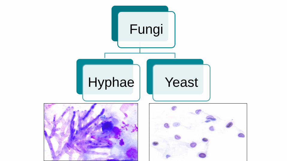
Fungi
YeastHyphae

Categorizing Fungal Hyphae
Hyphae
Pigmented hyphae
Dematiaceous molds
Non-septate hyaline hyphae
Zygomycetes
Septate hyaline hyphae
Aspergillus and others
Pseudohypaewith Yeast (Candida)

Assessing Fungal Hyphae
● True hyphae or pseudohyphae● Pigmented or non-pigmented (hyaline)?● Septate or non-septate?
● Branching -○ Acute angle, right angle, variable?○ Dichotomous (equal) vs. non-dichotomous (variable)?
● Arrangement - uniform/parallel vs. disorganized● Other structures
○ Fruiting Bodies (conidia, phialides, vesicle)○ Pseudohyphae○ Yeast
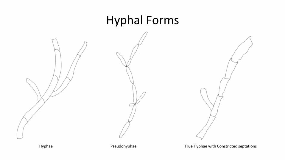
Hyphal Forms
Hyphae Pseudohyphae True Hyphae with Constricted septations

Cervical Pap ThinPrep 29 Female
What is this?

• Oval budding yeast (3-6 µm)• Pseudohyphae• True hyphae (less common)• Blastoconidia (rare)
Candida
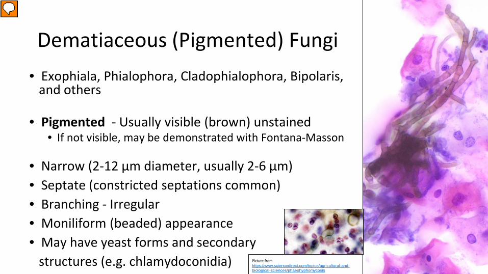
• Exophiala, Phialophora, Cladophialophora, Bipolaris, and others
• Pigmented - Usually visible (brown) unstained• If not visible, may be demonstrated with Fontana-Masson
• Narrow (2-12 µm diameter, usually 2-6 µm)• Septate (constricted septations common)• Branching - Irregular• Moniliform (beaded) appearance• May have yeast forms and secondary
structures (e.g. chlamydoconidia)
Dematiaceous (Pigmented) Fungi
Picture from https://www.sciencedirect.com/topics/agricultural-and-biological-sciences/phaeohyphomycosis
Presenter
Presentation Notes
Chlamydoconidia - large, thick-walled areas of vesicular swelling, >25 µm

Zygomycetes• Mucor spp., Rhizomucor sp., Rhisopuz spp.,
and others broad non-pigmented hyphae
• Non-pigmented• Non-septate (really Pauci-septate)• Broad (5-25 µm diameter, avaerage 12 µm)
• Branching - Irregularly-spaced, non-dichotomous, variable angles ( often 90°)
• Twisted, folded, ribbon-like morphology

Hyaline Fungi
• Aspergillus, Fusarium, Paecilomyces, Acremonium, Scedosporium, Pseudallescheria, and many, many others)
• Non-pigmented• Septate• Narrow (2-12 µm diameter, usually 2-8 µm)
• …

Aspergillus
● Organized● Branching –
○Parallel○Dichotomous (equal)○Acute angle (45°)
● May have fruiting bodies
● Disorganized● Branching –
○Variable orientation○Non-dichotomous (unequal)○Variable angles (45° & 90°)
● May see fruiting bodies
Other Hyaline Fungi

Esophageal Brush 56 F w/ SLE on prednisone.
Diagnosis?

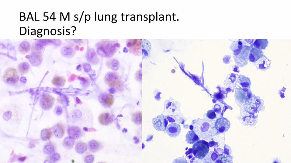
BAL 54 M s/p lung transplant.Diagnosis?
BAL 54 M s/p lung transplant.Diagnosis?

Categorizing Yeast
Yeast
Small
Histoplasma Cryptococcus Pneumocystis Candida
Large
Blastomyces Cryptococcus Coccidioidies

Assessing Yeast
● Size - Small, large, variable● Shape - Round, oval, irregular● Other morphology
● Wall - Thin, thick, refractile● Capsule, pseudocapsule, none● Division – Budding (Broad-based, narrow-based), Fission● Intracellular, extracellular, both● Internal structures
● Stains:● Mucin (Mucicarmine)● Pigment (Fontana-Masson)
Penicillium marneffei (E) - Courtesy of Vicki Schnadig at UTMBParacoccidiioides (*) - Courtesy of Diane Dziedzic at microworld.org *
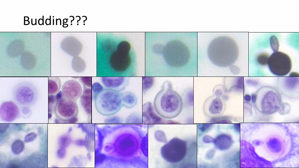
Budding???
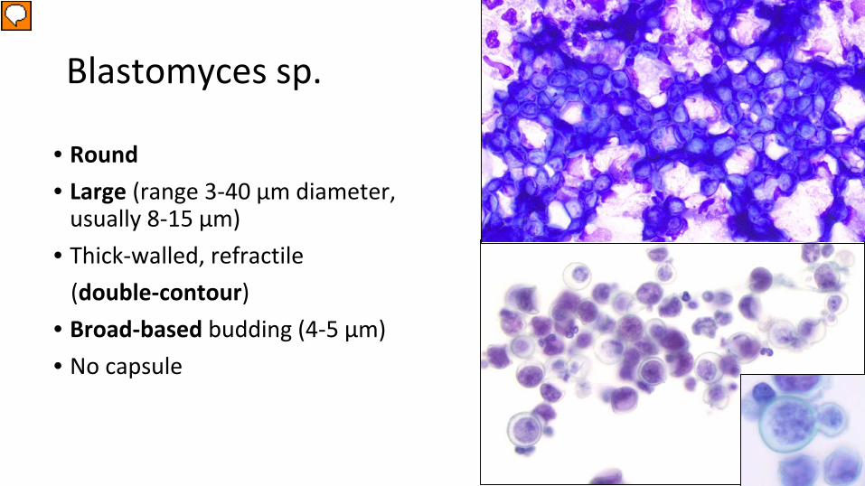
Blastomyces sp.
• Round• Large (range 3-40 µm diameter,
usually 8-15 µm)• Thick-walled, refractile
(double-contour)• Broad-based budding (4-5 µm)• No capsule
Presenter
Presentation Notes
Ohio & Mississippi river valleys, and great lakes

• Variable shapes (Round, oval, collapsed crescents)
• Variable sizes (range 2-20 µm diameter, usually 4-10)
• Thin-walled, Narrow-based budding• Encapsulated and unencapsulated forms
• Capsule is clear on Romanowsky, Pap• Capsule stains pink on Mucicarmine
• Contains melanin-like pigment• Stains brown with Fontana-Masson• Especially good for unencapsulated small
forms
Cryptococcus sp.
Courtesy of V. Schnadig
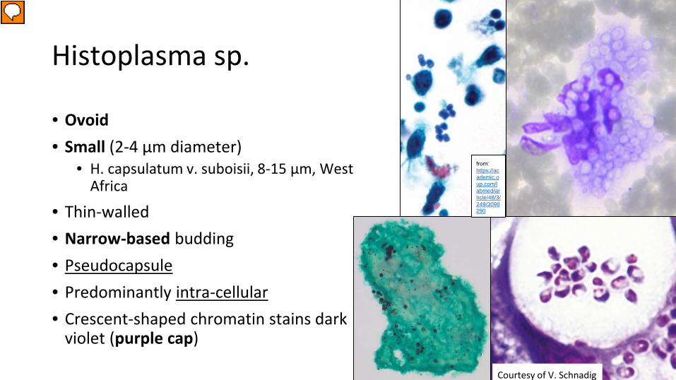
Histoplasma sp.
• Ovoid• Small (2-4 µm diameter)
• H. capsulatum v. suboisii, 8-15 µm, West Africa
• Thin-walled• Narrow-based budding• Pseudocapsule• Predominantly intra-cellular• Crescent-shaped chromatin stains dark
violet (purple cap)
Courtesy of V. Schnadig
from: https://academic.oup.com/labmed/article/48/3/249/3098290
Presenter
Presentation Notes
https://academic.oup.com/labmed/article/48/3/249/3098290

Pneumocystis jiroveci
• Round to ovoid cysts • Collapsed crescent, cup appearance
• Small (3-7 µm diameter)• Thin membrane• No capsule, pseudocapsule• No budding!• In “foamy exudate” (collection of cysts)• Romanowsky, Pap - clear cyst with dark dots
(nuclei, intra- and extra-cystic bodies)• GMS - Dot, comma, or parenthesis-shaped
(thickening in membrane)
Presenter
Presentation Notes
Former parasite, now Fungus (genetics); Doesn’t grow in culture!!! Infiltrate on CXR Dot is thickening in membrane
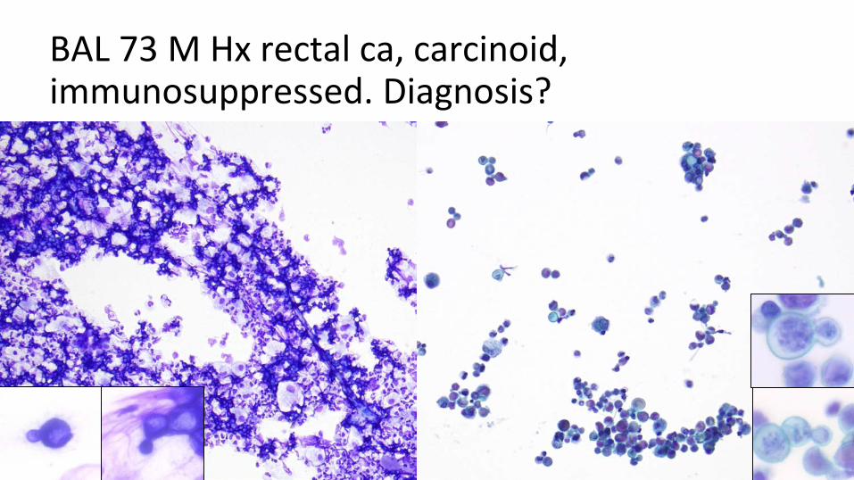
BAL 73 M Hx rectal ca, carcinoid, immunosuppressed. Diagnosis?

BAL 55 M w/ HIV and diffuse pneumonitis. Diagnosis?

BAL 49 M w/ HIV, immunosuppressed.Diagnosis?
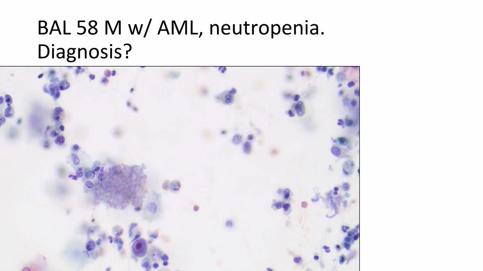
BAL 58 M w/ AML, neutropenia.Diagnosis?

Additional Reference:
(from Diagnostic Pathology of Infectious Disease 2nd Ed.
Kradin)

Differentiating Hyphal Fungal Organisms
Zygomycetes
Non-pigmentedBroadNon/Pauci-septateIrregular, uneven, 90°xTwisted, folded, ribbon-like
Aspergillus
Non-pigmentedNarrowSeptateRegular, even, 45°xParallel, radiating branches+/- fruiting bodies
Other Hyalohyphomycetes
Non-pigmentedNarrowSeptateIrregular, uneven xHaphazard arrangement x+/- fruiting bodies
Phaeohyphomycetes
PigmentedNarrowSeptate (constricted)Irregular, uneven xHaphazard arrangemet x+/- Yeast-like forms and chlamydoconidia
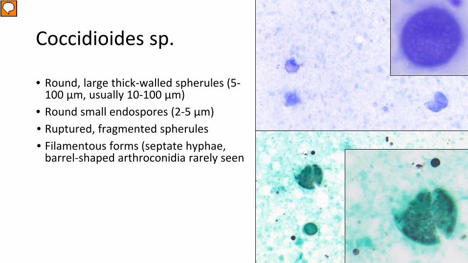
• Round, large thick-walled spherules (5-100 µm, usually 10-100 µm)
• Round small endospores (2-5 µm) • Ruptured, fragmented spherules• Filamentous forms (septate hyphae,
barrel-shaped arthroconidia rarely seen
Coccidioides sp.
Presenter
Presentation Notes
Technically not a dimorph; Culture at 25, 30, 37 only produces filamentous forms Lobar pneumonia and LAD Sphereules (PAS-, GMSv); Endospores (PAS+, GMS+)

Paracoccidioides sp.
• Round to oval • Large yeast (3-30 µm)• Narrow based budding• Multiple bud attached to single
parent cell (ships wheel)
• Round to oval • Small (3 µm diameter, 2.5-5 µm long)• Transverse septum (fission, not bud)• Pseudocapsule• Predominantly intracellular
• If extracellular - elongate up to 8 µm, have septae, become curved
Penicillium marneffei
Penicillium marneffei (E) - Courtesy of Vicki Schnadig at UTMBParacoccidiioides (*) - Courtesy of Diane Dziedzic at microworld.org
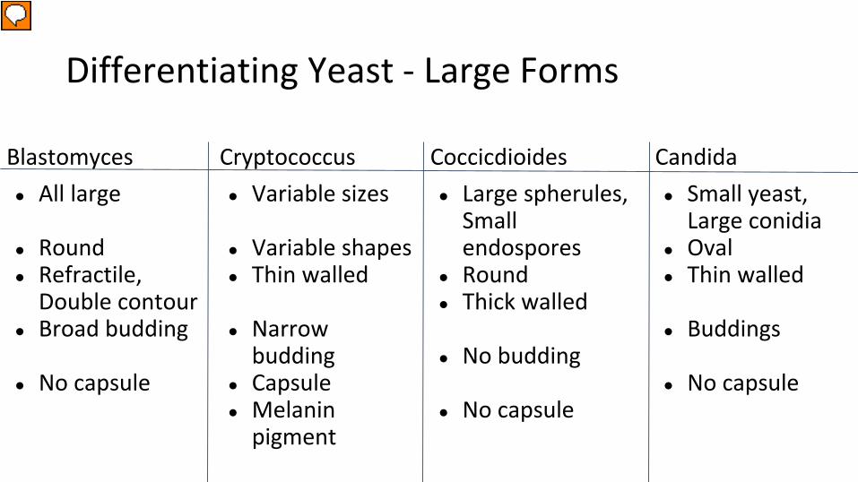
Differentiating Yeast - Large Forms
Blastomyces● All large
● Round● Refractile,
Double contour● Broad budding
● No capsule
Cryptococcus● Variable sizes
● Variable shapes● Thin walled
● Narrow budding
● Capsule● Melanin
pigment
Coccicdioides● Large spherules,
Small endospores
● Round● Thick walled
● No budding
● No capsule
Candida● Small yeast,
Large conidia● Oval● Thin walled
● Buddings
● No capsule
Presenter
Presentation Notes
Candida have blastoconidia
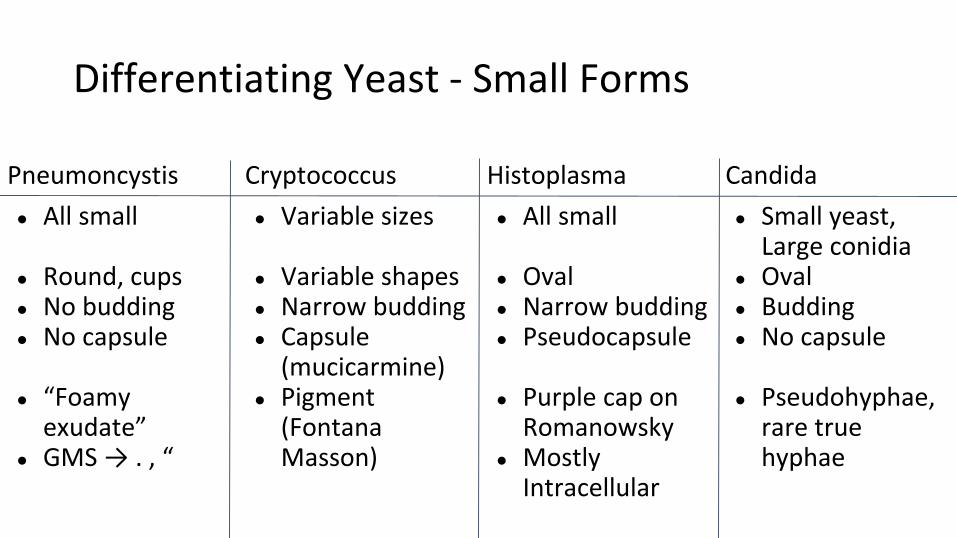
Differentiating Yeast - Small Forms
Pneumoncystis● All small
● Round, cups● No budding● No capsule
● “Foamy exudate”
● GMS → . , “
Cryptococcus● Variable sizes
● Variable shapes● Narrow budding● Capsule
(mucicarmine)● Pigment
(Fontana Masson)
Histoplasma● All small
● Oval● Narrow budding● Pseudocapsule
● Purple cap on Romanowsky
● Mostly Intracellular
Candida● Small yeast,
Large conidia● Oval● Budding● No capsule
● Pseudohyphae, rare true hyphae

Answers To Cases

Esophageal Brush 56 F w/ SLE on prednisone.
Diagnosis:
Fungal organisms consistent with Candida
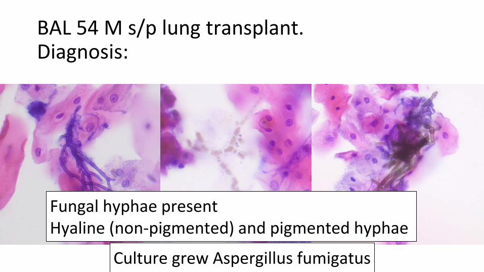
BAL 54 M s/p lung transplant.Diagnosis:
Fungal hyphae presentHyaline (non-pigmented) and pigmented hyphae
Culture grew Aspergillus fumigatus
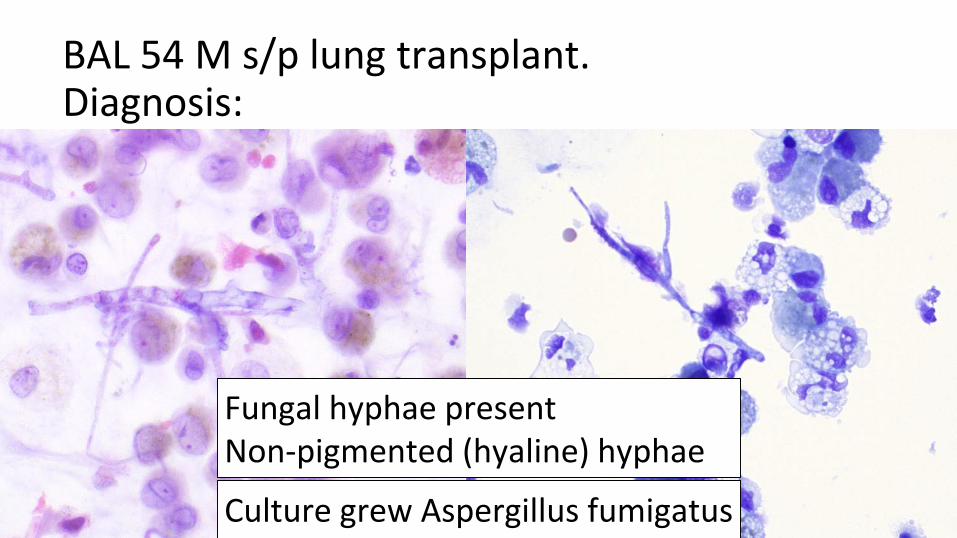
BAL 54 M s/p lung transplant.Diagnosis:
Fungal hyphae presentNon-pigmented (hyaline) hyphae
Culture grew Aspergillus fumigatus

BAL 73 M Hx rectal ca, carcinoid, immunosuppressed. Diagnosis:
C/W Blastomyces
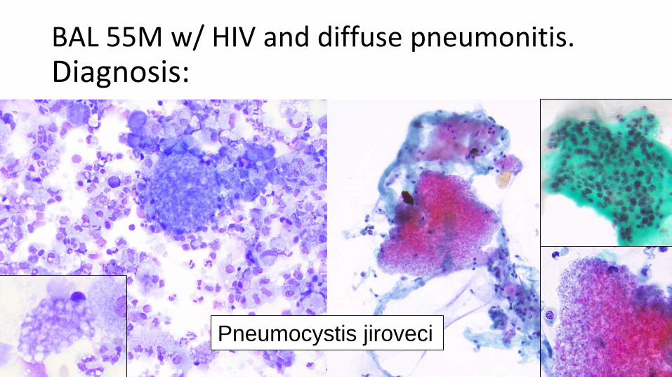
BAL 55M w/ HIV and diffuse pneumonitis.Diagnosis:
Pneumocystis jiroveci

BAL 49 M w/ HIV, immunosuppressed.Diagnosis:
Cryptococcus sp.

BAL 58 M w/ AML, neutropenia.Diagnosis:
Pneumocystis & CMV
Don’t forget to look for the second organism!