Infection Biology of Moniliophthora perniciosa on Theobroma cacao
12
Infection Biology of Moniliophthora perniciosa on Theobroma cacao and Alternate Solanaceous Hosts Jean-Philippe Marelli & Siela N. Maximova & Karina P. Gramacho & Seogchan Kang & Mark John Guiltinan Received: 27 May 2009 / Accepted: 13 November 2009 / Published online: 21 November 2009 # Springer Science + Business Media, LLC 2009 Abstract The C-biotype of Moniliophthora perniciosa is the causal agent of witches’ broom disease of Theobroma cacao L. While this disease is of major economic impor- tance, the pathogenicity mechanisms and plant responses underlying the disease are difficult to study given the cacao tree’ s long life cycle and the limited availability of genetic and genomic resources for this system. The S-biotype of M. perniciosa infects solanaceous hosts, particularly pepper (Capsicum annuum) and tomato (Solanum lycopersicum). These species are much more amenable for performing studies of mechanisms underpinning host-pathogen inter- actions as compared to cacao. A phylogenetic analysis performed in this study demonstrated that S-biotype strains clustered with C-biotype strains, indicating that these bio- types are not genetically distinct. A comparative analysis demonstrated that disease progression in tomato infected with the S- biotype is similar to that described for cacao infected with the C- biotype. The major symptoms ob- served in both systems are swelling of the infected shoots and activation and proliferation of axillary meristems. Cellular changes observed in infected tissues correspond to an increase in cell size and numbers of xylem vessels and phloem parenchyma along the infected stem. Observations revealed that fungal colonization is biotrophic during the first phase of infection, with appearance of calcium oxalate crystals in close association with hyphal growth. In summary, despite different host specificity, both biotypes of M. perniciosa exhibit similar disease-related character- istics, indicating a degree of conservation of pathogenicity mechanisms between the two biotypes. Keywords Moniliophthora perniciosa . Theobroma cacao L. . Solanum lycopersicum . Capsicum annuum . Fungal pathogenicity . Plant defense Introduction Moniliophthora (= Crinipellis) perniciosa (Stahel) Sing., is a basidiomycete fungus [2], closely related to the common button mushroom Agaricus bisporus [36]. The fungus is a pathogen of Theobroma cacao L., the chocolate tree, and the disease’ s common name, witches’ broom, is due to the appearance of dried and distorted shoots on the infected trees [44]. Moniliophthora perniciosa is indigenous to the Amazon basin region in association with several plant species in the genus Theobroma (Malvaceae) and also in plants from families unrelated to the Malvaceae [4]. Multiple biotypes of M. perniciosa are defined based on host adaptation: C-biotype infecting T. cacao ; L- biotype Communicated by: Schuyler S. Korban J.-P. Marelli : S. Kang Department of Plant Pathology, The Pennsylvania State University, Buckhout Lab, 16802 University Park, PA, USA S. N. Maximova : M. J. Guiltinan (*) Department of Horticulture, The Pennsylvania State University, 422 Life Sciences Building, 16802 University Park, PA, USA e-mail: [email protected] K. P. Gramacho CEPLAC/CEPEC/SEFIT, Caixa postal 07 45600-000 Itabuna, Bahia, Brazil Present Address: J.-P. Marelli Mars Center for Cocoa Science, Caixa postal 55 45630-000 Itajuipe, Bahia, Brazil Tropical Plant Biol. (2009) 2:149–160 DOI 10.1007/s12042-009-9038-1
Transcript of Infection Biology of Moniliophthora perniciosa on Theobroma cacao

Infection Biology of Moniliophthora perniciosaon Theobroma cacao and Alternate Solanaceous Hosts
Jean-Philippe Marelli & Siela N. Maximova &
Karina P. Gramacho & Seogchan Kang &
Mark John Guiltinan
Received: 27 May 2009 /Accepted: 13 November 2009 /Published online: 21 November 2009# Springer Science + Business Media, LLC 2009
Abstract The C-biotype of Moniliophthora perniciosa isthe causal agent of witches’ broom disease of Theobromacacao L. While this disease is of major economic impor-tance, the pathogenicity mechanisms and plant responsesunderlying the disease are difficult to study given the cacaotree’s long life cycle and the limited availability of geneticand genomic resources for this system. The S-biotype of M.perniciosa infects solanaceous hosts, particularly pepper(Capsicum annuum) and tomato (Solanum lycopersicum).These species are much more amenable for performingstudies of mechanisms underpinning host-pathogen inter-actions as compared to cacao. A phylogenetic analysisperformed in this study demonstrated that S-biotype strainsclustered with C-biotype strains, indicating that these bio-types are not genetically distinct. A comparative analysis
demonstrated that disease progression in tomato infectedwith the S- biotype is similar to that described for cacaoinfected with the C- biotype. The major symptoms ob-served in both systems are swelling of the infected shootsand activation and proliferation of axillary meristems.Cellular changes observed in infected tissues correspondto an increase in cell size and numbers of xylem vessels andphloem parenchyma along the infected stem. Observationsrevealed that fungal colonization is biotrophic during thefirst phase of infection, with appearance of calcium oxalatecrystals in close association with hyphal growth. Insummary, despite different host specificity, both biotypesof M. perniciosa exhibit similar disease-related character-istics, indicating a degree of conservation of pathogenicitymechanisms between the two biotypes.
Keywords Moniliophthora perniciosa .Theobroma cacao L. .
Solanum lycopersicum .Capsicum annuum .
Fungal pathogenicity. Plant defense
Introduction
Moniliophthora (=Crinipellis) perniciosa (Stahel) Sing., isa basidiomycete fungus [2], closely related to the commonbutton mushroom Agaricus bisporus [36]. The fungus is apathogen of Theobroma cacao L., the chocolate tree, andthe disease’s common name, witches’ broom, is due to theappearance of dried and distorted shoots on the infectedtrees [44]. Moniliophthora perniciosa is indigenous to theAmazon basin region in association with several plantspecies in the genus Theobroma (Malvaceae) and also inplants from families unrelated to the Malvaceae [4].Multiple biotypes of M. perniciosa are defined based onhost adaptation: C-biotype infecting T. cacao ; L- biotype
Communicated by: Schuyler S. Korban
J.-P. Marelli : S. KangDepartment of Plant Pathology,The Pennsylvania State University,Buckhout Lab,16802 University Park, PA, USA
S. N. Maximova :M. J. Guiltinan (*)Department of Horticulture, The Pennsylvania State University,422 Life Sciences Building,16802 University Park, PA, USAe-mail: [email protected]
K. P. GramachoCEPLAC/CEPEC/SEFIT,Caixa postal 07 45600-000 Itabuna, Bahia, Brazil
Present Address:J.-P. MarelliMars Center for Cocoa Science,Caixa postal 55 45630-000 Itajuipe, Bahia, Brazil
Tropical Plant Biol. (2009) 2:149–160DOI 10.1007/s12042-009-9038-1
associated with members of the Bignoniaceae [19]; H-biotype that can infect members of the Malpighiaceae andS-biotype associated with members of the Solanaceae [5].Based on morphological and molecular data, the H-biotypewas reclassified to a new species, Crinipellis brasiliensis[9]. The S-biotype was originally discovered on Solanumrugosum and Solanum lasiantherum, both solanaceousweeds present near cocoa farms in the Amazon [5].Infection with S-biotype basidiospores of cultivated sola-naceous like Solanum lycopersicum (tomato) and Capsicumannuum (pepper) in greenhouse conditions, induced stemswelling and hypertrophied buds [5]. However, in nature,M. perniciosa is not epidemic on cultivated solanaceous,indicating that the annual life style of these crops is notconducive for the full completion of the pathogen life cycle[14]. Cross infectivity between the C- and S-biotypes hasnot been observed under controlled experiments but,morphological and genetic evidence suggests that thesebiotypes belong to a single species [9, 20].
In nature, basidiospores of M. perniciosa, are the onlyknown infectious propagules, germinating on young cacaotissues such as apical and axillary meristems, young leaves,developing flowers, and fruits [30, 36], and subsequentlypenetrating plant tissues directly or entering throughstomata [15, 43]. The pathogen exhibits two phases in itsdisease cycle, the biotrophic phase and the necrotrophicphase. Germinated basidiospores form a swollen (5–20µm)monokaryotic hyphae that grows intercellularly as abiotroph [13] and causes symptoms that include shootswelling, petiole swelling, and loss of apical dominance[30]. The biotrophic phase usually lasts for about 60 days inartificially inoculated plants [40]. In the necrotrophic phaseof infection, from the apex downwards, the green broombecomes brown and dies within a week [30, 42]. This phaseis associated with the appearance of intra-cellular hyphae,which is dikaryotic and exhibits clamp connections [37].When the dried broom is exposed to alternate periods of dryand wet weather, basidiocarps emerge to complete the lifecycle [34].
A witches’ broom epidemic recently occurred in 1989 inBahia, Brazil, which was once the second most productivecacao-producing region in the world. In the subsequent tenyears, cocoa bean production in this region has declinedfrom 300,000 tons to 130,000 tons causing a loss of $235million annually and socioeconomic hardship in the region[6, 30]. The development of effective control measuresagainst M. perniciosa is one of the major challenges inboosting cacao production in South America. Equallycritical is avoiding the introduction of the pathogen toAfrica and Asia where the disease has not yet beenobserved. A better understanding of the physiological andgenetic mechanisms underpinning the development of thisdisease is vital for protecting global cacao production from
witches’ broom. However, research on M. perniciosa/T.cacao interactions has been difficult because of cacao’sslow and long life cycle, irreproducible infection efficien-cies under laboratory conditions, very limited genetic/genomic resources, and the complexity of M. perniciosa’sdisease cycle [8, 12].
To address this problem, this study aimed to use the S-biotype of M. perniciosa on cultivated solanaceous. Be-cause Solanum lycopersicum (tomato), in particular, hasbeen extensively used as a model plant for basic biologicalresearch, many molecular and genetic tools are available andcan be used to elucidate some of the questions remaining onthe effect of this pathogen on the plant’s physiology.
As the first step of this study, phylogenic relationships,colonization patterns, and the disease cycles of the S and Cbiotypes, were investigated. Cellular mechanisms underly-ing the development of the typical symptoms of thebiotrophic phase (stem swelling and release of axillarybuds) were also studied via morphometric analyses of theaffected areas. Lastly, utilizing transgenic tomato plantsharboring an auxin responsive reporter gene (DR5-GUSauxin responsive promoter), changes of this hormoneoccurring during infection, were monitored.
Results
Symptomology in Cacao, Pepper, and Tomato
As previously described [42], infected cacao plantsexhibited the first symptom of infection about 30 days postinoculation (dpi), in the form of a slight swelling of thestems (Fig. 1a). Activation of axillary meristems andswelling near the infection point was observed during laterstages of infection during the biotrophic phase of thedisease (Fig. 1b).
The developmental switch from the biotrophic to thenecrotrophic phase of infection occurred at about 100 dpi,causing a total necrosis of the shoot within 2-3 days(Fig. 1c). Exposing the dried shoot to conditions conducivefor sporulation caused basidiocarps to form on the drybranches within 270 dpi (Fig. 1d).
The pepper variety, Tequila Sunrise, was used forverifying the pathogenicity of S-biotype strain 73-6-01(Fig. 1e to h). Witches’ broom symptoms (stem swellingand activation of the axillary meristems) were recorded at17 dpi (Fig. 1e). The biotrophic phase lasted up to 90 dpi inpepper (Fig. 1f) Total necrosis of the infected shoot wasobserved at 105 dpi, but the progression was slower than incacao (Fig. 1g). When the dry shoot was exposed to sporu-lation conditions, basidiocarps appeared at 210 dpi (Fig. 1h).
The same general progression of symptoms was alsoobserved in tomato plants inoculated with S-biotype strain
150 Tropical Plant Biol. (2009) 2:149–160
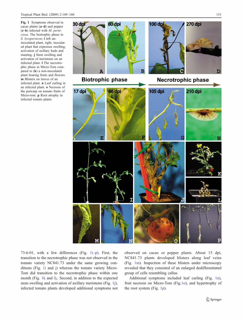
73-6-01, with a few differences (Fig. 1i–p). First, thetransition to the necrotrophic phase was not observed in thetomato variety NC841.73 under the same growing con-ditions (Fig. 1i and j) whereas the tomato variety Micro-Tom did transition to the necrotrophic phase within onemonth (Fig. 1k and l),. Second, in addition to the expectedstem swelling and activation of axillary meristems (Fig. 1j),infected tomato plants developed additional symptoms not
observed on cacao or pepper plants. About 15 dpi,NC841.73 plants developed blisters along leaf veins(Fig. 1m). Inspection of these blisters under microscopyrevealed that they consisted of an enlarged dedifferentiatedgroup of cells resembling callus.
Additional symptoms included leaf curling (Fig. 1n),fruit necrosis on Micro-Tom (Fig.1o), and hypertrophy ofthe root system (Fig. 1p).
Fig. 1 Symptoms observed incacao plants (a–d) and pepper(e–h) infected with M. perni-ciosa. The biotrophic phase inS. lycopersicon; i left un-inoculated plant, right, inoculat-ed plant that expresses swelling,activation of axillary buds andstunting. j Stem swelling andactivation of meristems on aninfected plant. l The necrotro-phic phase in Micro-Tom com-pared to (k) a non-inoculatedplant bearing fruits and flowers.m Blisters on leaves of aninfected plant. n Leaf curling inan infected plant. o Necrosis ofthe pericarp on tomato fruits ofMicro-tom. p Root atrophy ininfected tomato plants
Tropical Plant Biol. (2009) 2:149–160 151
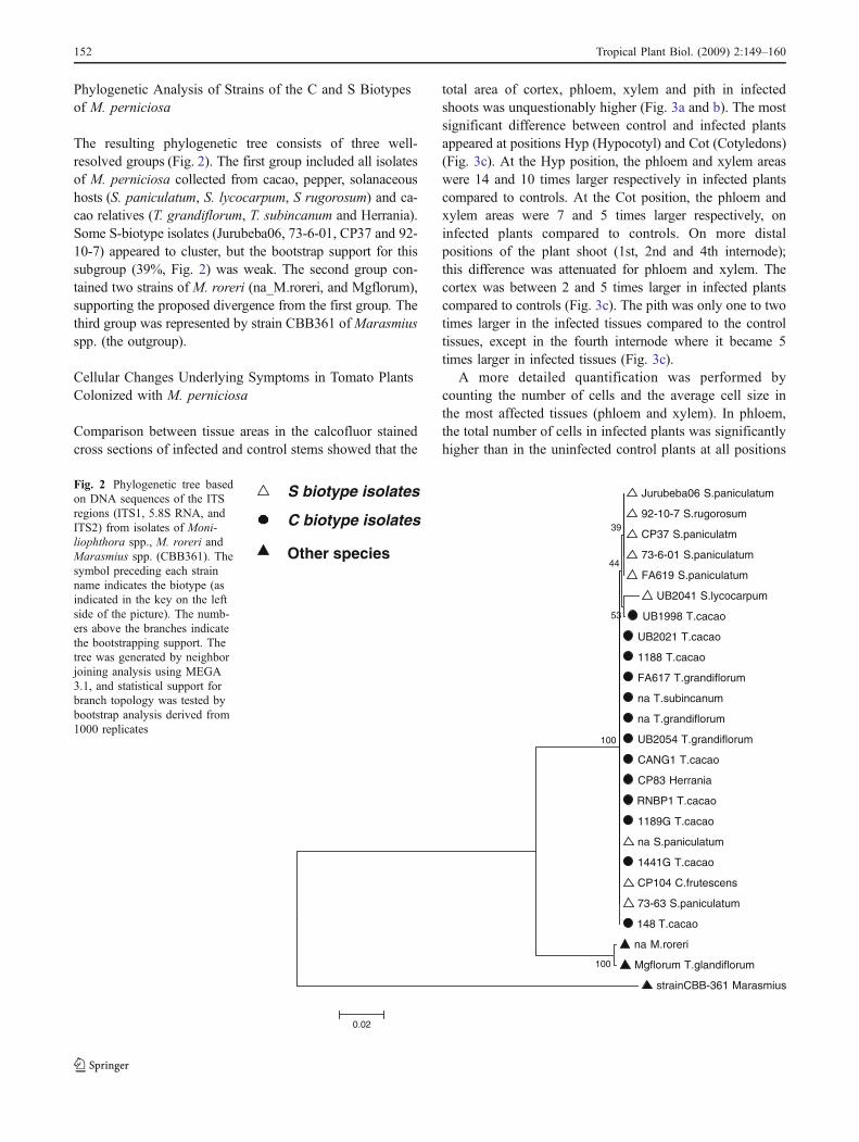
Phylogenetic Analysis of Strains of the C and S Biotypesof M. perniciosa
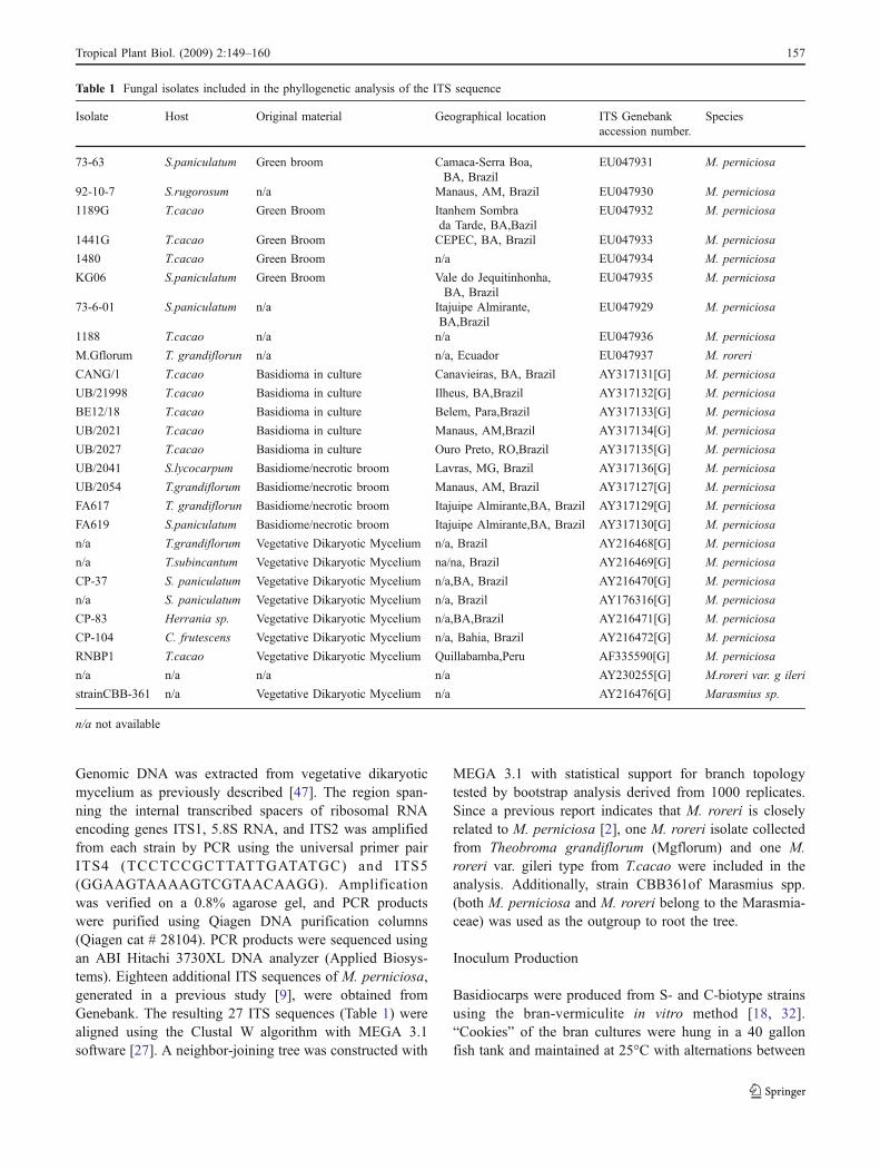
The resulting phylogenetic tree consists of three well-resolved groups (Fig. 2). The first group included all isolatesof M. perniciosa collected from cacao, pepper, solanaceoushosts (S. paniculatum, S. lycocarpum, S rugorosum) and ca-cao relatives (T. grandiflorum, T. subincanum and Herrania).Some S-biotype isolates (Jurubeba06, 73-6-01, CP37 and 92-10-7) appeared to cluster, but the bootstrap support for thissubgroup (39%, Fig. 2) was weak. The second group con-tained two strains of M. roreri (na_M.roreri, and Mgflorum),supporting the proposed divergence from the first group. Thethird group was represented by strain CBB361 ofMarasmiusspp. (the outgroup).
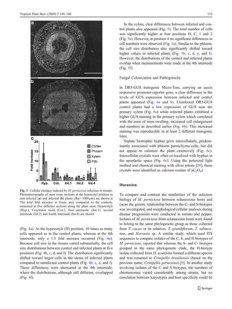
Cellular Changes Underlying Symptoms in Tomato PlantsColonized with M. perniciosa
Comparison between tissue areas in the calcofluor stainedcross sections of infected and control stems showed that the
total area of cortex, phloem, xylem and pith in infectedshoots was unquestionably higher (Fig. 3a and b). The mostsignificant difference between control and infected plantsappeared at positions Hyp (Hypocotyl) and Cot (Cotyledons)(Fig. 3c). At the Hyp position, the phloem and xylem areaswere 14 and 10 times larger respectively in infected plantscompared to controls. At the Cot position, the phloem andxylem areas were 7 and 5 times larger respectively, oninfected plants compared to controls. On more distalpositions of the plant shoot (1st, 2nd and 4th internode);this difference was attenuated for phloem and xylem. Thecortex was between 2 and 5 times larger in infected plantscompared to controls (Fig. 3c). The pith was only one to twotimes larger in the infected tissues compared to the controltissues, except in the fourth internode where it became 5times larger in infected tissues (Fig. 3c).
A more detailed quantification was performed bycounting the number of cells and the average cell size inthe most affected tissues (phloem and xylem). In phloem,the total number of cells in infected plants was significantlyhigher than in the uninfected control plants at all positions
Jurubeba06 S.paniculatum
92-10-7 S.rugorosum
CP37 S.paniculatm
73-6-01 S.paniculatum
FA619 S.paniculatum
UB2041 S.lycocarpum
UB1998 T.cacao
UB2021 T.cacao
1188 T.cacao
FA617 T.grandiflorum
na T.subincanum
na T.grandiflorum
UB2054 T.grandiflorum
CANG1 T.cacao
CP83 Herrania
RNBP1 T.cacao
1189G T.cacao
na S.paniculatum
1441G T.cacao
CP104 C.frutescens
73-63 S.paniculatum
148 T.cacao
na M.roreri
Mgflorum T.glandiflorum
strainCBB-361 Marasmius
100
53
39
44
100
0.02
C biotype isolates
S biotype isolates
Other species
Fig. 2 Phylogenetic tree basedon DNA sequences of the ITSregions (ITS1, 5.8S RNA, andITS2) from isolates of Moni-liophthora spp., M. roreri andMarasmius spp. (CBB361). Thesymbol preceding each strainname indicates the biotype (asindicated in the key on the leftside of the picture). The numb-ers above the branches indicatethe bootstrapping support. Thetree was generated by neighborjoining analysis using MEGA3.1, and statistical support forbranch topology was tested bybootstrap analysis derived from1000 replicates
152 Tropical Plant Biol. (2009) 2:149–160
(Fig. 4a). At the hypocotyls (H) position, 10 times as manycells appeared as in the control plants, whereas at the 4thinternode, only a 1.5 fold increase occurred (Fig. 4a).Because cell size in the tissues varied substantially, the cellsize distributions between control and infected plants at fivepositions (Fig. 4b, c, d, and f). The distribution significantlyshifted toward larger cells in the stems of infected plantscompared to uninfected control plants (Fig. 4b, c, d, and f).Those differences were attenuated at the 4th internode,where the distributions, although still different, overlapped(Fig. 4f).
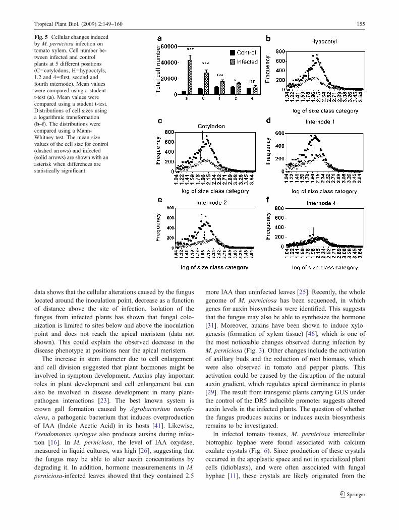
In the xylem, clear differences between infected and con-trol plants also appeared (Fig. 5). The total number of cellswas significantly higher at four positions H, C, 1 and 2(Fig. 5a). However, at position 4 no significant differences incell numbers were observed (Fig. 5a). Similar to the phloem,the cell size distribution also significantly shifted towardhigher values in infected plants (Fig. 5b, c, d, e, and f).However, the distributions of the control and infected plantsoverlap when measurements were made at the 4th internode(Fig. 5f).
Fungal Colonization and Pathogenicity
In DR5-GUS transgenic Micro-Tom, carrying an auxinresponsive promoter-reporter gene, a clear difference in thelevels of GUS expression between infected and controlplants appeared (Fig. 6a and b). Uninfected DR5-GUScontrol plants had a low expression of GUS near theprimary xylem (Fig. 6a) while infected plants exhibited ahigher GUS staining in the primary xylem which correlatedwith the zone of stem swelling, increased cell enlargementand numbers as described earlier (Fig. 6b). This increasedstaining was reproducible in at least 2 different transgeniclines.
Septate biotrophic hyphae grew intercellularly, predom-inantly associated with phloem parenchyma cells, but didnot appear to colonize the plant extensively (Fig. 6c).Intercellular crystals were often co-localized with hyphae inthe apoplastic space (Fig. 6c). Using the polarized lightmethod and chemical staining with silver nitrate [28], thesecrystals were identified as calcium oxalate (CaC2O4).
Discussion
To compare and contrast the similarities of the infectionbiology of M. perniciosa between solaneceous hosts andcacao the genetic relationship between the C- and S-biotypeswas investigated, and morphological/cellular analyses duringdisease progression were conducted in tomato and pepper.Isolates of M. perniciosa from solanaceous hosts were foundto belong to the same phylogenetic group as those collectedfrom T. cacao or its relatives, T. grandiflorum, T. subinca-tum, and Herrania sp. A similar study, which used ITSsequences to compare isolates of the C, S, and H biotypes ofM. perniciosa, reported that whereas the S- and C- biotypesgrouped in the same phylogenetic clade, the H-biotypeisolate collected from H. acutifolia formed a different speciesand was renamed to Crinipellis brasiliensis (based on theprevious name, Crinipellis perniciosa) [9]. In another studyinvolving isolates of the C and S biotypes, the numbers ofchromosomes varied considerably among strains, but nocorrelation between karyotypes and host specificity could be
Fig. 3 Cellular changes induced by M. perniciosa infection in tomato.Photomicrographs of stem cross sections at the hypocotyl position innon-infected (a) and infected (b) plants (Bar=1000µm) are shown. cThe total fold increase in tissue area compared to the controls,measured at five different sections along the plant stem, Hypocotyls(Hyp.), Cotyledon node (Cot.), first internode ,(Int.1), secondinternode (Int.2) and fourth internode (Int.4) are shown
Tropical Plant Biol. (2009) 2:149–160 153
established [38]. Based on AFLP and RAPD analyses, the S-biotype formed a distinct group from the C-biotype [3, 35].However, in both studies only one or two isolates of the S-biotype were included. In contrast, in this study nine isolatesof S-biotype strains collected from four different solanaceoushosts were characterized. The clustering pattern between S-and C-biotype isolates suggests that these biotypes are notgenetically distinct. In previous studies. cross inoculationsperformed on cacao and tomato using both S- and C-biotypes, only the S-biotype could induce symptoms ontomato and only the C-biotype could induce symptoms oncacao [5]. This suggests that host specificity inM. perniciosacould be determined by a complex recognition system that isnot yet known. The available genome sequence [31] willallow studies to understand such question.
The symptoms observed on cacao and solanaceousshowed very strong similarities (Fig. 1). The tomato varietyMicro-Tom could switch to the necrotrophic phase whereasthis phase was never observed on NC841-73. This couldindicate that the tomato variety NC841-73 does not expressthe components necessary for the switch to occur.
In infected tomato plants, M. perniciosa induced theenlargement of stem tissues, particularly in phloem and
xylem regions (Figs. 3, 4 and 5). This is consistent withprevious studies of cacao where xylem and phloemparenchyma tissues were significantly larger in infectedplants [33]. Recent studies with susceptible and resistantcacao clones also showed that the radius of the xylem andphloem in the stems of susceptible plants was 50% largerthan that of uninfected plants (Gramacho et al., personalcommunication). In tomato plants infected with M. perni-ciosa, the enlargement of the host’s vascular tissue wasassociated with a higher cell number in the phloem andxylem of the infected tissues as compared to control tissues(Figs. 4a and 5a), suggesting that stem enlargement is causedby increased cell division in those tissues (hyperplasia).
The cells were larger in infected phloem and xylemtissues as shown by the significant upward shifts in the cellsize distribution in the infected plants (Figs. 4 and 5). Thisresult also indicates an increase in cell sizes in the infectedplants (hypertrophy). To present knowledge, this is the firstpublished account of the involvement of both hypertrophyand hyperplasia in the stem swelling caused by M.perniciosa in tomato. The major effect was observed inthe hypocotyl and attenuated in the upper parts of the stem.Since inoculation occurred on the first emerging leaves, this
Fig. 4 Cellular changes inducedby M. perniciosa infection ontomato phloem. Cell numberbetween infected and controlplants at 5 different positions(C=cotyledons, H=hypocotyls,1,2 and 4=first, second andfourth internode). Mean valueswere compared using a studentt-test (a). Distributions of cellsizes using a logarithmic trans-formation (b–f). The distribu-tions were compared using aMann-Whitney test. The meansize values of the cell size forcontrol (dashed arrows) andinfected (solid arrows) areshown with an asterisk whendifferences are statisticallysignificant
154 Tropical Plant Biol. (2009) 2:149–160
data shows that the cellular alterations caused by the funguslocated around the inoculation point, decrease as a functionof distance above the site of infection. Isolation of thefungus from infected plants has shown that fungal colo-nization is limited to sites below and above the inoculationpoint and does not reach the apical meristem (data notshown). This could explain the observed decrease in thedisease phenotype at positions near the apical meristem.
The increase in stem diameter due to cell enlargementand cell division suggested that plant hormones might beinvolved in symptom development. Auxins play importantroles in plant development and cell enlargement but canalso be involved in disease development in many plant-pathogen interactions [23]. The best known system iscrown gall formation caused by Agrobacterium tumefa-ciens, a pathogenic bacterium that induces overproductionof IAA (Indole Acetic Acid) in its hosts [41]. Likewise,Pseudomonas syringae also produces auxins during infec-tion [16]. In M. perniciosa, the level of IAA oxydase,measured in liquid cultures, was high [26], suggesting thatthe fungus may be able to alter auxin concentrations bydegrading it. In addition, hormone measuremenents in M.perniciosa-infected leaves showed that they contained 2.5
more IAA than uninfected leaves [25]. Recently, the wholegenome of M. perniciosa has been sequenced, in whichgenes for auxin biosynthesis were identified. This suggeststhat the fungus may also be able to synthesize the hormone[31]. Moreover, auxins have been shown to induce xylo-genesis (formation of xylem tissue) [46], which is one ofthe most noticeable changes observed during infection byM. perniciosa (Fig. 3). Other changes include the activationof axillary buds and the reduction of root biomass, whichwere also observed in tomato and pepper plants. Thisactivation could be caused by the disruption of the naturalauxin gradient, which regulates apical dominance in plants[29]. The result from transgenic plants carrying GUS underthe control of the DR5 inducible promoter suggests alteredauxin levels in the infected plants. The question of whetherthe fungus produces auxins or induces auxin biosynthesisremains to be investigated.
In infected tomato tissues, M. perniciosa intercellularbiotrophic hyphae were found associated with calciumoxalate crystals (Fig. 6). Since production of these crystalsoccurred in the apoplastic space and not in specialized plantcells (idioblasts), and were often associated with fungalhyphae [11], these crystals are likely originated from the
Fig. 5 Cellular changes inducedby M. perniciosa infection ontomato xylem. Cell number be-tween infected and controlplants at 5 different positions(C=cotyledons, H=hypocotyls,1,2 and 4=first, second andfourth internode). Mean valueswere compared using a studentt-test (a). Mean values werecompared using a student t-test.Distributions of cell sizes usinga logarithmic transformation(b–f). The distributions werecompared using a Mann-Whitney test. The mean sizevalues of the cell size for control(dashed arrows) and infected(solid arrows) are shown with anasterisk when differences arestatistically significant
Tropical Plant Biol. (2009) 2:149–160 155

fungus. Consistent with this observation, as compared tonon-inoculated control plants, increased calcium oxalatecrystals was observed in susceptible genotypes of cacaoinfected with M. perniciosa [7]. A sharp decrease incrystals was also observed during the rapid necrosis leadingto the necrotrophic phase, suggesting a role of oxalate inprogrammed cell death [7]. Crystals of calcium oxalatewere also found associated with saprophytic hypha of M.perniciosa growing on a glass slide [10]. Moreover, theoxalo acetate acetylhydrolase gene (oah), essential foroxalic acid biosynthesis, was upregulated in biotrophicmycelium grown on cocoa meristem extract [39].
The data presented here clearly demonstrates that the S-biotype of M. perniciosa is pathogenic to S. lycopersicum(tomato) and Capsicum annuum (pepper) and producessymptoms similar to those of the witches’ broom disease,such as activation of axillary buds, cell enlargement andmultiplication, oxalate crystal production and switch to anecrotrophic phase (Fig. 1). The fact that in some tomatovarieties M. perniciosa does not present a transition to thenecrotrophic phase could be studied further since transfer-ring this trait to cacao could consist in a very efficientcontrol strategy.
A close observation of the interaction between the S-biotype of M. perniciosa and the solanaceous plantsprovided an insight into pathogenesis mechanisms. Thisinsight is potentially useful for developing methods tocontrol the disease in cacao. For example, tomato canpotentially be used to test the efficacy of various controlstrategies such as biological control agents, transgenicexpression of pathogenesis-related proteins (e.g. chitinase),and novel target-based chemicals. The feasibility of largescale experiments and the availability of various types ofmaterials for genetic mapping in tomato plants is anadvantage that can shorten the time necessary to elucidatethe mechanisms of the M. perniciosa infection process. Theinformation extracted from the infection biology of M.perniciosa in tomato plants could be very useful forunderstanding complex mechanisms for finding target-based control agents applicable to cacao in controlprograms.
Methods
Moniliophthora perniciosa Isolates
All isolates of M. perniciosa used in this study (Table 1) aremaintained in a solution of MES 0.01 M and 16% glycerolin liquid nitrogen as part of the Moniliophthora collectionat the Pennsylvania State University (APHIS permit # 72545).For the S-biotype, basidiocarps were produced from themonosporadic strain 73-6-01, which was collected frominfected Solanum paniculatum near the Mars Center forCocoa Science in Bahia (Brazil). An infected cacao plant inBahia was the source of strain 73SP16 (C-biotype) that wassubsequently single spored. Spores or mycelium were platedon Potato Dextrose Agar (Difco) and grown at 25°C for1 month for genomic DNA extraction.
Phylogenetic Analysis
To determine the phylogenetic relationships between the Sand C biotypes, nine isolates from the Penn State Collectionand 18 isolates from the Genebank were analyzed (Table 1).
Fig. 6 Fungal colonization on infected tomato tissue and auxinlocalization. a Presence of GUS expression on control plant (b) GUSexpression on infected plant. Plants were sectioned six weeks afterinoculation and strained for GUS following the standard GUS stainingprotocol [24]. Bar=100µm (c) Biotrophic fungal hyphae growing inthe phloem parenchyma cells near calcium oxalate clusters. Bar=50µm
156 Tropical Plant Biol. (2009) 2:149–160
Genomic DNA was extracted from vegetative dikaryoticmycelium as previously described [47]. The region span-ning the internal transcribed spacers of ribosomal RNAencoding genes ITS1, 5.8S RNA, and ITS2 was amplifiedfrom each strain by PCR using the universal primer pairITS4 (TCCTCCGCTTATTGATATGC) and ITS5(GGAAGTAAAAGTCGTAACAAGG). Amplificationwas verified on a 0.8% agarose gel, and PCR productswere purified using Qiagen DNA purification columns(Qiagen cat # 28104). PCR products were sequenced usingan ABI Hitachi 3730XL DNA analyzer (Applied Biosys-tems). Eighteen additional ITS sequences of M. perniciosa,generated in a previous study [9], were obtained fromGenebank. The resulting 27 ITS sequences (Table 1) werealigned using the Clustal W algorithm with MEGA 3.1software [27]. A neighbor-joining tree was constructed with
MEGA 3.1 with statistical support for branch topologytested by bootstrap analysis derived from 1000 replicates.Since a previous report indicates that M. roreri is closelyrelated to M. perniciosa [2], one M. roreri isolate collectedfrom Theobroma grandiflorum (Mgflorum) and one M.roreri var. gileri type from T.cacao were included in theanalysis. Additionally, strain CBB361of Marasmius spp.(both M. perniciosa and M. roreri belong to the Marasmia-ceae) was used as the outgroup to root the tree.
Inoculum Production
Basidiocarps were produced from S- and C-biotype strainsusing the bran-vermiculite in vitro method [18, 32].“Cookies” of the bran cultures were hung in a 40 gallonfish tank and maintained at 25°C with alternations between
Table 1 Fungal isolates included in the phyllogenetic analysis of the ITS sequence
Isolate Host Original material Geographical location ITS Genebankaccession number.
Species
73-63 S.paniculatum Green broom Camaca-Serra Boa,BA, Brazil
EU047931 M. perniciosa
92-10-7 S.rugorosum n/a Manaus, AM, Brazil EU047930 M. perniciosa
1189G T.cacao Green Broom Itanhem Sombrada Tarde, BA,Bazil
EU047932 M. perniciosa
1441G T.cacao Green Broom CEPEC, BA, Brazil EU047933 M. perniciosa
1480 T.cacao Green Broom n/a EU047934 M. perniciosa
KG06 S.paniculatum Green Broom Vale do Jequitinhonha,BA, Brazil
EU047935 M. perniciosa
73-6-01 S.paniculatum n/a Itajuipe Almirante,BA,Brazil
EU047929 M. perniciosa
1188 T.cacao n/a n/a EU047936 M. perniciosa
M.Gflorum T. grandiflorun n/a n/a, Ecuador EU047937 M. roreri
CANG/1 T.cacao Basidioma in culture Canavieiras, BA, Brazil AY317131[G] M. perniciosa
UB/21998 T.cacao Basidioma in culture Ilheus, BA,Brazil AY317132[G] M. perniciosa
BE12/18 T.cacao Basidioma in culture Belem, Para,Brazil AY317133[G] M. perniciosa
UB/2021 T.cacao Basidioma in culture Manaus, AM,Brazil AY317134[G] M. perniciosa
UB/2027 T.cacao Basidioma in culture Ouro Preto, RO,Brazil AY317135[G] M. perniciosa
UB/2041 S.lycocarpum Basidiome/necrotic broom Lavras, MG, Brazil AY317136[G] M. perniciosa
UB/2054 T.grandiflorum Basidiome/necrotic broom Manaus, AM, Brazil AY317127[G] M. perniciosa
FA617 T. grandiflorun Basidiome/necrotic broom Itajuipe Almirante,BA, Brazil AY317129[G] M. perniciosa
FA619 S.paniculatum Basidiome/necrotic broom Itajuipe Almirante,BA, Brazil AY317130[G] M. perniciosa
n/a T.grandiflorum Vegetative Dikaryotic Mycelium n/a, Brazil AY216468[G] M. perniciosa
n/a T.subincantum Vegetative Dikaryotic Mycelium na/na, Brazil AY216469[G] M. perniciosa
CP-37 S. paniculatum Vegetative Dikaryotic Mycelium n/a,BA, Brazil AY216470[G] M. perniciosa
n/a S. paniculatum Vegetative Dikaryotic Mycelium n/a, Brazil AY176316[G] M. perniciosa
CP-83 Herrania sp. Vegetative Dikaryotic Mycelium n/a,BA,Brazil AY216471[G] M. perniciosa
CP-104 C. frutescens Vegetative Dikaryotic Mycelium n/a, Bahia, Brazil AY216472[G] M. perniciosa
RNBP1 T.cacao Vegetative Dikaryotic Mycelium Quillabamba,Peru AF335590[G] M. perniciosa
n/a n/a n/a n/a AY230255[G] M.roreri var. g ileri
strainCBB-361 n/a Vegetative Dikaryotic Mycelium n/a AY216476[G] Marasmius sp.
n/a not available
Tropical Plant Biol. (2009) 2:149–160 157
a 10-hr period at 100% humidity and a 14-hr period at 50%humidity. Water mist from an ultra-sonic humidifiersupplied humidity. After 2 to 3 months, basidiocarps wereharvested and the inoculum was collected by attaching themushroom caps onto Petri-dish lids with petroleum jellyand placing them over flasks containing 5–10 ml of MES0.01 M, 16% glycerol buffer. The resulting spore suspen-sion was then frozen in liquid nitrogen until used.
Plant Material and Sterilization
Seeds of S. lycopersicum (cvs. New Yorker, NC841.73 andMicro-Tom), and Capsicum annum (cv. Tequila Sunrise)were surface-sterilized with 2% NaOCl (commercialbleach) for 20 min and washed 3 times with sterile distilledwater for 2 min each time. Seeds were pre-germinated inthe dark over moist sterile filter paper for 3–4 days at 25ºC.After germination, seeds were sown in 100 ml potscontaining Scotts© ready-earth soil and grown at 25ºCwith 16:8 h light-dark periods at 60% humidity. Seedlingsof cacao (cv. Comum) were sown and kept in a greenhouseunder the same conditions. The plants were fertilized once aweek with 1/10 Hoagland solution (160 ppm).
Plant Infection
The method of [45] was used to infect plants. Using ahemacytometer, inoculum concentration was adjusted to 6×10⁵ spores per ml with a solution of 0.01 M MES and0.25 % agar. A 10µl drop of inoculum was placed on theyoungest emerging leaf using a micro-pipette. The plantswere then placed in the dark for 48 h in a dew chamber(Percival # Model E-54U DL) at temperatures ranging from25–27ºC and 100% humidity. Under these conditions smallwater droplets accumulated on the leaves. The rate of sporegermination was evaluated before and after plant inocula-tion by plating 10µl inoculum on a water agar plate thatwas incubated under the same conditions as the inoculatedplants. The plants were subsequently transferred to agrowth chamber set at 25ºC with a relative humidity of60% and a 16:8 h light: dark period for 2–3 months. Imagesof infected plants were taken every week using a C-4000zoom digital camera (Olympus) to record morphologicalchanges.
Morphometric Analysis
To investigate the cellular basis of stem swelling, the firstemerging leaf of ten day-old tomato plants (NC841.73) wasinoculated in a completely randomized experiment with 10replicates per treatment. Among the controls and the treatedplants, 3 plants were randomly selected and stem samples(five 1-cm long sections) were collected 8 weeks post
inoculation. The middle of the hypocotyl (H), the cotyle-donary node (C) and internodes 1, 2 and 4 were used asreference points. Stem sections were fixed in formaldehydeacetic alcohol (50% ethanol, 5% glacial acetic acid, 10%formalin, and 35% water) for 24 h at room temperature.Fixed samples were dehydrated by treating them with aseries of ethanol solutions: 50% (1 h), 70% (15 min), 95%(8.5 h), and 100% (1.5 h). Paraffin embedding wasconducted using a Shandon Citadel 2000 paraffin processor(Thermo Scientific). Five to ten µm cross-sections weregenerated using a Leica 2000 paraffin microtome (LeicaMicrosystems) and placed onto positively charged slides(Thermo Scientific Shandon, NC9180670). Sections werecleared with xylene and the cell walls and fungal hyphaewere stained with calcofluor (0.01%). Alternatively, sec-tions were stained with Pianeze IIIB to specifically stainfungal hyphae and observations were conducted via bright-field microscopy as previously described [17, 48]. Fungalhyphae stained with Pianeze III B [17] appeared pink whenobserved using difference interference contrast (DIC). Amicroscope connected to an AxioCam MRm digital camera(Zeiss) was used to collect the images. The presence ofcalcium oxalate crystals was observed under polarized lightand by staining with silver nitrate as previously described[28].
To measure the total area of whole sections, mosaics of10x images were taken using a Olympus BX60 microscopewith a motorized stage, under UV light and taken with aHamamatsu cooled digital camera (ORCA 100, Model#C4742-95). For precise cell measurements, an invertedAxiovert 200 Zeiss microscope (Zeiss) with UV filter wasused.
Individual images were processed manually using atablet PC to correct for image artifacts and measured usingthe “analyze particles” function on Image J V. 1.38X [1].The total area of each tissue type, cell size distribution, celldensity (number of cells per unit area ) and total number ofcells (cell density X total area) were quantified andcompared between infected and control plants. In total, thisanalysis involved six biological replicates for each of thefive positions studied. Cell numbers between control andinfected plants, and cell size distribution (logarithmictransformations) were compared using a Student t-test anda Mann-Whitney test respectively using the statisticalpackage R [22].
Genetic Transformation of Tomato Plantswith the GUS Gene Under the Control of the DR5Auxin Inducible Promoter
The DR5 construct is a synthetic promoter inducible byauxin and consists of 7 tandem repeats (TGTCTC) spacedby 5 bp [21]. Agrobacterium mediated transformation
158 Tropical Plant Biol. (2009) 2:149–160
(ATMT) of tomato (var. Micro-Tom) was performed at theUniversity of California Davis Plant Transformation Facil-ities. Transgenic plants expressing GUS were grown andinfected as described above. Six weeks after inoculation,plants were sectioned at the sites of swelling and stained forGUS activity as previously described [24].
Acknowledgements We would like to thank Dr. Majid Foolad (ThePennsylvania State University, Department of Horticulture) forproviding tomato seeds (NC841.73). Thanks to Dr Paul Backmanfor useful comments and for providing lab space to work with thepathogen under containment conditions. Thanks also to Ruth Haldeman,Susan Mogargee and Elaine Kunze for technical assistance withmicroscopy and to Dr. Sook-Young Park for her technical assistancewith molecular techniques. This work was supported in part by theAmerican Cocoa Research Institute Endowed Program in the MolecularBiology of Cocoa, byGrants toM.J.G. from theWorld Cocoa Foundationand the United States Department of Agriculture Office of InternationalResearch Programs, and by support from the Departments of PlantPathology and Horticulture, Agricultural Experiment Station of ThePennsylvania State University (CRIS 3550).
References
1. Abramoff MD, Magelhaes PJ, Ram SJ (2004) Image processingwith imageJ. Biophotonics 11(7):36–42
2. Aime MC, Phillips-Mora W (2005) The causal agents of witches’broom and frosty pod rot of cacao (chocolate, Theobroma cacao)form a new lineage of Marasmiaceae. Mycologia 97(5):1012–1022
3. Anderbrhan T, Furtek DB (1994) Random amplified polymorphicDNA (RAPD) analysis of Crinipellis perniciosa isolates fromdifferent hosts. Plant Pathol 43:1020–1027
4. Bastos CN, Anderbrhan T, Almeida LC (1988) Morphological andpathological comparison of Crinipellis perniciosa isolates. Fito-patol bras 13(3):2002–2006
5. Bastos CN, Evans HC (1985) A new pathotype of Crinipellisperniciosa (Witches’ broom disease) on solanaceous hosts. PlantPathol 34:306–312
6. Bowers JH, Bailey BA, Hebbar PK, S.Sanogo, and Lumsden RD(2001) The impact of plant diseases on world chocolateproduction. In APS net features. Available via http://www.apsnet.org/online/feature/cacao/
7. Ceita GO, Macedo JNA, Santos TB, Alemanno L, Gesteira AS,Micheli F, Mariano AC, Gramacho KP, Silva DC, Meinhardt L,Mazzafera P, Guimaraes Pereira GA, Cascardo JCM (2007)Involvement of calcium oxalate degradation during programmedcell death in Theobroma cacao tissues triggered by the hemi-biotrophic fungus Moniliophthora perniciosa. Plant Sci 173:106–117
8. Cronshaw DK, Evans HC (1978) Witches’ broom disease ofcocoa (Crinipellis perniciosa) in Ecuador. II- Methods ofinfection. Ann Appl Biol 89:193–200
9. de Arruda MCC, Sepulveda GF, Miller RNG, Ferreira MASV,Santiago DVR, Resende MLV, Dianese JC, Felipe MSS (2005)Crinipellis brasiliensis, a new species based on morphologicaland molecular data. Mycologia 97(6):1348–1361
10. do Rio MCS, de Oliveira BV, de Tomazella DPT, da Silva JAF,Pereira GAG (2008) Production of calcium oxalate crystals by thebasidiomycete Moniliophthora perniciosa, the causal agent ofWitches’ Broom disease of cacao. Curr Microbiol 56(4):363–370
11. Dutton MV, Evans CS (1996) Oxalate production by fungi: its rolein pathogenicity and ecology in the soil environment. Can JMicrobiol 42(9):881–895
12. Evans HC (1978) Witches’ broom disease of cocoa (Crinipellisperniciosa) in Ecuador. I- The fungus. Ann Appl Biol 89:185–192
13. Evans HC (1980) Pleomorphism in Crinipellis perniciosa, causalagent of witches’ broom disease of cocoa. Trans Br Mycol Soc 74(3):515–523
14. Evans HC, Barreto RW (1996) Crinipellis perniciosa: a muchinvestigated but little understood fungus. Mycologist 10(2):58–61
15. Frias GA, Purdy LH (1991) Infection biology of Crinipellisperniciosa on vegetative flushes of cacao. Plant Dis 75:552–556
16. Glickmann E, Gardan L, Jacquet S, Hussain S, Elasri M, Petit A,Dessaux Y (1998) Auxin production is a common feature of mostpathovars of Pseudomonas syringae. Mol Plant Microbe Interact11(2):156–162
17. Gramacho KP (1999) Disease resistance and pathogenic variabil-ity in the fusiform rust-slash pine pathosystem. Dissertation,University of Florida
18. Griffith GW, Hedger JN (1993) A novel method for producingbasidiocarps of the cocoa pathogen Crinipellis perniciosa using abran-vermiculite medium. Neth J Plant Pathol 99:227–230
19. Griffith GW, Hedger JN (1994) Spatial distribution of mycelia ofthe liana (L-) biotype of the garic Crinipellis perniciosa (Stahel)Singer in tropical forest. New Phytol 127:243–259
20. Griffith GW, Nicholson J, Negnninger A, Birch RN, Hedger JN(2003) Witches’ brooms and frosty pods: two major pathogens ofcacao. New Zeal J Bot 41:423–435
21. Hagen G, Guilfoyle T (2002) Auxin-responsive gene expression:genes, promoters and regulatory factors. Plant Mol Biol 49:373–385
22. Ihaka R, Gentleman R (1996) R: a language for data analysis andgraphics. J Comput Graph Stat 5(3):299–314
23. Jameson PE (2000) Cytokinins and auxins in plant-pathogeninteractions—An overview. Plant Growth Regul 32(2–3):369–380
24. Jefferson RA, Kavanagh TA, Bevan MW (1987) Gus fusions—beta-glucuronidase as a sensitive and versatile gene fusion markerin higher-plants. Embo J 6(13):3901–3907
25. Kilaru A, Bailey BA, Hasenstein KH (2007) Moniliophthoraperniciosa produces hormones and alters endogenous auxin andsalicylic acid in infected cocoa leaves. FEMS Microbiol Lett 274(2):238–244
26. Krupasagar V, Sequeira L (1969) Auxin destruction byMarasmiusperniciosus. Am J Bot 56(4):390–397
27. Kumar S, Tamura K, Nei M (2004) MEGA3: integrated softwarefor molecular evolutionary genetics analysis and sequencealignment. Brief Bioinform 5:150–163
28. Kurrein F, Green G, Rowles S (1975) Localized deposition ofcalcium oxalate around a pulmonary Aspergillus niger fungusball. Am J Clin Pathol 64:556–563
29. Leyser O (2003) Regulation of shoot branching by auxin. TrendsPlant Sci 8(11):541–545
30. Meinhardt LW, Rincones J, Bailey BA, Aime MC, Griffith GW,Zhang DP, Pereira GAG (2008) Moniliophthora perniciosa, thecausal agent of witches’ broom disease of cacao: what’s new fromthis old foe? Mol Plant Pathol 9(5):577–588
31. Mondego JMC et al. (2008) A genome survey of Moniliophthoraperniciosa gives new insights into witches’ broom disease ofcacao. BMC Genomics, 9
32. Niella GR, Resende MI, Castro HA, Silva LHCP, Carvalho JA(1999) Aperfeicoamento da metodologia de producao artificial debasidiocarpos de Crinipellis perniciosa. Fitopatol bras 23:523–527
33. Orchard J, Collin HA, Hardwick K, Isaac S (1994) Changes inmorphology and measurement of cytokinin levels during thedevelopment of witches’ broom on cocoa. Plant Pathol 43:65–72
Tropical Plant Biol. (2009) 2:149–160 159
34. Pires ABL, Gramacho KP, Silva DC, Goes-Neto A, Silva MM,Muniz-Sobrinho JS, Porto RF, Villela-Dias C, Brendel M,Cascardo JCM, Pereira GAG (2009) Early development ofMoniliophthora perniciosa basidiomata and developmentallyregulated genes. BMC Microbiol 9:18
35. Ploetz R, Shnell RJ, Ying Z, Zheng Q, Olano CT, Matamayor JC,Johnson ES (2005) Analysis of molecular diversity in Crinipellisperniciosa with AFLP markers. Eur J Plant Pathol 111:317–326
36. Purdy LH, Schmidt RA (1996) Status of cacao witches broom:biology, epidemiology, andmanagement. AnnRev Phyto 34:573–594
37. Radzali BM, Hamish AC, Issac S, Hardwick K (1996) Effects ofthe fungus Crinipellis perniciosa, causal agent of witches’ broomdisease, on cell and tissue cultures of cocoa (Theobroma cacao L.).Plant Pathol 45:145–154
38. Rincones J, Mazotti GD, Grifith GW, Pomela A, Figueira A, LealGA, Queiroz MV, Pereira JF, Azevedo RA, Pereira GAG,Meinhardt LW (2006) Genetic variability and chromosome-length polymorphisms of the witches’ broom pathogen Crinipellisperniciosa from various parts in South America. Mycol Res110:821–832
39. Rincones J, Scarpari LM, Carazzolle MF, Mondego JMC,Formighieri EF, Barau JG, Costa GGL, Carraro DM, BrentaniHP, Vilas-Boas LA, de Oliveira BV, Sabha M, Dias R, JlM C,Azevedo RA, Meinhardt LW, Pereira GAG (2008) Differentialgene expression between the biotrophic-like and saprotrophicmycelia of the witches’ broom pathogen Moniliophthora perni-ciosa. Mol Plant Microbe Interact 21(7):891–908
40. Scarpari LM, Meinhardt LW, Mazzafera P, Pomella AWV,Schiavinato MA, Cascardo JCM, Pereira GAG (2005) Biochem-ical changes during the development of witches’broom: the mostimportant disease of cocoa in Brazil caused by Crinipellisperniciosa. J Exp Bot. 56(413):865–877
41. Schwalm K, Aloni R, Langhans M, Heller MW, Stich S, UllrichCI (2003) Flavonoid-related regulation of auxin accumulation inAgrobacterium tumefaciens-induced plant tumors. Planta 218:163–178
42. Silva SDVM, Luz EDMN, Almeida LC, Gramacho KP, BezerraJL (2002) Redescicao da sintomatologia causada por Crinipellisperniciosa em cacaueiro. Agrotropica 14(1):1–10
43. Sreenivasan TN, Dabydeen S (1989) Modes of penetration ofyoung cocoa leaves by Crinipellis perniciosa. Plant Dis 73:478–481
44. Stahel G (1919) Contribution to the knowledge of witchbroomdisease. Trop Agr 4(6):167–176
45. Surujdeo S, Umaharan P, Butler DR, Sreenivasan TN (2003) Anoptimized screening method for identifying levels of resistance toCrinipellis perniciosa in cocoa (Theobroma cacao). Plant Pathol52:464–475
46. Taiz L, Zeiger E (1998) Plant physiology. Sinauer Associates,Inc., Sunderland
47. Thomson D, Henry RJ (1993) Single step protocol for preparationof plant tissue for analysis by PCR. Biotechniques 19:394–397
48. Vaughan RE (1914) A method for the differential staining offungus and host cells. Ann Mo Bot Gard 1:241–242
160 Tropical Plant Biol. (2009) 2:149–160