Infection and inflammation - Higher Education | … and inflammation • The two cannot be separated...
60
Infection and inflammation John Buscombe
Transcript of Infection and inflammation - Higher Education | … and inflammation • The two cannot be separated...

Infection and inflammation
John Buscombe

Infection and inflammation
• The two cannot be separated by bone
scanning
• Sensitive but not specific
• Two or three phase bone scan may help
• Infection
– Spontaneous
– Malunion of fracture
– Around prosthetic joints
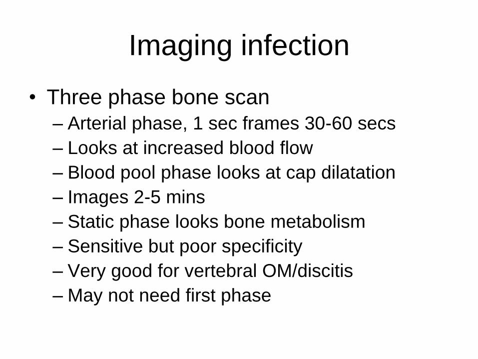
Imaging infection
• Three phase bone scan
– Arterial phase, 1 sec frames 30-60 secs
– Looks at increased blood flow
– Blood pool phase looks at cap dilatation
– Images 2-5 mins
– Static phase looks bone metabolism
– Sensitive but poor specificity
– Very good for vertebral OM/discitis
– May not need first phase

3 phase bone scan
Disease Dynamic Static
Osteomyelitis Pos Pos
Cellulitis Pos Neg
Non infected
Bone
Neg Pos

False positives in 3 phase bone
• Recent but treated infection
• Fracture
• Non-infected malunion
• Inflammatory arthritis
– Look for synovial uptake
– Could be septic arthritis

Need for more specific agents
• Ga-67 may have uptake in fractures, low grade
uptake in normal bone and BM
• Labelled WBCs High sensitivity and specificity,
not good in vertebral infection
• HIG Ok for arms and legs
• AGAB generally good
• PET non-specific but excellent localisation
• Prosthetic joint may be different
• Charcot’s joints difficult

Ga-67 in bones
• Will be very sensitive
• Problem with imaging as counts can be
low in periphery
• Imaging out to 7 days possible
• Some uptake in metabolically active bones
• May be best for vertebral infections
• SPECT possible

Ga-67 in E.coli spinal OM

Prosthetic joints
• Need to exclude infection
• Radiology little help
• CT and MRI affected by metal inplant and
cement
• Bone scan negative means infection
unlikely
• Knee different from hip
• Cemented different from uncemented

?infected knee or synovitis

? Infected TKR

?infected TKR-SPECT

Ga-67 in infected TKR

Infected knee
Tc-99m HMPAO WBCs

Spinal TB and Pott’s #

Chest Ga-67
• Maybe most commonly used test
• Does not depend on WBC function
• May be best in TB
• However uptake non-specific
• In HIV Bowel uptake very intense-no pathology
• May be looking at more than one pathology

Ga-67 in AIDS

Ga-67 in PCP and KS

Dual Time F-18 FDG Imaging
M. Sathekge Steve Biko Hospital

F-18 FDG PET-CT
M. Sathekge Steve Biko Hospital

F-18 FDG in TB
M. Sathekge Steve Biko Hospital

F-18 FDG lung abcess
M. Sathekge Steve Biko Hospital

Inflammatory bowel disease
• Still probably underused
• Can be used to aid establishing diagnosis
• Esp small bowel Crohn’s
• Mostly used for follow-up
– ?post op adhesions or reactivated IBD

IBD-agents
• Ga-67 – Too non specific
• In-111 – High sensitivity and specificity imaging at 4 & 24
hours, quantifiable
• Tc-99m HMPAO – High sensitivty and specificity, imaging 1 & 3 hours,
semi quantifiable
• Best of the rest – Antibodies not proven maybe Tc-99m Il2

The big battle
• In-111 WBC
• High accuracy
• NO bowel activity
• Years of experience
• Faecal In-111
activity over 48
hours quantifies
disease activity
• Tc-99m HMPAO
• High accuracy
• Imaging 1 & 3
hours
• Low dosimetry
• Semiquantification
possible
• Years of experience

In-111 WBC in IBD

Small bowel Crohn’s

Tc-99m HMPAO WBC in UC

IBD-special cases
• With Tc-99m HMPAO WBCs
• Later imaging will find Crohn’s abscess
– Activity in bowel moves, abscess does not image up to 24 hours
• Pelvic disease
– Do squat/outlet view
• Connecting abscess
– Focal area of uptake adjacent to bowel that then decreases or disappears

PUO SPECT-CT
• Roach et al 2006 NMC
• Looked at 50 scans including bone and Ga-67 SPECT-CT
• 16% of patients had minor change 11% major change c/w SPECT alone
• Almost all to do with localisation and improved specificity
• Specificity itself improved by 26%

Bar-Shalom et al JNM 2006
SPECT/CT for suspected bone infection on GS. A 56-y-old woman presented
with fever, low back pain, and infected scar 1 mo after spinal surgery and was
referred for GS for suspected vertebral osteomyelitis. (A) Planar posterior whole-
body GS image (left) shows prominent abnormal uptake in left lower back,
corresponding in part to regions of increased irregular uptake seen on planar
posterior whole-body 99mTc-MDP image (right) along operated vertebrae. (B)
Transaxial GS SPECT/CT image (left) localizes abnormal uptake on GS (center)
to paravertebral soft-tissue abscess seen on corresponding CT image (right),
thus defining soft-tissue infection without osteomyelitis. There was no evidence of
vertebral osteomyelitis on follow-up CT 4 wk later
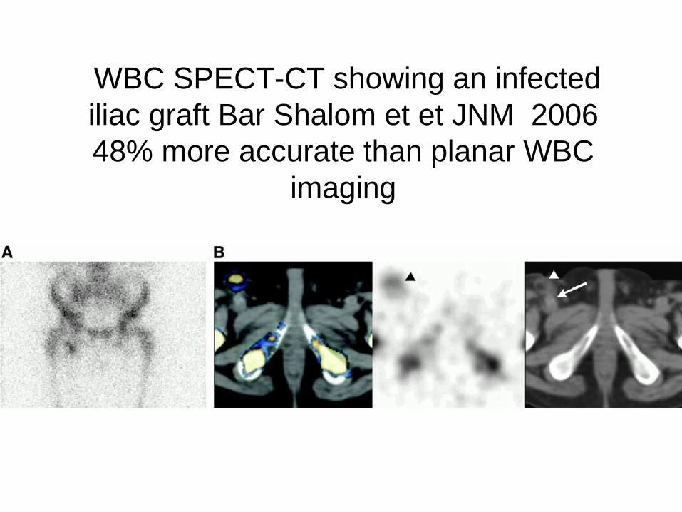
WBC SPECT-CT showing an infected
iliac graft Bar Shalom et et JNM 2006
48% more accurate than planar WBC
imaging

Specific results for infection
• Inquie et al J Comp Assist Tom 2007
• 16 patients (11 In-111 WBC and 6 Ga--67)
• SPECT/CT images yielded "added value" for anatomical localization in 65%, diagnostic confidence in 71%, and altered interpretations in 47% of cases

Ga-67 in infected Tx
Nowosinska CNM in press 2013

Patient with Ga-67 SPECT-CT
Patient with
infected
renal
transplant
SPECT-CT
confirms
uptake in
peri-nephric
fat

In-111 WBC in iliac A mycotic
aneurysm

PET in FUO
• Blockmans et al Clin Infect Dis
• Leuvan department
• 58 patients with FUO studies, final
diagnosis in 38
• 40% of scans unhelpful in diagnosis
• Results similar to those from Ga-67 in 40
patients studied with both scans only
helpful in 42% for each tracer

PET and FUO
• Bleeker-Rovers et al EJNMMI 2004
• Nijmegen group
• 35 patients with FUO imaged
• Diagnosis conformed in 19
• 37% of scans clinically useful
• 65% of the positive scans clinically useful
• PPV 87%, NPV 95%

PET vs In-111 WBC
• Kjaer et al EJNMMI 2004
• Copenhagen group
• 19 patients had In-111 WBC and F-18
FDG
• FDG counted as useful if found infection or
malignancy (WBC infection)

Image from
Blockmans
et al
Showing F-
18 FDG
uptake in
spinal TB

Image from
Blockmans
et al
Showing F-
18 FDG
uptake in
infective
aortitis

Cyptococcus in patient with HD
treated with Chemo

Giant cell arteritis

Peritonitis
FDG PET in inflammation
• Increasing use in non infected
inflammation
• Quantifying uptake can monitor progress
• Able to look at burden of inflammation
• Some special cases
– Cardiac sarcoid
– RA on peripheral joints

Sarcoid
• Disseminated inflammatory disease
• Characterised by granuloma
• Various patterns
– Salivary/lacrimal glands
– Lymph nodes
– CNS
– Skin
– Joint
– Pulmonary- the most dangerous

Imaging in sarcoid
• Normally diagnosis clinical followed by
biopsy
• 50% of patients have raised serum ACE
• If lymph nodes involved may see
symmetrical enlarged mediastinal/hilar
nodes the lambda pattern
• Since 1966 Ga-67 citrate used
– Not very trendy
– High radiation dose

Ga-67 in sarcoid
Panda sign,
lacrimal and
salivary glands
Lamba sign
mediastinum and
hilar nodes
Diffuse lung uptake
Lymphadanopathy
(symmetrical)
Joints
Liver-diffuse

Bitran grading in sarcoid-Ga-67

Use of F-18 FDG
• Lymphocytes very FDG avid
• Much improved resolution
• Lower radiation dose (5mSv vs 18mSv)
• Confirm sites of active disease esp in the
abdomen
• Quantify uptake which may be useful in
treatment monitoring

FDG vs Ga-67 • Nishiyama et el JNM
2006
• 18 sarcoid patients
imaged with Ga-67 and
FDG.
• Pulmonary disease Ga-
67 81%, FDG 100% -
mean SUVmax 7
• Extra-pulmonary disease
Ga 48%, FDG 90% mean
SUVmax 5 A= Ga-67
B= F-18 FDG
C= F-18 FDG post therapy

Using FDG to monitor therapy
• Sobic-Saronovic, Clin Nucl Med 2013
• 30 patients imaged before and after
steroids for active sarcoid
• Observed reduction in sites and intensity
of activity
• Correlated well with clinical symptoms
• SUVmax 8.5 to 4.9 (p<0.05)
• Serum ACE did not predict response

Cardiac sarcoid
• Cardiac sarcoid may occur with other sites
or be isolated
• Can result in heart failure and arrythmias
• Cause of unexpected cardiac death
• Recently a growing role for cardiac F-18
FDG
• Has been proposed both for diagnosis and
to monitor any response to therapy

Imaging cardiac sarcoid
• Patient preparation vital
• Patient need 24hrs high
fat/low carbohydrate diet
• IHD should be excluded by
MIBI/Rb-82
• Images should be gated
• No myocardial uptake or
diffuse uptake normal.
• Focal uptake is cardiac
sarcoid
Before Tx After
steroids

Review of FDG in cardiac
sarcoid • Youssef et al JNM 2012
• Systematic review of 7 studies of FDG in
cardiac sarcoid
• 164 patients with sarcoid scanned 50%
had cardiac involvement
• Sensitivity of FDG 89% (95% CI 76-06%)
• Specificity of FDG 78% (95% CI 68-86%)

Using FDG in RA
• Beckers et al JNM 2004
• 21 patients with active RA
• FDG imaging with views of knees and
hands
• FDG positive in 68% joints though 75% of
joints swollen and 79% painful
• Good correlation with increased blood flow
on Doppler ultrasound

FDG uptake in RA
Beckers et al JNM 2004
Normal
Patient
with RA

Monitoring response
• Vijavant et al WJR 2012. 17 newly
diagnosed RA and 11 newly diagnosed
sero-neg arthropathy
• Good correlation between symptoms and
sites of increased uptake of FDG
• Change in SUVmax correlated well with
clinical response and change in CRP

FDG before and after Tx
Vijavant et al WJR 2012

RA and PET in 2004-stll true
2013 • However, much work remains to be done
to gain more detailed information and to
clarify the impact of 18F-FDG PET on
diagnosis and therapy of RA, in
comparison with state-of-the-art MRI,
ultrasound, and three-phase bone
scanning. Eventually, we may be able to
define indications for 18F-FDG PET to
improve and adjust RA management.-
Wilfred Brenner