Infection and Activation of Monocytes by Marburg and...
9
JOURNAL OF VIROLOGY, 0022-538X/01/$04.000 DOI: 10.1128/JVI.75.22.11025–11033.2001 Nov. 2001, p. 11025–11033 Vol. 75, No. 22 Copyright © 2001, American Society for Microbiology. All Rights Reserved. Infection and Activation of Monocytes by Marburg and Ebola Viruses UTE STRO ¨ HER, 1 ELMAR WEST, 1 HARALD BUGANY, 1 HANS-DIETER KLENK, 1 HANS-JOACHIM SCHNITTLER, 2 AND HEINZ FELDMANN 1,3 * Institut fu ¨r Virologie, Philipps-Universita ¨t, D-35037 Marburg, 1 and Institut fu ¨r Physiologie und Pathophysiologie, Carl-Gustav-Carus-Universita ¨t, D-01307 Dresden, 2 Germany, and Canadian Science Centre for Human and Animal Health, Winnipeg, Manitoba R3E 3R2, Canada 3 Received 18 July 2001/Accepted 26 July 2001 In this study we investigated the effects of Marburg virus and Ebola virus (species Zaire and Reston) infections on freshly isolated suspended monocytes in comparison to adherent macrophages under culture conditions. Our data showed that monocytes are permissive for both filoviruses. As is the case in macrophages, infection resulted in the activation of monocytes which was largely independent of virus replication. The activation was triggered similarly by Marburg and Ebola viruses, species Zaire and Reston, as indicated by the release of the proinflammatory cytokines interleukin-1 (IL-1), tumor necrosis factor , and IL-6 as well as the chemokines IL-8 and gro-. Our data suggest that infected monocytes may play an important role in the spread of filoviruses and in the pathogenesis of filoviral hemorrhagic disease. Marburg virus (MBGV) and Ebola virus (EBOV), family Filoviridae, cause severe hemorrhagic fevers in humans and nonhuman primates. Outbreaks such as the latest ones in the Democratic Republic of the Congo (MBGV) and Uganda (EBOV) are unpredictable and a matter of considerable public health concern to the affected as well as neighboring countries. The Zaire species of EBOV shows the highest mortality in humans, whereas the Reston species may be apathogenic, based on very limited data. The clinical syndrome of MBGV and EBOV hemorrhagic fever is characterized by generalized fluid distribution problems, hypotension, coagulation disor- ders, variable degrees of hemorrhage, and widespread focal tissue destruction (6, 18, 23, 26, 28, 29). Morphological studies on postmortem material indicate that mononuclear phagocytic cells are the primary targets for filovirus replication (10, 11, 30, 39, 40). Cultured human macrophages are highly susceptible to infection with MBGV, the prototype filovirus, resulting in ac- tivation, massive virus production, and finally cell lysis (5). Endothelial cells are considered as secondary targets. Filoviruses replicate in human umbilical cord vein endothe- lial cells, causing endothelial cell lysis (32). This effect may in part be associated with the cytotoxicity of the viral transmem- brane glycoprotein (3, 38), which mediates virus attachment to target cells (33, 37). Postmortem observations on humans have demonstrated viral antigen in endothelial cells of several or- gans (39, 40), but widespread destruction of endothelial cells is not always obvious in the final stages of the disease in exper- imentally infected nonhuman primates (10, 11, 30). The mechanisms of disease development during filovirus infections are still poorly understood. Following infection via small lesions, virus particles enter lymph vessels or the vascular system directly. The primary organ tropism (lymph nodes, spleen, and liver) may be explained by the direct access of viral particles to sessile macrophages without penetration of the cell or the tissue barrier (31). The pantropism of filovirus infec- tions, typically occurring during late disease stages, is mecha- nistically unexplained. Extravasation of infected circulating cells, such as monocytes, might be a mechanism for the spread of the virus and the occurrence of focal tissue destruction. This requires the activation of the extravasating cells and the endo- thelium that is normally mediated through cytokines and che- mokines (15, 21). However, neither infection nor activation of monocytes by filoviruses has been experimentally demon- strated yet. In this study we compared the effects of filovirus infections on freshly isolated suspended monocytes and adherent macro- phages under culture conditions. Monocytes could be estab- lished as permissive cells for MBGV and EBOV. Both cell subsets, the suspended monocytes and the adherent macro- phages, became activated upon infection, as evidenced by the release of cytokines and chemokines. The activation seemed to be largely independent of viral replication. Based on our data, the infected monocytes may play a key role in the spread of the virus and therefore sufficiently explain the pantropism and the associated focal tissue destruction. Furthermore, the virus-in- duced cytokine and chemokine release from activated mono- cytes may trigger an instability of the endothelium in the in- fected host. MATERIALS AND METHODS Viruses and cell lines. In this study we used the Musoke strain of MBGV, the Mayinga strain of the Zaire species of EBOV (EBOV-Zaire), and the Reston strain of the Reston species of EBOV (EBOV-Reston). All virus stocks were kindly provided by the Special Pathogens Branch, Centers for Disease Control and Prevention, Atlanta, Ga. Virus stocks were freshly prepared in Vero E6 cells (ATCC 1568). Harvesting was performed when no obvious cytopathic effect was seen. Mock-infected Vero E6 cells were treated the same way in order to prepare a control (mock stock). Vero E6 cells were cultured in Dulbecco’s modified Eagle’s medium (DMEM) (Gibco-BRL, Karlsruhe, Germany) supplemented with 10% fetal calf serum (Biochrom, Berlin, Germany), penicillin (100 U/ml), streptomycin (100 g/ml), and L-glutamine (2 mM). For virus propagation, DMEM with 2% fetal calf serum was used. * Corresponding author. Mailing address: Canadian Science Centre for Human and Animal Health, 1015 Arlington St., Winnipeg, Mani- toba R3E 3R2, Canada. Phone: (204) 789-6019. Fax: (204) 789-2140. E-mail: [email protected]. 11025 on July 13, 2018 by guest http://jvi.asm.org/ Downloaded from
Transcript of Infection and Activation of Monocytes by Marburg and...

JOURNAL OF VIROLOGY,0022-538X/01/$04.00�0 DOI: 10.1128/JVI.75.22.11025–11033.2001
Nov. 2001, p. 11025–11033 Vol. 75, No. 22
Copyright © 2001, American Society for Microbiology. All Rights Reserved.
Infection and Activation of Monocytes by Marburgand Ebola Viruses
UTE STROHER,1 ELMAR WEST,1 HARALD BUGANY,1 HANS-DIETER KLENK,1
HANS-JOACHIM SCHNITTLER, 2 AND HEINZ FELDMANN1,3*
Institut fur Virologie, Philipps-Universitat, D-35037 Marburg,1 and Institut fur Physiologie und Pathophysiologie,Carl-Gustav-Carus-Universitat, D-01307 Dresden,2 Germany, and Canadian Science Centre
for Human and Animal Health, Winnipeg, Manitoba R3E 3R2, Canada3
Received 18 July 2001/Accepted 26 July 2001
In this study we investigated the effects of Marburg virus and Ebola virus (species Zaire and Reston)infections on freshly isolated suspended monocytes in comparison to adherent macrophages under cultureconditions. Our data showed that monocytes are permissive for both filoviruses. As is the case in macrophages,infection resulted in the activation of monocytes which was largely independent of virus replication. Theactivation was triggered similarly by Marburg and Ebola viruses, species Zaire and Reston, as indicated by therelease of the proinflammatory cytokines interleukin-1� (IL-1�), tumor necrosis factor �, and IL-6 as well asthe chemokines IL-8 and gro-�. Our data suggest that infected monocytes may play an important role in thespread of filoviruses and in the pathogenesis of filoviral hemorrhagic disease.
Marburg virus (MBGV) and Ebola virus (EBOV), familyFiloviridae, cause severe hemorrhagic fevers in humans andnonhuman primates. Outbreaks such as the latest ones in theDemocratic Republic of the Congo (MBGV) and Uganda(EBOV) are unpredictable and a matter of considerable publichealth concern to the affected as well as neighboring countries.
The Zaire species of EBOV shows the highest mortality inhumans, whereas the Reston species may be apathogenic,based on very limited data. The clinical syndrome of MBGVand EBOV hemorrhagic fever is characterized by generalizedfluid distribution problems, hypotension, coagulation disor-ders, variable degrees of hemorrhage, and widespread focaltissue destruction (6, 18, 23, 26, 28, 29). Morphological studieson postmortem material indicate that mononuclear phagocyticcells are the primary targets for filovirus replication (10, 11, 30,39, 40). Cultured human macrophages are highly susceptible toinfection with MBGV, the prototype filovirus, resulting in ac-tivation, massive virus production, and finally cell lysis (5).Endothelial cells are considered as secondary targets.
Filoviruses replicate in human umbilical cord vein endothe-lial cells, causing endothelial cell lysis (32). This effect may inpart be associated with the cytotoxicity of the viral transmem-brane glycoprotein (3, 38), which mediates virus attachment totarget cells (33, 37). Postmortem observations on humans havedemonstrated viral antigen in endothelial cells of several or-gans (39, 40), but widespread destruction of endothelial cells isnot always obvious in the final stages of the disease in exper-imentally infected nonhuman primates (10, 11, 30).
The mechanisms of disease development during filovirusinfections are still poorly understood. Following infection viasmall lesions, virus particles enter lymph vessels or the vascularsystem directly. The primary organ tropism (lymph nodes,
spleen, and liver) may be explained by the direct access of viralparticles to sessile macrophages without penetration of the cellor the tissue barrier (31). The pantropism of filovirus infec-tions, typically occurring during late disease stages, is mecha-nistically unexplained. Extravasation of infected circulatingcells, such as monocytes, might be a mechanism for the spreadof the virus and the occurrence of focal tissue destruction. Thisrequires the activation of the extravasating cells and the endo-thelium that is normally mediated through cytokines and che-mokines (15, 21). However, neither infection nor activation ofmonocytes by filoviruses has been experimentally demon-strated yet.
In this study we compared the effects of filovirus infectionson freshly isolated suspended monocytes and adherent macro-phages under culture conditions. Monocytes could be estab-lished as permissive cells for MBGV and EBOV. Both cellsubsets, the suspended monocytes and the adherent macro-phages, became activated upon infection, as evidenced by therelease of cytokines and chemokines. The activation seemed tobe largely independent of viral replication. Based on our data,the infected monocytes may play a key role in the spread of thevirus and therefore sufficiently explain the pantropism and theassociated focal tissue destruction. Furthermore, the virus-in-duced cytokine and chemokine release from activated mono-cytes may trigger an instability of the endothelium in the in-fected host.
MATERIALS AND METHODS
Viruses and cell lines. In this study we used the Musoke strain of MBGV, theMayinga strain of the Zaire species of EBOV (EBOV-Zaire), and the Restonstrain of the Reston species of EBOV (EBOV-Reston). All virus stocks werekindly provided by the Special Pathogens Branch, Centers for Disease Controland Prevention, Atlanta, Ga. Virus stocks were freshly prepared in Vero E6 cells(ATCC 1568). Harvesting was performed when no obvious cytopathic effect wasseen. Mock-infected Vero E6 cells were treated the same way in order to preparea control (mock stock). Vero E6 cells were cultured in Dulbecco’s modifiedEagle’s medium (DMEM) (Gibco-BRL, Karlsruhe, Germany) supplementedwith 10% fetal calf serum (Biochrom, Berlin, Germany), penicillin (100 U/ml),streptomycin (100 �g/ml), and L-glutamine (2 mM). For virus propagation,DMEM with 2% fetal calf serum was used.
* Corresponding author. Mailing address: Canadian Science Centrefor Human and Animal Health, 1015 Arlington St., Winnipeg, Mani-toba R3E 3R2, Canada. Phone: (204) 789-6019. Fax: (204) 789-2140.E-mail: [email protected].
11025
on July 13, 2018 by guesthttp://jvi.asm
.org/D
ownloaded from
Endotoxin test. Prior to use, all virus stocks and media were analyzed forendotoxin presence using the Limulus amebocyte lysate test (QCL-1000; Bio-Whittaker, Walkersville, Md.). All compounds and media used in this studycontained less than 0.3 EU/ml, which was less or equivalent to the amounts foundin the mock stock used as controls for the experiments.
Inactivation of virus stocks. Virus stocks were inactivated by exposure to UVlight for 1 h. Proper inactivation was controlled by the incubation of Vero E6cells with the inactivated virus particles and subsequent screening for the pres-ence of viral proteins (immunofluorescence) and viral RNA (reverse transcrip-tion [RT]-PCR targeting virus-specific transcripts). The UV-inactivated stockswere used at the same dilutions as the noninactivated stocks.
Isolation of PBMC. Peripheral blood mononuclear cells (PBMC) consisting ofmonocytes and lymphocytes were separated from the buffy coats of healthy blooddonors using Ficoll-Hypaque density gradient centrifugation (nonpooled).
(i) Monocytes. Blood monocytes were isolated by countercurrent centrifugalelutriation of PBMC-rich fractions using a JE-6B rotor (Beckman).
(ii) Macrophages. Freshly prepared PBMC were seeded into 24-well cultureplates (Primaria; Falcon), and the monocytes were allowed to adhere. After anadsorption period of 1 h, the monolayers were washed extensively to remove anynonadherent cells. The cells were incubated at 37°C in a humidified (95%) 5%CO2–air atmosphere for 7 days prior to infection. The monocytes and macro-phages were cultured in RPMI 1640 (Linaris, Wertheim-Bettingen, Germany)containing 5% human AB serum, penicillin (100 U/ml), streptomycin (100 �g/ml), L-glutamine (2 mM), nonessential amino acids (2 mM), and pyruvate (2mM).
Infection of mononuclear phagocytic cells. (i) Monocytes. A total of 106 freshmononuclear cells were infected in suspension (test tube) using a multiplicity ofinfection (MOI) of 10. Subsequently, the cells were washed with phosphate-buffered saline (PBS), seeded into 24-well culture plates, and incubated at 37°Cin a CO2 incubator (humidity 95%) for the appropriate time (up to 7 dayspostinfection).
(ii) Macrophages. The infection was performed on day 7 postseeding at anMOI of 10. After an adsorption period of 1 h, the inoculum was replaced by newmedium (RPMI containing 5% human AB serum), and the cultures were incu-bated for various times at 37°C in a CO2 incubator (humidity 95%).
Antibody treatment. Macrophages were infected as described above. Follow-ing inoculation, fresh medium containing anti-human tumor necrosis factor al-pha (TNF-�) antibody (5 �g/ml) (R&D Systems, Weisbaden, Germany) wasadded.
Immuno-Plaque assay. Confluent Vero E6 cells grown on cover slips wereinfected using a 10-fold dilution series. Following virus adsorption for 45 min at37°C, the cells were washed three times with PBS. The infected cells wereoverlaid with DMEM containing 0.4% low-melting-point agarose and 2% fetalcalf serum. At 48 h postinfection, the cells were fixed with 2% formaldehyde inPBS overnight, washed with PBS, and permeabilized with 0.1% Triton X-100 inPBS for 15 min. Subsequently, the cells were incubated for 1 h at room temper-ature with the desired antibody (anti-MBGV NP, 1:1,000; anti-EBOV, 1:200)diluted in PBS. The samples were washed three times with PBS and incubated foranother hour with the appropriate indocarbocyanine (Cy3)- or fluorescein iso-thiocyanate (FITC)-conjugated secondary antibody. Following washing (threetimes), cover slips were mounted with Supermount (BioGenex, Hamburg, Ger-many), and the immunostained plaques were counted.
Detection of cytokines. The supernatants from the infected or mock-infectedcell cultures were removed, clarified from cell debris by centrifugation (8,000 �g, 4°C, 10 min), and stored at �80°C. All samples were tested in duplicate for thepresence of cytokines after being thawed only once using commercial enzyme-linked immunosorbent assay (ELISA) systems (human interleukin-6 [IL-6],TNF-� ELISA kit, and human IL-8 enzyme immunoassay [EIA] from Promocell,Heidelberg, Germany; human gro-� ELISA from R&D Systems). Since thesamples were not inactivated, the analyses were conducted in a biosafety level 4containment laboratory.
RT-PCR for cytokine-specific transcripts. The supernatants from the infectedor mock-infected cells were removed, and the cells were lysed using a guani-dinium isothiocyanate-based buffer system (RNeasy kit; Qiagen, Hilden, Ger-many). At this point the samples were taken out of the containment laboratory,and RNA isolation was performed using the Qiagen kit. The RNA concentra-tions were adjusted to 100 ng/�l prior to use in RT-PCR. Reverse transcriptionand PCR were carried out in the same tube using the OneStep RT-PCR kit(Qiagen) according to the manufacturer’s guidelines. RT-PCR was performedwith a Perkin Elmer cycler (model 2400) using the following protocol: 1 cycle at50°C for 30 min and 95°C for 15 min, followed by 19 to 32 cycles at 94°C for 30 s,65°C for 30 s, and 72°C for 1 min. The cycle numbers were adjusted to allowsemiquantitative analysis of the amplification products after ethidium bromide
staining. Table 1 shows the sequences of the oligonucleotides and the cyclenumbers used for the different target mRNAs.
RESULTS
Replication of filoviruses in monocytes . Human peripheralblood monocytes were infected with MBGV and EBOV-Zaireat an MOI of 10. Within several hours, the infection of themonocytes resulted in the formation of cell clumps indicativeof activation (Fig. 1A). Immunostaining done at day 2 postin-fection with a virus-specific antiserum demonstrated the infec-tion of the monocytes (Fig. 1B). The supernatants were col-lected at different days postinfection, and the virus titers weredetermined by plaque assay in Vero E6 cells (Fig. 1C). Noobvious difference in infection of suspended monocytes andadherent macrophages between the viruses was noted. Thedata clearly demonstrated that the circulating monocyte was apermissive target cell for filoviruses and that the infection didnot require cell adhesion to the substrates.
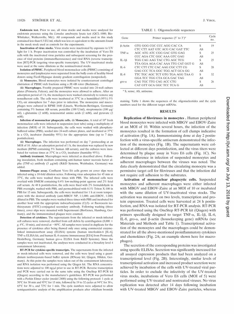
Activation of mononuclear phagocytic cells. Suspendedmonocytes and adherent macrophages were either infectedwith MBGV and EBOV-Zaire at an MOI of 10 or incubatedwith the same dilution of UV-inactivated virus stocks. Theactivation was measured on two levels, transcription and pro-tein expression. Treated cells were harvested at 24 h postin-fection, and RNA was isolated for RT-PCR analysis. RT-PCRwas performed using the OneStep RT-PCR kit (Qiagen) withprimers specifically designed to target TNF-�, IL-1�, IL-6,IL-8, gro-�, and �-actin (housekeeping gene) mRNAs (seeMaterials and Methods and Table 1). Transcriptional activa-tion of the monocytes and the macrophages could be demon-strated for all the above-mentioned proinflammatory cytokinesand chemokines (Fig. 2A; an example shown here for macro-phages).
The secretion of the corresponding proteins was investigatedusing specific ELISAs. Secretion was significantly increased forall assayed expression products that had been analyzed on atranscriptional level (Fig. 2B). Interestingly, similar levels oftranscriptional activation and increased product secretion wereachieved by incubation of the cells with UV-treated viral par-ticles. In order to exclude the infectivity of the UV-treatedvirus stocks, incubations of Vero E6 cells (MOI of 5) wereperformed using UV-treated and nontreated viruses. No virusreplication was detected after 14 days following incubationwith UV-treated MBGV and EBOV-Zaire particles, whereas
TABLE 1. Oligonucleotide sequences
Gene Primer sequence (5� to 3�)a Cycleno.
�-Actin GTG GGG CGC CCC AGG CAC CA S 19CTC CTT AAT GTC ACG CAC GAT TTC AS
TNF-� AGC ATG ATC CGG GAC GTG GAG S 25CCC AGA CTC GGC AAA GTC GAG AS
IL-1� TGG CAG AAG TAC CTG AGC TCG S 25TTA GGA AGA CAC AAA TTG CAT GGT G AS
IL-6 CTC CTT CTC CAC AAG CGC CTT CG S 28GAG CCC TCA GGC TGG ACT GCA GG AS
IL-8 TTC TGC AGC TCT GTG TGA AGG TAA G S 32GGA TCC TGG CTA GCA GAC TAG AS
gro-� CAG TGC TTG CAG ACC CTG S 26CAT GTT GCA GGC TCC TCA G AS
a S, sense; AS, antisense.
11026 STROHER ET AL. J. VIROL.
on July 13, 2018 by guesthttp://jvi.asm
.org/D
ownloaded from

monolayers infected with nontreated virus were uniformly an-tigen positive 2 days postinfection (Fig. 2C). In addition, RT-PCR targeting virus-specific transcripts confirmed completeinactivation of UV-treated virus stocks (data not shown). Thedata suggest that activation is largely independent of virusreplication.
TNF-� by itself can induce expression of mediators in mono-nuclear phagocytic cells (24, 34). To exclude the possible in-fluence of TNF-� on the production of IL-6, IL-1�, IL-8, andgro-�, we infected macrophages in the absence or presence ofa neutralizing anti-human TNF-� antibody. The supernatantswere collected 24 h postinfection and assayed for the differentmediators. Under these conditions, virus-mediated inductionof IL-6, IL-8, and gro-� (Fig. 3) was only slightly affected,indicating that cytokine and chemokine expression is indeedlargely induced by virus infection and not triggered by theproduction of TNF-�.
Time course of activation. In general, MBGV and EBOV-Zaire showed a similar activation profile in both subsets ofmononuclear phagocytic cells. Therefore, the kinetics of virus-induced activation was studied using MBGV. Monocytes anddifferentiated macrophages were infected at an MOI of 10.The supernatants and cells were harvested at different timepoints postinfection. RT-PCR was performed as described pre-viously, but the number of cycles was adjusted to 19 to 32 inorder to be semiquantitative. All target genes (TNF-�, IL-1�,IL-6, IL-8, and gro-�) were transcriptionally activated begin-ning at 3 h postinfection. TNF-� reached its maximum at about3 to 6 h postinfection, whereas gro-� showed a very weakincrease at 3 h postinfection and increased to peak valueswithin 24 h. IL-6 and IL-8 remained at an increased level for alonger period and showed a slight decrease at 24 h postinfec-tion (Fig. 4A; an example shown here for macrophages). Theslower response in activation of gro-� can also be seen fromthe level of protein secretion, which showed an increased re-lease beginning at 6 h postinfection, in contrast to TNF-�,which was detected after only 3 h (Fig. 4B). Interestingly, IL-6and, to a lesser extent, IL-8 secretion was delayed compared tothe transcriptional activation of the corresponding genes. TheIL-8 background was high but significantly lower on the tran-script and protein levels in the mock-infected compared to thevirus-infected cells (Fig. 4).
The data from the time course studies compared favorablywith the results with the UV-inactivated virus (Fig. 2). Activa-tion seemed to start earlier than virus replication, indicatingthat replication is not needed to trigger this event. In order toconfirm this conclusion, we performed RT-PCR using primersthat specifically target virus-specific RNA transcripts. At 3 hpostinfection, we could not detect transcripts of viral genes,which supported the conclusion that replication (and most
FIG. 1. Filovirus replication in mononuclear phagocytic cells. (Aand B) Infection of monocytes. Monocytes were infected with MBGV(MBG) and EBOV-Zaire (EBO Z) and -Reston (EBO R) at an MOIof 10. A mock infection served as a control. Cultures were checked bylight microscopy (A) and immunostaining (B). On day 1 postinfection(1d pi), activation could be demonstrated by clumping of monocytes
(A). Immunostaining was performed on day 2 postinfection, as shownhere for MBGV using an anti-MBGV antiserum (B). (C) Kinetic ofvirus growth. Virus growth was determined by immunoplaque assay.The supernatants of infected cultures were 10-fold diluted, and con-fluent monolayers of Vero E6 cells were infected. On day 2 postinfec-tion, cells were stained with specific antisera, and the titer was deter-mined. Open circles, infected macrophages; solid squares, infectedmonocytes.
VOL. 75, 2001 FILOVIRUS INFECTION OF MONOCYTES 11027
on July 13, 2018 by guesthttp://jvi.asm
.org/D
ownloaded from

11028 STROHER ET AL. J. VIROL.
on July 13, 2018 by guesthttp://jvi.asm
.org/D
ownloaded from

likely transcription) was not necessary for target cell activation(data not shown).
Infection of mononuclear phagocytic cells with EBOV-Reston. Monocytes and macrophages were infected withMBGV, EBOV-Zaire, and EBOV-Reston at an MOI of 10. Asdemonstrated for MBGV and EBOV-Zaire previously (Fig. 1to 4), EBOV-Reston infection also resulted in the activation ofboth subsets of mononuclear phagocytic cells (Fig. 5). In gen-eral, the levels of activation did not differ significantly fromthose activated by MBGV and EBOV-Zaire infections.
DISCUSSION
As hypothesized previously, filoviruses may enter throughminor lesions on the skin and/or mucous membranes and sub-sequently access the blood directly or via the lymphatic system(31). Studies on experimentally infected animals have demon-strated that filovirus infections follow a specific order, startingwith the blood or lymph fluid, the interstitium, and the paren-chyma, with the crucial pathogenic events taking place in thebloodstream (30). On the basis of in vivo studies, mononuclearphagocytic cells, especially macrophages, seem to be the pri-mary target cells for filoviruses (10, 11, 30, 39, 40). The vastmajority of mononuclear phagocytic cells in the bloodstreamare monocytes. Given the fulminant course of a filovirus hem-
orrhagic fever, virus production in this subset of mononuclearphagocytic cells could be the major source of the high viremia(up to 108 PFU/ml) in naturally and experimentally infectedhosts during the critical early stages of the infection. Highviremia may subsequently allow the direct infection of impor-tant secondary target cells, such as endothelial cells (10, 11, 30,32, 39).
Filovirus infection leads to activation of monocytes, as dem-onstrated by clump formation (Fig. 1A) and the release ofmediators (Fig. 2 to 4). In addition, cytokines (e.g., TNF-� andIL-1�) and other proinflammatory mediators are known toincrease the expression of various cell adhesion molecules onendothelial cells (ICAM-1, VCAM-1, E-selectin, and P-selec-tin) that are involved in diapedesis of leukocytes (2, 15). TheC-X-C chemokines IL-8 and gro-� are not the only chemoat-tractants for neutrophils. IL-8 also triggers firm adhesion ofmonocytes to activated vascular endothelium, suggesting a po-tential role in monocyte recruitment (12, 20). Therefore, ex-travasation of activated, infected monocytes may be a mecha-nism for MBGV and EBOV to spread from the bloodstreaminto organ tissues during the second stage of infection. Thiscould explain the pantropism associated with severe filovirusinfections, and this would not necessarily depend on damage tothe endothelium, a feature that is not observed in all infected
FIG. 2. Mode of activation. Activation of suspended monocytes and adherent macrophages was determined on the level of transcription andexpression. Cells were incubated with the same dilutions of UV-treated or untreated MBGV (MOI of 10), EBOV-Zaire (MOI of 10), or mockstocks (negative control). (A) Transcriptional activation. Cells were harvested 24 h postinfection, and RNA was isolated using a commercial kit.RT-PCR was performed with specific oligonucleotides for the indicated proinflammatory cytokines and chemokines. �-Actin was used as ahousekeeping gene in order to verify that equal amounts of RNA were used in the RT-PCR assays (an example shown here for macrophages).To get semiquantitative results, the number of cycles was adjusted to 19 to 32 cycles. Products were run on a 2% agarose gel and visualizedfollowing ethidium bromide staining. (B) Activation of expression. Supernatants were harvested 24 h postinfection and assayed by ELISA for thepresence of mediators. The amount of mediators released is given in picograms per milliliter. (C) Inactivation of virus stock. In order to proveproper inactivation of virus stocks by UV treatment, Vero E6 cells were incubated with the same dilutions of UV-treated and untreated MBGV(MOI of 5) and EBOV-Zaire (MOI of 5). Cells were prepared for antigen detection by fixation and inactivation with 2% paraformaldehyde ondays 2 (upper and middle panel) and 14 (bottom panel) postinfection. For indirect immunofluorescence, cells were subsequently permeabilizedwith 0.1% Triton X-100 and incubated with anti-MBGV and anti-EBOV antisera (1:1,000 and 1:200 dilution, respectively) followed by Cy3- orFITC-labeled secondary antibodies.
VOL. 75, 2001 FILOVIRUS INFECTION OF MONOCYTES 11029
on July 13, 2018 by guesthttp://jvi.asm
.org/D
ownloaded from

hosts (10, 11, 29, 30). A similar role for monocytes has beendiscussed for the spread of other virus infections, such as hu-man immunodeficiency virus (25) and visna virus (27).
Clumping of monocytes (Fig. 1A) may be an important ob-servation for the pathogenesis of filovirus hemorrhagic fever.Monocyte clumps in vessels in vivo could dramatically influ-ence the rheology of the bloodstream, especially in smallvenules, which could lead to thrombus formation, as has beenobserved in infected patients. Intravascular clump formationcould also activate coagulation, a general phenomenon ob-served in many clinical cases as well as in experimentally in-fected monkeys (6, 18, 23, 26, 28, 29). Alterations of the co-agulation pathways, particularly the intrinsic one, wereobserved in infected monkeys (8, 9).
The release of cytokines as a result of monocyte and mac-rophage activation (Fig. 2 to 4) is also involved in the devel-opment of shock (1). Shock can be caused by many things,
including bacterial (e.g., endotoxin) and viral infection (4). Ithas been shown that supernatants of filovirus-infected macro-phages increased paraendothelial permeability in endothelialmonolayers, an effect that was mainly driven by TNF-� (5).Increased paraendothelial permeability is one of the most im-portant events during shock development (13, 22), and shock isknown to be the major cause of death in filovirus hemorrhagicfever (6, 18, 23, 26, 28, 29). Elevated levels of mediators,including TNF-�, were detected in the acute-phase sera ofseveral EBOV-infected patients (35) and in infected animals(17). Increased TNF-� values were found to correlate with theseverity of Junin virus infections causing Argentine hemor-rhagic fever (16). Therefore, it seems reasonable to assumethat massive cytokine release triggered by filovirus-inducedactivation of mononuclear phagocytic cells may induce a sys-temic inflammatory response syndrome, which can be pro-voked by cytokine treatment and can be observed during en-dotoxin-induced shock (1, 4).
As mentioned above, virus-induced activation of monocyteswill most likely result in the release of other proinflammatorymediators (e.g., histamine and serotonin), proteases (e.g., elas-tase), and peroxide (H2O2). These factors might be of increas-ing significance at the final stages of the disease, when infectedmonocytes/macrophages undergo cell lysis (5). Recently it waspostulated that cell detachment and cytotoxicity were causedby the viral transmembrane glycoprotein and may play a crucialrole in EBOV pathogenesis (3, 38). Although filoviruses doreplicate cytolytically in target cells, it remains open as to whatextent the cytolysis of target cells contributes to disease devel-opment. However, a cytotoxic effect on mononuclear phago-cytic cells could further promote a pathological release ofcytokines and other mediators and interfere with effective hostimmune responses.
The initial activation of monocytes and macrophages seemsto be largely independent of virus replication (Fig. 2). How-ever, a sustained release of mediators seems to require ongo-ing viral replication (14) or, as in our experiments, the contin-uous presence of inactivated virus. Therefore, replication maynot be contributing directly to the early critical events in patho-genesis. If this is true, specific viral products may be importantfactors in the activation by binding to cell surface receptors,which subsequently trigger signal cascades. The different formsof the viral glycoproteins expressed by EBOV and MBGV arecandidates for such products. The functions of the solubleglycoproteins have not been finally determined, whereas theviral transmembrane glycoprotein functions in receptor bind-ing and fusion (7, 19). These recently described soluble prod-ucts should be investigated in future work with respect totarget cell activation.
MBGV and EBOV-Zaire are human pathogenic filoviruses,whereas EBOV-Reston may be apathogenic for humans, basedon very limited data (6, 29). Therefore, propagation and acti-vation of human mononuclear phagocytic cells by EBOV-Reston were not necessarily expected (Fig. 5). Similar mech-anisms triggered by EBOV-Reston underline the importanceof infection and activation of monocytes by filoviruses inpathogenesis. Differences in susceptibility of target cells amongspecies as well as the lower cleavability of the EBOV-Restonvirus transmembrane glycoprotein in human-derived cell lines
FIG. 3. Mode of activation. Macrophages were infected withMBGV at an MOI of 10 with (�) or without (�) a neutralizinganti-TNF-� monoclonal antibody (5 �g/ml). A mock infection wasperformed as a negative control. Supernatants were harvested 24 hpostinfection and assayed by ELISA for the indicated mediators (pi-cograms per milliliter).
11030 STROHER ET AL. J. VIROL.
on July 13, 2018 by guesthttp://jvi.asm
.org/D
ownloaded from

(36) may contribute to lower pathogenicity in humans com-pared to nonhuman primates (7, 19).
In conclusion, it is reasonable to assume that mononuclearphagocytic cells, especially monocytes, play a central role in the
pathogenesis of filovirus hemorrhagic fever by mediating virusspread through extravasation and by triggering a cascade ofinterrelated pathological host responses through virus-inducedactivation. This may lead to an impairment of the immune
FIG. 4. Kinetics of activation. Suspended monocytes and adherent macrophages were infected with MBGV at an MOI of 10. A mock infectionserved as a control. (A) Transcriptional activation. Cells were harvested at the indicated time points postinfection, and RNA was isolated usinga commercial kit. RT-PCR and product analyses were performed as described in the legend to Fig. 2 (an example shown here for macrophages).(B) Activation of expression. Supernatants were harvested at the indicated times and assayed by ELISA for the presence of the indicatedmediators. The amount of products released is given in picograms per milliliter.
VOL. 75, 2001 FILOVIRUS INFECTION OF MONOCYTES 11031
on July 13, 2018 by guesthttp://jvi.asm
.org/D
ownloaded from

FIG. 5. Infection of mononuclear phagocytic cells with EBOV-Reston. Suspended monocytes and adherent macrophages were infected withMBGV, EBOV-Zaire, and EBOV-Reston at an MOI of 10. A mock infection served as a control. (A) Transcriptional activation. Cells wereharvested 24 h postinfection, and RNA was isolated using a commercial kit. RT-PCR and product analyses were performed as described in thelegend to Fig. 2 (an example shown here for macrophages). (B) Activation of expression. Supernatants were harvested 24 h postinfection andassayed by ELISA for the indicated mediators (picograms per milliliter).
11032 STROHER ET AL. J. VIROL.
on July 13, 2018 by guesthttp://jvi.asm
.org/D
ownloaded from

system, homeostasis, and the barrier function of the endothe-lium, finally leading to shock and death. Neutralizing antibod-ies directed against key inflammatory cytokines and chemo-kines might be able to help the host to overcome the earlyevents in infection and thus allow time for the specific immuneresponse needed to clear the virus.
ACKNOWLEDGMENTS
We thank Daryl Dick for editorial comments.This work was supported by grants from the Deutsche Forschungs-
gemeinschaft (SFB 535; Schn 430/1-1, Kl 238/7-1), the Canadian In-stitutes of Health Research (MOP-43921), and the European Com-munity (INCO-grant ERBIC 18 CT9803832). Ute Stroher performedthis work in partial fulfillment of the requirements for a Ph.D. degreefrom the Philipps-Universitat, Marburg, Germany.
REFERENCES
1. Brouckaert, P., and W. Fiers. 1996. Tumor necrosis factor and the systemicinflammatory response syndrome. Curr. Top. Microbiol. Immunol. 216:167–187.
2. Butcher, E. C. 1993. Specificity of leukocyte-endothelial interactions anddiapedesis: physiologic and therapeutic implications of an active decisionprocess. Res. Immunol. 144:695–698.
3. Chan, S. Y., M. C. Ma, and M. A. Goldsmith. 2000. Differential induction ofcellular detachment by envelope glycoproteins of Marburg and Ebola(Zaire) viruses. J. Gen. Virol.. 81:2155–2159.
4. Dinarello, C. A. 1996. Cytokines as mediators in the pathogenesis of septicshock. Curr. Top. Microbiol. Immunol. 216:133–165.
5. Feldmann, H., H. Bugany, F. Mahner, H. D. Klenk, D. Drenckhahn, andH. J. Schnittler. 1996. Filovirus-induced endothelial leakage triggered byinfected monocytes/macrophages. J. Virol. 70:2208–2214.
6. Feldmann, H., A. Sanchez, and H. D. Klenk. 1998. Filoviruses, p. 651–664. InL. H. Collier, A. Balows, and M. Sussman (ed.), Topley and Wilson’s mi-crobiology and microbial infections, 9th ed. Arnold, London, England.
7. Feldmann, H., V. E. Volchkov, V. A. Volchkova, and H. D. Klenk. 1999. Theglycoproteins of Marburg and Ebola virus and their potential roles in patho-genesis. Arch. Virol. Suppl. 15:159–169.
8. Fisher-Hoch, S. P., G. S. Platt, G. Lloyd, D. I. Simpson, G. H. Neild, and A. J.Barrett. 1983. Haematological and biochemical monitoring of Ebola infec-tion in rhesus monkeys: implications for patient management. Lancet ii:1055–1058.
9. Fisher-Hoch, S. P., G. S. Platt, G. H. Neild, T. Southee, A. Baskerville, R. T.Raymond, G. Lloyd, and D. I. Simpson. 1985. Pathophysiology of shock andhemorrhage in a fulminating viral infection (Ebola). J. Infect. Dis. 152:887–894.
10. Geisbert, T. W., L. E. Hensley, T. R. Gibb, K. E. Steele, N. K. Jaax, and P. B.Jahrling. 2000. Apoptosis induced in vitro and in vivo during infection byEbola and Marburg viruses. Lab. Investig. 80:171–186.
11. Geisbert, T. W., P. B. Jahrling, M. A. Hanes, and P. M. Zack. 1992. Asso-ciation of Ebola-related Reston virus particles and antigen with tissue lesionsof monkeys imported to the United States. J. Comp. Pathol. 106:137–152.
12. Gerszten, R. E., E. A. Garcia-Zepeda, Y. C. Lim, M. Yoshida, H. A. Ding,M. A. Gimbrone, Jr., A. D. Luster, F. W. Luscinskas, and A. Rosenzweig.1999. MCP-1 and IL-8 trigger firm adhesion of monocytes to vascular en-dothelium under flow conditions. Nature 398:718–723.
13. Glauser, M. P. 1996. The inflammatory cytokines. New developments in thepathophysiology and treatment of septic shock. Drugs 52:9–17.
14. Gupta, M., S. Mahanty, R. Ahmed, and P. E. Rollin. 2001. Monocyte-derivedhuman macrophages and peripheral blood mononuclear cells infected withebola virus secrete MIP-1alpha and TNF-alpha and inhibit poly-IC-inducedIFN-alpha in vitro. Virology 284:20–25.
15. Haraldsen, G., D. Kvale, B. Lien, I. N. Farstad, and P. Brandtzaeg. 1996.Cytokine-regulated expression of E-selectin, intercellular adhesion mole-cule-1 (ICAM-1), and vascular cell adhesion molecule-1 (VCAM-1) in hu-man microvascular endothelial cells. J. Immunol. 156:2558–2565.
16. Heller, M. V., M. C. Saavedra, R. Falcoff, J. I. Maiztegui, and F. C. Molinas.1992. Increased tumor necrosis factor-alpha levels in Argentine hemorrhagicfever. J. Infect. Dis. 166:1203–1204.
17. Ignatyev, G. M. 1999. Immune response to filovirus infections. Curr. Top.Microbiol. Immunol. 235:205–217.
18. Klenk, H. D. (ed.). 1999. Marburg and Ebola viruses. Curr. Top. Microbiol.Immunol. 235:1–225.
19. Klenk, H. D., V. E. Volchkov, V. A. Volchkova, and H. Feldmann. 1999. Thepolymorphism of the Ebola virus glycoprotein and its potential role in patho-genesis. Nova Acta Leopoldina 307:141–149.
20. Luscinskas, F. W., R. E. Gerszten, E. A. Garcia-Zepeda, Y. C. Lim, M.Yoshida, H. A. Ding, M. A. Gimbrone, Jr., A. D. Luster, and A. Rosenzweig.2000. C-C and C-X-C chemokines trigger firm adhesion of monocytes tovascular endothelium under flow conditions. Ann. N. Y. Acad. Sci. 902:288–293.
21. Luster, A. D. 1998. Chemokines–chemotactic cytokines that mediate inflam-mation. N. Engl. J. Med. 338:436–445.
22. Maier, R. V., and E. M. Bulger. 1996. Endothelial changes after shock andinjury. New Horiz. 4:211–223.
23. Martini, G. A., and R. Siegert. 1971. Marburg virus disease, 1st ed. Springer-Verlag, Berlin, Germany.
24. Netea, M. G., C. H. Selzman, B. J. Kullberg, J. M. Galama, A. Weinberg,A. F. Stalenhoef, J. W. Van der Meer, and C. A. Dinarello. 2000. Acellularcomponents of Chlamydia pneumoniae stimulate cytokine production inhuman blood mononuclear cells. Eur. J. Immunol. 30:541–549.
25. Orenstein, J. M., M. S. Meltzer, T. Phipps, and H. E. Gendelman. 1988.Cytoplasmic assembly and accumulation of human immunodeficiency virustypes 1 and 2 in recombinant human colony-stimulating factor-1-treatedhuman monocytes: an ultrastructural study. J. Virol. 62:2578–2586.
26. Pattyn, S. R. 1978. Ebola virus hemorrhagic fever, 1st ed. Elsevier/North-Holland, Amsterdam, The Netherlands.
27. Peluso, R., A. Haase, L. Stowring, M. Edwards, and P. Ventura. 1985. ATrojan Horse mechanism for the spread of visna virus in monocytes. Virol-ogy 147:231–236.
28. Peters, C. J., and J. W. LeDuc. 1999. Ebola: the virus and the disease.J. Infect. Dis. 179(Suppl. 1):S1–S288.
29. Peters, C. J., A. Sanchez, P. E. Rollin, T. G. Ksiazek, and F. A. Murphy. 1996.Filoviridae: Marburg and Ebola viruses, p. 1161–1176. In B. N. Fields, D. M.Knipe, et al. (ed.), Virology, 3rd ed. Raven Press, Philadelphia, Pa.
30. Ryabchikova, E. I., L. V. Kolesnikova, and S. V. Luchko. 1999. An analysisof features of pathogenesis in two animal models of Ebola virus infection.J. Infect. Dis. 179(Suppl. 1):S199–S202.
31. Schnittler, H. J., and H. Feldmann. 1998. Marburg and Ebola hemorrhagicfevers: does the primary course of infection depend on the accessibility oforgan-specific macrophages? Clin. Infect. Dis. 27:404–406.
32. Schnittler, H. J., F. Mahner, D. Drenckhahn, H. D. Klenk, and H. Feldmann.1993. Replication of Marburg virus in human endothelial cells. A possiblemechanism for the development of viral hemorrhagic disease. J. Clin. Inves-tig. 91:1301–1309.
33. Takada, A., C. Robison, H. Goto, A. Sanchez, K. G. Murti, M. A. Whitt, andY. Kawaoka. 1997. A system for functional analysis of Ebola virus glycopro-tein. Proc. Natl. Acad. Sci. USA 94:14764–14769.
34. Vasilescu, C., D. Berger, K. Buttenschon, M. Seidelmann, and H. G. Beger.1996. Endotoxin-induced release of interleukin 6 and interleukin 1 beta inhuman blood is independent of tumor necrosis factor alpha. Eur. Surg. Res.28:55–62.
35. Villinger, F., P. E. Rollin, S. S. Brar, N. F. Chikkala, J. Winter, J. B.Sundstrom, S. R. Zaki, R. Swanepoel, A. A. Ansari, and C. J. Peters. 1999.Markedly elevated levels of interferon (IFN)-gamma, IFN-alpha, interleukin(IL)-2: IL-10, and tumor necrosis factor-alpha associated with fatal Ebolavirus infection. J. Infect. Dis. 179(Suppl. 1):S188–S191.
36. Volchkov, V. E., H. Feldmann, V. A. Volchkova, and H. D. Klenk. 1998.Processing of the Ebola virus glycoprotein by the proprotein convertasefurin. Proc. Natl. Acad. Sci. USA 95:5762–5767.
37. Yang, Z., R. Delgado, L. Xu, R. F. Todd, E. G. Nabel, A. Sanchez, and G. J.Nabel. 1998. Distinct cellular interactions of secreted and transmembraneEbola virus glycoproteins. Science 279:1034–1037.
38. Yang, Z. Y., H. J. Duckers, N. J. Sullivan, A. Sanchez, E. G. Nabel, and G. J.Nabel. 2000. Identification of the Ebola virus glycoprotein as the main viraldeterminant of vascular cell cytotoxicity and injury. Nat. Med. 6:886–889.
39. Zaki, S. R., and C. S. Goldsmith. 1999. Pathologic features of filovirusinfections in humans. Curr. Top. Microbiol. Immunol. 235:97–116.
40. Zaki, S. R., and C. J. Peters. 1997. Viral hemorrhagic fevers, p. 347–364. InD. H. Connor (ed.), Pathology of infectious diseases. Appleton and Lange,Stamford, Conn.
VOL. 75, 2001 FILOVIRUS INFECTION OF MONOCYTES 11033
on July 13, 2018 by guesthttp://jvi.asm
.org/D
ownloaded from