Infantile tibia vara - · PDF fileInfantile tibia vara is a developmental disorder of ......
15
The PDF of the article you requested follows this cover page. This is an enhanced PDF from The Journal of Bone and Joint Surgery 1993;75:130-143. J Bone Joint Surg Am. WB Greene Infantile tibia vara This information is current as of April 16, 2011 Reprints and Permissions Permissions] link. and click on the [Reprints and jbjs.org article, or locate the article citation on to use material from this order reprints or request permission Click here to Publisher Information www.jbjs.org 20 Pickering Street, Needham, MA 02492-3157 The Journal of Bone and Joint Surgery
Transcript of Infantile tibia vara - · PDF fileInfantile tibia vara is a developmental disorder of ......

The PDF of the article you requested follows this cover page.
This is an enhanced PDF from The Journal of Bone and Joint Surgery
1993;75:130-143. J Bone Joint Surg Am.WB Greene
Infantile tibia vara
This information is current as of April 16, 2011
Reprints and Permissions
Permissions] link. and click on the [Reprints andjbjs.orgarticle, or locate the article citation on
to use material from thisorder reprints or request permissionClick here to
Publisher Information
www.jbjs.org20 Pickering Street, Needham, MA 02492-3157The Journal of Bone and Joint Surgery

130 THE JOURNAL OF BONE AND JOINT SURGERY
Infantile Tibia Vara*BY WALTER B. GREENE, M.D.t, CHAPEL HILL, NORTH CAROLINA
An Instructional cottrse Lecture, The American Academy of Orthopaedic Surgeons
Infantile tibia vara is a developmental disorder ofgrowth that affects the medial aspect of the proximaltibial physis. Blount’s4 article in 1937 prompted necogni-tion of this disorder, and, in fact, infantile tibia vana isoften called “Blount’s disease.” Blount also describedan adolescent form of tibia vara. It is now generallyaccepted that although the two forms have some similaranatomical and epidemiological features, the disorderof adolescent tibia vana should be considered differentfrom that of infantile tibia vana.
Children with infantile tibia vana have no apparentabnormality at birth, they are generally healthy, andearly growth of the legs is within normal limits. Thesechildren are usually brought to a physician at fourteento thirty-six months old for evaluation of bow legs. Atypical history is that the genu varum had gotten worsesince walking began. Infantile tibia vara is found morefrequently in children who are black, female, and obeseand who started walking at an early age (Fig. 1)12 227293 38.
Nine to 43 per cent of patients have been reportedto have an affected parent or sibling2- , but the spec-trum of infantile tibia vana within a family is usuallyconsistent with a multifactonial rather than a mendelianpattern of inheritance. The disorder is localized to theproximal pant of the tibia, and any genetic or familialinfluence is probably related to factors such as obesityor age at the initiation of walking rather than to anenzymatic defect of bone growth.
The etiology of infantile tibia vana is best explainedas abnormal compression on the medial aspect of theproximal tibial physis, causing retardation of growthfrom that area on increased growth from the pnoxi-mal aspect of the fibula and the lateral aspect of theproximal pant of the tibia, or both33. The association ofearly walking, black race, female gender, and obesity inthese children supports that explanation. If a child be-gins walking at an early age, when the knees are stillaligned in marked varus, then weight-bearing compres-sive forces will be greater on the medial aspect of the
*printed with permission of The American Academy of Ortho-
paedic Surgeons. This article will appear in Instructional CourseLectures, Voume 42, The American Academy of Orthopaedic Sur-geons, Rosemont, Illinois, March 1993.
tUniversity of North Carolina at Chapel Hill, CB 7055, 237Burnett-Womack Building, Chapel Hill, North Carolina 27599-7055.
physis7. Black and female children walk at an earlier agethan white and male children do’, and this factor mayaccount for the greaten preponderance of black childrenand girls with infantile tibia vara. Obesity also increasescompression at the physis. These clinical observationswere supported by a study that used finite element anal-ysis. In this study, Cook et al.7 calculated that 20 degreesof genu varum in a two-year-old child of normal weightnetands growth from the medial aspect of the proximaltibial physis. Obesity and increased height, as seen in anolden child, decreased the degree of genu varum neces-sany to cause abnormal compression on the medial sideof the proximal tibial physis.
Histological changes observed in the medial pon-tion of the tibial physis are consistent with increasedcompression and reduced growth in these areas. Thesehistological changes include (1) islands of denselypacked cartilage cells showing more hypentrophy thannormal, (2) islands of almost acellulan fibrous cartilage,and (3) abnormal groups of capillary vessels’425.
Continued compression results in limited growth ofboth the physis and the epiphysis. The medial aspectof the epiphysis becomes narrowed, and longitudinalgrowth from the medial aspect of the metaphysis is in-hibited. Persistent internal tibial torsion also occurs, theetiology of which is uncertain, but I speculate that it maybe related to a relative overgrowth of the fibula thatblocks normal development of external tibial tonsion ’.Eventually, changes in the growth plate become irne-versible and premature bridging of the medial aspect ofthe proximal tibial physis occurs. When patients are nottreated, severe degenerative joint disease develops dun-ing early adulthood2.
Differential Diagnosis and Examination
For the onthopaedic surgeon, genu vanum as a di-agnostic question is most common in the fourteento thirty-six-month-old child. Bowleg deformity thatcauses concern before that time occurs in children whohave either extremely short stature and an obvious skel-etal dysplasia or some other systemic problem that haspreviously been evaluated and diagnosed.
Evaluation of the fourteen to thirty-six-month-oldchild with genu vanum should include screening ofdevelopmental milestones and plotting of the child’s

r ‘I
INFANTILE TIBIA VARA 131
VOL. 75-A, NO. I, JANUARY 1993
height and weight on standard growth charts. Physicalexamination includes a brief survey of the entire lowerextremity. The degrees of genu varum and tibial ton-sion are specifically measured and recorded. Knee mo-tion and ligamentous instability are also assessed. In theolder child with untreated infantile tibia vara, mild lax-ity of the lateral collateral ligament is common, but afourteen to thirty-six-month-old child with this condi-tion usually has ligamentous stability that is within nor-mal limits.
The next decision in the process of evaluation iswhether radiographs of the knees are warranted. I obtainradiognaphs for the following reasons: (1) genu varumthat is relatively severe for the child’s age;(2) genu varumthat, according to the patient’s history, has not improvedon has gotten worse over the previous three to fourmonths; (3) excessive internal tibia! torsion; (4) a heightless than the twenty-fifth percentile; (5) a positive familyhistory for genu vanum; or (6) marked asymmetry of limbalignment. These factors make it more likely for the childto have some condition other than physiological genuvarum.
Two studies have charted the normal developmentof the femoral-tibial angle and are helpful in the deter-mination of whether a child’s genu vanum is a matterfor concern. Salenius and Vankka37 used radiographs tomeasure the femoral-tibial angle in a relatively homo-geneous Finnish population. Engel and Staheli’ derivedmeasurements from clinical photographs of a moreheterogeneous population of children in Seattle, Wash-ington. Both studies demonstrated that normal kneealignment progresses from 10 to 15 degrees of varus atbirth to a maximum or peak valgus angulation of 10to 15 degrees at the age of three to three and one-half years. The studies differed in terms of the age atwhich neutral femonal-tibial alignment was reached. En-gel and Staheli observed neutral femonal-tibial align-ment when their patients were an average of twelve tofourteen months old, but Salenius and Vankka notedneutral alignment when their patients were an averageof twenty to twenty-two months old. On the basis ofclinical measurement of genu varum with a goniometen,I have observed that neutral femonal-tibial alignmentusually develops by fourteen months old.
For the fourteen-month to three-year-old child withno history of previous trauma on infection, the diffenen-tial diagnosis of genu vanum includes physiological bowlegs, infantile tibia vara, hypophosphatemic rickets, met-aphyseal chondrodysplasia, and focal fibrocartilaginousdysplasia. Physiological bow legs is the most commoncause of genu vanum in this age group. These childrenhave genu vanum that persists after eighteen months oldbut their bowleg alignment will spontaneously resolve,usually before the age of three. The primary consider-ation in these children is to rule out other disorders andto reassure the parents.
Typical radiographic characteristics of physiological
FIG. 1
A five-year-old black girl with bilateral infantile tibia vara. Thepatient started walking at seven months old. The weight was fortykilograms (seventeen kilograms above the ninety-fifth percentile forage).
bow legs include symmetrical involvement, a normal-appearing growth plate, and medial bowing of both theproximal pant of the tibia and the distal part of thefemur’7. The radiographic appearance and the femoral-tibia! angle may be similar in physiological bow legs andinfantile tibia vana, particularly in a child who is less thantwo years old. The metaphyseal-diaphyseal angle, how-ever, can be helpful in the differentiation of these twoconditions (Figs. 2 and 3). In the study of Levine andDrennan25, physiological bow legs was the ultimate diag-nosis in forty-nine of fifty-two children who had ametaphyseal-diaphyseal angle of 1 1 degrees or less; how-ever, infantile tibia vana developed in all children with ametaphyseal-diaphyseal angle of 12 degrees on more.
Subsequent studies have shown that measurementof the metaphyseal-diaphyseal angle is reproducible be-tween observers’3’9. The mean intnaobserven difference,however, is 2.0 ± 2.0 degrees’9, and some onthopaedicsurgeons advocate that the metaphyseal-diaphyseal an-gle should be at least 14 to 16 degrees before a diagno-sis of infantile tibia vara is presumed and treatment isprescribed9”.
Hypophosphatemid rickets is the most common typeof rickets in the United States. Its unique sex-linkeddominant inheritance pattern may lead to early recog-

FI;. 2
I 32 W. B. GREENE
THE JOURNAL OF BONE AND JOINT SURGERY
Measurement of the metaphyseal-diaphyseal angle: the angle be-tween a line drawn perpendicular to the axis of the tibia and a linedrawn through the medial and lateral beaks of the tibial metaphysis.(Adapted from: Levine, A. M., and Drennan. J. C.: Physiologicalbowing and tibia vara. The metaphyseal-diaphyseal angle in themeasurement of howleg deformities. J. Bone and Joint Surg., 64-A:1l59,Oct. 1982.)
nition, but in many cases the diagnosis is made after thechild starts walking. Short stature and genu varum areclinical features that stimulate parental concern and avisit to a physician. The height at the initial diagnosis isusually less than the tenth percentile and always lessthan the twenty-fifth percentile’5. Abnormal genu varumis observed in 95 per cent of patients who have hypo-phosphatemic rickets’9.
Metaphyseal chondrodysplasia is an inherited disor-den of bone growth that also causes bowing of the lowerextremities. In the most common type of metaphysealchondrodysplasia (Schmidt pattern), height and limbalignment are within normal limits at birth, but genuvarum persists and retarded growth becomes obvious inthe preschool years.
Hypophosphatemic rickets and metaphyseal chon-
drodysplasia are often misdiagnosed. The key to theidentification of these disorders is the radiographic ap-pearance of the physis. Both are characterized by widen-ing or rachitic-like changes at the physis (Fig. 4). Thesechanges, however, are not as extreme as those seen innutritional or vitamin D-deficient rickets. Low serumphosphorus levels distinguish hypophosphatemic ricketsfrom metaphyseal chondrodysplasia.
Focal fibrocartilaginous dysplasia is an uncommondisorder that causes a unilateral and progressive tibiavara33. These children demonstrate indentation of themedial aspect of the tibia at the junction of the metaph-ysis and the diaphysis. Bone in this indented area showsdense fibrous tissue and a relatively greater amountof dense lamellar bone. The genu varum usually pro-gresses, but recurrent deformity is uncommon after val-gus osteotomy.
Radiographic Classification
In 1952, Langenskiold25 described a six-stage ra-diographic classification of infantile tibia vara that was
FIG. 3
Anteroposterior radiograph of a two-year-old girl with persistentgenu varum that measured as 10 degrees with a goniometer. Themetaphyseal-diaphyseal angle measures 3 degrees on the right and5 degrees on the left. The growth plates have a normal appear-ance. The diagnosis was physiological bow legs. and no treatment wasnecessary.
I II III IV V VI
INFANTILE TIBIA VARA 133
VOL. 75-A, NO. I, JANUARY 1993
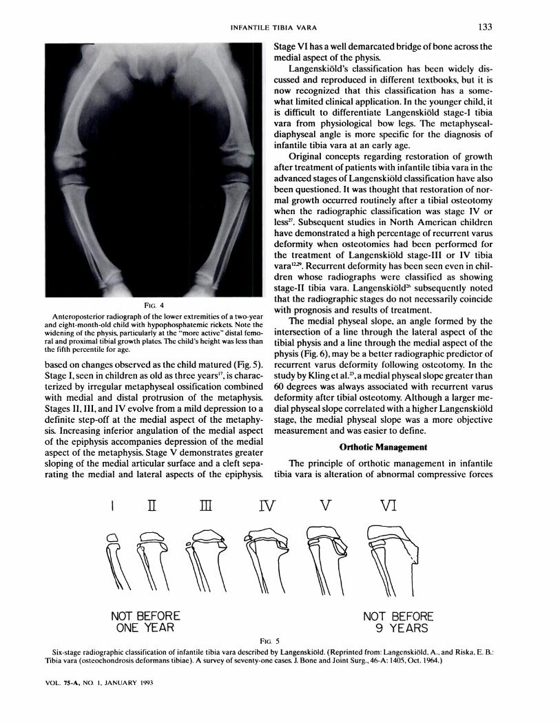
FIG. 4
Anteroposterior radiograph of the lower extremities of a two-yearand eight-month-old child with hypophosphatemic rickets. Note thewidening of the physis, particularly at the “more active” distal femo-ral and proximal tibial growth plates. The child’s height was less thanthe fifth percentile for age.
based on changes observed as the child matured (Fig. 5).Stage I, seen in children as old as three years’7, is chanac-tenized by irregular metaphyseal ossification combinedwith medial and distal protrusion of the metaphysis.Stages II, III, and IV evolve from a mild depression to adefinite step-off at the medial aspect of the metaphy-sis. Increasing inferior angulation of the medial aspedtof the epiphysis accompanies depression of the medialaspect of the metaphysis. Stage V demonstrates greatensloping of the medial articulan surface and a cleft sepa-rating the medial and lateral aspects of the epiphysis.
Stage VI has a well demarcated bridge of bone across themedial aspect of the physis.
Langenskiold’s classification has been widely dis-cussed and reproduced in different textbooks, but it isnow recognized that this classification has a some-what limited clinical applidation. In the younger child, itis difficult to differentiate Langenskiold stage-I tibiavara from physiological bow legs. The metaphyseal-diaphyseal angle is more specific for the diagnosis ofinfantile tibia vara at an early age.
Original concepts regarding restoration of growthafter treatment of patients with infantile tibia vara in theadvanced stages of Langenskiold classification have alsobeen questioned. It was thought that restoration of non-mal growth occurred routinely after a tibial osteotomywhen the radiographic classification was stage IV onless27. Subsequent studies in North American childrenhave demonstrated a high percentage of recurrent vanusdeformity when osteotomies had been performed forthe treatment of Langenskiold stage-Ill on IV tibiavara’2 . Recurrent deformity has been seen even in chil-dren whose nadiographs were classified as showingstage-I! tibia vara. Langenskiold ” subsequently notedthat the radiographic stages do not necessarily coincidewith prognosis and results of treatment.
The medial physeal slope, an angle formed by theintersection of a line through the lateral aspect of thetibial physis and a line through the medial aspect of thephysis (Fig. 6), may be a better radiographic predictor ofrecurrent varus deformity following osteotomy. In thestudy by Kling et al! , a medial physeal slope greater than60 degrees was always associated with recurrent vanusdeformity after tibia! osteotomy. Although a larger me-dial physeal slope correlated with a higher Langenskioldstage, the medial physeal slope was a more objectivemeasurement and was easier to define.
Orthotic Management
The principle of onthotic management in infantiletibia vara is alteration of abnormal compressive forces
NOT BEFOREONE YEAR
FIG. S
NOT BEFORE9 YEARS
Six-stage radiographic classification of infantile tibia vara described by Langenskiold. (Reprinted from: LangenskiOld, A., and Riska, E. B.:Tibia vara (osteochondrosis deformans tibiae). A survey of seventy-one cases. J. Bone and Joint Surg., 46-A: 1405, Oct. 1964.)

Fli . 6
134 w. B. GREENE
THE JOURNAL OF BONE AND JOINT SURGERY
Measurement of the medial physeal slope. (Adapted from: Kling,T. F. Jr.: Volk. A. G.: Dias. L.: Morgan. R. C.. Jr.: and DeRosa, G. P.:Infantile Blount’s disease treated with osteotomy: follow-up to matu-rity. Orthop. Trans., 14: 634. 1990.)
so that normal growth will resume and the genu varumwill be corrected. Bracing does not always accomplishthis goal; however, a trial with a brace before the childis three years old does not seem to compromise theresults of tibial osteotomy.
Bracing is the treatment of choice for a child four-teen to thirty months old with infantile tibia vara (Fig.7). Because it takes approximately one year for thephysician to know whether brace treatment has beensuccessful. the treatment of the thirty to thirty-six-month-old child should include consideration of otherfactors. For children of this age. I prescribe an orthosisif the medial physeal slope is less than 50 degrees. Irecommend a tibial osteotomy when the medial phys-cal slope is greater than 60 degrees (Fig. 8). With a me-dial physeal slope of 50 to 60 degrees, orthotic treatmentis selected only after consideration of other elements.Obesity. female gender, and a poor social situation arepoor prognostic signs for successful bracing.
The orthosis prescribed for children with infantiletibia vara is an above-the-knee brace with a free ankle,single medial upright. and no hinge joint at the knee
(Fig. 9). A cuff around the knee pulls the leg into valgusangulation. A hinge joint at the knee is not needed forsitting activities in these young children. Elimination ofthe knee joint from the brace makes it easier to alignthe cuff, makes the brace more adaptable for subse-quent growth, and allows easy adjustment of the medialupright. Every six to twelve weeks, the medial up-right can be bent to gain further valgus alignment at theknee.
Although some texts recommend only nighttimebracing’74, I believe that weaning of the brace twenty-two to twenty-three hours a day provides greaten poten-tial for correction of infantile tibia vara. In fact, if a childis not able to tolerate full-time brace wean, then the timeout of the brace should be while the child is sleeping.Standing and walking increase the compressive, growth-inhibiting forces, and the physis needs protection duringthese activities.
Because the radiographic parameters that permitearly differentiation of physiological bow legs from in-fantile tibia vara are of recent vintage, the results oforthotic management in infantile tibia vara are still be-ing defined. Early results have indicated that brace then-apy for infantile tibia vara is successful in approximately50 per cent of cases29. It is my impression that the re-sults are even better if an above-the-knee brace is worntwenty-three hours a day.
Anteroposterior radiograph of a one-year and six-month-old girl.The metaphyseal-diaphyseal angle is 20 degrees on the right. and themedial aspect of the proximal tibial physis also has an inferior slope.Although diagnosis of infantile tibia vara could not be made conclu-sively at this time, the patient had a high probability of having thisdisorder. A knee-ankle-foot orthosis was prescribed.

Fi;, 9
INFANTILE TIBIA VARA 135
VOL. 75-A. NO. I. JANUARY 1993
Anteroposterior standing radiograph of the lower extremity of atwo-year and six-month-old boy. Even though the patient was rela-tively young. he was a poor candidate for brace therapy, at least onthe left side. The left tibia had a medial physeal slope of 60 degrees.Internal tibial torsion was also evident in the left leg. To makeradiographs centered at the knees. the radiology technician fre-quently externally rotates a leg with marked internal tibial torsion.This makes the fibula appear relatively posterior to the tibia.
Operative Treatment
Osteotomy of the proximal part of the tibia is mdi-cated for the child who is first seen for treatment af-ten age three, for the child thirty to thirty-six monthsold who is a poor candidate for brace therapy, and forthe three-year-old child who has persistent genu varumdespite brace therapy. Multiple techniques have beendescribed for the performance of this procedure in chil-dnen5’22242635 ’ ’4’. All involve placement of the osteot-omy distal to the tibial tubencle to prevent damage tothe tibia! apophysis and subsequent genu recurvatum.Concomitant osteotomy of the fibula is necessary topermit adequate correction of the genu varum and in-ternal tibia! torsion.
Preoperative assessment before a tibial osteotomyincludes judgment of the risk of recurrent varus defor-
mity, a problem that occurs more often than was onigi-nally suspected’22 2’3”. If the child is at increased risk forrecurrent varus deformity. the operation should be al-tened to diminish that risk. Factors that correlate withrecurrent varus deformity include massive obesity, a ra-diognaphic classification of Langenskiold stage III orgreaten, a medial physeal slope of greater than 60 de-grees, and a child who is more than five years old 2” ”.
Concepts that concern age as a risk factor havechanged during the previous decade. Three studies fromdifferent centers in the United States have confirmedthat children more than five years old with infantile tibiavana have a very high rate of recurrent varus deformityfollowing tibial osteotomy2’35. Ferriter and Shapiro’2observed a recurrence rate of 76 per cent in children un-dengoing osteotomy at five years old or older. comparedwith a 31 per dent recurrence rate in those having open-ative treatment before that age. Loder and Johnston29found that 88 per cent of children less than four yearsold were successfully treated with one osteotomy. butonly 32 per cent of children more than four years oldhad an adequate result after a single osteotomy. Moreimportantly, the percentage of good results is decreased
The knee-ankle-foot orthosis used for patients with infantile tibiavara. The single medial upright has no knee joint. The lateral cuffpulls the knee into valgus angulation. The brace can he easily ad-justed as the genu varum corrects.

1/I.FIG. 10-A FIG. 10-B Fi;. 10-C
Figs. 10-A, 10-B, and 10-C: Radiographs of a four-year and eleven-month-old morbidly obese girl (body weight, thirty-eight kilograms) inwhom recurrent varus deformity quickly developed following bilateral tibial osteotomy at the age of three years and ten months. Tomogramsshowed a probable medial physeal bar.
Fig. 10-A: Anteroposterior radiograph of the left knee.Fig. 10-B: Anteroposterior radiograph of the left knee three months after bridge resection and realignment osteotomy. The operation was
difficult and lasted for seven hours. However, knee alignment was maintained. The right leg was operated on six months later.Fig. 10-C: Anteroposterior radiograph of both legs, 1.5 years after bridge resection on the right and 2.3 years after bridge resection on the
left. Recurrent varus deformity developed in the right leg and the result was considered a failure. Growth and alignment of the left leg weremaintained.
I 36 W. B. GREENE
THE JOURNAL OF BONE AND JOINT SURGERY
TABLE I
INoIcA1IoNS FOR PREOPERATIVE TOMOGRAMS
OR FOR MooIIIAIIoN OF TIBIAL OSTEOTOMY, OR BOTh,
IN INFANTILE TIBIA VARA
Age of more than live yearsMedial physeal slope greater than 60 degreesLangcnskiold stage-IV radiographic changesBody weight greater than ninety-fifth percentileBlack girl close to meeting above criteriaMedial physcal slope of SO to 59 degrees in a patient close to
meeting above criteria
in children who are five years of age on older ”. Thiscontrasts with the guidelines originally described byLangenskiold . His data, however, were derived froma homogeneous population of white Finnish children,whereas in the United States the population is moreheterogeneous and the majority of children with infan-tile tibia vara are of the black race and are thereforeskeletally more mature.
One possible cause of recurrent varus deformity af-ten tibia! osteotomy is an osseous ban bridging the me-dial aspect of the tibial physis. This physeal ban may notbe visible on plain nadiognaphs; therefore, my preopera-tive evaluation includes two-millimeter thin-slice tomo-grams on computed tomography scans on patients whohave at least one of the following risk factors: (1) anage of five years on olden, (2) a medial physeal slopeof more than 60 degrees, (3) Langenskiold stage-IV ra-diognaphic changes, (4) body weight greater than theninety-fifth percentile, (5) a black girl who is close to
meeting the criteria just described, and (6) a medial-physeal slope of 50 to 59 degrees in a patient who isclose to meeting the criteria just described (Table I).Unfortunately, the medial aspect of the proximal tibia!physis in infantile tibia vara takes a serpentine descend-ing pathway. This can make identification of a physealban challenging, even when the radiographic images areadjusted to the plane of the deformity.
If a physeal bridge of bone is identified, then resec-tion of the osseous bar with interposition of fat, meth-ylmethacrylate, on medical-grade elastomen should beconsidened3 . Tibia! realignment is done at the sameoperation. Physeal bar resection in infantile tibia vara,however, is difficult and the results are unpredictable(Figs. 10-A, 10-B, and 10-C). The edge of the normalgrowth plate can be hand to find and the resection maybe larger than one would anticipate from the preopera-tive radiographic studies. In three children with infantiletibia vana undergoing this procedure, Loden and John-sto&’ noted inconsistent results, with one good, onefain, and one poor outcome. An osseous bridge greaterthan 50 pen cent of the width of the growth plate isusually listed as a contraindication to physeal ban exci-sion, but I now restrict physeal ban resection in childrenwith infantile tibia vana to cases in which the osseousbridge occupies less than 30 pen cent of the growthplate.
Even with no apparent physeal bar, vanus deformitymay recur after tibia! osteotomy. In this situation, themedial portion of the proximal tibia! physis has been

U I’ -FIG. I 1-A FI;. I 1-B Fl;. 1 1-C
Figs. 11-A. Il-B. and Il-C: Radiographs of a three-year and eight-month-old boy with persistent tibia vara (between Langenskiold stages 11and III).
Fig. 11-A: Preoperative anteroposterior standing radiograph. The medial physeal slope measures 61 degrees. The body weight wasacceptable. being at the eightieth percentile. The patient was thought to be at some increased risk for recurrent varus deformity.
Fig. I 1-B: Anteroposterior radiograph of the leg nine days after a valgus derotation osteotomy. The osteotomy was aligned so that theanatomical knee axis was in increased valgus (17 degrees).
Fig. 11-C: Anteroposterior standing radiograph of both knees at the age of nine years and three months. The over-all alignment wassatisfactory. but observation needed to be continued through the remainder of growth.
INFANTILE TIBIA VARA 137
VOL. 75-A. NO. I. JANUARY 1993
abnormally compressed for so long that its growth p0-tential remains limited even after realignment of thetibia. The result is continued overgrowth from the lat-era! portion of the tibia! physis and recurrent genuvarum. For children who are at increased risk for recur-rent varus deformity but who have no obvious physea!bar (Table I), I modify the operation with one of threetechniques: (1) increased valgus alignment of the oste-otomy, (2) stapling of the lateral aspect of the tibia!physis, or (3) hemiepiphyseodesis of the lateral aspectof the tibia! physis and an epiphyseodesis of the proxi-mal part of the fibu!a. Options 2 and 3 are combinedwith a realignment tibial osteotomy and are used formore severe problems.
Positioning of the osteotomy in excessive valgus isprimarily used in a three or four-year-old child who hasincreased risk factors for recurrence; that is, obesity, fe-male gender, and an increased medial physea! slope.This position minimizes compression across the disorga-nized physis and provides time for normal growth toresui’ne. In the best-case scenario, symmetrical growthwill resume when the tibia has drifted back into a phys-iologica! alignment. The degree of ovenconrection is de-termined according to the surgeon’s judgment, but itis geiiera!ly S to 10 degrees more valgus angulation
than is normal for the child’s age (Figs. I I-A, 1 I-B, and11-C). Overcorrection should not be done in a childwithout risk factors, because in such cases a persistentor even increasing valgus deformity can occur. This sit-uation is similar to the valgus deformity that developsafter a proximal tibial metaphyseal fracture in a youngchild.
The child between the ages of five and eight yearswho does not have a demonstrable physeal bar needsmore prolonged protection of the medial portion of thetibia! physis. This is particularly true for a girl with amedial physeal slope of greater than 60 degrees or whois massively obese. or both’. In this situation, the tibia isrealigned and staples are inserted across the lateral por-tion of the proximal tibia! physis, a concept initiallysuggested to me by Griffin”. Staples inhibit growth fromthe lateral aspect of the tibia! physis and thereby allowmore time for the media! aspect of the physis to re-cupenate from previous years of abnormal compression.When the tibia starts to demonstrate increased valgusangu!ation. the staples are removed to allow equivalentgrowth from both sides of the physis. Initial results withthis technique for the older child with infantile tibia varahave been promising, but more experience will be nec-essany before the success rate of this technique can be

FIG. 12-C
138 w. B. GREENE
THE JOURNAL OF BONE AND JOINT SURGERY
FI3. 12-A FIG. 12-B
Figs. 12-A. I 2-B. and 12-C: Radiographs of a six-year and eleven-month-old morbidly obese girl (weight, 49.8 kilograms - twenty kilogramsabove the ninety-fifth percentile for age). The tibia vara was LangenskiOld stage IV. and the medial physeal slope was 75 degrees. The patientwas at marked risk for recurrent varus deformity. Treatment consisted of valgus derotation osteotomy and stapling of the lateral aspect of theproximal tibial physis. Because of the patient’s size. the osteotomy was fixed with two large. threaded Steinmann pins.
Fig. I 2-A: Anteroposterior standing radiograph of the left knee.Fig. 12-B: Anteroposterior standing radiograph of the knee after injection of radiopaque contrast medium into the knee joint. The medial
aspect of the tihial epiphysis shows a large unossified region. Lateral subluxation of the tibia and relative overgrowth of the medial femoralcondyle are clearly outlined. The medial meniscus does not appear hypertrophic.
Fig. I 2-C: Anteroposterior radiograph of the left knee 1 .2 years after a tibial osteotomy. Alignment is well maintained. but the growth plateis closing. The patient was subsequently treated with a contralateral proximal tibial epiphyseodesis. removal of staples. and completion of a leftlateral proximal tibial epiphyseodesis. Evaluation prior to the epiphyseodesis revealed an advanced skeletal age (chronological age, eightyears: bone age. eleven years). Therefore. ultimate reduction of leg length was expected to be minimum.
determined (Figs. 12-A, 12-B, and 12-C).Epiphyseodesis of the lateral aspect of the tibia!
physis and the proximal part of the fibula should becombined with a realignment osteotomy in a child whois first seen at the age of eight on nine years on in a youn-ger child with recurrent varus deformity and a medialphyseal bridge that is too large to be nesected. In thissituation, a contralatenal epiphyseodesis should be con-sidered to prevent or correct a leg-length discrepancy.
With advanced changes, the anticulan surface of themedial tibia! plateau is markedly depressed. In thesechildren, an opening wedge osteotomy of the epiphysisshould be considered”'2539. Langenskiold reported somegood long-term results with the use of this technique inolder children who were first seen with a markedly de-pressed medial tibia! plateau25. My experience with thisprocedure is limited to one case. With a five-year follow-up, the result was rated as fair. Relative overgrowth of
the medial femona! condyle may limit the success of thisprocedure (Fig. 12-C).
Neurovascular Complications
The risk of neunovascu!ar complications following aproximal tibia! osteotomy is greaten in children (Fig. 13)than in adults3’3242. Steel et al.42 initially described thisproblem when they reported nine cases of neurovascu-lan complications in forty-six children who had under-gone proximal tibia! osteotomy. As described in thisreport, the primary symptom was “severe pain in thefront of the leg, unremitting and intractable.” Steel et a!.attributed the neurological deficits in these children tocompression of the anterior tibia! artery as it perforatedthe interosseous membrane, and they recommended im-mediate repositioning of the leg into vanus angulation.These signs and symptoms, however, are also consistentwith a compartment syndrome and, in my opinion, this

FIG. 13
FIG. 14-A FIG. 14-B
Fig. 14-A: Outline of the cuts for an opening-closing chevronosteotomy.
Fig. 14-B: At the completion of the osteotomy, the lateral wedge isinserted medially. Fixation of the osteotomy can be accomplishedwith a cast alone, one pin, or two cross-pins. The type of fixationdepends on the stability of the osteotomy, the patient’s size and age,and the degree of obesity.
INFANTILE TIBIA VARA 139
VOL. 75-A, NO. 1, JANUARY 1993
A child in whom a compartment syndrome developed in all fourcompartments after a bilateral tibial osteotomy for infantile tibiavara at age eight. The problem was not recognized until the thirdpostoperative day. Compartment releases necessitated closure withskin-grafting. Recurrent varus deformity developed, and muscle re-covery was limited to grade-2 strength in the gastrocnemius.
is the most common cause of neunovascular compromisefollowing proximal tibia! osteotomy in children.
The anterior compartment is most vulnerable fol-lowing a realignment operation for infantile tibia vana.This osteotomy is performed distal to the tibia! tuber-cle, and the resultant soft-tissue trauma is the probablecause of this increased susceptibility. I routinely per-form a prophylactic subcutaneous release of the ante-nor compartment fascia at the completion of the tibia!osteotomy. Careful and continued postoperative assess-ment is also necessary. This may be challenging in ayoung child whose natural apprehension of doctors isenhanced by postoperative pain.
If symptoms and signs of circulatory compromiseoccur, or if the child is having more pain than expected,the cast should be bivalved and compartment pressuresshould be measured35’32. A compartment pressure of lessthan thirty millimeters of mercury calls for continuedobservation. If the pressure measurements are greatenthan forty-five millimeters of mercury, the patient isimmediately returned to the operating room for fasciot-omy. If the compartment pressures are between thirtyand forty-five millimeters of mercury, the patient may
be observed if examination demonstrates intact neuro-muscular function and if no subsequent deteriorationoccurs.
Technique of Osteotomy
I prefer an opening-closing chevron osteotomy inpatients who have infantile tibia vana (Figs. 14-A and14-B). This osteotomy is a modification of the domeosteotomy and has the advantage of providing greatenstability and minimum change in leg lengths. The the-oretical disadvantage is that a slightly longer periodof cast immobilization (approximately two weeks) isneeded for incorporation of the wedge segment. This,however, does not cause a problem in children, and theadditional period of reduced stress on the medial aspectof the physis may be advantageous. Before the osteot-omy is done, paper cutouts are made and a template that

,J ._... ‘ -
Fu;. 15-A FIG. 15-B FIG. 15-C
Figs. IS-A. 15-B. and 15-C: Radiographs of a four-year and eight-month-old boy with body weight at the seventieth percentile. Femoral-tibialalignment is 14 degrees of varus, and the medial physeal slope measures 52 degrees. Even though the patient was close to age five, the otherrisk factors were absent or only marginally increased. Therefore. treatment was a valgus rotational osteotomy.
Fig. 15-A: Preoperative radiograph.Fig. 1 S-B: Intraoperative anteroposterior radiograph of the tibia after completion of the opening-closing chevron osteotomy. Alignment was
stable, and the single smooth pin used for fixation may not have been needed in this patient.Fig. IS-C: Anteroposterior standing radiograph five years after osteotomy.
140 w. B. GREENE
THE JOURNAL OF BONE AND JOINT SURGERY
outlines the desired lateral wedge is prepared.The operation is performed with the patient in a
supine position. A sandbag is positioned underneath theipsilatera! hip to facilitate exposure of the fibula. The legis prepped from the toes to the proximal aspect of thethigh. Prepping of the foot allows more accurate assess-ment of tibia! torsion and permits evaluation of thedorsalis pedis and posterior tibia! pulses after the pneu-matic tourniquet has been deflated.
The operation starts with osteotomy of the fibula.The middle third of the fibula is exposed via the intervalbetween the lateral and the posterior compartments. Thepeniosteum of the fibula is sharply incised and is circum-ferentially elevated, with care taken to prevent injury tothe adjacent peronea! vessels. A one-centimeter segmentof fibula is removed with a reciprocating saw. The fibulais cut obliquely in a supenolateral-to-infenomedial di-rection. This allows the distal portion of the fibula toslide past the proximal fragment as the leg is broughtfrom a varus to a valgus position.
A hockey-stick incision for the tibia! osteotomy be-gins four to five dentimeters distal to the tibia! tuber-cle. Staying immediately lateral to the anterior spine ofthe tibia, the incision extends to the tibia! tubencle andthen curves in a lateral direction toward the Gurdy tu-berc!e. The peniosteum is sharply incised immediately
adjacent to the anterior compartment muscles. Immedi-ately distal to the tibia! tuhencle, the peniosteum is trans-versely incised and then circumferentia!!y elevated sothat curved retractors can be positioned to protect theposterior soft tissues. Because of its triangular shape,more care is required at the postero!atera! and pos-teromedial edges of the tibia to ensure that the dissec-tion remains subpeniostea!.
The osseous cuts are outlined on the anterior surfaceof the tibia with an osteotome or cautery. The apex ofthe osteotomy is immediately distal to the tibia! tuber-cle. An anterior-to-posterior drill-hole is made at thispoint to minimize the risk of the osteotomy extend-ing beyond the desired location. The osteotomy is com-p!eted with an oscillating saw and the lateral wedge isremoved.
Depending on the child’s age and degree of obesity,as well as the stability of the osteotomy, a single pin, twocrossed pins, or no internal fixation may be used (Figs.15-A, 15-B, and 15-C). Depending on the same factors,I select either smooth or threaded pins. Smooth pins areeasier to remove and, in older children, this may be donein the outpatient clinic with the aid of a sedative medi-cation and local anesthesia. In addition, smooth pinsshould cause less damage if they are inserted across thephysis. Threaded pins are easier to insert and have less

INFANTILE TIBIA VARA 141
VOL. 75.A, NO. 1, JANUARY 1993
of a tendency to loosen but, in children, general anes-thesia is usually needed for removal.
If pins are used for fixation, pre-dnl!ing of the di-aphysis with small drill-bits makes pin insertion easierand more precise. For the media! pin, a small stab mci-sion is made approximately one centimeter distal to theinsertion site. For the lateral pin, the anterior compart-ment muscles can be retracted enough to allow inser-tion. Optimum pin position is such that both pins crossthe osteotomy site and exit through the proximal cortexwithout crossing the physis. This pin position, however,is not always necessary. The decision concerning what isacceptable is based on the age of the child and theinherent stability of the osteotomy.
After optimum positioning of the drill-bits at the Os-teotomy site has been confirmed, the tibia is swung intothe desired position of valgus and external rotation. Thelateral wedge is inserted medially in a position that willmaintain the desired degree of correction. Appropriate-size Steinmann pins replace the drill-bits and are drivenacross the osteotomy site.
The pneumatic tourniquet is deflated and circula-tion of the foot is assessed. Doppler examination maybe necessary. If the circulation is abnormal, the pins areremoved and the tibia is restored to its preoperativealignment to eliminate the possibility of compression ofthe anterior tibia! artery. I have never experienced thatsituation and, as previously noted, I believe that a corn-partment syndrome is more likely to cause neurovascu-lan compromise after tibia! osteotorny in children.
If the circulation is satisfactory, anteroposterior andlateral nadiographs of the proximal part of the tibiaare obtained to assess osseous apposition, pin position,and tibia! alignment. With operating room drapes anda small lower extremity, it may be difficult to includeenough of the distal aspect of the femur on the intra-operative radiognaphs to measure the anatomical kneeaxis. In this situation, the amount of correction can beassessed by measurement of the “articulodiaphyseal”angle on the preoperative and intnaopenative radio-graphs. This angle is formed by a line drawn tangentialto the anticular surface of the proximal part of the tibiaand a line drawn through the axis of the diaphysis. Thedifference between the angles on the preoperative andintnaoperative nadiographs is the degree of valgus con-rection that has been obtained.
When satisfactory alignment has been confirmed,the Steinmann pins are positioned to facilitate removaland minimize such problems as pin-track infectionand skin ulceration. For tibia! osteotomies, particularlyin obese children, I prefer to bury the pins under theskin rather than leaving them protruding. Smooth pinsshould be bent and cut to prevent subsequent migration.The pin inserted through the lateral dontex is drilledthrough the proximal pant of the tibia and the overlyingskin. This pin is then drilled retrograde until its distal tipis flush with the lateral cortex. The pin inserted through
the media! side of the distal part of the tibia is leftprominent at its distal end.
Subcutaneous fasciotomy of the anterior compart-rnent is performed with long Metzenbaurn scissors. Thefascia is incised by pushing of the scissors as far distallyas possible, usually to the junction of the middle anddistal thirds of the leg. This type of fasciotomy is quicklydone, is associated with minimum operative morbidity,and should markedly diminish the risk of compartmentsyndrome. If possible, the thick peniosteum of the tibiais reapproximated to increase the stability of the oste-otomy, to decrease hematoma formation, and to en-hance bone union. If bleeding from the osteotomy haslargely abated, the skin can be closed with a runningsubcuticulan absorbable suture. Otherwise, skin closureshould be with interrupted sutures and suction drainageshould be utilized.
Closure of the fibular incision starts with inspectionof the osteotomy site for possible damage to the pero-neal vessels. The fascia is left open but a more extensivecompartment release is not performed. The guidelinesjust described for subcuticu!an versus interrupted su-tures for skin closure are used for this incision as we!!.
An above-the-knee cast is applied with the kneeflexed 45 degrees and the ankle in a neutral posture.Knee flexion relaxes the posterior neurovascular struc-tunes and minimizes slippage of the cast in these chi!-dren, whose limbs are relatively short but often large incircumference. Before the anesthesia is discontinued,radiographs are repeated to confirm that the cast wasapplied without displacement of the osteotorny.
Young children are allowed only bed-to-chair activ-ities for approximately four weeks. A three to four-year-old child rarely has the coordination and upper bodystrength to become adept on crutches in this four-weekperiod. For the older child, crutch ambulation is delayeduntil the fourth to sixth postoperative day. Attempts atambulation before that time are usually counterproduc-tive, because the operative pain has not abated enoughto make therapy sessions effective or efficient. Thesechildren are usually discharged from the hospital on thesecond or third postoperative day, and the therapy ses-sions are arranged on an outpatient basis.
The cast is changed approximately four weeks afterthe operation. If nadiographs show satisfactory interimhealing, the pins are removed and an above-the-kneecast is applied in a position to allow weight-bearingactivities. Usually eight to ten weeks of postoperativeimmobilization is sufficient. Although osteotornies healfaster in children than they do in adults, osteotomies andfractures in children must be protected long enough tominimize the risk of fracture that accompanies a quickresumption of vigorous play activities.
Recurrent Varus Deformity
Preoperative and postoperative discussions with theparents should emphasize the need for periodic assess-

142 W. B. GREENE
THE JOURNAL OF BONE AND JOINT SURGERY
ment of the child until growth is completed. Recur-rent varus deformity may occur within a relatively shortperiod on it may occur on a delayed basis, panticu-larly during the adolescent growth spurt. In any case,early identification will allow for appropriate treat-ment before irreversible damage to the anticulan surfaceoccurs.
If the varus deformity recurs quickly after the oste-otomy, I evaluate the patient with computed tomogra-phy scans to determine whether a ban of bone is crossingthe physis. The decision to do a physeal bridge resectionon another procedure is based on guidelines previouslydiscussed. For the olden child, the best option is fne-quently a lateral hemiepiphyseodesis of the proximalpant of the tibia. If the vanus deformity is recognizedsoon enough, a realignment osteotomy of the tibia canbe avoided. An epiphyseodesis of the contralateral limbmay be necessary to correct or minimize the problem ofleg-length discrepancy.
The need for a realignment osteotomy of the tibiain a person who is at on near completion of growthdepends on the degree of vanus deformity. A hip-knee-ankle mechanical axis of greaten than 10 degrees ofvanus donsistent!y doncentrates forces on the media!
side of the knee joint and probably predisposes the pa-tient to early osteoarthnosis2. Realignment tibia! oste-otomy is usually recommended for these patients. Forthe child who has less than 5 degrees of mechanicalvanus alignment, the joint mechanics are satisfactoryand osteotomy is not warranted. At the present time, itis unclear whether a 5 to 10-degree hip-knee-ankle me-chanical axis will excessively load the anticular surfaceenough to cause osteoarthnosis at an early age.
Conclusions
Infantile tibia vana is a localized disorder of growthsecondary to abnormal compression of the medial as-pect of the proximal tibia! physis. An above-the-kneebrace may successfully treat infantile tibia vana in ayoung child, and a standard tibia! osteotomy usuallyrestores normal growth and joint mechanics in a three-year-old child. For an older child with infantile tibiavana, the operative procedure is more complicated andthe possibility of a good result is markedly decreased.We must continue to work with primary cane physiciansand public health nurses so that children with persistentgenu varum are evaluated at an early age, preferablybefore they are two years old.
References
1. Bateson, E. M.: The relationship between Blount’s disease and bow legs. British J. Radiol., 41: 107-114. 1968.2. Bathfield, C. A., and Beighton, P. H.: Blount disease. A review of etiological factors in 110 patients. C/in. Orthop., 135: 29-33, 1978.3. Beck, C. L.; Burke, S. W.; Roberts, J. M.; and Johnston, C. E., II: Physeal bridge resection in infantile Blount disease. J. Pediat. Orthop., 7:
161-163. 1987.
4. Blount, W. P.: Tibia vara. Osteochondrosis deformans tibiae.J. Bone andJoint Surg., 19: 1-29,Jan. 1937.S. Canale, S. T., and Harper, M. C.: Biotrigonometric analysis and practical applications of osteotomies of tibia in children. In Instructional
Course Lectures, The American Academy ofOrthopaedic Surgeons. Vol. 30, pp. 85-101. St. Louis, C. V. Mosby, 1981.6. Capute, A. J.; Shapiro, B. K.; Palmer, F. B.; Ross, Alan; and Wachtel, R. C.: Normal gross motor development: the influences of race, sex
and socio-economic status. Devel. Med. and Child Neurol., 27: 635-643, 1985.7. Cook, S. D.; Lavernia, C. J.; Burke, S. W.; Skinner, H. B.; and Haddad, R. J., Jr.: A biomechanical analysis of the etiology of tibia vara.
J. Pediat. Orthop., 3: 449-454. 1983.8. Dietz, F. R., and Weinstein, S. L.: Spike osteotomy for angular deformities of the long bones in children. J. Bone and Joint Surg., 70-A:
848-852. July 1988.
9. Drennan, J. C.: Pediatric knee disorders. Instructional Course Lecture at the Annual Meeting of The American Academy of Orthopae-die Surgeons, Washington, D.C., Feb. 21, 1992.
10. Engel, G. M., and Staheli, L. T.: The natural history of torsion and other factors influencing gait in childhood. A study of the angle ofgait. tibial torsion, knee angle. hip rotation, and development of the arch in normal children. C/in. Orthop., 99: 12-17, 1974.
1 1 . Feldman, M. D., and Schoenecker, P. L.: Use of Drennan’s metaphyseal-diaphyseal angle in evaluating bow legs. Read at the AnnualMeeting of The American Academy of Orthopaedic Surgeons, Washington, D.C., Feb. 21, 1992.
12. Femter, Pierce, and Shapiro, Frederic: Infantile tibia vara: factors affecting outcome following proximal tibial osteotomy. J. Pediat.Orthop., 7: 1-7, 1987.
13. Foreman, K. A., and Robertson, W. W., Jr.: Radiographic measurement of infantile tibia vara. J. Pediat. Orthop., 5: 452-455. 1985.14. Golding, J. S. R., and McNeil-Smith, J. D. G.: Observations on the etiology of tibia vara. J. Bone and Joint Surg., 4S-B(2): 320-325. 1963.15. Greene, W. B., and Kahler, S. G.: Hypophosphatemic rickets: still misdiagnosed and inadequately treated. Southern Med. J., 78: 1179-
1 184, 1985.
16. Gregosiewicz, Andrzej; Wosko, Ignacy; Kandzierski, Grzegorz; and Drabik, Zbigniew: Double-elevating osteotomy of tibiae in thetreatment of severe cases of Blount’s disease. J. Pediat. Orthop., 9: 178-181, 1989.
17. Griffin, P. P.: The lower limb. In Pediatric Orthopaedics, edited by W. W. Lovell and R. B. Winter. Vol. 2, pp. 881-909. Philadelphia. J. B.Lippincott. 1980.
18. Griffin, Paul: Personal communication. Chapel Hill. North Carolina.19. Henderson, R. C.; Lechner, C. T.; DeMasi, R. A.; and Greene, W. B.: Variability in radiographic measurement of bowleg deformity in
children. .1. Pediat. Orthop., 10: 491-494. 1990.20. Hofmann, Aaron; Jones, R. E.; and Herring, J. A.: Blount’s disease after skeletal maturity. .1. Bone and Joint Surg.. 64-A: 1004-1009,
Sept. 1982.21. Johnson, F.; Leitl, S.; and Waugh, W.: The distribution of load across the knee. A comparison of static and dynamic measurements.
J. Bone andJoint Surg., 62-B(3): 346-349. 1980.22. Johnston, C. E., II: Infantile tibia vara. C/in. Orthop., 255: 13-23, 1990.
INFANTILE TIBIA VARA 143
23. Kling, T. F., Jr.; VoIk, A. G.; Dias, L.; Morgan, R. C., Jr.; and DeRosa, G. P.: Infantile Blount’s disease treated with osteotomy: follow-upto maturity. Orthop. Trans., 14: 634-635, 1990.
24. Kruse, R. W.; Bowen, J. R.; and Heithoff, Steven: Oblique tibial osteotomy in the correction of tibial deformity in children. J. Pediat.Orthop., 9: 476-482, 1989.
25. Langenskiold, A.: Tibia vara. (Osteochondrosis deformans tibiae.) A survey of 23 cases. Acta Chir. Scandinavica, 103: 1-22, 1952.
26. Langenski#{246}ld, Anders: Tibia vara. A critical review. C/in. Orthop., 246: 195-207, 1989.27. Langenskiold, A., and Riska, E. B.: Tibia vara (osteochondrosis deformans tibiae). A survey of seventy-one cases. J. Bone and Joint
Surg., 46-A: 1405-1420, Oct. 1964.28. Levine, A. M., and Drennan, J. C.: Physiological bowing and tibia vara. The metaphyseal-diaphyseal angle in the measurement of bow-
leg deformities.J. Bone andJoint Surg., 64-A: 1158-1163, Oct. 1982.29. Loder, R. T., and Johnston, C. E., II: Infantile tibia vara. J. Pediat. Orthop., 7: 639-646, 1987.30. Matsen, F. A., III: Pathophysiology of compartment syndromes. In Instructional Course Lectures, The American Academy of Orthopae-
dic Surgeons. Vol. 38, pp. 463-466. Park Ridge, Illinois, The American Academy of Orthopaedic Surgeons, 1989.31 . Matsen, F. A., III, and Staheli, L. T.: Neurovascular complications following tibial osteotomy in children. A case report. C/in. Orthop.,
1 10: 210-214. 1975.32. Matsen, F. A., III, and Veith, R. G.: Compartmental syndromes in children.J. Pediat. Orthop., 1: 33-41, 1981.33. Olney, B. W.; Cole, W. G.; and Menelaus, M. B.: Three additional cases of focal fibrocartilaginous dysplasia causing tibia vara. .1. Pediat.
Orthop., 10: 405-407, 1990.34. O’Neill, D. A., and MacEwen, G. D.: Early roentgenographic evaluation of bowlegged children. J. Pediat. Orthop., 2: 547-553, 1982.35. Rab, G. T.: Oblique tibial osteotomy for Blount’s disease (tibia vara). J. Pediat. Orthop., 8: 715-720, 1988.36. Richardson, E. G.: Miscellaneous nontraumatic disorders. In Campbe//’s Operative Orthopaedics, edited by A. H. Crenshaw. Ed. 7, pp.
1005-1088. St. Louis. C. V. Mosby. 1986.37. Salenius, Pentti, and Vankka, Eila: The development of the tibiofemoral angle in children. J. Bone and Joint Surg., 57-A: 259-261,
March 1975.33. Schoenecker, P. L.; Meade, W. C.; Pierron, R. L.; Sheridan, J. J.; and Capelli, A. M.: Blount’s disease: a retrospective review and
recommendations for treatment. J. Pediat. Orthop., 5: 181-186, 1985.39. Siffert, R. S.: Intraepiphyseal osteotomy for progressive tibia vara: case report and rationale of management. J. Pediat. Orthop., 2:
81-85, 1982.40. Smith, C. F.: Current concepts review. Tibia vara (Blount’s disease). J. Bone andJoint Surg., 64-A: 630-632, April 1982.41. Staheli, L. T.: The lower limb. In Lovel/ and Winter’s Pediatric Orthopaedics, edited by R. T. Morrissy. Ed. 3, pp. 741-766. Philadelphia,
J. B. Lippincott, 1990.42. Steel, H. H.; Sandrow, R. E.; and Sullivan, P. D.: Complications of tibial osteotomy in children for genu varum or valgum. Evidence that
neurological changes are due to ischemia. J. Bone and Joint Surg., 53-A: 1629-1635, Dec. 1971.
VOL. 75.A, NO. 1, JANUARY 1993