Induction of micronuclei and other nuclear abnormalities ... · sively with increasing the duration...
8
EKOLOGIJA. 2010. Vol. 56. No. 3–4. P. 124–131 DOI: 10.2478/v10055-010-0018-4 © Lietuvos mokslų akademija, 2010 © Lietuvos mokslų akademijos leidykla, 2010 Induction of micronuclei and other nuclear abnormalities in blue mussels Mytilus edulis aſter 1-, 2-, 4- and 8-day treatment with crude oil from the North Sea Janina Baršienė 1 *, Laura Andreikėnaitė 1 , Anne Bjornstad 2 1 Institute of Ecology of Nature Research Centre, Akademijos Str. 2, LT-08412 Vilnius, Lithuania 2 International Research Institute of Stavanger, Mekjarvik 12, N-4070 Randaberg, Norway * Corresponding author. E-mail: [email protected] e genotoxicity and cytotoxicity of crude oil processed from the Statord B platform in the North Sea were studied in gills of blue mussels (Mytilus edulis), exposed to 0.5 ppm concentration of the dispersed oil for 1, 2, 4 and 8 days. Induction of micronuclei (MN) and nuclear buds (NB) was assessed as the crude oil genotoxicity endpoints; induction of fragmented-apoptotic (FA) and bi-nucleated (BN) cells indicated the cytotoxicity potential of the oil. Time-related MN elevation incidences were detected in all experimental groups compared to the control group of mussels. e elevation of micronuclei increased progres- sively with increasing the duration of exposure – from 1.9-fold aſter 1 day to 2.4-fold aſter 8 days of exposure. ere was no significant occurrence of nuclear buds in the experimen- tal groups. A significant induction of fragmented-apoptotic cells (P = 0.0115) was detected in M. edulis gills aſter a 4-day exposure and of bi-nucleated cells aſter an 8-day treatment (P = 0.0232). Key words: micronuclei, nuclear abnormalities, crude oil, blue mussel INTRODUCTION Environmental contaminants released from oil industry are well known as the primary source of persistent toxicity in aquatic ecosystems. Certain substances, such as crude oil, polycyclic aromatic hydrocarbons, heavy metals and alkyl- phenols, are usually present in petroleum industry wastewa- ter and attract particular attention because of their potential mutagenic and carcinogenic properties. e hazardous sub- stances may occur below the detection limit; however, they act as genotoxins, inducing genomic instability at very low con- centrations. Furthermore, contaminants, usually discharged in complex mixtures, can provoke interactions among un- known substances and lead to deviations in genotoxicity re- sponses to pollution (Jha, 2008). erefore, the need to devel- op and standardize tests for the monitoring of their biological effects is urgent in polluted marine ecosystems. is poses a major challenge for petroleum industry managers to protect indigenous populations and provide methodological instru- ments to monitor the effects of hazardous substances. Marine organisms could be exposed to genotoxic compounds via several routes. Genotoxins can bind to DNA molecules and trigger off a damaging chain of biological changes, such as an impaired enzyme function or general metabolism, cyto- toxicity, immunotoxicity, reproduction disturbances, growth inhibition, or carcinogenesis (Ohe et al., 2004). Oil spills can result in a wide distribution of petroleum hydrocarbons in the marine environment, seriously impacting DNA of filter- feeding bivalve populations (Hamoutene et al., 2002). Bi- valves have a limited ability to metabolize petroleum hydro- carbons and accumulate comparatively high levels of these compounds in their tissues (Dyrynda et al., 1997). Hazardous effects of various polyaromatic hydrocarbons (PAHs) arise typically as a result of oxidative biotransformation producing highly DNA-reactive metabolites. ese metabolites are re- cognized as carcinogenic and mutagenic compounds (Wood- head et al., 1999). e micronucleus (MN) test, one of those most frequently used in environmental genotoxicity studies, has served as an index of cytogenetic damage for over 30 years (Fenech et al., 2003). is is a sensitive and fast test to detect genomic al- terations due to clastogenic effects and impairments of mi- totic spindle. e MN test was originally developed for the analysis of chemical genotoxicity in mammals (Heddle et al., 1991), and later it has been successfully adapted to species from other groups, including aquatic organisms. Micronuclei
Transcript of Induction of micronuclei and other nuclear abnormalities ... · sively with increasing the duration...

EKOLOGIJA. 2010. Vol. 56. No. 3–4. P. 124–131DOI: 10.2478/v10055-010-0018-4© Lietuvos mokslų akademija, 2010© Lietuvos mokslų akademijos leidykla, 2010
Induction of micronuclei and other nuclear abnormalities in blue mussels Mytilus edulis aft er 1-, 2-, 4- and 8-day treatment with crude oil from the North Sea
Janina Baršienė1*,
Laura Andreikėnaitė1,
Anne Bjornstad2
1 Institute of Ecology ofNature Research Centre,Akademijos Str. 2, LT-08412 Vilnius,Lithuania
2 International Research Instituteof Stavanger, Mekjarvik 12,N-4070 Randaberg, Norway
* Corresponding author. E-mail: [email protected]
Th e genotoxicity and cytotoxicity of crude oil processed from the Statfj ord B platform in the North Sea were studied in gills of blue mussels (Mytilus edulis), exposed to 0.5 ppm concentration of the dispersed oil for 1, 2, 4 and 8 days. Induction of micronuclei (MN) and nuclear buds (NB) was assessed as the crude oil genotoxicity endpoints; induction of fragmented-apoptotic (FA) and bi-nucleated (BN) cells indicated the cytotoxicity potential of the oil. Time-related MN elevation incidences were detected in all experimental groups compared to the control group of mussels. Th e elevation of micronuclei increased progres-sively with increasing the duration of exposure – from 1.9-fold aft er 1 day to 2.4-fold aft er 8 days of exposure. Th ere was no signifi cant occurrence of nuclear buds in the experimen-tal groups. A signifi cant induction of fragmented-apoptotic cells (P = 0.0115) was detected in M. edulis gills aft er a 4-day exposure and of bi-nucleated cells aft er an 8-day treatment (P = 0.0232).
Key words: micronuclei, nuclear abnormalities, crude oil, blue mussel
INTRODUCTION
Environmental contaminants released from oil industry are well known as the primary source of persistent toxicity in aquatic ecosystems. Certain substances, such as crude oil, polycyclic aromatic hydrocarbons, heavy metals and alkyl-phenols, are usually present in petroleum industry wastewa-ter and attract particular attention because of their potential mutagenic and carcinogenic properties. Th e hazardous sub-stances may occur below the detection limit; however, they act as genotoxins, inducing genomic instability at very low con-centrations. Furthermore, contaminants, usually discharged in complex mixtures, can provoke interactions among un-known substances and lead to deviations in genotoxicity re-sponses to pollution (Jha, 2008). Th erefore, the need to devel-op and standardize tests for the monitoring of their biological eff ects is urgent in polluted marine ecosystems. Th is poses a major challenge for petroleum industry managers to protect indigenous populations and provide methodological instru-ments to monitor the eff ects of hazardous substances. Marine organisms could be exposed to genotoxic compounds via several routes. Genotoxins can bind to DNA molecules and
trigger off a damaging chain of biological changes, such as an impaired enzyme function or general metabolism, cyto-toxicity, immunotoxicity, reproduction disturbances, growth inhibition, or carcinogenesis (Ohe et al., 2004). Oil spills can result in a wide distribution of petroleum hydrocarbons in the marine environment, seriously impacting DNA of fi lter-feeding bivalve populations (Hamoutene et al., 2002). Bi-valves have a limited ability to metabolize petroleum hydro-carbons and accumulate comparatively high levels of these compounds in their tissues (Dyrynda et al., 1997). Hazardous eff ects of various polyaromatic hydrocarbons (PAHs) arise typically as a result of oxidative biotransformation producing highly DNA-reactive metabolites. Th ese metabolites are re-cognized as carcinogenic and mutagenic compounds (Wood-head et al., 1999).
Th e micronucleus (MN) test, one of those most frequently used in environmental genotoxicity studies, has served as an index of cytogenetic damage for over 30 years (Fenech et al., 2003). Th is is a sensitive and fast test to detect genomic al-terations due to clastogenic eff ects and impairments of mi-totic spindle. Th e MN test was originally developed for the analysis of chemical genotoxicity in mammals (Heddle et al., 1991), and later it has been successfully adapted to species from other groups, including aquatic organisms. Micronuclei
125Nuclear abnormalities in blue mussels exposed to crude oil
can be produced from chromosomal fragments or the whole chromosome that lags during cell division due to the lack of a centromere, damage in a centromere or a defect in cytokine-sis (Heddle et al., 1991).
Increased MN levels have been described in zones af-fected by oil spills (Parry et al., 1997; Harvey et al., 1999; Baršienė et al., 2004, 2006a, 2006b). A signifi cant elevation of genotoxicity was observed in mussels 30 days post-oil-spill, and the persistence of the cytogenetic damage lasted up to 100 days (Parry et al., 1997) or even 7 months (Baršienė et al., 2006a). A statistically signifi cant increase of micronuclei le-vels was found in oysters and fi sh caged in Haven oil spill zones 10 years aft er an oil spill (Bolognesi et al., 2006a). Increased genotoxicity was described in mussels from oil terminal and marine port zones in the Baltic Sea (Baršienė, Baršytė Lovejoy, 2000; Baršienė, 2002), in a Mediterranean commercial port zone (Magni et al., 2006), in zones of the Venice lagoon, polluted by aromatic hydrocarbons (Venier, Zampieron, 2005), coastal Mediterranean ecosystems in Croatia (Klobučar et al., 2008), aft er dredging contaminated by PAHs and Pb sediments (Bocchetti et al., 2008).
A positive relationship between MN frequencies and PAH concentrations (mainly alkylated homologues) was de-scribed in inter-tidal mussels Perna perna from a Brazilian coast chronically contaminated with oil, particularly aft er the major MF–380 oil spill in January 2000. In the PAH pro-fi le, the predominance of petrogenic compounds, especially phenanthrenes, was identifi ed. Signifi cant elevations of MN incidence were observed for ΣPAH above 1000 μg/kg, and the levels close to or above 300 μg/kg are able to initiate an in-crease in MN frequency (Francioni et al., 2007).
In addition to the MN test, nuclear buds, fragmented-apoptotic, bi-nucleated cells and some other nuclear abnor-malities were successfully used for assessing the pollutant eff ects in mussels (Venier et al., 1997; Dolcetti, Venier, 2002; Izquierdo et al., 2003; Carvalho Pinto Silva et al., 2005; Venier, Zampieron, 2005; Baršienė, Rybakovas, 2006; Baršienė et al., 2006a, 2006b, 2006c, 2006d, 2008; Baršienė, Andreikėnaitė, 2007; Koukouzika, Dimitriadis, 2008). Signifi cantly increased levels of micronuclei, nuclear buds and fragmented apoptotic cells were found in bivalves inhabiting the Baltic Sea close to the Būtingė oil terminal (Baršienė et al., 2006a) and to the Russian oil platform D-6 (Baršienė et al., 2008).
Formation of nuclear buds may refl ect the unequal ca-pacity of organisms to expel damaged, amplifi ed, failed rep-lication or improperly condensed DNA, chromosome frag-ments without telomeres and centromeres from the nucleus (Lindberg et al., 2007). Elimination of cytogenetic damage by apoptosis and necrosis is a key process which occurs at diff erent rates in various organisms (Micič et al., 2002). Nev-ertheless, there is an information gap in our knowledge on formation of nuclear buds and bi-nucleated cells in marine organisms, which could be useful markers for pollution ef-fects assessment and for validating the battery of the genoto-xicity and cytotoxicity test in aquatic media.
Despite progress in the development of biomarker ap-proach, there is a lack of laboratory-controlled studies with environmentally realistic doses of genotoxic and cytotoxic compounds used to refl ect their damage formation in indig-enous species inhabiting chronically contaminated oil plat-form areas. Recent fi ndings of high levels of micronuclei and other nuclear abnormalities found in fi sh and mussels aft er a 3-week exposure to crude oil processed from the Statfj ord B platform (Baršienė et al., 2006e; Bolognesi et al., 2006b; Baršienė, Andreikėnaitė, 2007) has raised concerns as to the environmental genotoxicity and cytotoxicity of crude oil from the North. An experimental treatment with 0.5 ppm of the Statfj ord B crude oil signifi cantly increased the incidence of micronuclei (P = 0.0288) and fragmented-apoptotic (FA) cells (P = 0.0115). Th e co-exposure to 0.5 ppm of oil spiked with a mixture of alkylphenols (Σ = 0.1 ppm) and 0.1 ppm of PAHs induced the highest level of MN (up to 3.6‰; a 2.8-fold increase versus the control level), nuclear buds (up to 1.5‰; a 4-fold increase versus the control) and FA cells (up to 2.4‰; a 4-fold increase versus the control level) (Baršienė, Andreikėnaitė, 2007). Increased MN levels were observed aft er caging Atlantic cod (Gadus morhua) and blue mussels (Mytilus edulis) in the areas of oil platforms or in indigenous fi sh species inhabiting the oil platform areas in the North Sea (Hylland et al., 2008; our unpublished data).
Th e main objective of the present study was to evaluate genotoxicity and cytotoxicity in mussels Mytilus edulis af-ter a 1-, 2-, 4- and 8-day treatment with 0.5 ppm crude oil processed from the Statfj ord B platform in the North Sea. In-duction of micronuclei (MN) and nuclear buds (NB) in gills was used as genotoxicity endpoints, while induction of bi-nucleated (BN) and fragmented-apoptotic (FA) cells in gills of M. edulis was utilized as cytotoxicity endpoints.
MATERIALS AND METHODS
Experimental treatmentBlue mussels (Mytilus edulis) were colleted in Førlandsfj or-den (Southern Norway). Th is location had been frequently used as a reference site in previous ecotoxiocological stud-ies (e. g., Baršienė et al., 2004, Baršienė, Andreikėnaitė, 2007). Following 9 days of acclimation, the mussels were exposed to a continuous fl ow system at the IRIS Biomiljø, Norway (as de-scribed by Sundt et al., 2006) for 1, 2, 4 and 8 days, to a nomi-nal concentration of 0.5 ppm of dispersed North Sea crude oil (i. e. Statfj ord B oil). Control mussels received only fi ltered seawater (salinity 34‰, temperature 10 ºC). Oil dispersions were made mechanically by passing oil and seawater through a high pressure mixing valve. Th e composition of PAHs in Statfj ord B oil is listed in Table.
Ten specimens were used for the genotoxicity and cytotox-icity analysis from each of experimental and control groups. Via the fl ow through system, 10 litres/min of the sea water for all groups, i. e. more than one litre/kg biomass/min, was created in 600-litre glass fi ber tanks. A Watson Marlow 2 058

126 Janina Baršienė, Laura Andreikėnaitė, Anne Bjornstad
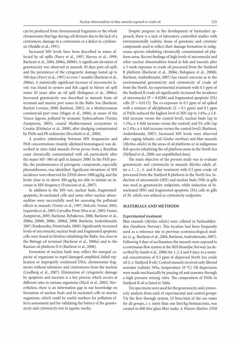
Analysis of crude oil genotoxicity and cytotoxicity end-pointsTh e blind scoring of micronuclei and other nuclear abnor-malities was performed on coded slides without knowledge of the origin of samples. Micronuclei (MN) were identifi ed according to the following criteria: (1) round and ovoid-shaped non-refractory particles in the cytoplasm, (2) colour and structure similar to those of chromatin, (3) a diameter of 1/3–1/20 of the main nucleus, (4) particles completely separated from the main nucleus (Fig. 1). Nuclear buds, bi-nucleated and fragmented-apoptotic cells were identifi ed using the criteria described by M. Fenech with co-authors (2003). Th e morphological features of studied nuclear abnor-malities are shown in Fig. 1. For each specimen of mussels, 2 000 cells with intact cytoplasm were scored. Th e frequency of micronuclei and other nuclear abnormalities was evalu-ated as the number of abnormalities per 1 000 cells scored (Baršienė et al., 2004).
Statistical analysis was carried out using the PRISM sta-tistical package. Th e mean and the standard error were cal-culated for each experimental group. Th e non-parametric Mann–Whitney U-test was used to compare alteration fre-quencies between control and treatment groups. One-way ANOVA and post hoc Tukey’s test were performed to deter-mine whether signifi cant diff erences were present among the treatment groups.
RESULTS
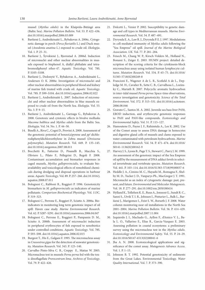
Crude oil genotoxicity (MN and NB)Th e frequency of MN in gills of blue mussels varied from 1.4 MN/1000 cells in the pre-exposure group to 3.4 MN/1000 cells in those aft er an 8-day treatment with Statfj ord B crude oil. Exposure time-dependent elevation of MN incidences was detected in mussels from 1-, 2-, 4- and 8-day treatment groups (Fig. 2). A statistically signifi cant increase of MN val-ues was found in mussels aft er a 1-day (P = 0.0185), 2-day (P = 0.0039), 4-day (P = 0.0068) and 8-day (P < 0.0001) exposure to crude oil. Lower levels of induction of nuclear buds compared to MN incidences were noted, and there were no statistically signifi cant diff erences in nuclear bud fre-quency between pre-exposure and exposure groups. A com-paratively low induction of nuclear buds (NB) was recorded in the 4-day treatment group (Fig. 2). One-way analysis of MN variance (ANOVA) with post hoc Tukey’s test showed P = 0.0033, F = 4.615, whilst the analysis of NB variance showed P = 0.2638, F = 1.357.
Crude oil cytotoxicity (FA and BN)Th e values of fragmented-apoptotic (FA) cells were lower than of nuclear buds. In the pre-exposure group, the level of FA was equal to 0.67 FA/1000 cells. Aft er a 4-day expo-sure to crude oil, the value reached 1.43 FA/1000 cells and aft er a 2-day exposure 1.42 FA/1 000 cells (Fig. 2). Neverthe-less, as was seen in the study results, signifi cant diff erences
Ta b l e . The PAH composition recorded in Statfj ord B crude oil by GC-MS and some characteristics of the PAH molecules
Compound / quantity Statfj ord μg/g oil Mass (M/Z) Log Kow
Naphthalene 1 147.0 128.2 3.3
C1-Naphthalenes 3 787.3 142.2 3.9
C2-Naphthalenes 5 288.7 156.2 4.4
C3-Naphthalenes 3 830.3 170.2 4.9
Acenaphthylene 10.0 152.2 4.1
Acenapthene 9.7 154.2 4.0
Fluorene 135.9 166.2 4.2
Anthracene 252.9 178.2 4.6
Phenanthrene – 178.2 4.6
C1-Phenanthrenes 460.2 192.2 5.1
C2-Phenanthrenes 439.4 206.0 –
Dibenzothiophene 91.9 184.2 4.4
C1-Dibenzothiophene 196.5 198.2 –
C2-Dibenzothiophene 232.9 212.2 –
Fluoranthene 2.6 202.0 5.1
Pyrene 8.6 202.0 5.1
Chrysene 23.9 228.2 5.7
C1-Chrysene 37.9 242.2 –
C2-Chrysene 41.5 256.2 –
Benzo(a)anthracene 3.3 228.2 5.7
Benzo(b)fl uoranthene 7.7 252.3 6.4
Benzo(k)fl uoranthene – 252.3 6.5
Benzo(b + k)fl uoranthene 6.9 – –
Benzo(a)pyrene 4.7 252.3 6.3
Indeno(1,2,3,cd)pyrene – 276.3 6.9
Benzo(g,h,i)perylene 1.7 276.3 7.0
Dibenzo(a,h)anthracene – 278.3 6.7
Sum PAH 16 020.9
Sum PAH in1 ppm dose (μg) 16.0
Note. Log Kow
is the logarithm of the octano-to-water coeffi cient Kow
. LogKow
values on alkylated
PAH are hard to fi nd. A thumb rule is to add from 0.3 to 0.5 log units per methyl group added;
see also http://logkow.cisti.nrc.ca. The 1 ppm oil dosage would equal a quantity of 1 mg
oil/kg seawater.
peristaltic pump with Watson Marlow Maprene 0.76 mm pump tubes was used for the crude oil calibration and intake into the tanks. Th e exposure components were introduced into the infl ow of sea water in each tank. Suitable water cur-rents, component’s infl ow speed and improved water circula-tion were adjusted by a nozzle.
Preparation of slidesA branch of mussel gills was placed in a big drop of 3 : 1 etha-nol acetic acid solution on clean microscopic slide and gently nipped with tweezers for 2–3 min (until cells spread within a drop). Th en the cell suspension was soft ly smeared on a surface of the slide. Dried slides were fi xed in methanol for 10 min and stained with 5% Giemsa solution in phosphate buff er pH = 6.8. Th e stained slides were analyzed under the Olympus BX51 light microscope at the fi nal magnifi cation of 1000×.

127Nuclear abnormalities in blue mussels exposed to crude oil
(P = 0.0115) were found only between mussels from the 4-day oil treatment and the pre-exposure groups. Th e frequency of bi-nucleated (BN) cells ranged from 1.05 BN/1000 cells in the pre-exposure group to 3.02 BN/1000 cells in those aft er an 8-day oil treatment (Fig. 2). Th e BN levels showed the follow-ing trend: pre-exposure < 1-day < 2-day < 4-day oil exposure mussel groups. Th e non-parametric Mann–Whitney U-test showed that BN induction in gills of mussels aft er an 8-day exposure to Statfj ord B oil signifi cantly diff ered from the pre-exposure level (P = 0.0232). Th e one-way ANOVA analysis of BN variance, followed by the Tukey test, yielded P = 0.0266, F = 3.042; analysis of FA cells: P = 0.4636, F = 0.9148.
DISCUSSION
Off shore oil and gas installations are the major contributors to pollution in the North Sea. Water, crude oil, heavy met-als, alkylphenols and other compounds are usually either co-produced or added during extraction processes in plat-forms (Røe, 1999). Our recent analysis of environmental ge-notoxicity within the water column monitoring station near the Statfj ord B oil platform revealed a clear gradient-related MN increase in mussel haemocytes and liver erythrocytes of
Fig. 1. a – micronucleus, b – cell with nuclear bud,
c – fragmented-apoptotic cell, d – bi-nucleated cell in blue mussels gills
Fig. 2. Frequency of micronuclei (MN), nuclear buds (NB), fragmented-apoptotic
(FA) and bi-nucleated (BN) cells in mussel gills exposed to 0.5 ppm of crude oil.
Diff erences between pre-exposure and exposure groups: * P < 0.05, ** P < 0.001,
*** P < 0.0001

128 Janina Baršienė, Laura Andreikėnaitė, Anne Bjornstad
Atlantic cod caged in 2004 for 6 week in the area of the plat-form. Signifi cantly increased MN levels were detected in mus-sels and cod deployed 500 meters from the platform, com-pared to the reference site (Hylland et al., 2008). Nevertheless, there was no signifi cant induction of nuclear buds, bi-nucle-ated and fragmented-apoptotic cells in mussel haemolymph and Atlantic cod liver erythrocytes (our unpublished data). In the Statfj ord B platform area, the concentrations of PAHs and alkylphenols were low compared to the other coastal sites. Only the biomarkers used to monitor impacts such as EROD, DNA adducts, PAH metabolites, etc., lysosomal destabiliza-tion in mussel hepatopancreas and micronuclei induction in haemocytes indicated that caged mussels suff ered from con-taminants in the studied area of the North Sea and thus were validated as sensitive biomarkers to be used to monitor low levels of petroleum contaminants (Hylland et al., 2008).
In the present study, the genotoxicity and cytotoxicity of crude oil processed from the Statfj ord B platform in the North Sea were studied in gills of mussels exposed to 0.5 ppm of oil for 1, 2, 4 and 8 days. Signifi cantly higher MN frequencies were determined in all experimental groups compared to the pre-exposure group of mussels; an 1.9-fold elevation of micronu-clei was found aft er a 1-day, 2.1-fold aft er a 2-day, 2.3-fold aft er a 4-day and 2.4-fold aft er an 8-day exposure. Th erefore, a high potential of crude oil to quickly induce an irreversible DNA damage in mussels was revealed by the MN test. Nevertheless, in a short-term exposure, there was no signifi cant increment of nuclear buds. Data of our previous study had shown that the capacity of mussels to expel a damaged amplifi ed, failed replication or improperly condensed DNA, chromosome frag-ments without telomeres and centromeres from the nucleus appeared aft er a 3-week exposure of mussels to the Statfj ord B crude oil (Baršienė, Andreikėnaitė, 2007).
Investigation of crude oil cytotoxicity revealed a signifi -cant incidence of fragmented-apoptotic cells in mussels aft er a 4-day exposure and bi-nucleated cells in mussel gills aft er an 8-day treatment. Th us, the results presented here confi rm the genotoxicity and cytotoxicity of crude oil, and it is worth noting that formation of damage in bivalve mollusks (except nuclear buds) can appear soon aft er oil spillage in the marine environment.
Th e genotoxicity and cytotoxicity of crude oil from the Statfj ord B platform had earlier been proven in a 3-week treatment of turbot Scophthalmus maximus (Baršienė et al., 2006e; Bolognesi et al., 2006b) and in blue mussels (Baršienė, Andreikėnaitė, 2007). A signifi cant elevation of MN levels aft er a 3-week treatment with 0.5 ppm of dispersed crude oil was found in turbot blood and kidney erythrocytes, to-gether with elevation of nuclear buds and bi-nucleated cells in kidney erythrocytes (Baršienė et al., 2006e). A statistically signifi cant induction of MN and fragmented-apoptotic cells was registered in mussels exposed to 0.5 ppm of crude oil or to spiked 0.5 ppm of the oil. A signifi cant increase of nuclear buds was observed aft er exposure to spiked 0.5 ppm of crude oil (Baršienė, Andreikėnaitė, 2007). Time-related changes of
MN and other nuclear abnormalities were observed in Atlan-tic cod liver erythrocytes aft er 3-, 14- and 24-day treatments with diff erent concentrations of crude oil from the North Sea. Th e induction of MN was highest aft er 14 days and of nuclear buds, fragmented-apoptotic and bi-nucleated cells aft er a 24-day exposure to 0.25 ppm, to 1 ppm, or to 1 ppm spiked oil (our unpublished data).
Crude oil consists of diff erent components such as various hydrocarbons, heavy metals, nitrogen-oxygen compounds. Th e content of components diff ers depending on oil process-ing sites (Wake, 2005). In order to assess the potential genoto-xicity and cytotoxicity of oil from the Minija well (Lithuania), we performed a 10-day exposure of freshwater bivalve Ano-donta anatina and perch Perca fl uviatilis to 0.25 ppm, 0.5 ppm and 1 ppm of crude oil. Nuclear abnormalities were studied in bivalve gills and perch peripheral blood. Th e non-parametric Mann–Whitney U-test revealed the largest diff erences in MN induction among mussels, perch from the control groups and those treated with 0.5 ppm. Treatment with 0.25 ppm crude oil resulted in MN induction only in bivalves. Th e frequency of nuclear buds was not increased in the exposed groups of mussels and fi sh. Exposure of A. anatina and P. fl uviatilis to 1 ppm of Lithuanian crude oil did not increase genotoxicity levels, but signifi cantly elevated the induction of bi-nucleated cells in gills of bivalves and the incidence of fragmented-ap-optotic cells in fi sh blood (Baršienė et al., 2006c). A species-specifi c pattern in genotoxicity response to crude oil expo-sure was shown in bivalve mollusks (Hamoutene et al., 2002) and in fi sh (Baršienė et al., 2006c). DNA damage in mussel Mytilus galloprovincialis signifi cantly increased aft er a 12-day exposure to Arabian light crude oil, but did not induce damage in clams Mya arenaria (Hamoutene et al., 2002). Th ese studies demonstrated a potential environmental geno-toxicity resulting from diff erent oil spills as well as pointed to interspecies sensitivity.
In the crude oil processed from the Statfj ord B platform, polycyclic aromatic hydrocarbons (PAH) constitute about 1.5% of the total weight. Naphthalene and its derivatives prevailed in the crude oil and showed the highest bioavail-ability in fi sh (Sundt et al., 2006). A time-dependent ability of naphthalene and benzo[a]pyrene (concentrations 0.1, 0.3, 0.9, or 2.7 μM for both compounds) to induce micronuclei in mature erythrocytes aft er a short-term exposure (2, 4, 6 and 8 h) has been recorded in juvenile Dicentrarchus labrax fi sh. A signifi cant induction of MN and other nuclear abnor-malities was observed aft er 4-h of treatment with 0.3, 0.9 and 2.7 μM, upon a 6-h exposure to 0.9 and 2.7 μM, an 8-h expo-sure to 0.3, 0.9 and 2.7 μM of naphthalene (Gravato, Santos, 2002). Signifi cantly increased levels of nuclear abnormalities were described in D. labrax erythrocytes aft er 2-h of expo-sure to 0.1, 0.9, and 2.7 μM benzo[a]pyrene (B[a]P), aft er 4-h of exposure to 0.1 and 0.9 μM. Interestingly, 6-h and 8-h ex-posures to low B[a]P concentrations (0.1 and 0.3 μM) caused the highest increase of nuclear abnormalities as compared to control (Gravato, Santos, 2002).

129Nuclear abnormalities in blue mussels exposed to crude oil
In eel Anguilla anguilla, treatment with 0.3, 0.9 and 2.7 μM of naphthalene induced micronuclei and other nu-clear abnormalities (Teles et al., 2003). An increased fre-quency of MN was observed in mussels aft er 15-day expo-sure to 0.1 μg/L of phenanthrene (Koukouzika, Dmitriadis, 2008), in zebra mussel Dreissena polymorpha aft er 2-, 3- and 4-day treatments with diff erent concentrations (2 μg/L and 10 μg/L) of B[a]P (Binelli et al., 2008).
Benzo[a]pyrene was classifi ed as a progenotoxin which aft er metabolic activation becomes a very aggressive DNA-damaging agent (Johnson, 1992). Genotoxic eff ects of B[a]P and dimethylbenz[a]antracene had been demonstrated earli-er in gills and hemolymph of marine molluscs (Burgeot et al., 1995; Bolognesi et al., 1996; Venier et al., 1997; Siu et al., 2004). Comet and MN assays have indicated clear dose- and time-dependent responses to B[a]P exposure in Mytilidae bi-valve Perna viridis (Siu et al., 2004). DNA strand breaks were signifi cantly diff erent from control levels from 1- to 6-day treatment and then a gradual decrease to control levels upon a 20-day exposure in scallops Chlamys farreri treated with 0.5 and 3 μg/L B[a]P (Pan et al., 2008). Data were reported on the genotoxicity of 10 polycyclic aromatic hydrocarbons (anthra-cene, benz[a]anthracene, 7,12-dimethylbenz[a]anthracene, dibenz[a,h]anthracene, dibenz[a,c]anthracene, 3-methyl-cholanthrene, benzo[a]pyrene, benzo[e]pyrene, chrysene and pyrene) in mice skin cells, and it was pointed out that the genotoxicity of these compounds in general correlated with their carcinogenicity (Nishikawa et al., 2005).
Th e extensive development of off shore oil industry in the North Sea and arctic regions requires an integrated system of well-developed methods of estimating the ecological risk of oil contamination in marine ecosystems. However, there is a lack of validated tests, simple, cost-eff ective screening me-thods, which could be applied eff ectively, taking into consider-ation environmentally realistic routes of contamination in oil platform areas. Th e present study results have demonstrated the genotoxic and cytotoxic potentials of the North Sea crude oil in mussels, a cosmopolitan and ecologically relevant or-ganism. Validation of the micronuclei test and the approval of other nuclear abnormalities as biomarkers of environmental genotoxicity and cytotoxicity were performed using diff erent marine fi sh and mussel species in laboratory treatments with crude oil from diff erent platforms, with a mixture of alkyl-phenols, PAHs, wastewater, fl ame retardants or endocrine dis-ruptor concentrations. Contaminant-species-tissue-exposure time-specifi c responses have been described (Baršienė et al., 2005, 2006a, 2006c, 2006d; Andreikėnaitė et al., 2007; Baršienė, Andreikėnaitė, 2007). Th us, the developed approach and the collected information should also help to fi ll gaps in our understanding of the ecological signifi cance of crude oil pollution in marine ecosystems. Th e endpoints used in our study may serve as early warning signs off ering an evidence of genotoxicity risks in wildlife species and for describing the cytological mechanisms of crude oil toxicity. Th is biomarker system could be successfully used for the assessment of back-
ground levels before off shore oil installations, as well as with-in the frames of environmental quality monitoring. Labora-tory-controlled experiments, active monitoring approaches, and in situ assessment of DNA damage in various tissues, especially in gonads of target species, will help to achieve a substantial progress in the assessment of early responses as well as short- or long-term adaptations to chronic pollution originating from petroleum installations.
ACKNOWLEDGEMENTS
Th is work was supported by the European Commission (Re-search Directorate General, Environment Program – Marine Ecosystems) through the project “Biological Eff ects of Envi-ronmental Pollution in Marine Coastal Ecosystems” (contract EVK3-CT2000-00025). We are grateful to Odd Ketil Andersen for helpful scientifi c interactions.
Received 10 June 2010Accepted 17 November 2010
References
1. Andreikėnaitė L., Baršienė J., Vosylienė M. Z. 2007. Studies of micronuclei and other nuclear abnormalities in blood of rainbow trout (Oncorhynchus mykiss) treated with heavy metal mixture and road maintenance salts. Acta Zoologica Lituanica. Vol. 17. P. 213–219.
2. Baršienė J., Baršytė Lovejoy D. 2000. Environmental ge-notoxicity in Klaipėda port area. International Review of Hydrobiology. Vol. 85. P. 663–672.
3. Baršienė J. 2002. Genotoxic impacts in Klaipėda marine Port and Būtingė oil terminal areas (Baltic Sea). Marine Environmental Research. Vol. 54. P. 475–479. doi:10.1016/S0141-1136(02)00160-5
4. Baršienė J., Lazutka J., Šyvokienė J., Dedonytė V., Ryba ko-vas A., Bjornstad A., Andersen O. K. 2004. Analysis of mi-cronuclei in blue mussels and fi sh from the Baltic and the North Seas. Environmental Toxicology. Vol. 19. P. 365–371. doi:10.1002/tox.20031
5. Baršienė J., Dedonytė V., Rybakovas A., Broeg K., Forlin L., Gercken J., Kopecka J., Balk L. 2005. Environmental muta-genesis in diff erent zones of the Baltic Sea. Acta Zoologica Lituanica. Vol. 15. P. 90–95.
6. Baršienė J., Rybakovas A. 2006. Cytogenetic and cytotoxic eff ects in gill cells of the blue mussel Mytilus edulis from the Baltic coast and aft er 1–3-day maintenance in laboratory. Acta Zoologica Lituanica. Vol. 16. P. 191–197.
7. Baršienė J., Schiedek D., Rybakovas A., Šyvokienė J., Ko-pecka J., Forlin L. 2006a. Cytogenetic and cytotoxic eff ects in gill cells of the blue mussel Mytilus spp. from diff erent zones of the Baltic Sea. Marine Pollution Bulletin. Vol. 53. P. 469–478. doi:10.1016/j.marpolbul.2005.11.015
8. Baršienė J., Lehtonen K., Koehler A., Broeg K., Vourinen P. J., Lang T., Pempkowiak J., Šyvokienė J., Dedonytė V., Ryba-kovas A., Repečka R., Vountisjarvi H., Kopecka J. 2006b. Biomarker responses in fl ounder (Platichthys fl esus) and
130 Janina Baršienė, Laura Andreikėnaitė, Anne Bjornstad
mussel (Mytilus edulis) in the Klaipėda–Būtingė area (Baltic Sea). Marine Pollution Bulletin. Vol. 53. P. 422–436. doi:10.1016/j.marpolbul.2006.03.009
9. Baršienė J., Andreikėnaitė L., Rybakovas A. 2006c. Cyto ge-netic damage in perch (Perca fl uviatilis L.) and Duck mus-sel (Anodonta anatina L.) exposed to crude oil. Ekologija. Vol. 1. P. 25–31.
10. Baršienė J., Šyvokienė J., Bjornstad A. 2006d. Induction of micronuclei and other nuclear abnormalities in mus-sels exposed to bisphenol A, diallyl phthalate and tetra-bromodiphenyl ether-47. Aquatic Toxicology. Vol. 78S. P. S105–S108.
11. Baršienė J., Dedonytė V., Rybakovas A., Andreikėnaitė L., Andersen O. K. 2006e. Investigation of micronuclei and other nuclear abnormalities in peripheral blood and kidney of marine fi sh treated with crude oil. Aquatic Toxicology. Vol. 78S. P. S99–S104. doi:10.1016/j.aquatox.2006.02.022
12. Baršienė J., Andreikėnaitė L. 2007. Induction of micronu-clei and other nuclear abnormalities in blue mussels ex-posed to crude oil from the North Sea. Ekologija. Vol. 53. No. 3. P. 9–15.
13. Baršienė J., Andreikėnaitė L., Garnaga G., Rybakovas A. 2008. Genotoxic and cytotoxic eff ects in bivalve mollusks Macoma balthica and Mytilus edulis from the Baltic Sea. Ekologija. Vol. 54. No. 1. P. 44–50.
14. Binelli A., Riva C., Cogni D., Provini A. 2008. Assessment of the genotoxic potential of benzo(a)pyrene and pp’-dichlo-rodiphenyldichloroethylene in Zebra mussel (Dreissena polymorpha). Mutation Research. Vol. 649. P. 135–145. doi:10.1016/j.mrgentox.2007.08.011
15. Bocchetti R., Fattorini D., Pisanelli B., Macchia S., Oliviero L., Pilato F., Pellegrini D., Regoli F. 2008. Contaminant accumulation and biomarker responses in caged mussels, Mytilus galloprovincialis, to evaluate bio-availability and toxicological eff ects of remobilized chemi-cals during dredging and disposal operations in harbour areas. Aquatic Toxicology. Vol. 89. P. 257–266. doi:10.1016/j.aquatox.2008.07.011
16. Bolognesi C., Rabboni R., Roggieri P. 1996. Genotoxicity biomarkers in M. galloprovincialis as indicators of marine pollutants. Comparison Biochemical Physiology. Vol. 113C. P. 319–323.
17. Bolognesi C., Perrone E., Roggieri P., Sciutto A. 2006a. Bio-indicators in monitoring long term genotoxic impact of oil spill: Haven case study. Marine Environmental Research. Vol. 62. P. S287–S291. doi:10.1016/j.marenvres.2006.04.047
18. Bolognesi C., Perrone E., Roggieri P., Pampanin D. M., Sciutto A. 2006b. Assessment of micronuclei induction in peripheral erythrocytes of fi sh exposed to xenobiotics under controlled conditions. Aquatic Toxicology. Vol. 78S. P. S93–S98. doi:10.1016/j.aquatox.2006.02.015
19. Burgeot T., His E., Galgani F. 1995. Th e micronucleus assay in Crassostrea gigas for the detection of seawater genotoxic-ity. Mutation Research. Vol. 343. P. 125–140.
20. Carvalho Pinto-Silva C. R., Creppy E., Matias W. 2005. Micronucleus test in mussels Perna perna fed with the tox-ic dinofl agellate Prorocentrum lima. Archives of Toxicology. Vol. 79. P. 422–426.
21. Dolcetti L., Venier P. 2002. Susceptibility to genetic dam-age and cell types in Mediterranean mussels. Marine Envi-ronmental Research. Vol. 54. P. 487–491.
22. Dyrynda E. A., Law R. J., Dyrynda P. E. J. 1997. Modulations in cell-mediated immunity of Mytilus edulis following the “Sea Empress” oil spill. Journal of the Marine Biological Association. UK. Vol. 77. P. 281–284.
23. Fenech M., Chang W. P., Kirsch-Volders M., Holland N., Bonassi S., Zeiger E. 2003. HUMN project: detailed de-scription of the scoring criteria for the cytokinesis-block micronucleus assay using isolated human lymphocyte cul-tures. Mutation Research. Vol. 534. P. 65–75. doi:10.1016/S1383-5718(02)00249-8
24. Francioni E., Wagener A de L. R., Scofi eld A de L., Dep-ledge M. H., Cavalier B., Sette C. B., Carvalhosa L., Lozins-ky C., Mariath R. 2007. Polycyclic aromatic hydrocarbon in inter-tidal mussel Perna perna: Space-time observations, source investigation and genotoxicity. Science of the Total Environment. Vol. 372. P. 515–531. doi:10.1016/j.scitotenv.2006.08.046
25. Gravato C., Santos M. A. 2002. Juvenile sea bass liver P450, EROD induction, and erythrocytic genotoxic responses to PAH and PAH-like compounds. Ecotoxicology and Environmental Safety. Vol. 51. P. 115–127.
26. Hamoutene D., Payne J. F., Rahimtula A., Lee K. 2002. Use of the Comet assay to assess DNA damage in hemocytes and digestive gland cells of mussels and clams exposed to water contaminated with petroleum hydrocarbons. Marine Environmental Research. Vol. 54. P. 471–474. doi:10.1016/S0141-1136(02)00162-9
27. Harvey J. S., Lyons B., Page T. S., Stewart C., Parry J. M. 1999. An assessment of the genotoxic impact of the Sea Empress oil spill by the measurement of DNA adduct levels in select-ed invertebrate and vertebrate species. Mutation Research. Vol. 441. P. 103–114. doi:10.1016/S1383-5718(99)00037-6
28. Heddle J. A., Cimino M. C., Hayashi M., Romagna F., Shel-by M. D., Tucker J. D., Vanparys Ph., MacGregor J. T. 1991. Micronuclei as an index of cytogenetic damage: past, pre-sent, and future. Environmental and Molecular Mutagenesis. Vol. 18. P. 277–291. doi:10.1002/em.2850180414
29. Hylland K., Tollefsen K. E., Ruus A., Jonsson G., Sundt R. C., Sanni S., Utvik T. I. R., Johnsen I., Pinturier L., Balk L., Bar-šienė J., Marigomez I., Feist S. W., Borseth J. F. 2008. Water column monitoring near oil installations in the North Sea 2001–2004. Marine Pollution Bulletin. Vol. 56. P. 414–429. doi:10.1016/j.marpolbul.2007.11.004
30. Izquierdo J. I., Machado G., Ayllon F., d’Amico V. L., Ba-la L. O., Vallarino E., Elias R., Garcia-Vazquez E. 2003. Assessing pollution in coastal ecosystems: a preliminary survey using the micronucleus test in the Mytilus edulis. Ecotoxicology and Environmental Safety. Vol. 55. P. 24–29. doi:10.1016/S0147-6513(02)00041-6
31. Jha A. N. 2008. Ecotoxicological applications and sig-nifi cance of the comet assay. Mutagenesis Advance Access. Vol. 1. P. 1–15.
32. Johnson B. T. 1992. Potential genotoxicity of sediments from the Great Lakes. Environmental Toxicology. Water Quality International. Vol. 7. P. 373–390.

131Nuclear abnormalities in blue mussels exposed to crude oil
33. Klobučar G. I. V., Štambuk A., Hylland K., Pavlica M. 2008. Detection of DNA damage in haemocytes of Mytilus gallo-provincialis in the coastal ecosystems of Kaštela and Trogir bays, Croatia. Sciences of the Total Environment. Vol. 405. P. 330–337. doi:10.1016/j.scitotenv.2008.05.015
34. Koukouzika N., Dimitriadis V. K. 2008. Aspects of the use-fulness of fi ve marine pollution biomarkers, with emphasis on MN and lipid content. Marine Pollution Bulletin. Vol. 56. P. 941–949. doi:10.1016/j.marpolbul.2008.01.043
35. Lindberg H. K., Wang X., Järventaus H., Falck G. C. M., Norppa H., Fenech M. 2007. Origin of nuclear buds and micronuclei in normal and folate-deprived human lympho-cytes. Mutation Research. Vol. 617. P. 33–45. doi:10.1016/j.mrfmmm.2006.12.002
36. Magni P., de Falco G., Falugi C., Franzoni M., Monteverde M., Perrone E., Sigro M., Bolognesi C. 2006. Genotoxicity biomarkers and acetylcholinesterase activity in natural populations of Mytilus galloprovincialis along a pollution gradient in the Gulf of Oristano (Sardinia, west-ern Mediterranean). Environmental Pollution. Vol. 142. P. 65–72. doi:10.1016/j.envpol.2005.09.018
37. Mičic M., Bihari N., Jaksic Z., Muller E. G., Batel R. 2002. DNA damage and apoptosis in the mussel Mytilus gallopro-vincialis. Marine Environmental Research. Vol. 53. P. 243–262.
38. Nishikawa T., Nakamura T., Fukushima A., Takagi Y. 2005. Further evaluation of the skin micronucleus test: Results obtained using 10 polycyclic aromatic hydrocar-bons. Mutation Research. Vol. 588. P. 56–63. doi:10.1016/j.mrgentox.2005.09.004
39. Ohe T., Watanabe T., Wakabayashi K. 2004. Mutagens in surface waters: a review. Mutation Research. Vol. 567. P. 109–149. doi:10.1016/j.mrrev.2004.08.003
40. Pan L., Miao J., Wang J., Liu J. 2008. AHH activity, tissue dose and DNA damage in diff erent tissues of the scallop Chlamys farreri exposed to benzo[a]pyrene. Environmental Pollution. Vol. 153. P. 192–198. doi:10.1016/j.envpol.2007.07.022
41. Parry J. M., Harvey J. S., Lyons B. P. 1997. Th e application of genetic toxicology in the analysis of the consequences of a major marine pollution incident. Mutation Research. Vol. 379. Issue 1. P. S91.
42. Røe T. U. 1999. Chemical characterisation of produced water from four off shore oil production platforms in the North Sea. Chemosphere. Vol. 39. P. 2593–2606.
43. Siu W. H. L., Cao J., Jack R. W., Wu R. S. S., Richardson B. J., Xu L., Lam P. K. S. 2004. Application of the comet and mi-cronucleus assays to the detection of B[a]P genotoxicity in haemocytes of the green-lipped mussel (Perna viridis). Aquatic Toxicology. Vol. 66. P. 381–392. doi:10.1016/j.aquatox.2003.10.006
44. Sundt R. C., Pampanin D. M., Larsen B. K., Brede C., Herz-ke D., Bjornstad A., Andersen O. K. 2006. Th e BEEP Stavan-ger workshop: mesocosm exposures. Aquatic Toxicology. Vol. 78S. P. S5–S12. doi:10.1016/j.aquatox.2006.02.012
45. Teles M., Pacheco M., Santos M. A. 2003. Anguilla anguil-la L. liver ethoxyresorufi n O-deethylation, Glutathione S-transferase, erythrocytic nuclear abnormalities, and en-docrine responses to naphthalene and β-naphthofl avone.
Ecotoxicology and Environmental Safety. Vol. 55. P. 98–107. doi:10.1016/S0147-6513(02)00134-3
46. Venier P., Maron S., Canova S. 1997. Detection of micro-nuclei in gill cells and haemocytes of mussels exposed to benzo[a]pyrene. Mutation Research. Vol. 390. P. 33–44.
47. Venier P., Zampieron C. 2005. Evidence of genetic da-mage in grass gobies and mussels from the Venice la-goon. Environment International. Vol. 31. P. 1053–1064. doi:10.1016/j.envint.2005.05.016
48. Wake H. 2005. Oil refi neries: a review of their ecological impacts on the aquatic environment. Estuarine, Coastal and Shelf Science. Vol. 62. P. 131–140. doi:10.1016/j.ecss.2004.08.013
49. Woodhead R. J., Law R. J., Matthiessen P. 1999. Polycyclic aromatic hydrocarbons in surface sediments around England and Wales, and their possible biological signifi -cance. Marine Pollution Bulletin. Vol. 9. P. 773–790.
Janina Baršienė, Laura Andreikėnaitė, Anne Bjornstad
BRANDUOLIO PAŽAIDŲ SUSIFORMAVIMAS MIDIJŲ MYTILUS EDULIS ŽIAUNŲ LĄSTELĖSE PO 1, 2, 4 IR 8 PARŲ POVEIKIO ŠIAURĖS JŪROJE IŠGAUNAMA ŽALIAVINE NAFTA
S a n t r a u k aGenotoksinis ir citotoksinis Statfj ord B naft os platformos (Šiau-rės jūra) žaliavinės naft os poveikis laike įvertintas midijų Mytilus edulis žiaunų ląstelėse. Moliuskai 1, 2, 4 ir 8 paras veikti 0,5 ppm Statfj ord B gręžinio naft a. Genotoksinis teršalų poveikis buvo ver-tinamas pagal mikrobranduolių (MB) ir branduolio pumpurų (BP) susiformavimą, o citotoksinis – pagal dvibranduolių (DB) ir fragmentuotų-apoptozinių (FA) ląstelių dažnį. Po 1, 2, 4 ir 8 parų ekspozicijos nustatytas laipsniškas, atsižvelgus į poveikio trukmę, MB ir DB parametrų didėjimas: kontrolinė gr.< po 1 paros < po 2 parų < po 4 parų < po 8 parų. Po 8 parų nustatytas MB kiekis buvo 2,4 karto, FA – 2 kartus, o DB – 3 kartus didesnis nei kontrolėje. Statistiškai patikimi MB dažnių skirtumai rasti tarp kontrolinės bei pirmos (P = 0,0185), antros (P = 0,0039), ketvirtos (P = 0,0068) ir aštuntos paros (P < 0,0001) M. edulis grupių. Statistiškai patikimų branduolio pumpurų skirtumų nerasta, tuo tarpu po 4 parų nusta-tyta statistiškai patikima FA ląstelių indukcija (P = 0,0115), o po 8 parų ekspozicijos – DB (P = 0,0232).
Raktažodžiai: mikrobranduoliai, branduolio pažaidos, žaliavi-nė naft a, midijos