
1 genesis 46: 104-111 Induction of apoptosis by Drosophila Myc ...
Induction of DNA Synthesis and Apoptosis in Cardiac Myocytes by E1A Oncoprotein Yan Liu and Richard N. Kitsis Departments of Medicine (Cardiology) and Cell Biology, Albert Einstein College of Medicine, Bronx, New York 10461
Abstract. Beginning during the second half of gesta- tion, increasing numbers of cardiac myocytes withdraw from the cell cycle such that DNA synthesis is no longer detectable in these cells by neonatal day 17 in vivo. The mechanisms that exclude these and other terminally differentiated cells from the cell division cycle are poorly understood. To begin to explore the molecular basis of the barrier to G1/S progression in cardiac myo- cytes, we used adenoviruses to express wild-type and mutant E1A proteins in primary cultures from embry- onic day 20 rats. While most of these cardiac myocytes are ordinarily refractory to DNA synthesis, even in the presence of serum growth factors, expression of wild-type E1A stimulates DNA synthesis in up to 94% or almost all successfully transduced cells. Rather than complete the cell cycle, however, these cells undergo apoptosis. Apoptosis is limited to those cells that engage in DNA
synthesis, and the kinetics of the two processes suggest that DNA synthesis precedes apoptopsis. Mutations in E1A that disable it from binding Rb and related pocket proteins have little effect on its ability to stimulate DNA synthesis in cardiac myocytes. In contrast, mu- tants that are defective in binding the cellular protein p300 stimulate DNA synthesis 2.4-4.1-fold less effi- ciently, even in the context of retained E1A pocket protein binding. In the absence of EIA pocket protein binding, the usual situation in the cell, loss of p300 binding severely decreases the ability of E1A to stimu- late DNA synthesis. These results suggest that the bar- rier to G1/S progression in cardiac myocytes is medi- ated, at least in part, by the same molecules that gate the G1/S transition in actively cycling cells, and that p300 or related family members play an important role in this process.
M OST adult mammalian cells exist in a quiescent state (GO), characterized by the absence of net DNA synthesis and cell division. While some
GO ceils retain the ability to resume proliferative growth in response to certain signals, others that have undergone terminal differentiation are permanently excluded from the cell cycle. The mechanisms that prevent cell cycle re- entry in terminally differentiated cells are unclear. Striated muscle cells, including skeletal and cardiac myocytes, ex- emplify the terminally differentiated state. In skeletal muscle, terminal differentiation involves three distinct processes: commitment, in which proliferating myoblasts withdraw irreversibly from the cell cycle; fusion of com- mited myoblasts into multinucleated myocytes; and induc- tion of muscle specific genes (Nguyen et al., 1983). MyoD, myogenin, myf-5, and MRF 4 comprise a family of skeletal myogenic determination factors (MDFs), 1 whose members
Please address all correspondence to Richard N. Kitsis, Departments of Medicine (Cardiology) and Cell Biology, Albert Einstein College of Medicine, 1300 Morris Park Avenue, Bronx, NY. Tel.: (718) 430-2609; Fax: (718) 430-8991. e-mail: [email protected]
1. Abbreviat ions used in this paper: BrdU, 5-bromo-2'-deoxy-uridine; CR, conserved region; MDF, myogenic determination factor; MLC2v, ventric- ular myosin light chain 2; TUNEL, terminal deoxynucleotidyl transferase nick end labeling.
are sufficient to accomplish all three of these steps, as evi- denced by their ability to convert a variety of nonmuscle cells to skeletal myocytes (reviewed in Weintraub, 1993). MDFs are basic helix-loop-helix transcription factors that transactivate the expression of multiple muscle-specific genes. In addition, these proteins exhibit intrinsic growth suppressive properties in both myogenic and nonmyogenic cells (Sorrentino et al., 1990; Crescenzi et al., 1990). MDF- induced growth inhibition requires member(s) of the ret- inoblastoma (Rb) family of pocket proteins (Gu et al., 1993), which also include p107 and p130. Although MDFs bind directly to pocket proteins in vivo (Gu et al., 1993; Schneider et al., 1994), the precise mechanism by which they suppress growth remains unclear, and recent work suggests that these effects may be indirect (Halevy et al., 1995; Skapek et al., 1995; Parker et al., 1995).
While both skeletal and cardiac myocytes~are special- ized contractile cells, they differ fundamentally from a de- velopmental perspective. Skeletal myoblasts .~rise from the somites around midgestation (Holtzer et al., 1957); in contrast, cardiac myoblasts originate earlier and from the lateral plate mesoderm (Rawles, 1943; reviewed in Litvin et al., 1992). Furthermore, in contrast to skeletal myogene- sis, where cell cycle withdrawal precedes fusion and the expression of muscle-specific genes (Nadal-Ginard, 1978), fully functioning cardiac myocytes develop from their pre-
© The Rockefeller University Press, 0021-9525/96/041325/10 $2.00 The Journal of Cell Biology, Volume 133, Number 2, April 1996 325-334 325
Dow
nloaded from http://rupress.org/jcb/article-pdf/133/2/325/1265722/325.pdf by guest on 24 N
ovember 2021
cursors in the absence of cell cycle withdrawal or cell fusion (Goldstein et al., 1974). As ontogeny progresses, however, an increasing proportion of cardiac myocytes withdraw from the cell cycle so that, by neonatal days 15-17, DNA synthesis is not detectable in cardiac myocytes in vivo (Claycomb, 1975; Rumyantsev, 1977; Kim et al., 1994). Forced expression of c-myc (Jackson et al., 1990) and SV-40 T antigen (Field, 1988; Behringer et al., 1988; Katz et al., 1992) in transgenic hearts beginning early in fetal life can induce cardiac myocyte hyperplasia and/or tumorigenesis; however, these genetic perturbations by themselves are in- sufficient to allow the direct establishment of permanent lines of differentiated cardiac myocytes (Jackson et al., 1990; Steinhelper et al., 1990). The molecules, which induce and maintain terminally differentiation in cardiac myocytes, have not been identified; specifically, the skeletal MDFs are absent (reviewed in Olson, 1993).
To begin to understand the molecular basis of terminal differentiation in cardiac myocytes, we have exploited two properties of adenoviruses. First, these viruses are able to infect primary cultures of cardiac myocytes with high effi- ciency (Kirshenbaum et al., 1993; Kass-Eisler et al., 1993). Second, adenoviruses express an endogenous oncogene, E1A, which facilitates G1 to S progression in nontermi- nally differentiated cells (Howe et al., 1990) and whose structure-function relationships have been particularly well delineated (reviewed in Bayley and Mymryk, 1994). We reasoned that if E1A were able to surmount the G1/S block in cardiac myocytes, it might be possible to explore the molecular nature of this barrier by assaying the ability of various E1A mutants to stimulate DNA synthesis in cardiac myocytes. Although the products of the EI~ . . . . stimulate DNA synthesis in nonterminally differentiated cells, their ability to transform these cells is quite limited because they also induce apoptosis or programmed cell death through a p53-dependent mechanism (Debbas and White, 1993; Lowe and Ruley, 1993). E1A-induced apop- tosis can be inhibited by the products of the adenovirus E1B gene and the cellular bcl-2 gene (Rao et al., 1992; re- viewed in White, 1993). Both the ability of E1A to induce cellular DNA synthesis and apoptosis map to residues at the extreme amino terminus and in conserved regions (CR) 1 (amino acids 40-80) and 2 (amino acids 120-138). These E1A functions are thought to be mediated through its interactions with several host proteins. These include p300, a transcriptional adaptor molecule (reviewed in Mo- ran, 1993; Eckner, et al., 1994), which requires residues at the extreme amino terminus of E1A and the carboxy end of CR1 for full binding, as well as pocket proteins whose binding maps to residues in CR2 and the amino portion of CR1 (Wang et al., 1993). E1A mutants defective for p300 binding alone are modestly impaired in their abilities to stimulate cellular DNA synthesis in cycling cells while those unable to bind pocket proteins alone perform this function normally. Mutants defective for binding both of these classes of proteins, however, are completely crippled in this regard (Howe et al., 1990). Expression of E1A in skeletal myoblasts inhibits the activation of muscle-spe- cific genes and differentiation of these cells (Webster et al., 1988; Enkemann et al., 1990; Braun et al., 1992; Mym- ryk et al., 1992; Caruso et al., 1993; Taylor et al., 1993). The effect of E1A expression in striated muscle cells that
have already undergone terminal differentiation is not known, however.
In this study, we demonstrate that expression of E1A is sufficient to stimulate DNA synthesis in embryonic day 20 cardiac myocytes, most of which are ordinarily refractory to this process, even in serum-containing media. Rather than undergoing mitosis, however, these same cells un- dergo apoptosis. Finally, mutations disabling E1A from binding p300--both in the presence and absence of pocket protein binding--decrease the ability of this oncoprotein to stimulate DNA synthesis in these cells. These results suggest that p300 and/or related family members contrib- ute to the regulation of DNA synthesis in cardiac myo- cytes.
Materials and Methods
Primary Rat Cardiac Myocyte Cultures
Cultures of cardiac myocytes were prepared from the hearts of embryonic day 20 Sprague-Dawley fetuses (Taconic Farms, Germantown, NY) as de- scribed (Kass-Eisler et al., 1993). After enzymatic dissociation, cells were pre-plated for 1 h to enrich for myocytes (90-95% of cells after this step). Cells were then plated at a density of 700/mm 2 onto 35- or 60-mm tissue culture dishes (Primaria, Falcon; Becton Dickinson & Co., Lincoln Park, N J) and cultured in media consisting of Hanks' salts plus MEM vitamin stock, MEM amino acids, MEM nonessential amino acids, 2 mM L-glu- tamine, 0.67 mM glycine, 0.92 mM hypoxanthine (all from GIBCO BRL, Gaithersburg, MD), 19.6 mM NaHCO3 (pH 7.1-7.2), penicillin, strepto- mycin, and 10% (vol/vol) FBS (Hyclone Laboratories, Logan, UT) at 37°C, 5% CO2. 12 h after plating, cells were washed twice with media be- fore the addition of fresh media.
Adenoviruses Mutant human type 5 adenoviruses, derived from dl309 (Jones and Schenk, 1979), which lacks a functional E3 gene, but is phenotypically normal in cultured cells, were used throughout these experiments, dl309/ E1B- (kindly provided by Dr. Eileen White, Center for Advanced Bio- technology and Medicine, Piscataway, NJ) contains the wild-type E1A gene, which is transcribed into alternatively spliced 12S and 13S mRNAs encoding 243- and 289-residue proteins, respectively, but lacks a func- tional EIB gene. d1520/E1B- and its derivatives (generous gifts from Dr. Stanley T. Bayley, McMaster University, Hamilton, Ontario, Canada) contain an E1A gene in which the 5' splice site needed to generate the 13S transcript has been mutated, resulting in the production of only the 12S transcript and the 243-amino acid protein; these viruses also lack a func- tional EIB gene. Derivatives of d1520EIB- containing additional muta- tions in the E1A gene include dlllO1/520EIB- (deletion of residues 14-25), dlllO8/520EIB- (deletion of residues 124-127), and dlO1/O8/520E1B- (with both of these deletions) (Howe et al., 1990). dl312 lacks functional E1 and E3 genes (Jones and Shenk, 1979). Viruses were grown up on 293 cells, purified on CsCI gradients, and then titered by plaque assay on 293 cells (Jones and Shenk, 1979).
Infection Ceils were infected 36 h after plating at a multiplicity of infection (MOI) of 10 or 20 plaque-forming units (pfu) per cell as specified in a volume of 1 ml for 60-mm plates and 0.5 ml for 35-mm plates of serum-free media and incubated at 37°C, 5% CO2. Plates were gently swirled every 15 min. After 1.5 h, serum-containing media was added to plates so that the final serum concentration was 10%. Infection efficiency of myocyte cultures was de- termined by double immunofluorescence for E1A (Ab-1 from Oncogene Science [Uniondale, NY]; used at 1:20 dilution) and ventricular myosin light chain 2 (MLC2v; lwaki et al., 1990), which was generously provided by Dr. Ken Chien (University of California at San Diego) and used at 1:20 dilution or desmin (Sigma Chemical Co., St. Louis, MO; used at 1:10 dilu- tion). Alternatively, staining was performed with a polyclonal adenoviral antibody (gift from Dr. Erik Falck-Pedersen, Cornell University Medical College, New York; used at 1:1,000 dilution). Steady-state E1A levels
The Journal of Cell Biology, Volume 133, 1996 326
Dow
nloaded from http://rupress.org/jcb/article-pdf/133/2/325/1265722/325.pdf by guest on 24 N
ovember 2021
were assessed by fractionating cellular lysates on 10% SDS-polyacryl- amide gels, transferring to nitrocellulose membranes, and immunoblotting using the E1A antibody at a 1:200 dilution. Bands were detected by en- hanced chemiluminescence (Amersham Corp., Arlington Heights, IL) and quantified by laser densitometry of autoradiograms exposed in the linear range (Molecular Dynamics, Sunnyvale, CA).
5-Bromo-2 " Deoxy- Uridine Incorporation
Nuclear 5-bromo-2'-deoxy-uridine (BrdU) incorporation and cytoplasmic muscle-specific antigens were simultaneously visualized using double indi- rect immunofluorescence. Cultures were pulsed with BrdU (Amersham) at a final concentration of 20 p~M for the time period indicated. At the end of the pulse, cells on coverslips were fixed in 3.7% (vol/vol) formaldehyde for 10 min at room temperature, permeabilized with 70% ethanol for 30 min at -20°C, and incubated with 10% normal goat serum followed by a mouse monoclonal anti-BrdU antibody (undiluted; Amersham) and the rabbit polyclonal antibody against either MLC2v or desmin as noted above. Primary antibodies were then detected with rhodamine-conjugated goat anti-mouse IgG and FITC-conjugated goat anti-rabbit | gG (both used at 1:40; The Jackson Laboratory, Bar Harbor, ME), mounted with Flu-mount (Fisher Scientific, Pittsburgh, PA), and examined using an MRC 600 confocal microscope (Bio Rad, Hercules, CA) with a Kr/Ar laser.
Nucleosomal Ladder Assay
At the times indicated after infection, cells were harvested by scraping into the media. After centrifugation at 500 g for 5 min at 4°C, cells were lysed in 10 mM Tris, pH 8.0, 5 mM EDTA, 100 mM NaCl, 0.5% (vol/vol) SDS, and 1 }xg/ml proteinase K (Sigma Chemical Co.) at 37°C for 5 h on an or- bital rocker followed by the dropwise addition of 4 M NaC1 to a final con- centration of 1 M and incubation at 4°C overnight (Mymryk et al., 1994). After centrifugation at 12,000 g for 30 min at 4°C, the supernatants were recovered, extracted with an equal volume of 25:24:1 phenol (Tris-satu- rated, pH 8)/chloroform/isoamyl alcohol, and the combination of high mo- lecular weight and fragmented DNAs were isopropanol precipitated, washed in 70% ethanol, resuspended in water, digested with 100 p~g/ml RNase A for 30 min at 37°C, size fractionated on 1.2% agarose gels, and stained with ethidium bromide.
In situ Apoptosis Assay
48 h after infection, apoptotic cells were identified in situ using the termi- nal deoxynucleotidyl transferase UTP nick end labeling (TUNEL) assay (Gavrieli et al., 1992). After fixation in 10% neutral buffered formalin for 10 min at room temperature and permeabilization in 70% ethanol for 30 min at -20°C, the TUNEL assay was performed on cells on coverslips us- ing the commercially available Fluorescein Apop Tag kit (Oncor, Gaith- ersburg, MD) according to the manufacturer's recommendations. In some experiments, indirect immunofluorescence was simultaneously used to identify striated myocytes by staining them with F 59 (Miller et al., 1989), a mouse monoclonal antisarcomeric myosin antibody (1:5 dilution; gener- ous gift from Dr. Frank Stockdale, Stanford University, Palo Alto, CA), followed by rhodamine conjugated goat anti-mouse IgG (1:40 dilution; The Jackson Laboratory). In other experiments, cells were simultaneously scored for apoptosis and de novo DNA synthesis by pulsing with BrdU for the time period indicated, fixing the cells as for the TUNEL protocol, fol- lowed by sequentially performing TUNEL and BrdU staining.
Resul ts
E1A Stimulates DNA Synthesis in Cardiac Myocytes
By embryonic day 20, most cardiac myocytes in the intact heart have withdrawn from the cell cycle (Rumyantsev, 1977; Kim et al., 1994). When placed into primary culture, only a low percentage of these cells exhibit evidence of on- going DNA synthesis even in growth factor-supplemented or serum-containing media. To determine whether DNA synthesis could be stimulated in this population of cells, cultures were infected with dl309/E1B-, a human type 5 adenovirus containing a wild-type EIA allele but lacking a
functional E1B gene. We chose this particular virus to per- mit the effects of E1A to be assessed independently of the products of the E1B gene, which also interact with host proteins involved in cell cycle control. In preliminary stud- ies, we confirmed by immunoblot that this virus does in fact produce E1A as early as 8 h after infection of cardiac myocytes (data not shown). In addition, the efficiency with which d/309/E1B- infects cardiac myocytes at MOI 10 was 70--80%, as determined by double immunofluorescence for E1A and MLV2v, a striated myocyte specific protein, 12 h after infection (data not shown). Control plates were infected with d/312, which lacks a functional E1A gene, or mock infected. Cells were pulsed with BrdU between 24 and 26 h after infection, after which they were fixed and analyzed by indirect immunofluorescence. Fig. 1 shows double staining for BrdU (orange, rhodamine) and MLC2v (green, FITC). Mock-infected myocytes (a) and those in- fected with d1312 (b) exhibited no evidence of BrdU incor- poration by immunofluorescence. In contrast, 70--80% of the myocytes in plates infected with d/309/E1B- (c) showed marked nuclear staining for BrdU. Thus, E1A is sufficient to stimulate DNA synthesis in a large proportion of cardiac myocytes that do not ordinarily synthesize DNA.
EIA Induces Apoptosis in Cardiac Myocytes
To determine whether EiA-stimulated cardiac myocytes go on to mitosis, cells infected with dl309/E1B- or d/312 or mock infected, as above, were observed for longer periods of time. Beginning 36 h after infection, increasing numbers of cells were observed to round up and detach from the surface of the plates infected with dl309/E1B-, but not those infected with d/312 or mock infected. By 72 h after infection, >70% of the cells on E IA infected plates were found floating in the media. Since E IA has been shown to induce apoptosis in cycling cells, we wished to determine whether cells in the d/309/EiB--infected plates were un- dergoing apoptotic death. Therefore, at various times after infection, DNA was assayed for nucleosomal laddering, a hallmark of apoptosis (Fig. 2). Only high molecular weight, unfragmented DNA was recovered from mock- and d/312-infected plates at all time points. In contrast, cells infected with d/309/E1B- exhibited nucleosomal lad- ders 48 and 72 h after infection. This experiment shows that cell death in EIA-infected plates occurred by apopto- sis and that E1A by itself is sufficient to induce this process.
In rodent hearts, myocytes are outnumbered ~4:1 by nonmyocytes (predominantly fibroblasts). For this reason, various strategies to enrich for myocytes are used when preparing primary myocyte cultures from dissociated hearts. Although at the time of plating our cultures typi- cally contain 90--95% myocytes, as judged by staining for sarcomeric myosin (data not shown), there is always a small degree of nonmyocyte contamination. Therefore, we wished to ascertain whether the apoptosis induced by EIA in these cultures actually occured in myocytes. Cultures that had been mock-infected or infected with d/312 or dl309/E1B- 48 h earlier were fixed and assayed on a cell- by-cell basis for the presence of sarcomeric myosin and apop- tosis. The TUNEL in situ apoptosis assay is based on the increased number of DNA termini in nuclei of apoptotic cells, as compared with normal or necrotic cells. These
Liu and Kitsis ElA-induced DNA Synthesis and Apoptosis in Cardiocytes 327
Dow
nloaded from http://rupress.org/jcb/article-pdf/133/2/325/1265722/325.pdf by guest on 24 N
ovember 2021

Figure 1. E1A-induced DNA synthesis in cardiac myocytes. Primary cultures of embryonic day 20 cardiac myocytes were plated on cov- erslips in 6-well dishes. Cells were mock-infected (a) or infected with adenoviruses dl312 (b) or dl309/E1B- (c) 36 h after plating, pulsed with 20 ixM BrdU between 24 and 26 h after infection, fixed, and stained for BrdU (orange, rhodamine) and MLC2v (green, FITC). Du- plicate coverslips were examined by confocal microscopy. In plates infected with dl309/EiB-, 70-80% of myocytes were BrdU positive. In the field shown in c, six myocyte nuclei were scored as positive. In contrast, in plates infected with d1312 or mock infected, no BrdU- positive myocytes were found out of >300 myocytes scored. This experiment was performed using four independent preparations of cells with similar results.
were identif ied by covalently at taching digoxigenin-tagged d U T P to 3' ends with terminal deoxytransferase and sub- sequent immunostaining for digoxigenin. As shown in Fig. 3 c, myosin-posi t ive cells (red, rhodamine) from plates in- fected with dl309/E1B- show clear evidence of nuclear staining (green, FITC) indicative of ongoing apoptosis . In
contrast, no nuclear staining was seen in mock- (Fig. 3 a) or dl312-infected (Fig. 3 b) plates. In addit ion, only occa- sional myosin negative cells were found to exhibit nuclear staining, suggesting that nonmyocytes in these cultures were not as efficiently infected by the virus and/or more resistant to E1A- induced apoptosis, as compared with
Figure 2. Time course of E1A- induced apoptosis in primary cul- tures of cardiac myocytes. Pri- mary cultures of embryonic day 20 cardiac myocytes were plated on 60-mm dishes and mock in- fected (lanes 1-5) or infected with adenoviruses dl312 (lanes 6-9) or dI309/E1B- (lanes 10-13) 36 h af- ter plating. Cells were harvested 0-96 h after infection as indicated. DNA was isolated, size fraction- ated on a 1.2% agarose gel, stained with ethidium bromide, and visualized under ultraviolet light. M, 1-kb mol wt standard. This experiment was performed using three independent prepara- tions of cells with similar results.
The Journal of Cell Biology, Volume 133, 1996 328
Dow
nloaded from http://rupress.org/jcb/article-pdf/133/2/325/1265722/325.pdf by guest on 24 N
ovember 2021
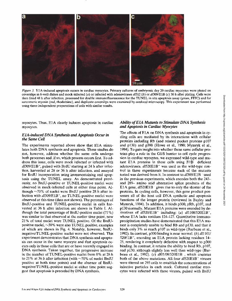
Figure 3. E1A-induced apoptosis occurs in cardiac myocytes. Primary cultures of embryonic day 20 cardiac myocytes were plated on coverslips in 6-well dishes and mock-infected (a) or infected with adenoviruses d/312 (b) or d/309/E1B (c) 36 h after plating. Cells were then fixed 48 h after infection, processed for double immunofluorescence for the TUNEL in situ apoptosis assay (green, FITC) and for sarcomeric myosin (red, rhodamine), and duplicate coverslips were examined by confocal microscopy. This experiment was performed using three independent preparations of cells with similar results.
myocytes. Thus, E I A clearly induces apoptosis in cardiac myocytes.
EIA-induced D N A Synthesis and Apoptosis Occur in the Same Cell
The experiments reported above show that E1A stimu- lates both DNA synthesis and apoptosis. These studies do not, however, address whether the same cells undergo both processes and, if so, which process occurs first. To ad- dress this issue, cells were mock infected or infected with dl309/E1B-, pulsed with BrdU starting at 24 h after infec- tion, harvested at 28 or 36 h after infection, and assayed for BrdU incorporation using immunostaining and apop- tosis using the TUNEL assay. As demonstrated previ- ously, no BrdU-positive or TUNEL-positive nuclei were observed in mock-infected cells at either time point. Al- though ~70% of nuclei were BrdU positive 28 h after in- fection with dl309/E1B-, no TUNEL-positive nuclei were observed at this time (data not shown). The percentages of BrdU-positive and TUNEL-positive nuclei in cells har- vested at 36 h after infection are shown in Table I. Al- though the total percentage of BrdU-positive nuclei (71%) was similar to that observed at the earlier time point, now 21% of total nuclei were TUNEL positive. Of the BrdU- positive nuclei, ,'~30% were also TUNEL positive, examples of which are shown in Fig. 4. Notably, however, BrdU- negative/TUNEL-positive nuclei were not observed. This experiment demonstrates that DNA synthesis and apopto- sis can occur in the same myocyte and that apoptosis oc- curs only in those cells that are or have recently engaged in DNA synthesis. Taken together, the progressive increase in the number of TUNEL-positive nuclei from 0% at 28 h to 21% at 36 h after infection (with ~70% of nuclei BrdU positive at both time points) and the absence of BrdU- negative/TUNEL-positive nuclei at either time point sug- gest that apoptosis is preceded by DNA synthesis.
Ability o f E1A Mutants to Stimulate DNA Synthesis and Apoptosis in Cardiac Myocytes
The effects of E I A on DNA synthesis and apoptosis in cy- cling cells are mediated by its interactions with cellular proteins including Rb (and related pocket proteins p107 and p130) and p300 (Howe et al., 1990; Mymryk et al., 1994). To gain insight into whether these same cellular pro- teins play a role in the G1/S barrier to cell cycle progres- sion in cardiac myocytes, we expressed wild-type and mu- tant E1A proteins in these cells using E I B - deficient adenoviruses, d1520EIB- was used as the wild-type con- trol in these experiments because each of the mutants tested was derived from it. In contrast to dl309fE1B- used in the previous experiments, which encodes both the 243- and 289- amino acid alternative splice products of the E1A gene, d/520EIB- gives rise to only the shorter of the proteins. In cycling cells, however, this gene product pos- sesses all of the host cell DNA synthetic and apoptosis functions of the longer protein (reviewed in Bayley and Mymryk, 1994). In addition, it binds p300, pRb, p107, and p130 normally. Mutant E I A proteins were encoded by de- rivatives of dI520/E1B- including: (a) dlllOS/520E1B-, whose E I A lacks residues 124-127. Quantitative immuno- precipitation studies have demonstrated that this E1A mu- tant is completely unable to bind Rb and p130, and that it binds only 3% as much p107 as wild-type (Barbeau et al., 1992). In contrast, p300 binding is near normal. (b) dlllO1/ 520E1B-, encoding an E1A protein lacking residues 14- 25, rendering it completely defective with respect to p300 binding. In contrast, it retains the ability to bind Rb, p107, and p130, although slightly less well than wild-type (Bar- beau et al., 1992). (c) dlO1/O8/520E1B-, which contains both of the above mutations. All four dI520EIB- viruses were titered on 293 cells to establish the concentrations of infective particles in each stock. Cultured cardiac myo- cytes were infected with these viruses, pulsed with BrdU
Liu and Kitsis EIA-induced DNA Synthesis and Apoptosis in Cardiocytes 329
Dow
nloaded from http://rupress.org/jcb/article-pdf/133/2/325/1265722/325.pdf by guest on 24 N
ovember 2021
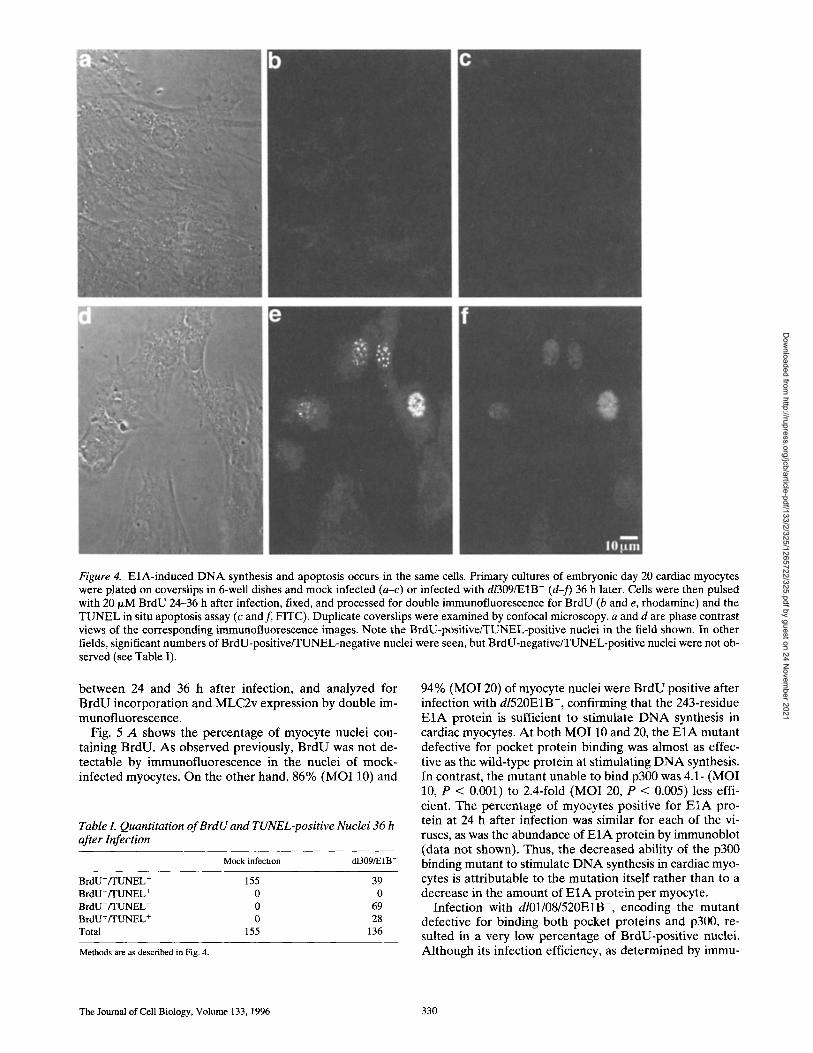
Figure 4. EiA-induced DNA synthesis and apoptosis occurs in the same cells. Primary cultures of embryonic day 20 cardiac myocytes were plated on coverslips in 6-well dishes and mock infected (a-c) or infected with dl309/E1B- (d-f) 36 h later. Cells were then pulsed with 20 I~M BrdU 24-36 h after infection, fixed, and processed for double immunofluorescence for BrdU (b and e, rhodamine) and the TUNEL in situ apoptosis assay (c and f, FITC). Duplicate coverslips were examined by confocal microscopy, a and d are phase contrast views of the corresponding immunofluorescence images. Note the BrdU-positive/TUNEL-positive nuclei in the field shown. In other fields, significant numbers of BrdU-positive/TUNEL-negative nuclei were seen, but BrdU-negative/TUNEL-positive nuclei were not ob- served (see Table I).
between 24 and 36 h after infection, and analyzed for BrdU incorporation and MLC2v expression by double im- munofluorescence.
Fig. 5 A shows the percentage of myocyte nuclei con- taining BrdU. As observed previously, BrdU was not de- tectable by immunofluorescence in the nuclei of mock- infected myocytes. On the other hand, 86% (MOI 10) and
Table L Quantitation of BrdU and TUNEL-positive Nuclei 36 h after Infection
Mock infection dI309/E1B-
B r d U - / T U N E L - 155 39
B r d U - f I ' U N E L + 0 0 BrdU+/TUNEL - 0 69 BrdU+/TUNEL + 0 28
Total 155 136
Methods are as described in Fig. 4.
94% (MO120) of myocyte nuclei were BrdU positive after infection with d1520E1B-, confirming that the 243-residue E1A protein is sufficient to stimulate DNA synthesis in cardiac myocytes. At both MOI 10 and 20, the E1A mutant defective for pocket protein binding was almost as effec- tive as the wild-type protein at stimulating DNA synthesis. In contrast, the mutant unable to bind p300 was 4.1- (MOI 10, P < 0.001) to 2.4-fold (MOI 20, P < 0.005) less effi- cient. The percentage of myocytes positive for E I A pro- tein at 24 h after infection was similar for each of the vi- ruses, as was the abundance of E I A protein by immunoblot (data not shown). Thus, the decreased ability of the p300 binding mutant to stimulate DNA synthesis in cardiac myo- cytes is attributable to the mutation itself rather than to a decrease in the amount of E1A protein per myocyte.
Infection with dlO1/O8/520E1B-, encoding the mutant defective for binding both pocket proteins and p300, re- suited in a very low percentage of BrdU-positive nuclei. Although its infection efficiency, as determined by immu-
The Journal of Cell Biology, Volume 133, 1996 330
Dow
nloaded from http://rupress.org/jcb/article-pdf/133/2/325/1265722/325.pdf by guest on 24 N
ovember 2021
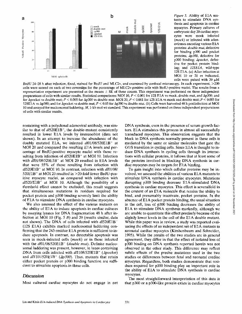
Figure 5. Ability of E1A mu- tants to stimulate DNA syn- thesis and apoptosis in cardiac myocytes. Primary cultures of embryonic day 20 cardiac myo- cytes were mock infected (mock) or infected with aden- oviruses encoding various EIA proteins: double rout, defective for binding p300 and pocket proteins; Ap300, defective for p300 binding; zlpocket, defec- tive for pocket protein bind- ing; and 12SEIA, wild-type 12S E1A. (a) After infection at MOI 10 or 20 as indicated, cells were pulsed with 20 I~M
BrdU 24-28 h after infection, fixed, stained for BrdU and MLC2v, and examined by confocal microscopy. In each experiment, >200 cells were scored on each of two coverslips for the percentage of MLC2v-positive cells with BrdU-positive nuclei. The results from a representative experiment are presented as the means --- SE of these counts. This experiment was performed on three independent preparations of cells with similar results. Statistical comparisons: MOI 10, P < 0.001 for 12S E1A vs mock, double mut, and Ap300, and for Apocket vs double mut; P < 0.005 for Ap300 vs double mut. MOI 20, P < 0.001 for 12S E1A vs mock and double mut; P < 0.005 for 12SEIA vs Ap300; and for Apocket vs double mut; P < 0.05 for Ap300 vs double mut. (b) Cells were harvested 48 h postinfection at MOI 10 and assayed for nucleosomal laddering. M, 1-kb mol wt standard. This experiment was performed on three independent preparations of cells with similar results.
nostaining with a polyclonal adenoviral antibody, was sim- ilar to that of d1520E1B-, the double-mutant consistently resulted in lower E1A levels by immunoblot (data not shown). In an attempt to increase the abundance of the doubly mutated E1A, we infected dlO1/O8/520E1B- at MOI 20 and compared the resulting E1A levels and per- centage of BrdU-positive myocyte nuclei with those re- sulting from infection of d1520E1B- at MOI 10. Infection with dlO1/O8/520E1B- at MOI 20 resulted in E1A levels that were 55% of those resulting from infection with d1520E1B- at MOI 10. In contrast, infection with dlO1/08/ 520E1B- at MOI 20 resulted in >20-fold fewer BrdU-pos- itive myocyte nuclei, as compared with infection with d1520/E1B- at MOI 10. Although the possibility of a threshold effect cannot be excluded, this result suggests that simultaneous mutations in residues required for pocket protein and p300 binding severely limit the ability of E1A to stimulate DNA synthesis in cardiac myocytes.
We also assessed the effect of the various mutants on the ability of E1A to induce apoptosis in cardiac cultures by assaying lysates for DNA fragmentation 48 h after in- fection at MOI 10 (Fig. 5 B) and 20 (results similar, data not shown). The DNA of cells infected with d1520E1B- (12S E1A) exhibits marked nucleosomal laddering con- firming that the 243-residue E1A protein is sufficient to in- duce apoptosis. In contrast, no detectable apoptosis was seen in mock-infected cells (mock) or in those infected with the dlO1/O8/520E1B- (double rout). Definite nucleo- somal laddering was present, however, in lanes containing DNA from cells infected with dlllO8/520E1B- (Apocket) and dlllO1/520E1B- (Ap300). Thus, mutants that retain either pocket protein or p300 binding function are suffi- cient to stimulate apoptosis in these cells.
Discuss ion
Most cultured cardiac myocytes do not engage in net
DNA synthesis, even in the presence of serum growth fac- tors. E1A stimulates this process in almost all successfully transduced myocytes. This observation suggests that the block to DNA synthesis normally present in these cells is mediated by the same or similar molecules that gate the G1/S transition in cycling cells. Since E1A is thought to in- duce DNA synthesis in cycling cells through its interac- tions with cellular proteins, it follows that at least some of the proteins involved in blocking DNA synthesis in car- diac myocytes may be targets for E1A binding.
To gain insight into which cellular proteins may be in- volved, we assessed the abilities of various E1A mutants to stimulate DNA synthesis in cardiac myocytes. Mutations disrupting p300 binding decrease E1A-stimulated DNA synthesis in cardiac myocytes. This effect is severalfold in the context of an E1A molecule that retains the ability to bind, and presumably inactivate, pocket proteins. In the absence of E1A pocket protein binding, the usual situation in the cell, loss of p300 binding decreases the ability of E1A to stimulate DNA synthesis markedly, although we are unable to quantitate this effect precisely because of the slightly lower levels in the cell of the E1A double mutant. While this paper was in review, a study was reported eval- uating the effects of an independent set of E1A mutants in neonatal cardiac myocytes (Kirshenbaum and Schneider, 1995). While the results of the two studies are in general agreement, they differ in that the effect of isolated loss of p300 binding on DNA synthesis reported herein was not observed in the other study. This difference may reflect subtle effects of the precise mutations used in the two studies or differences between fetal and neonatal cardiac myocytes. Regardless, both studies demonstrate that resi- dues required for p300 binding play an important role in the ability of E1A to stimulate DNA synthesis in cardiac myocytes.
The most straightforward interpretation of this data is that p300 or a p300-1ike protein exists in cardiac myocytes
Liu and Kitsis E1A-induced DNA Synthesis and Apoptosis in Cardiocytes 331
Dow
nloaded from http://rupress.org/jcb/article-pdf/133/2/325/1265722/325.pdf by guest on 24 N
ovember 2021
and, in the absence of being sequestered by E1A, contrib- utes to the inhibition of cellular DNA synthesis. We has- ten to emphasize, however, that since the identities and functions of proteins in cardiac myocytes that interact with E1A residues required for p300 binding have not yet been determined, this interpretation must be regarded as condi- tional. In addition, this model assumes that the absence of DNA synthesis in cardiac myocytes results from active re- pression rather than simply being the default mode. This view is supported by skeletal myoblast/myocyte hetero- karyon experiments demonstrating dominance of the phe- notype characterized by the absence of net DNA synthesis (Clegg and Hauschka, 1987). It remains to be determined whether this is also the case in cardiac myocytes in light of experiments demonstrating the lack of dominance of car- diac gene expression in fusions between cardiac myocytes and fibroblasts (Evans et al., 1994).
p300 is the prototype member of a family of bromo- domain-containing transcriptional adaptor molecules that are thought to transactivate the expression of target genes by interacting simultaneously with specific enhancer bind- ing proteins and components of the basal transcriptional apparatus (Eckner et al., 1994; discussed in Arany et al., 1994). Members of the p300 family appear to be expressed ubiquitously. How then can the presence of p300 family members in cycling as well as terminally differentiated cells be reconciled with the possibility that these proteins play an important role in excluding the latter from the cell cycle? One possibility is that different p300 family mem- bers exist in proliferating and postmitotic cells. Another is that the activity of the same p300 family member is differ- entially modulated in these cells by changes in abundance, posttranslational modifications, or interactions with other proteins. The answers to these questions will become clearer as the different p300 family members and their modes of regulation become better defined.
If the role postulated above for p300 in cardiac myo- cytes is correct, one pathway by which it might act to in- hibit DNA synthesis is suggested by recent work in termi- nally differentiating keratinocytes, demonstrating that it plays a role in activating the promoter of the gene encod- ing p21 clP1/wAFt (Missero et al., 1995). This protein, in turn, is a member of the family of universal cyclin-depen- dent kinase inhibitors and blocks DNA synthesis and cell cycle progression (reviewed in Sherr, 1994). Interestingly, p21CIP1/wAFI is also induced during differentiation of C2C12 myotubes (Halevy et al., 1995; Parker et al., 1995; Missero et al., 1995).
It is well established that Rb plays an important role in regulating the G1/S transition in mammalian cells, as well as in the terminal differentiation of skeletal myocytes (Gu et al., 1993; Schneider et al., 1994), neurons (Lee et al., t994), and lens fiber cells (Pan and Griep, 1994; Morgen- besser et al., 1994; and Fromm et al., 1994). Although its presence in wild-type cardiac myocytes has not been for- mally documented, it is present in extracts of whole-heart tissue and in cardiac myocytes that express an SV-40 T an- tigen transgene (Kim et al., 1994). Therefore, it was some- what surprising that the E IA mutant that is completely de- fective for Rb and p130 binding and binds p107 only 3% as well as the wild-type protein (Barbeau et al., 1992) retains the ability to stimulate DNA synthesis in cardiac myocytes
almost as efficiently as the wild type. One possible expla- nation for this observation is that the expression of E1A leads to the inactivation of Rb through an alternative mechanism. In support of this, E1A has been shown in other cells to induce the phosphorylation of Rb through a mechanism independent of its ability to bind this pocket protein (Wang et al., 1991).
As in cycling cells containing functional p53, the end re- sult of E1A overexpression in cardiac myocytes is apopto- sis. Direct evidence that both processes occur in the same cells is shown in Fig. 4. In addition, the data demonstrate that: (a) while ~70% of nuclei are BrdU positive both 28 and 36 h after infection, the number of TUNEL-positive nuclei increases from 0 to 21% during the same period of time; and (b) while many BrdU-positive nuclei are TUNEL- negative 36 h after infection, all TUNEL-positive nuclei are BrdU positive (Table I). Taken together, these find- ings suggest that DNA synthesis precedes apoptosis.
Although tight associations between forced DNA syn- thesis and apoptosis have been observed in the present study and previously (Wu and Levine, 1994; Qin et al., 1994; Shan and Lee, 1994; Morgenbesser et al., 1994), a cause and effect relationship between these two processes has not been demonstrated. Alternatively, the temporal association between these two processes could reflect shared proximal signaling events (discussed in Evan et al., 1995). Conversely, it is clear that a block to DNA synthesis is not required for E1A-induced apoptosis because E1A elicits apoptosis in cycling cells as well as in those which do not ordinarily engage in DNA synthesis. Thus, while in some situations apoptosis may represent the resolution of conflicting positive and negative growth signals, this does not appear to be the case with E1A-induced apoptosis. Moreover, the outlines of a plausible scenario by which E1A could induce apoptosis have emerged from known epistatic relationships: E1A increases steady-state levels of p53 through transcriptional (Braithwaite et al., 1990) and posttranscriptional mechanisms (Lowe et al., 1993). p53, in turn, is known to activate the expression of the bax gene (Miyashita and Reed, 1995), whose product can promote apoptosis (Oltvai et al., 1993). Further work is needed to determine whether these events constitute the actual sig- naling pathway mediating E1A-induced apoptosis.
We acknowledge the expert technical assistance of Soo Jin Lee in prepa- ration of the fetal cardiac myocyte cultures, the advice of Michael Cam- mer regarding confocal microscopy, and the generosity of Drs. Stanley Bayley, Eileen White, Ken Chien, and Erik Falck-Pedersen for reagents.
This work was supported by grants to R.N. Kitsis from the National In- stitutes of Health (HL-02699), the American Heart Association, New York City Affiliate, and the Cardiovascular Research Foundation. R.N. Kitsis is the Charles and Tamara Krasne Faculty Scholar in Cardiovascu- lar Research of the Albert Einstein College of Medicine.
Received for publication 10 March 1995 and in revised form 3 January
1996.
References
Arany, Z., W.R. Sellers, D.M. Livingston, and R. Eckner. 1994. E1A-associated p300 and CREB-associated CBP belong to a conserved family of coactiva- tors. Cell. 77:799-800.
Barbeau, D., R.C. Marcellus, S. Bachetti, S.T. Bayley, and P.E. Branton. 1992. Quantitative analysis of regions of adenovirus E1A products involved in in- teractions with cellular proteins. Biochem. Cell Biol. 70:1123-1134.
Bayley, S.T., and J.S. Mymryk. 1994. Adenovirus E1A proteins and transforma-
The Journal of Cell Biology, Volume 133, 1996 332
Dow
nloaded from http://rupress.org/jcb/article-pdf/133/2/325/1265722/325.pdf by guest on 24 N
ovember 2021
tion. Int. J. Oncology. 5:425-444. Behringer, R.R., J.J. Peschon, A. Messing, C.L. Gartside, S.D. Hauschka, R.D.
Palmiter, and R.L. Brinster. 1988. Heart and bone tumors in transgenic mice. Proc. Natl. Acad. Sci. USA. 85:2648-2652.
Braithwaite, A., C. Nelson, A. Skulimowski, J. McGovern, D. Pigott, and J. Jen- kins. 1990. Transactivation of the p53 oncogene by E1A gene products. Vi- rology. 177:595~505.
Braun, T., E. Bober, and H.H. Arnold. 1992. Inhibition of muscle differentia- tion by the adenovirus E1A protein: repression of the transcriptional activat- ing function of the HLH protein Myf-5. Genes & Dev. 6:888-902.
Caruso, M., F. Martelli, A. Giordano, and A. Felsani. 1993. Regulation of MyoD gene transcription and protein function by the transforming domains of the adenovirus E1A oncoprotein. Oncogene. 8:267-278.
Claycomb, W.C. 1975. Biochemical aspects of cardiac muscle differentiation. J. Biol. Chem. 250:3229-3235.
Clegg, C.H., and S.D. Hauschka. 1987. Heterokaryon analysis of muscle differ- entiation: regulation of the postmitotic state. Z Cell Biol. 105:937-947.
Crescenzi, M., T.P. Fleming, A.B. Lassar, H. Weintraub, and S.A. Aaronson. 1990. MyoD induces growth arrest independent of differentiation in normal and transformed cells. Proc. Natl. Acad. Sci. USA. 87:8442-8446.
Debbas, M., and E. White. 1993. Wild-type p53 mediates apoptosis by E1A, which is inhibited by E1B. Genes & Dev. 7: 546-554.
Eckner, R., M.E. Ewen, D. Newsome, M. Gerdes, J.A. DeCaprio, J.B. Lawrence, and D.M. Livingston. 1994. Molecular cloning and functional analysis of the adenovirns E1A-associated 300-kD protein (p300) reveals a protein with properties of a transcriptional adaptor. Genes & Dev. 8:869-884.
Enkemann, S.A., S.F. Konieczny, and E.J. Taparowsky. 1990. Adenovirus 5 E1A represses muscle specific enhancers and inhibits expression of the myo- genic regulatory factor genes MyoD1 and myogenin. Cell, Growth Differ. 1: 375-382.
Evan, G.I., L. Brown, M. Whyte, and E. Harrington. 1995. Apoptosis and the cell cycle. Curr. Opin. Cell Biol, 7:825-834.
Evans, S.M., L.J. Tai, V.P. Tan, C.B. Newton, and K.R. Chien. 1994. Hetero- karyons of cardiac myocytes and fibroblasts reveal the lack of dominance of the cardiac muscle phenotype. Mol. Cell, Biol. 14:4269-4279.
Field, L.J. 1988. Atrial natriuretic factor SV-40 T antigen transgenes produce tumors and cardiac arrhythmias in mice. Science (Wash. DC). 239:1029- 1033.
Fromm, L., W. Shawlot, K. Gunning, J.S. Butel, and P.A. Overbeek. 1994. The retinoblastoma protein-binding region of simian virus 40 large T antigen al- ters cell cycle regulation in lenses of transgenic mice. Mol. Cell Biol. 14: 6743-6754.
Gavrieli, Y., Y. Sherman, and S.A. Ben-Sasson. 1992. Identification of pro- grammed cell death in situ via specific labeling of nuclear DNA fragmenta- tion. J. Cell Biol, 119:493-501.
Goldstein, M.A., W.C. Claycomb, and A. Schwartz. 1974. DNA synthesis and mitosis in well differentiated cardiac myocytes. Science (Wash. DC). 183: 212-213.
Gu, W., J.W. Schneider, G. Condorelli, S. Kaushal, V. Mahdavi, and B. Nadal- Ginard. 1993. Interaction of myogenic factors and the retinoblastoma pro- tein mediates muscle cell commitment and differentiation. Cell. 72:309-324.
Halevy, O., B.G. Novitch, D.B. Spicer, S.X. Skapek, J. Rhee, G.J. Hannon, D. Beach, and A.B. Lassar. 1995. Correlation of terminal cell cycle arrest of skeletal muscle with induction of p21 by MyoD. Science (Wash. DC). 267: 1018-1021.
Holtzer, H., J.M. Marshall, and H. Finck. 1957. An analysis of myogenesis by the use of fluorescent antimyosin. J. Biophys. Biochem. Cytol. 3:705-724.
Howe, J.A., J.S. Mymryk, C. Egan, P.E. Branton, and S.T. Bayley. 1990. Retino- blastoma growth suppressor and a 300-kDa protein appear to regulate cellu- lar DNA synthesis. Proc. Natl. Acad. Sci. USA. 87:5883-5887.
Iwaki, K., V.P. Sukhatme, H.E. Shubeita, and K.R. Chien. 1990. Alpha- and beta-adrenergic stimulation induces distinct patterns of immediate early gene expression in neonatal rat myocardial cells: fos//un expression is associ- ated with sarcomere assembly; Egr-1 induction is primarily an alpha-1 medi- ated response. Z Biol. Chem. 265:13809-13817.
Jackson, T., M.F. Allard, C.M. Sreenan, L.K. Doss, S.P. Bishop, and J.L. Swain. 1990. The c-myc proto-oncogene regulates cardiac development in trans- genic mice. Mol. Cell. Biol. 10:3709-3716.
Jones, N., and T. Shenk. 1979. Isolation of adenovirus type 5 host range dele- tion mutants defective for transformation of rat embryo cells. Cell. 17:683- 689.
Kass-Eisler, A.A., E. Falck-Pedersen, M. Alvira, J. Rivera, P.M. Buttrick, B.A. Wittenberg, L. Cipriani, and L.A. Leinwand. 1993. Quantitative determina- tion of adenovirus-mediated gene delivery to rat cardiac myocytes in vitro and in vivo. Proc. Natl. Acad. Sci. USA. 90:11498-11502.
Katz, E.B., M.E. Steinhelper, J.B. Delcarpio, A.I. Daud, W.C. Claycomb, and L.J. Field. 1992. Cardiomyocyte proliferation in mice expressing a-cardiac myosin heavy chain SV-40 T antigen transgenes. Am. J. Physiol. 262:H1867- H1876.
Kim, K.K., M.H. Soonpaa, A.I. Daud, G.Y. Koh, J.S. Kim, and L.J. Field. 1994. Tumor suppressor gene expression during normal and pathologic myocar- dial growth. J. Biol. Chem. 269:22607-22613.
Kirshenbaum, L.A., W.R. MacLellan, W. Mazur, B.A. French, and M.D. Schneider. 1993. Highly efficient gene transfer into adult ventricular myo- cytes by recombinant adenoviruses. Z Clin. Invest. 92:381-387.
Kirshenbaum, L.A., and M.D. Schneider. 1995. Adenovirus E I A represses car- diac gene transcription and reactivates DNA synthesis in ventricular myo- cytes, via alternative pocket protein- and p300-binding domains. J. Biol. Chem. 270:7791-7794.
Lee, E.Y.-H.P., N. Hu, S.-S.F. Yuan, L.A. Cox, A. Bradley, W.-H. Lee, and K. Herrup. 1994. Dual roles of the retinoblastoma protein in cell cycle regula- tion and neuron differentiation. Genes & Dev. 8:2008--2021.
Litvin, J., M. Montgomery, A. Gonzalez-Sanchez, J.G. Bisaha, and D. Bader. 1992. Commitment and differentiation of cardiac myocytes. Trends Cardio- vasc. Med. 2:27-32.
Lowe, S.W., and H.E. Ruley. 1993. Stabilization of the p53 tumor suppressor is induced by adenovirus 5 E1A and accompanies apoptosis. Genes & Dev. 7: 535-545.
Miller, J.B., S.B. Teal, and F.E. Stockdale. 1989. Evolutionarily conserved se- quences of striated muscle myosin heavy chain isoforms: epitope mapping by cDNA expression. Z Biol. Chem. 264:13122-13130.
Missero, C., E. Calautti, R. Eckner, J. Chin, L.H. Tsai, D.M. Livingston, and G.P. Dotto. 1995. Involvement of the cell cycle inhibitor Cipl /Wafl and the E1A-associated p300 protein in terminal differentiation. Proc. Natl. Acad. Aci. USA. 92:5451-5455.
Miyashita, T., and J.C. Reed. 1995. Tumor suppressor p53 is a direct transcrip- tional activator of the human bax gene. Cell 80:293-299.
Moran, E. 1993. DNA tumor virus transforming proteins and the cell cycle. Curr. Opin. Genet. Dev. 3:63-70.
Morgenbesser, S.D., B.O. Williams, T. Jacks, and R.A. DePinho. 1994. p53- dependent apoptosis produced by Rb-deficiency in the developing mouse lens. Nature (Lond.). 371:72-74.
Mymryk, J.S., R.W.H. Lee, and S.T. Bayley. 1992. Ability of adenovirus 5 E1A proteins to suppress differentiation of BC3H1 myoblasts correlates with their binding to a 300-kD cellular protein. Mol. Biol. Cell. 3:1107-1115.
Mymryk, J.S., K. Shire, and S.T. Bayley. 1994. Induction of apoptosis by ade- novirus type 5 El A in rat cells requires a proliferation block. Oncogene. 9: 1187-1193.
Nadal-Ginard, B. 1978. Commitment, fusion, and biochemical differentiation of a myogenic cell line in the absence of DNA synthesis. Cell, 15:855-864.
Nguyen, H.T., R.M. Medford, and B. Nadal-Ginard. 1983. Reversibility of mus- cle differentiation in the absence of commitment: analysis of a myogenic cell line temperature-sensitive for commitment. Cell, 34:281-293.
Olson, E.N. 1993. Regulation of muscle transcription by the MyoD family. The heart of the matter. Circ. Res. 72:1-6.
Oltvai, Z.N., C.L. Millman, and S.J. Korsmeyer. 1993. Bcl-2 heterodimerizes in vivo with a conserved homolog, bax, that accelerates programmed cell death. Cell, 74:609-619.
Pan, H., and A.E. Griep. 1994. Altered cell cycle regulation in the lens of HPV- 16 E6 or E7 transgenic mice: implications for tumor suppressor gene func- tion in development. Genes & Dev. 8:1285-1299.
Parker, S.B., G. Eichele, P. Zhang, A. Rawls, A.T. Sands, A. Bradley, E.N. OI- son, J.W. Harper, and S.J. Elledge. 1995. p53-independent expression of p21 clpl in muscle and other terminally differentiated cells. Science (Wash. DC). 267:1024-1027.
Qin, X.Q., D.M. Livingston, W.G. Kaelin, and P.D. Adams. 1994. Deregulated transcription factor E2F-1 expression leads to S phase entry and p53-medi- ated apoptosis. Proc. Natl. Acad. Sci. USA. 91:10918-10922.
Rao, L., M. Debbas, P. Sabbatini, D. Hockenberry, S. Korsmeyer, and E. White. 1992. The adenovirus E1A proteins induce apoptosis, which is inhib- ited by the E1B 19-kDa and Bcl-2 proteins. Proc. Natl. Acad. Sci. USA. 89: 7742-7746.
Rawles, M.E. 1943. The heart-forming areas of the early chick blastoderm. Physiol. Zool. 16:22-42.
Rumyantsev, P.P. 1977. Interrelationships of the proliferation and differentia- tion processes during cardiac myogenesis and regeneration. Int. Rev. Cytol. 51:187-273.
Sherr, C.J. 1994. G1 phase progression: cycling on cue. Cell 79:551-555. Schneider J.W., W. Gu, L. Zhu, V. Mahdavi, and B. Nadal-Ginard. 1994. Re-
versal of terminal differentiation mediated by p107 in Rb -/- muscle cells. Science (Wash. DC). 264:1467-1471.
Shan, B., and W.H. Lee. 1994. Deregulated expression of E2F-1 induces S-phase entry and leads to apoptosis. MoL Cell Biol. 14:8166-8173.
Skapek, S.X., J. Rhee, D.B. Spicer, and A.B. Lassar. 1995. Inhibition of myo- genic differentiation in proliferating myoblasts by cyclin Dl-dependent ki- nase. Science (Wash. DC). 267:1022-1024.
Steinhelper, M.E., N.A. Lanson, K.P. Dresdner, J.B. Delcarpio, A.L. Wit, W.C. Claycomb, and L.J. Field. 1990. Proliferation in vivo and in culture of differ- entiated atrial cardiomyocytes from transgenic mice. Am. J. Physiol. 259: H1826-H1834.
Sorrentino, V., R. Pepperkok, R.L. Davis, W. Ansorge, and L. Philipson. 1990. Cell proliferation inhibited by MyoD1 independently of myogenic differenti- ation. Nature (Loud.). 345:813-815.
Taylor, D.A., V.B. Kraus, J.J. Schwarz, E.N. Olson, and W.E. Kraus, 1993. E1A-mediated inhibition of myogenesis correlates with a direct physical in- teraction of E1A12S and basic helix-loop-helix proteins. Mol. Cell, Biol. 13: 4714-4727.
Wang, H.-G.H., G. Draetta, and E. Moran. 1991. E1A induces phosphorylation of the retinoblastoma protein independently of direct physical association between E1A and retinoblastoma proteins. Mol. Cell. Biol. 11:4253-4265.
Liu and Kitsis E1A-induced DNA Synthesis and Apoptosis in Cardiocytes 333
Dow
nloaded from http://rupress.org/jcb/article-pdf/133/2/325/1265722/325.pdf by guest on 24 N
ovember 2021
Wang, H-G.H., Y. Rikitake, M.C. Carter, P. Yaciuk, S.E. Abraham, B. Zeller, and E. Moran. 1993. Identification of specific adenovirus E1A N-terminal residues critical to the binding of cellular proteins and to the control of cell growth. J. Virology. 67:476--488.
Webster, K.A., G.E.O. Muscat, and L. Kedes. 1988. Adenovirus E1A products suppress myogenic differentiation and inhibit transcription from muscle-spe- cific promoters. Nature (Lond.). 332:553-557.
Weintraub, H. 1993. The MyoD family and myogenesis: redundancy, networks, and thresholds. Cell 75:1241-1244.
White, E. 1993. Regulation of apoptosis by the transforming genes of the DNA tumor virus adenovirus. Proc. Soc. Exp. BioL Med. 204:30-39.
Wu, X., and A.J. Levine. 1994. p53 and E2F-1 cooperate to mediate apoptosis. Proc. Natl. Acad. Sci. USA. 91:3602-3606.
The Journal of Cell Biology, Volume 133, 1996 334
Dow
nloaded from http://rupress.org/jcb/article-pdf/133/2/325/1265722/325.pdf by guest on 24 N
ovember 2021


