Indications and Interpretation of Serological Tests in the...
36
Indications and Interpretation Indications and Interpretation of Serological Tests in the of Serological Tests in the Context of Kidney Disease Context of Kidney Disease Prof. Peter Gergely Prof. Peter Gergely Semmelweis University Semmelweis University Central Laboratory of Immunology Central Laboratory of Immunology
Transcript of Indications and Interpretation of Serological Tests in the...

Indications and Interpretation Indications and Interpretation of Serological Tests in the of Serological Tests in the Context of Kidney DiseaseContext of Kidney Disease
Prof. Peter GergelyProf. Peter Gergely
Semmelweis UniversitySemmelweis University
Central Laboratory of ImmunologyCentral Laboratory of Immunology

General RemarksGeneral Remarks
Immunological tests are of Immunological tests are of limitedlimited value in the diagnosis of value in the diagnosis of kidney diseases, sincekidney diseases, since
clinical picture;clinical picture;
other laboratory tests (e.g. proteinuria, serum creatinine, other laboratory tests (e.g. proteinuria, serum creatinine, GFR etc); GFR etc);
renal histology (kidney biopsy) have a more important renal histology (kidney biopsy) have a more important role in establishing both the diagnosis and prognosis of role in establishing both the diagnosis and prognosis of kidney diseases, as well as in monitoring disease activitykidney diseases, as well as in monitoring disease activity
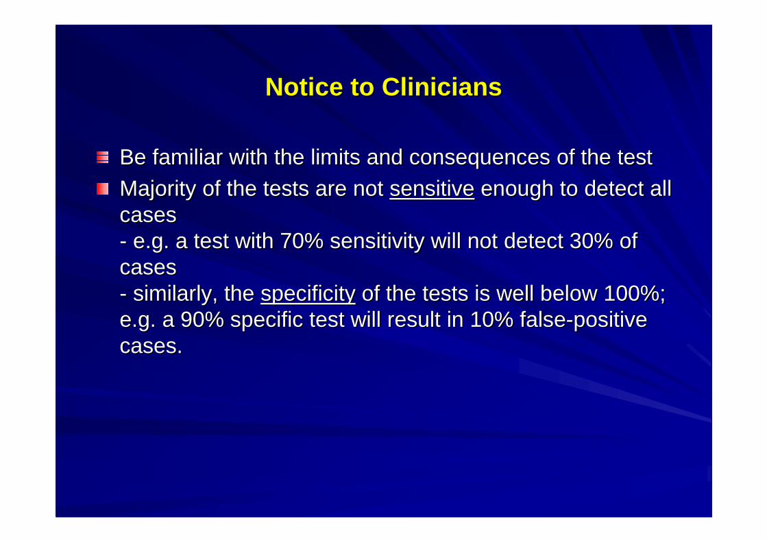
Notice to CliniciansNotice to Clinicians
Be familiar with the limits and consequences of the testBe familiar with the limits and consequences of the test
Majority of the tests are not Majority of the tests are not sensitivesensitive enough to detect all enough to detect all casescases-- e.g. a test with 70% sensitivity will not detect 30% of e.g. a test with 70% sensitivity will not detect 30% of casescases-- similarly, the similarly, the specificityspecificity of the tests is well below 100%; of the tests is well below 100%; e.g. a 90% specific test will result in 10% falsee.g. a 90% specific test will result in 10% false--positive positive cases.cases.

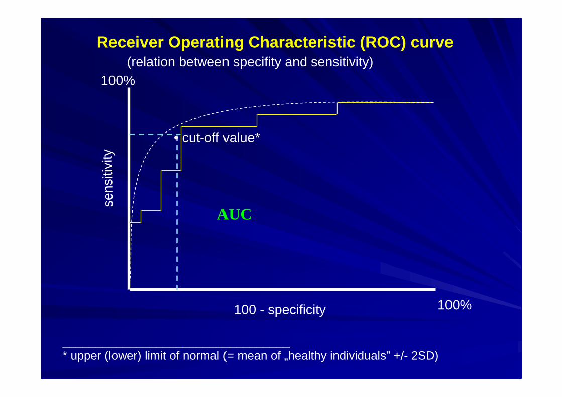
INFORMATIONAL INDICES
No. of patients with a positive testSensitivity =
Total No. of patients with the disease
No. of patients with a negative testSpecificity =
Total No. of patients without the disease
Positive predictive value (PPV) = The probability that a disease is present if a certain test is positive
Negative predictive value (NPV) = The probability that a disease is not present if a certain test isnegative
100%
100%
sens
itivi
ty
100 - specificity
AUC
Receiver Operating Characteristic (ROC) curve(relation between specifity and sensitivity)
• cut-off value*
__________________________________* upper (lower) limit of normal (= mean of „healthy individuals” +/- 2SD)

The effect of cut-off value on sensitivity and spec ificity
Test Cut-off Specificity Sensitivity
Anti-DNA 56 U/ml 85% 57%35 79% 70%26 50% 77%
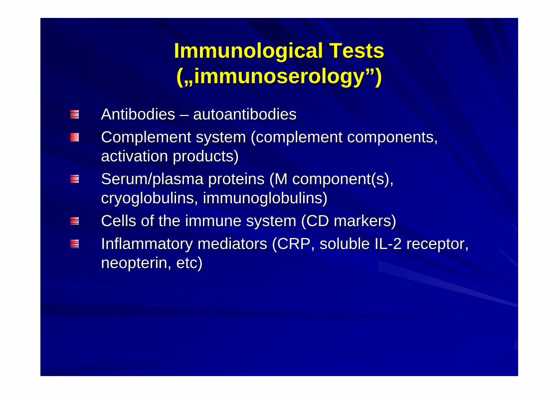
Immunological Tests Immunological Tests ((„„ immunoserologyimmunoserology ”” ))
Antibodies Antibodies –– autoantibodiesautoantibodies
Complement system (complement components, Complement system (complement components, activation products)activation products)
Serum/plasma proteins (M component(s), Serum/plasma proteins (M component(s), cryoglobulins, immunoglobulins)cryoglobulins, immunoglobulins)
Cells of the immune system (CD markers)Cells of the immune system (CD markers)
Inflammatory mediators (CRP, soluble ILInflammatory mediators (CRP, soluble IL--2 receptor, 2 receptor, neopterin, etc)neopterin, etc)

The Role of Autoantibody TestsThe Role of Autoantibody Tests
Antibody tests help differentiate Antibody tests help differentiate
1.1. Secondary nephritis due to systemic autoimmune diseases:Secondary nephritis due to systemic autoimmune diseases:–– Systemic lupus erythematosus (SLE)Systemic lupus erythematosus (SLE)–– SjSjöögrengren’’s syndrome (acute/chronic interstitial nephritis)s syndrome (acute/chronic interstitial nephritis)–– Rheumatoid arthritis (RA) Rheumatoid arthritis (RA) –– most common is secondary most common is secondary
amyloidosisamyloidosis–– SclerodermaScleroderma–– Mixed connective tissue disease (MCTD)Mixed connective tissue disease (MCTD)–– Systemic vasculitis (e.g. WegenerSystemic vasculitis (e.g. Wegener’’s granulomatosis, s granulomatosis,
microscopic polyangiitis, Churgmicroscopic polyangiitis, Churg--Strauss syndrome, BehStrauss syndrome, Behççetet’’s s disease, Schdisease, Schöönlein Henoch purpura)nlein Henoch purpura)
2.2. Secondary nephritis due to virusSecondary nephritis due to virus--induced cryoglobulinaemiainduced cryoglobulinaemia3.3. Glomerular basement membrane (GBM) antibodyGlomerular basement membrane (GBM) antibody--induced induced
nephritisnephritis4.4. Vascular kidney disease due to antiphospholipid syndrome Vascular kidney disease due to antiphospholipid syndrome
(APS)(APS)

Antinuclear antibody (ANA)Antinuclear antibody (ANA)
Target:Target: nuclear antigens nuclear antigens -- collective name of many collective name of many antibodies of various specificityantibodies of various specificity
Determination:Determination: by indirect immunofluorescence (IIF) or enzyme by indirect immunofluorescence (IIF) or enzyme immunoassay (EIA/ELISA)immunoassay (EIA/ELISA)
Why IIF?Why IIF?ANA titre (≥1:160)chromatin staining (e.g. anti-centromere)staining of mitotic proteins
ANA patternspecial ANA types (e.g. PCNA)cytoplasm staining (e.g. ribosomal)
cytoskeleton staining
Hep-2 cell

ANA patterns
a) homogenous b) rim like
c) speckled d) nucleolar

0102030405060708090
100
1 2 3 4 5 6 7
SLEOtherControl
% positive
IIF ANA ELISA
Positive ANA test in SLE, other autoimmunediseases, and healthy controls
Emlen & Neill, Arthritis Rheum 40:1612, 1997

Diagnostic role:Diagnostic role: ANA is frequently positive in systemic ANA is frequently positive in systemic autoimmune diseases. ANA test should be performed in case autoimmune diseases. ANA test should be performed in case of suspicion of systemic autoimmune disease. Diagnostic of suspicion of systemic autoimmune disease. Diagnostic criterium in SLE, frequently positive in Sjcriterium in SLE, frequently positive in Sjöögrengren’’s syndrome, s syndrome, mixed connective disease, scleroderma and others. Both mixed connective disease, scleroderma and others. Both specificity and sensitivity are not too high; a positive ANA tesspecificity and sensitivity are not too high; a positive ANA test t may be found in neoplasms, infections and other diseases:may be found in neoplasms, infections and other diseases:
SLE 80-100%Drug-induced LE 70%MCTD 100%Sjögren’s syndrome 70%Felty syndrome 70-100%Scleroderma 75-85%Myositis 30%RA 15-30%Chronic hepatitis 40-60%Pulmonary fibrosis 20%Tuberculosis 50%Neoplasms 10%Control <5%

02468
101214161820
5 10 15 20 40 >40
ANA positivity in healthy children and adults(IIF HEp-2, at 1:32 titre)
age groups
%
Craig et al, J Rheumatol 26:914, 1999
years
ANA includes many antibodies :
Anti-(double-stranded/native) DNA antibodyHistone antibodyCentromere antibodyENA (antibodies against extractable nuclear antigens)PCNA antibodyKu antibodyChromatin antibodiesand many others, e.g. coilin, DEK, single-stranded-DNA, IFI 16, Mi-2, nucleosoma, nucleolar antibodies
Type of ANA should be determined if the ANA test is positive
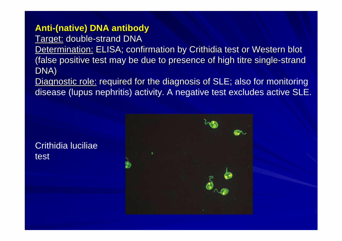
AntiAnti --(nat(nat ive)ive) DNDNAA antianti bodybodyTargetTarget:: doubledouble--strand DNAstrand DNADetermination:Determination: ELISA; confirmation by Crithidia test or Western blot ELISA; confirmation by Crithidia test or Western blot (false positive test may be due to presence of high titre single(false positive test may be due to presence of high titre single--strand strand DNA)DNA)Diagnostic role:Diagnostic role: required for the diagnosis ofrequired for the diagnosis of SLE; also for monitoring disease (lupus nephritis) activity. A negative test excludes active SLE.
Crithidia luciliaetest
Histone antibody
Target: histones (H1, H2, H3, H4) in nucleosomesDetermination: ELISADiagnostic role: histone antibodies may be present in many
systemic autoimmune diseases, e.g. in SLE, drug-induced lupus, RA, juvenile idiopathic arthritis (JIA), systemic scleroderma, etc. Diagnostic significance: diagnosis of drug-induced lupus.
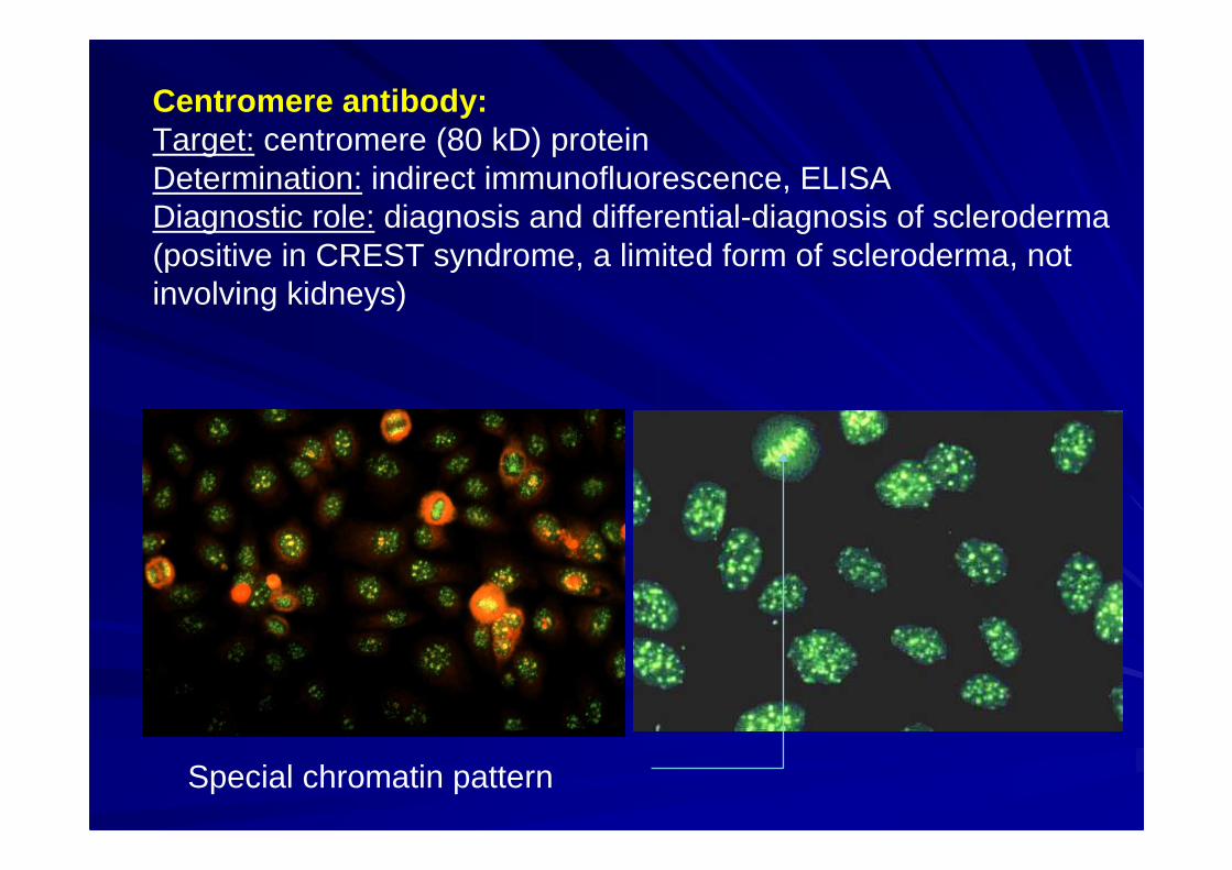
Centromere antibody:Target: centromere (80 kD) proteinDetermination: indirect immunofluorescence, ELISADiagnostic role: diagnosis and differential-diagnosis of scleroderma (positive in CREST syndrome, a limited form of scleroderma, not involving kidneys)
Special chromatin pattern

Antibodies to Extractable Nuclear Antigens Antibodies to Extractable Nuclear Antigens (ENA)(ENA)
A group of nuclear and nonA group of nuclear and non--nuclear antigens traditionally nuclear antigens traditionally named as ENA:named as ENA:
SSSS--A (Ro) and SSA (Ro) and SS--B (La)B (La)
SclScl--7070
JoJo--11
RNP (U1RNP (U1--RNP)RNP)
SmSm
Others: PCNA, Ku, MIOthers: PCNA, Ku, MI--2, PMScl, PM2, PMScl, PM--1.1.

SS-A (Ro) & SS-B (La) antibodiesTarget: non-histone nuclear proteinsDetermination: ELISADiagnostic role: diagnostic criterium for primary or secondary Sjögren’s syndrome (SS), frequently positive in SLE, subacute cutanous lupus (SCLE) and neonatal lupus. SS-A and B antibodies usually occur together.
Scl-70 antibodyTarget: DNA topoisomerase IDetermination: ELISADiagnostic role: positive in 50-70% of systemic forms of scleroderma (PSS); indicate a poor prognosis. Some patients with ‘primary’ Raynaud’s syndrome are positive; they have a high PSS risk

Jo-1 antibodyTarget: aminoacyl tRNA synthetaseDetermination: ELISADiagnostic role: positive in certain forms of myositis (polymyositis and/or dermatomyositis); such cases are also called anti-synthetase syndrome. Can be used for diagnosis/classification of myositis, and fibrosing alveolitis.
(U1)-RNP antibodyTarget: various nucleoproteinsDetermination: ELISADiagnostic role: in MCTD diagnostic criterium, frequentlypositive in SLE, less frequently in PSS.
Sm antibodyTarget: 29 kD nucleoproteinDeterminaton: ELISADiagnostic role: in SLE diagnostic and prognostic marker(central nervous system (CNS), kidney, skin involvement).

PCNA („proliferating cell nuclear antigen”) antibod y:specific for SLE; PCNA+ patients display renal and CNSinvolvement. Diagnostic significance: SLE (in particular if anti-DNA and Sm negative).
Typical nuclear stainingpattern in PCNA
Ku antibody: in polymyositis/scleroderma overlap syndrome.
Ribosomal protein antibodies: in systemic autoimmunediseases, in particular in SLE.

Rheumatoid factor (RF)Target: IgG moleculeDetermination: turbidimetry (IgM)Diagnostic role: classification criterium in RA, frequently positive in Sjögren’s syndrome and in cryoglobulinaemia. Both specificity and sensitivity are low (frequently negative in RA, and positive in many other autoimmune diseases including infections (e.g. poststreptococcalglomerulonephritis):
RA 50-90SLE 15-35Sjögren’s syndrome 75-95scleroderma 20-30Cryoglobulinaemia 40-100MCTD 50-60Viral infections 15-65Endocarditis 25-50Liver cirrhosis 15-40Sarcoidosis 3-33Pulmonary fibrosis 10-50Silicosis 30-50Neoplasms 5-25Control <5
Antibody to C1qTarget: C1q complement componentDetermination: ELISADiagnostic role: positive in hypocomplementaemic urticaria vasculitis (HUV). Positive in 17-58% of SLE cases (mostly in lupus nephritis). Positive in 88% of membranoproliferative glomerulonephritis (MPGN), and also in poststreptococcal glomerulonephritis

C3 Nephritic FactorTarget: activated factor B, a constituent of the C3 convertase, C3b,Bb.Determination: haemolytic assayDiagnostic role: present in membranoproliferative glomerulonephritis Type II (dense deposit disease ), but may also be present is Type I and III; the diagnostic significance of this test is uncertain (performed only rarely)

Antiphospholipid antibodies: Target: various phospholipids and phospholipid-binding glycoproteins (beta2-glycoprotein I).Determination: Antibodies against cardiolipin, phosphatidyl serine, β2-glycoprotein I and prothrombin are measured by ELISA, while lupus anticoagulant (LA) is measured in a functional (clotting) assay.Diagnostic significance: Diagnostic criterium (anti-cardiolipin, and anti-β2GPI antibody and/or lupus anticoagulant) in primary and secondary antiphospholipid syndrome. May be positive after infections (this is why a positive test should be repeatedafter 12 weeks)
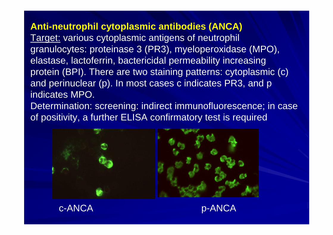
Anti-neutrophil cytoplasmic antibodies (ANCA)Target: various cytoplasmic antigens of neutrophil granulocytes: proteinase 3 (PR3), myeloperoxidase (MPO), elastase, lactoferrin, bactericidal permeability increasingprotein (BPI). There are two staining patterns: cytoplasmic (c) and perinuclear (p). In most cases c indicates PR3, and p indicates MPO.Determination: screening: indirect immunofluorescence; in case of positivity, a further ELISA confirmatory test is required
c-ANCA p-ANCA

Indication of ANCA screening: ANCA positive vasculitis group(Wegener’s granulomatosis, microscopic polyangiitis),differential-diagnosis of nephritis; inflammatory bowel diseases (IBD), primary sclerosing cholangitis, haemorrhagic alveolitis.
Proteinase 3 (PR3) antibody: highly specific for Wegener’sgranulomatosis (>95%), even for atypic forms (such as atypicneuritis and necrotising nephritis).
Myeloperoxidase (MPO) antibody: Positive in microscopicpolyangiitis (60-80%), in some focal necrotising glomerulonephritis, rapid progression glomerulonephritis (RPGN), rarely in Wegener’s granulomatosis, Churg-Strauss syndrome; Goodpasture syndrome, systemic autoimmune diseases.
The role of ANCA tests in monitoring is debated.

GBM antibodyAnti-GBM disease can present as a disease limited to RPGN or as the classic pulmonary-renal syndrome, known as Goodpasture’s syndrome.Target: glomerular basement membrane (type IV collagen α3 chain)Determination: ELISA (formerly: IIF in monkey/human kidney)Diagnostic role: confirmation of diagnosis obtained by renal biopsy
GBM antibody as seen by indirect immunofluorescence
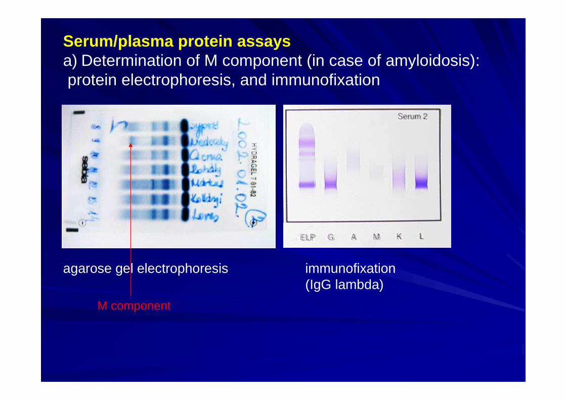
Serum/plasma protein assaysa) Determination of M component (in case of amyloidosis):protein electrophoresis, and immunofixation
agarose gel electrophoresis immunofixation(IgG lambda)
M component
b) Serum immunoglobulin concentrations: determined by turbidimetry; diagnostic role in case of M component. Elevated IgA1 levels and IgA-containing immune complexes are of doubtful diagnostic value in IgA nephropathyc) Cryoglobulin assay: required in primary (essential) mixed cryoglobulinaemia. Cryoprecipitate should be analysed, it may be monoclonal or mixed.This test may be positive in many autoimmune diseases (due to immune complexes in plasma). To rule out secondary cryoglobulinaemia, HCV antibody assay is mandatory.
A prerequisite of reliable result: to draw blood at 37°C and to store at this temperature until clot formation (for cca 2 hours).

Assay of complement components (C3, C4):Determination: haemolytic assay, immunodiffusion and turbidimetryDiagnostic role: For screening of complement deficiencies; monitoring of immune complex diseases (e.g. SLE, acute glomerulonephritis, and certain types of chronic glomerulonephritis) – in active stage their level decreases, while in inactive stage they tend to normalize.
Complement deteriorates at room temperature in serum or fluid; samples should be brought to the laboratory as soon as possible. Separate serum from clot and freeze at -70°C until test is perform ed. Both blood and fluid must be processed and frozen within 2 hours after specimen collection. Failure to process the specimen in this manner may lead to falsely decreased functional activity levels.

Low C3 levels:Low C3 levels:–– Severe recurrent bacterial infections due to C3 homozygous Severe recurrent bacterial infections due to C3 homozygous
deficiencydeficiency–– Absence of C3b inactivator factorAbsence of C3b inactivator factor–– Acute poststreptococcal glomerulonephritisAcute poststreptococcal glomerulonephritis–– Immune complex diseaseImmune complex diseasess–– Active SLEActive SLE–– Membranoproliferative glomerulonephritisMembranoproliferative glomerulonephritis (MPGN)(MPGN)–– Some other forms of nSome other forms of nephritisephritis–– EndEnd--stage liver diseasestage liver disease
Low C4 levels:Low C4 levels:–– SLESLE (exacerbation)(exacerbation)–– GGlomerulonephritislomerulonephritis (early stage)(early stage)–– Immune complex diseaseImmune complex diseasess–– CryoglobulinemiaCryoglobulinemia–– Inborn C4 deficiencyInborn C4 deficiency–– Hereditary angioneurotic edemaHereditary angioneurotic edema (HANE)(HANE)
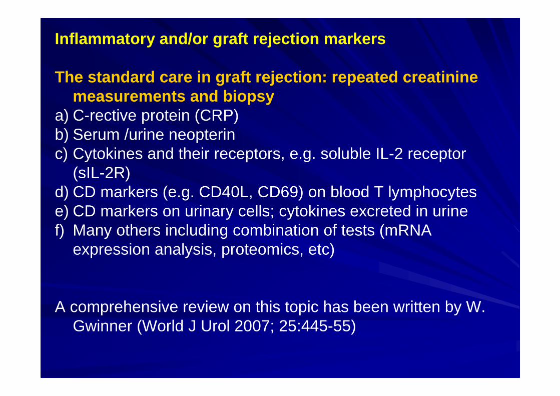
Inflammatory and/or graft rejection markers
The standard care in graft rejection: repeated crea tinine measurements and biopsy
a) C-rective protein (CRP)b) Serum /urine neopterinc) Cytokines and their receptors, e.g. soluble IL-2 receptor
(sIL-2R)d) CD markers (e.g. CD40L, CD69) on blood T lymphocytese) CD markers on urinary cells; cytokines excreted in urinef) Many others including combination of tests (mRNA
expression analysis, proteomics, etc)
A comprehensive review on this topic has been written by W. Gwinner (World J Urol 2007; 25:445-55)

Proposed tests in order to exclude or verify some systemic autoimmune diseases
SLE: ANA*, anti-DNA*, ENA (=anti-Sm, Scl-70, RNP, SS-A/B), anti-CL/LA, C3, C4
RA: RF, CRP, CCP, ANA, (C3, C4)
Juvenile idiopathic arthritis (JIA): RF, ANA, CRP
Sjögren’s syndrome: SS-A*, SS-B*, ANA*, RF
Scleroderma: Scl-70, anti-centromere, ANA, RNP
MCTD: ANA*, ENA (RNP*, Scl-70)
Myositis: ANA (Hep-2), ENA (Jo-1, Scl-70, RNP)
Vasculitis: ANCA, CRP, ANA, RF, cryoglobulin (C3, C4)
_________________
* a negative test makes diagnosis unlikely
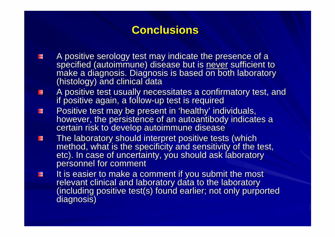
ConclusionsConclusions
A positive serology test may indicate the presence of a A positive serology test may indicate the presence of a specified (autoimmune) disease but is specified (autoimmune) disease but is nevernever sufficient to sufficient to make a diagnosis. Diagnosis is based on both laboratory make a diagnosis. Diagnosis is based on both laboratory (histology) and clinical data(histology) and clinical dataA positive test usually necessitates a confirmatory test, and A positive test usually necessitates a confirmatory test, and if positive again, a followif positive again, a follow--up test is requiredup test is requiredPositive test may be present in Positive test may be present in ‘‘healthyhealthy’’ individuals, individuals, however, the persistence of an autoantibody indicates a however, the persistence of an autoantibody indicates a certain risk to develop autoimmune diseasecertain risk to develop autoimmune diseaseThe laboratory should interpret positive tests (which The laboratory should interpret positive tests (which method, what is the specificity and sensitivity of the test, method, what is the specificity and sensitivity of the test, etc). In case of uncertainty, you should ask laboratory etc). In case of uncertainty, you should ask laboratory personnel for commentpersonnel for commentIt is easier to make a comment if you submit the most It is easier to make a comment if you submit the most relevant clinical and laboratory data to the laboratory relevant clinical and laboratory data to the laboratory (including positive test(s) found earlier; not only purported (including positive test(s) found earlier; not only purported diagnosis)diagnosis)