Indiana-ACC 2015 Poster Competition Abstracts · PDF file · 2015-09-03Indiana-ACC...
13
Indiana-ACC 2015 Poster Competition Abstracts
Transcript of Indiana-ACC 2015 Poster Competition Abstracts · PDF file · 2015-09-03Indiana-ACC...

Indiana-ACC 2015Poster Competition
Abstracts

Fellow in Training
Research Abstracts
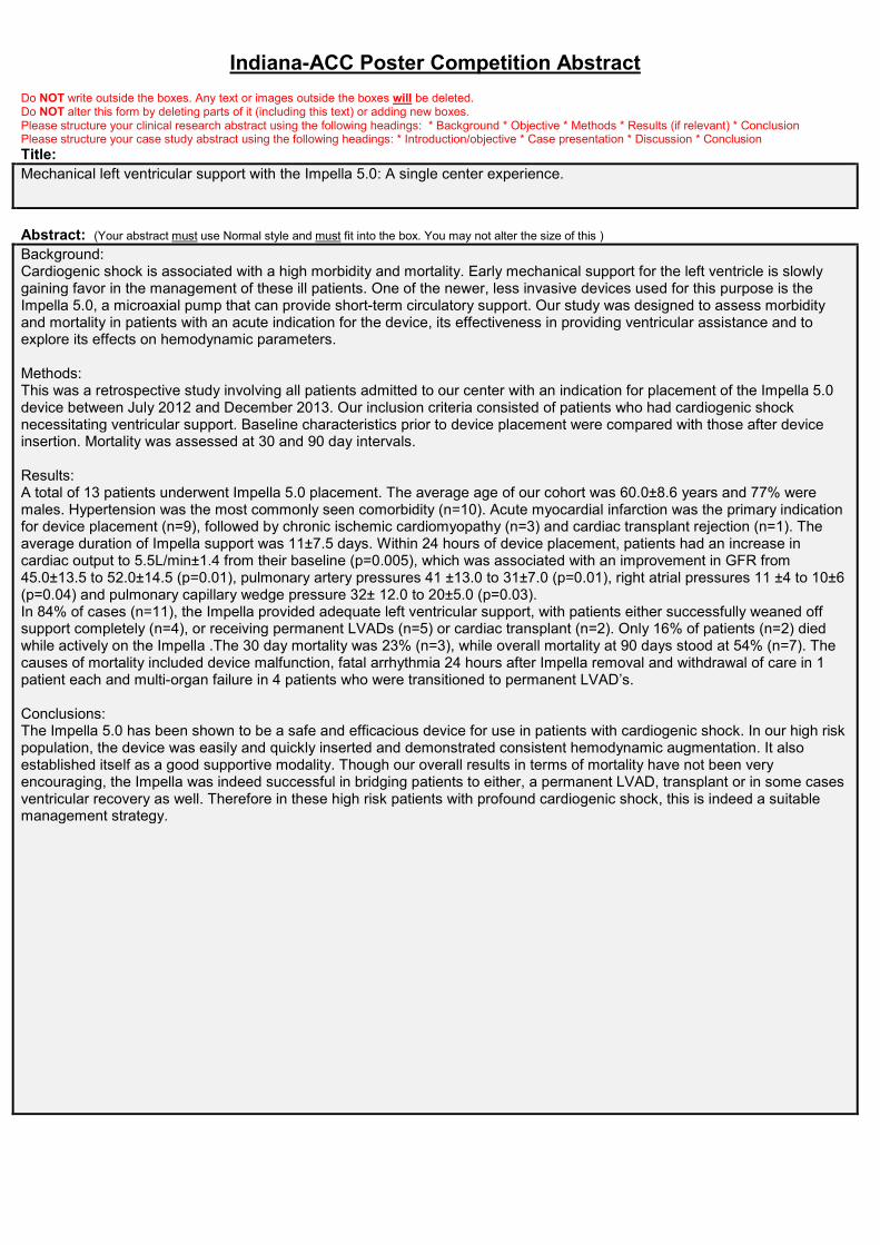
Indiana-ACC Poster Competition AbstractDo NOT write outside the boxes. Any text or images outside the boxes will be deleted.Do NOT alter this form by deleting parts of it (including this text) or adding new boxes.Please structure your clinical research abstract using the following headings: * Background * Objective * Methods * Results (if relevant) * ConclusionPlease structure your case study abstract using the following headings: * Introduction/objective * Case presentation * Discussion * ConclusionTitle:Mechanical left ventricular support with the Impella 5.0: A single center experience.
Abstract: (Your abstract must use Normal style and must fit into the box. You may not alter the size of this )Background:Cardiogenic shock is associated with a high morbidity and mortality. Early mechanical support for the left ventricle is slowlygaining favor in the management of these ill patients. One of the newer, less invasive devices used for this purpose is theImpella 5.0, a microaxial pump that can provide short-term circulatory support. Our study was designed to assess morbidityand mortality in patients with an acute indication for the device, its effectiveness in providing ventricular assistance and toexplore its effects on hemodynamic parameters.
Methods:This was a retrospective study involving all patients admitted to our center with an indication for placement of the Impella 5.0device between July 2012 and December 2013. Our inclusion criteria consisted of patients who had cardiogenic shocknecessitating ventricular support. Baseline characteristics prior to device placement were compared with those after deviceinsertion. Mortality was assessed at 30 and 90 day intervals.
Results:A total of 13 patients underwent Impella 5.0 placement. The average age of our cohort was 60.0±8.6 years and 77% weremales. Hypertension was the most commonly seen comorbidity (n=10). Acute myocardial infarction was the primary indicationfor device placement (n=9), followed by chronic ischemic cardiomyopathy (n=3) and cardiac transplant rejection (n=1). Theaverage duration of Impella support was 11±7.5 days. Within 24 hours of device placement, patients had an increase incardiac output to 5.5L/min±1.4 from their baseline (p=0.005), which was associated with an improvement in GFR from45.0±13.5 to 52.0±14.5 (p=0.01), pulmonary artery pressures 41 ±13.0 to 31±7.0 (p=0.01), right atrial pressures 11 ±4 to 10±6(p=0.04) and pulmonary capillary wedge pressure 32± 12.0 to 20±5.0 (p=0.03).In 84% of cases (n=11), the Impella provided adequate left ventricular support, with patients either successfully weaned offsupport completely (n=4), or receiving permanent LVADs (n=5) or cardiac transplant (n=2). Only 16% of patients (n=2) diedwhile actively on the Impella .The 30 day mortality was 23% (n=3), while overall mortality at 90 days stood at 54% (n=7). Thecauses of mortality included device malfunction, fatal arrhythmia 24 hours after Impella removal and withdrawal of care in 1patient each and multi-organ failure in 4 patients who were transitioned to permanent LVAD’s.
Conclusions:The Impella 5.0 has been shown to be a safe and efficacious device for use in patients with cardiogenic shock. In our high riskpopulation, the device was easily and quickly inserted and demonstrated consistent hemodynamic augmentation. It alsoestablished itself as a good supportive modality. Though our overall results in terms of mortality have not been veryencouraging, the Impella was indeed successful in bridging patients to either, a permanent LVAD, transplant or in some casesventricular recovery as well. Therefore in these high risk patients with profound cardiogenic shock, this is indeed a suitablemanagement strategy.

Indiana-ACC Poster Competition Abstract
Authors:
Hamza Ansari, Willie Robinson, Adnan Malik.
C
Author to Receive Correspondence - Contact Information
Full Name: Ansari Hamza ZLast First M.I.
Address: 418 Canal view way HStreet Address Apartment/Unit
#Indianapolis IN 46202
City State ZIP CodeWork Phone: 317-312-2497 Alternate Phone: 312-376-6171
E-mail Address: [email protected]
TrainingProgram: Krannert Institute of Cardiology, Indiana University.
_Yes_ I understand that submission of an abstract constitutes a commitment to be present Indiana-ACC Annual Meeting. Iunderstand that if I cannot be present that my poster will be withdrawn.

Fellow in Training
Case Abstracts
Indiana-ACC Poster Competition Abstract Do NOT write outside the boxes. Any text or images outside the boxes will be deleted. Do NOT alter this form by deleting parts of it (including this text) or adding new boxes. Please structure your clinical research abstract using the following headings: * Background * Objective * Methods * Results (if relevant) * Conclusion Please structure your case study abstract using the following headings: * Introduction/objective * Case presentation * Discussion * Conclusion
Title:
Endovascular repair of multiple large artery psudoaneurysms in a patient with possible HCV associated cryoglobulinemic vasculitits
Abstract: (Your abstract must use Normal style and must fit into the box. You may not alter the size of this )
Introduction: Management of multiple pseudoaneurysm (MPA) in a single patient by endovascular treatment has been rarely reported. To the best of knowledge this is the second case report in such a situation. The definite cause of MPA in our patient is not known and we speculate it to be secondary to HCV associated cryoglobulinemic vasculitis. Case report : An 18 year-old male presented with one month history of mass in the left anterior cervical region, odynophagia, hoarseness of voice and low grade fever with constitutional symptoms. On physical examination a large tender pulsatile mass of 7x6 cm in the left anterior cervical region and a similar 4x5cm mass was noticed in the left popliteal fossa. He was hypertensive but no significant difference was noted in both upper and lower limb blood pressures. Neurological and ophthalmological examinations were normal. No characteristic facies, arthralgia, skin laxity, skin bruising, oral or digital ulcers were noticed. Bacterial and fungal blood cultures were negative. Transesophageal echocardiogram surveillance for infective endocarditis was negative and no structural heart defects were seen. Patient was tested positive for HCV infection with high viral RNA load. His serum cryoglobulin level was markedly elevated (212 mg/L, normal < 60mg/L. Tuberculin skin test was positive however, CXR and sputum stain and culture for mycobacteria were negative. Pathergy test was negative. CT scan showed multiple fusiform aneurysms involving left common carotid, and bilateral subclavian arteries. CT angiography of lower limb arteries revealed large popliteal aneurysm. Vascular surgery consultation was obtained. Due to multifocality of the lesions, it was decided to proceed with endovascular technique. Conclusion: Giant cell arteritis and Takayasu arteritis are two large vessel vasculitides having a known association with aneurysm formation. The relative rarity of the former entity in India as well as an older age of the patient made this an unlikely diagnosis. Isolated multiple aneurysms are rare in Takayasu disease. The reported incidence of aneurysmal lesions in Takayasu arteritis varies from 4.9% to 31.9 %. The incidence of extracranial carotid artery aneurysm in TA varies from 1.8% to 3.9%. Therefore, Takayasu arteritis was kept lower down in the list of possibilities. Another interesting possibility considered was that of the vascular form of Ehlers-Danlos syndrome (v-EDS). Exuberant presence of aneurysms without significant arterial lumen narrowing , an uninvestigated family history of sudden cardiac death and absence of inflammatory signs of large vessel arteritis on CT are the pointers supporting v-EDS. Absence of clinical stigmata is not sufficient to rule out this possibility. Unfortunately, we were unable to identify the telltale collagen mutations due to financial constraints. The elephant in the room was the presence of HCV infection and significant cryoglobulinemia. Aneurysms have been reported only once before in the setting of cryoglobulinemic vasculitis. A putative mechanism may be immune-complex deposition and complement related medial damage leading to aneurysmal dilatation, although this remains conjectural at best. Our patient did not have typical manifestations of cryoglobulinemic vasculitis like arthralgia, neuropathy or purpura.

Indiana-ACC Poster Competition Abstract Authors:
Bagga S, Chakraborty S, Velu V
C
Author to Receive Correspondence - Contact Information
Full Name: Bagga Shiv
Last First M.I.
Address: Tradd St 3B
Street Address Apartment/Unit #
Carmel IN 46032
City State ZIP Code
Work Phone: Alternate Phone: 317-675-7695
E-mail Address: [email protected]
Training Program: Fellow Cardiac Electrophysiology, St. Vincent, Indianapolis
xxxI understand that submission of an abstract constitutes a commitment to be present Indiana-ACC Annual Meeting. I understand that if I cannot be present that my poster will be withdrawn.

Indiana-ACC Poster Competition AbstractDo NOT write outside the boxes. Any text or images outside the boxes will be deleted.Do NOT alter this form by deleting parts of it (including this text) or adding new boxes.Please structure your clinical research abstract using the following headings: * Background * Objective * Methods * Results (if relevant) * ConclusionPlease structure your case study abstract using the following headings: * Introduction/objective * Case presentation * Discussion * ConclusionTitle:Discovery of Partial Anomalous Pulmonary Venous Return associated with Sinus Venosus ASD during Oximetry Run
Abstract: (Your abstract must use Normal style and must fit into the box. You may not alter the size of this )Introduction: Partial anomalous pulmonary venous return (PAPVR) is a rare congenital cardiac defect defined as a left-to-rightshunt where one or more, but not all, of the pulmonary veins drain into a systemic vein or the right atrium. Right upper lobe(RUL) PAPVR is associated with a sinus venosus ASD (SVASD) in 80%-90% cases1. We present a case of RUL PAPVRassociated with a SVASD first suspected by oximetry during right heart catheterization.
Case Presentation: The patient is a 61 year old Caucasian female with a history of hypertension and obesity who was referredto cardiology clinic because of an abnormal echocardiogram. She had no functional limitations and was asymptomatic. Thestudy was ordered by her primary care physician because the patient reported a history of mitral valve prolapse. The echoshowed normal valves; however, the right atrium and right ventricle were enlarged. The RV function was preserved, but theestimated right ventricular systolic pressure was 35-40 mmHg. No bubble study was performed. A sleep study showed noevidence of sleep apnea. A right heart catheterization (RHC) with oximetry was ordered. The RHC showed no pulmonaryarterial hypertension (mean pulmonary artery pressure 20 mmHg) and normal pulmonary vascular resistance (1.8 Woodsunits). Oximetry run showed a higher than expected superior vena cava (SVC) saturation suggestive of partial anomalouspulmonary venous return (PAPVR). The Qp/Qs was calculated to be 1.0. To evaluate the pulmonary veins, a Cardiac CT wasordered which showed the right upper pulmonary vein (RUPV) attached to the superior vena cava. There was no evidence ofan ASD. A cardiac MRI was ordered to evaluate for any other cardiac abnormalities. The MRI demonstrated a sinus venosusASD, Qp/Qs of 2.2, and confirmed right chamber enlargement with RA volume of 28 m3 and mid-RV diastolic diameter of 5.3cm. The patient has been referred to cardiothoracic surgery and is currently waiting to be evaluated for surgery.
Discussion: Most adult cases of ASD remain unrecognized until adulthood when they become symptomatic of atrialarrhythmias, paradoxical embolism, or pulmonary hypertension. A low threshold for suspicion is critical for diagnosing a shunt,and a comprehensive approach must be undertaken when there is unexplained right sided chamber enlargement onechocardiography. Testing can include both invasive and non-invasive methods. Invasive testing with right heartcatheterization (RHC) is not necessary but can be useful to verify the presence or absence of pulmonary arterial hypertension.Non-invasive testing with transesophageal echocardiography, magnetic resonance imaging (MRI), or computed tomographicscanning helps to accurately define the anatomy.
During RHC, a left-to-right shunt should be suspected whenever pulmonary arterial oxygen saturation is > 80%2. Additionally,an oximetry run, measuring blood oxygen saturation at various locations on the right side of the heart, can help determine thelocation of the shunt and quantify the degree of shunting. A significant shunt is characterized by an oxygen step up of ≥ 5-7%between chambers. It should be noted that oxygen saturation in the superior vena cava (SVC) is typically lower (~5%) than theoxygen saturation in the inferior vena cava (IVC) because there is relatively low oxygen extraction from the kidneys; and theSVC oxygen saturation is normally between 70-80%.
Once a shunt is detected, the ratio of left-to-right shunting (Qp/Qs) should be estimated. Catheterization, echocardiography,and cardiac MRI can be used to estimate Qp/Qs. It should be noted that the oximetry method is not accurate in the setting ofextracardiac shunts, as in our case. However, echocardiography and cardiac MRI measure the flow across the aortic andpulmonic valves to determine the Qp/Qs thus are useful for quantifying both intra and extracardiac shunts.
Regarding the closure of ASD’s, the only Class I indication for closure of an ASD, either percutaneously or surgically, is forright atrial and RV enlargement with or without symptoms. Surgical or percutaneous closure of an ASD is reasonable (IIa) inthe presence of paradoxical-embolism or orthodeoxia-platypnea, and it can be considered (IIb) if Qp/Qs ≥ 1.1, pulmonaryvascular resistance less than 2/3 systemic vascular resistance, when responsive to vasodilator therapy, or test occlusion of thedefect. A sinus venosus, coronary sinus, or primum ASD should be repaired surgically rather than by percutaneous closure.Patients with severe irreversible PAH and no evidence of a left-to-right shunt should not undergo ASD closure. Surgical repairof PAPVR with SV ASD involves redirection of the APVR through the ASD into the left atrium. Repair of this anomaly is morecomplex than other types of ASD closures and carries the risk of stenosis of the SVC or pulmonary veins, residual shunting,and sinoatrial node dysfunction (SND)3.
Conclusion: A low threshold for suspicion is critical for diagnosing a shunt. Right sided chamber enlargement onechocardiography warrants thorough evaluation and may require invasive or non-invasive testing.
Indiana-ACC Poster Competition Abstract
Authors:
John R. Haas, MD; Zachary I. Hodes, MD; Michael C. Walls, MD; Ryan C. Oeltgen, MD
C
Author to Receive Correspondence - Contact Information
Full Name: Haas John RLast First M.I.
Address: 8333 Naab Rd Ste 400Street Address Apartment/Unit
#Indianapolis IN 46260
City State ZIP CodeWork Phone: 317-338-6024 Alternate Phone:
E-mail Address: [email protected]
TrainingProgram: St. Vincent Cardiovascular Disease Fellowship
_x__I understand that submission of an abstract constitutes a commitment to be present Indiana-ACC Annual Meeting. Iunderstand that if I cannot be present that my poster will be withdrawn.

Indiana-ACC Poster Competition AbstractDo NOT write outside the boxes. Any text or images outside the boxes will be deleted.Do NOT alter this form by deleting parts of it (including this text) or adding new boxes.Please structure your clinical research abstract using the following headings: * Background * Objective * Methods * Results (if relevant) * ConclusionPlease structure your case study abstract using the following headings: * Introduction/objective * Case presentation * Discussion * ConclusionTitle:Application of Transcatheter Valve-in-valve Techniques in Bioprosthetic Mitral Valve Dysfunction.
Abstract: (Your abstract must use Normal style and must fit into the box. You may not alter the size of this )
Introduction:Valve-in-valve implantation is effective therapy for dysfunctional bioprosthetic aortic valve. However, application tobioprosthetic mitral valves is still being developed. We report a patient with severe bioprosthetic mitral valve stenosis whosuccessfully underwent transcatheter valve-in-valve (TVIV) implantation.
Case Presentation:81 year old female presented with worsening dyspnea for 2 weeks. Medical history was significant for mitral valve diseasewith bioprosthetic valve replacement in 2007, and aortic stenosis with bioprosthetic valve replacement in 2013. TTE revealedheavily calcified and severely stenotic bioprosthetic mitral valve. LV systolic function was normal. Cardiac catheterization alsoshowed severe mitral stenosis, no significant CAD, and mild pulmonary hypertension. Surgery was declined due to high risk ofredo-redo-sternotomy. She underwent transcatheter mitral bioprosthetic valve replacement with Edwards Sapien XT 29 mmvalve via transapical approach. TEE pre-implant showed EOA of 0.9 cm2 and mean gradient of 11 mm Hg. TEE post-implantshowed mean gradient of 2 mm Hg and minimal perivalvular regurgitation. Patient was extubated same day as procedure, anddischarged on post-op day 8.
Discussion:Mitral TVIV implantation was first reported in 2009. Several case series have been published since then. In one study by A.Cheung et al., 23 patients with severe bioprosthetic mitral valve dysfunction underwent transapical TVIV implantation. Thetransvalvular gradient decreased from 11.1 ± 4.6 mm Hg to 6.9 ± 2.2 mm Hg (p = 0.014). Survival was 90.4% at medianfollow up of 753 days. Improvement from NYHA III-VI to NYHA I-II was seen in 95.4% of patients. Transcatheter mitralvalve-in-valve implantation for dysfunctional bioprosthesis has favorable clinical and hemodynamic outcomes, with minimaloperative morbidity and mortality. Therefore, it should be considered as lower risk alternative in selected high risk patientswith bioprosthetic mitral valve dysfunction.

Indiana-ACC Poster Competition Abstract
Authors:
Roja C. Pondicherry-Harish, Anjan Sinha
C
Author to Receive Correspondence - Contact Information
Full Name: Pondicherry-Harish Roja C.Last First M.I.
Address: 2042 Gable Lane Court 913Street Address Apartment/Unit
#Indianapolis IN 46228
City State ZIP CodeWork Phone: 404-519-8649 Alternate Phone:
E-mail Address: [email protected]
TrainingProgram: Indiana University
_x__I understand that submission of an abstract constitutes a commitment to be present Indiana-ACC Annual Meeting. Iunderstand that if I cannot be present that my poster will be withdrawn.

Indiana-ACC Poster Competition AbstractDo NOT write outside the boxes. Any text or images outside the boxes will be deleted.Do NOT alter this form by deleting parts of it (including this text) or adding new boxes.Please structure your clinical research abstract using the following headings: * Background * Objective * Methods * Results (if relevant) * ConclusionPlease structure your case study abstract using the following headings: * Introduction/objective * Case presentation * Discussion * ConclusionTitle:High Risk Transcatheter Aortic Valve Replacement with High Risk Percutaneous Coronary Intervention
Abstract: (Your abstract must use Normal style and must fit into the box. You may not alter the size of this )
Introduction
Severe symptomatic aortic stenosis is a very poor prognostic predictor if not corrected. Transcatheter aortic valve
replacement offers an alternative for patients that are deemed too high risk for traditional surgical aortic valve
replacement.
Case
51 year old woman presented to another hospital for severe progressive shortness of breath, dyspnea on exertion,
paroxysmal nocturnal dyspnea, and orthopnea over the past month. Her only significant past medical history
included Non-Hodgskin’s lymphoma that had been successfully treated with chemotherapy and radiation. At the
other hospital she became bradycardic and had one minute of CPR with resumption of spontaneous circulation.
She was subsequently intubated and transferred to our institution. On transthoracic echocardiogram she was found
to have severe aortic stenosis with preserved ejection fraction. She was able to be extubated and weaned off
vasopressors in the next few days. She then underwent left heart catheterization prior to being considered for
surgical aortic valve replacement. She was found to have seventy percent stenosis of her left main coronary artery,
seventy-five percent stenosis of her left circumflex, and two significant lesions in her right coronary artery. On CT
scan of her chest she was noted to have a very calcified aorta. The surgeons were consulted and decided that she
would be higher risk due to her calcified aorta but would be willing to proceed with surgical aortic valve
replacement and coronary artery bypass surgery. Unfortunately, while she was waiting for surgery she developed
low grade DIC with consumption of platelets. She also had ongoing anemia with possible fungemia. She then
suffered a cerebral vascular accident with significant right-sided weakness/neglect and right-sided vision loss. Due
to her ongoing comorbidities and her recent stroke she was deemed too high risk for surgery. In the interim her
neurological deficits improved. A discussion was then had regarding the risks and benefits of trying a TAVR and
PCI. She agreed and underwent successful stenting of her left main, left circumflex, and right coronary artery with
TAVR. She tolerated the procedure well with resolution of her symptoms. She was discharged home with home
physical therapy ten days post procedure.
Conclusion
This case illustrates that in carefully chosen patients, TAVR and PCI are acceptable alternatives to traditional surgical
aortic valve replacement and coronary artery bypass.

Indiana-ACC Poster Competition Abstract
Authors:
Robert Quade, MD
C
Author to Receive Correspondence - Contact Information
Full Name: Quade Robert RLast First M.I.
Address: 344 Neuman WayStreet Address Apartment/Unit
#Carmel IN 46032
City State ZIP CodeWork Phone: 3173387510 Alternate Phone:
E-mail Address: [email protected]
TrainingProgram: St. Vincent Hospital
_X__I understand that submission of an abstract constitutes a commitment to be present Indiana-ACC Annual Meeting. Iunderstand that if I cannot be present that my poster will be withdrawn.