Increased Serum Concentration of IgA2 Subclass and IgA2 ...
6
Eur J Clin Chem Clin Biochem 1997; 35(4):275-279 © 1997 by Walter de Gruyter · Berlin · New York Increased Serum Concentration of IgA2 Subclass and IgA2/IgAl Ratio: Specific Markers of Chronic Alcoholic Abuse? Dominique Meillet 1 , Frangoise Labrousse 2 , Marie Odile Benoit 3 , Alain Hernvann 4 , Lucile Mussel and Genevieve van Amerongen 6 1 Biochimie, Hopital de la Salpetriere, Paris, France 2 Biochimie, Hopital Laennec, Paris, France 3 Biochimie, Hopital Broussais, Paris, France 4 Biochimie, Hopital Cochin, Paris, France 5 Immunochirnie, Hopital de la Salpetiere, Paris, France 6 Biochimie, Hopital Raymond-Poincare, Garches, France 1 ~ 6 Groupe d'Evaluation et de Recherche des Biologistes de Γ Assistance Publique des Hopitaux de Paris (GERBAP) Summary: Enhanced serum IgA concentrations are common in alcoholic liver cirrhosis, but functional differences between IgA subclasses and their relation with interleukin-6 (IL-6) have not been described. Distinct immunoregula- tory mechanisms may exist that selectively affect one subclass. This possibility prompted us to investigate the distribution of IgAl and IgA2 subclasses in the serum of 25 heavy alcohol drinkers (alcohol: 80 to 200 g per day) without clinical disorders, in comparison with 35 patients affected by alcoholic liver cirrhosis, 29 viral hepatitis patients and 33 social drinkers as a control group. Mean (± SD) IgA2 concentration (0.56 ± 0.31 g/1) was signifi- cantly increased (p < 0.01) in heavy alcohol drinkers, with an IgA2/IgAl ratio of 0.33 ± 0.12, while the mean total IgA concentration was similar to the control group. Mean IgAl and IgA2 concentrations were significantly increased (p < 0.001) in alcoholic liver cirrhosis patients (6.13 ± 4.52 g/1 and 1.83 ± 1.93 g/1 respectively, with an IgA2/IgAl ratio of 0.32 ± 0.19) and viral hepatitis patients (3.66 ± 2.59 g/1 and 0.69 ± 0.67 g/1 respectively, with an IgA2/IgAl ratio of 0.21 ±0.14) High serum IL-6 concentrations (34 ± 33 ng/1) were correlated with elevated IgAl and IgA2 concentrations only in patients with alcoholic liver cirrhosis. IgA2 subclass and IgA2/IgAl ratio could therefore be used as markers of chronic alcohol abuse directly related to the extent and duration of the alcohol abuse and the effectiveness of alcohol withdrawal. Introduction 6) peripheral blood monoclonal cells have been reported Abnormalities in the immune systems of alcoholic liver in P atients with alcoholic liver cirrhosis < 10 )· IL ' 6 Ρ ΓΟ ' cirrhosis patients have been described (1 -4). However, duction Correlates closely with IgA serum levels. This their pathogenic significance and clinical implications abnormality may be related to overproduction of IgA remain unknown. Hypergammaglobulinaemia, mainly and i™™ 6 disturbances in patients with alcoholic enhancement of the serum IgA concentration, is a com- r dlsease · mon immunological finding in patients with alcoholic We have studied serum IgA, IgAl, IgA2 and IL-6 con- liver cirrhosis (5-7). It has been demonstrated that this centrations in heavy alcohol drinkers without clinical high immunoglobulin concentration is not due to abnor- liver disease in comparison with alcoholic liver cirrhosis mal catabolism of IgA (1—2). and viral hepatitis patients to clarify the significance of SwenOaw et al. have shown in patients with alcoholic the hi S h semm l ^ A2 concentration in relation to alcohol liver disease that the IgA2 subclass forms a major sub- consumption, class component contributing to the continuous pattern of IgA deposition in hepatic tissues (8). Patients and Methods Recently, high serum IgA2 concentrations were found in Patients heavy alcohol drinkers without alcoholic liver cirrhosis ^ b i ochemica i he p atic characteristics of the four groups are (9). This subclass may be a predictive marker for evolu- given in table 1. tion to alcoholic liver disease. - The control group comprised thirty-three healthy subjects (12 T , ,. . , . , . women and 21 men, mean age ± SD: 42 ± 12 years, range: 24- In addition, increased serum concentrations and in vitro 56 years) who were sodal ^^ (alcohol ingestion <40 g per spontaneous or induced production of interleukin-6 (IL- day) with normal hepatic characteristics.
Transcript of Increased Serum Concentration of IgA2 Subclass and IgA2 ...

Eur J Clin Chem Clin Biochem 1997; 35(4):275-279 © 1997 by Walter de Gruyter · Berlin · New York
Increased Serum Concentration of IgA2 Subclass and IgA2/IgAl Ratio:Specific Markers of Chronic Alcoholic Abuse?
Dominique Meillet1, Frangoise Labrousse2, Marie Odile Benoit3, Alain Hernvann4, Lucile Mussel andGenevieve van Amerongen6
1 Biochimie, Hopital de la Salpetriere, Paris, France2 Biochimie, Hopital Laennec, Paris, France3 Biochimie, Hopital Broussais, Paris, France4 Biochimie, Hopital Cochin, Paris, France5 Immunochirnie, Hopital de la Salpetiere, Paris, France6 Biochimie, Hopital Raymond-Poincare, Garches, France1 ~6 Groupe d'Evaluation et de Recherche des Biologistes de Γ Assistance Publique des Hopitaux de Paris (GERBAP)
Summary: Enhanced serum IgA concentrations are common in alcoholic liver cirrhosis, but functional differencesbetween IgA subclasses and their relation with interleukin-6 (IL-6) have not been described. Distinct immunoregula-tory mechanisms may exist that selectively affect one subclass. This possibility prompted us to investigate thedistribution of IgAl and IgA2 subclasses in the serum of 25 heavy alcohol drinkers (alcohol: 80 to 200 g per day)without clinical disorders, in comparison with 35 patients affected by alcoholic liver cirrhosis, 29 viral hepatitispatients and 33 social drinkers as a control group. Mean (± SD) IgA2 concentration (0.56 ± 0.31 g/1) was signifi-cantly increased (p < 0.01) in heavy alcohol drinkers, with an IgA2/IgAl ratio of 0.33 ± 0.12, while the meantotal IgA concentration was similar to the control group. Mean IgAl and IgA2 concentrations were significantlyincreased (p < 0.001) in alcoholic liver cirrhosis patients (6.13 ± 4.52 g/1 and 1.83 ± 1.93 g/1 respectively, with anIgA2/IgAl ratio of 0.32 ± 0.19) and viral hepatitis patients (3.66 ± 2.59 g/1 and 0.69 ± 0.67 g/1 respectively, withan IgA2/IgAl ratio of 0.21 ±0.14) High serum IL-6 concentrations (34 ± 33 ng/1) were correlated with elevatedIgAl and IgA2 concentrations only in patients with alcoholic liver cirrhosis. IgA2 subclass and IgA2/IgAl ratiocould therefore be used as markers of chronic alcohol abuse directly related to the extent and duration of the alcoholabuse and the effectiveness of alcohol withdrawal.
Introduction 6) peripheral blood monoclonal cells have been reported
Abnormalities in the immune systems of alcoholic liver in Patients with alcoholic liver cirrhosis <10)· IL'6 ΡΓΟ'cirrhosis patients have been described (1 -4). However, duction Correlates closely with IgA serum levels. Thistheir pathogenic significance and clinical implications abnormality may be related to overproduction of IgAremain unknown. Hypergammaglobulinaemia, mainly and i™™6 disturbances in patients with alcoholicenhancement of the serum IgA concentration, is a com- r dlsease·mon immunological finding in patients with alcoholic We have studied serum IgA, IgAl, IgA2 and IL-6 con-liver cirrhosis (5-7). It has been demonstrated that this centrations in heavy alcohol drinkers without clinicalhigh immunoglobulin concentration is not due to abnor- liver disease in comparison with alcoholic liver cirrhosismal catabolism of IgA (1—2). and viral hepatitis patients to clarify the significance ofSwenOaw et al. have shown in patients with alcoholic the hiSh semm l^A2 concentration in relation to alcoholliver disease that the IgA2 subclass forms a major sub- consumption,class component contributing to the continuous patternof IgA deposition in hepatic tissues (8). Patients and MethodsRecently, high serum IgA2 concentrations were found in Patientsheavy alcohol drinkers without alcoholic liver cirrhosis ^ biochemicai hepatic characteristics of the four groups are(9). This subclass may be a predictive marker for evolu- given in table 1.tion to alcoholic liver disease. - The control group comprised thirty-three healthy subjects (12T , ,. . , . , . women and 21 men, mean age ± SD: 42 ± 12 years, range: 24-In addition, increased serum concentrations and in vitro 56 years) who were sodal ̂ ^ (alcohol ingestion < 40 g per
spontaneous or induced production of interleukin-6 (IL- day) with normal hepatic characteristics.
276 Meillet et al.: Increased serum IgA2 in alcoholic liver disease
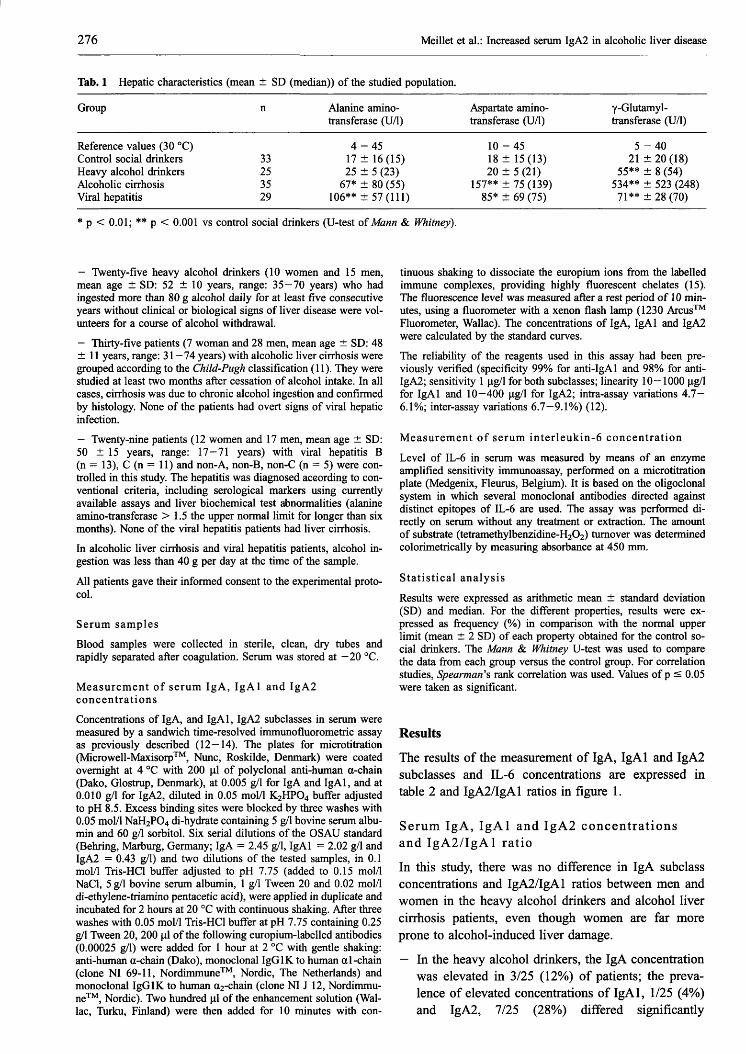
Tab. 1 Hepatic characteristics (mean ± SD (median)) of the studied population.
Group
Reference values (30 °C)Control social drinkersHeavy alcohol drinkersAlcoholic cirrhosisViral hepatitis
n
33253529
Alanine amino-transferase (U/l)
4-4517 ± 16(15)25 ± 5 (23)
67* ± 80 (55)106** ±57 (111)
Aspartate amino-transferase (U/l)
10-4518 ± 15(13)20 ± 5 (21)
157** ± 75 (139)85* ± 69 (75)
γ-Glutamyl-transferase (U/l)
5-4021 ±20(18)
55** ± 8 (54)534** ± 523 (248)71** ±28(70)
* p < 0.01; ** p < 0.001 vs control social drinkers (U-test of Mann & Whitney).
— Twenty-five heavy alcohol drinkers (10 women and 15 men,mean age ± SD: 52 ± 10 years, range: 35-70 years) who hadingested more than 80 g alcohol daily for at least five consecutiveyears without clinical or biological signs of liver disease were vol-unteers for a course of alcohol withdrawal.
— Thirty-five patients (7 woman and 28 men, mean age ± SD: 48±11 years, range: 31 —74 years) with alcoholic liver cirrhosis weregrouped according to the Child-Pugh classification (11). They werestudied at least two months after cessation of alcohol intake. In allcases, cirrhosis was due to chronic alcohol ingestion and confirmedby histology. None of the patients had overt signs of viral hepaticinfection.
— Twenty-nine patients (12 women and 17 men, mean age ± SD:50 ±15 years, range: 17—71 years) with viral hepatitis B(n = 13), C (n = 11) and non-A, non-B, non-C (n = 5) were con-trolled in this study. The hepatitis was diagnosed according to con-ventional criteria, including serological markers using currentlyavailable assays and liver biochemical test abnormalities (alanineamino-transferase > 1.5 the upper normal limit for longer than sixmonths). None of the viral hepatitis patients had liver cirrhosis.In alcoholic liver cirrhosis and viral hepatitis patients, alcohol in-gestion was less than 40 g per day at the time of the sample.
All patients gave their informed consent to the experimental proto-col.
Serum samples
Blood samples were collected in sterile, clean, dry tubes andrapidly separated after coagulation. Serum was stored at —20 °C.
Measurement of serum IgA, IgAl and IgA2concentrationsConcentrations of IgA, and IgAl, IgA2 subclasses in serum weremeasured by a sandwich time-resolved immunofluorometric assayas previously described (12—14). The plates for microtitration(Microwell-Maxisorp™, Nunc, Roskilde, Denmark) were coatedovernight at 4 °C with 200 μΐ of polyclonal anti-human α-chain(Dako, Glostrup, Denmark), at 0.005 g/1 for IgA and IgAl, and at0.010 g/1 for IgA2, diluted in 0.05 mol/1 K2HPO4 buffer adjustedto pH 8.5. Excess binding sites were blocked by three washes with0.05 mol/1 NaH2PO4 di-hydrate containing 5 g/1 bovine serum albu-min and 60 g/1 sorbitol. Six serial dilutions of the OSAU standard(Behring, Marburg, Germany; IgA = 2.45 g/1, IgAl = 2.02 g/1 andIgA2 = 0.43 g/1) and two dilutions of the tested samples, in 0.1mol/1 Tris-HCl buffer adjusted to pH 7.75 (added to 0.15 mol/1NaCl, 5 g/1 bovine serum albumin, 1 g/1 Tween 20 and 0.02 mol/1di-ethylene-triamino pentacetic acid), were applied in duplicate andincubated for 2 hours at 20 °C with continuous shaking. After threewashes with 0.05 mol/1 Tris-HCl buffer at pH 7.75 containing 0.25g/1 Tween 20, 200 μΐ of the following europium-labelled antibodies(0.00025 g/1) were added for 1 hour at 2 °C with gentle shaking:anti-human α-chain (Dako), monoclonal IgGlK to human α 1-chain(clone NI 69-11, Nordimmune™, Nordic, The Netherlands) andmonoclonal IgGlK to human a2-chain (clone NI J 12, Nordimmu-ne™, Nordic). Two hundred μΐ of the enhancement solution (Wai-lac, Turku, Finland) were then added for 10 minutes with con-
tinuous shaking to dissociate the europium ions from the labelledimmune complexes, providing highly fluorescent chelates (15).The fluorescence level was measured after a rest period of 10 min-utes, using a fluorometer with a xenon flash lamp (1230 Arcus™Fluorometer, Wallac). The concentrations of IgA, IgAl and IgA2were calculated by the standard curves.The reliability of the reagents used in this assay had been pre-viously verified (specificity 99% for anti-IgAl and 98% for anti-IgA2; sensitivity 1 μ^Ι for both subclasses; linearity 10—1000 μg/lfor IgAl and 10—400 μg/l for IgA2; intra-assay variations 4.7—6.1%; inter-assay variations 6.7-9.1%) (12).
Measurement of serum interleukin-6 concentration
Level of IL-6 in serum was measured by means of an enzymeamplified sensitivity immunoassay, performed on a microtitrationplate (Medgenix, Fleurus, Belgium). It is based on the oligoclonalsystem in which several monoclonal antibodies directed againstdistinct epitopes of IL-6 are used. The assay was performed di-rectly on serum without any treatment or extraction. The amountof substrate (tetramethylbenzidine-H2O2) turnover was determinedcolorimetrically by measuring absorbance at 450 mm.
Statistical analysis
Results were expressed as arithmetic mean ± standard deviation(SD) and median. For the different properties, results were ex-pressed as frequency (%) in comparison with the normal upperlimit (mean ± 2 SD) of each property obtained for the control so-cial drinkers. The Mann & Whitney U-test was used to comparethe data from each group versus the control group. For correlationstudies, Spearman's rank correlation was used. Values of p s 0.05were taken as significant.
Results
The results of the measurement of IgA, IgAl and IgA2subclasses and IL-6 concentrations are expressed intable 2 and IgA2/IgAl ratios in figure 1.
Serum IgA, IgAl and IgA2 concentrationsand IgA2/IgAl ratio
In this study, there was no difference in IgA subclassconcentrations and IgA2/IgAl ratios between men andwomen in the heavy alcohol drinkers and alcohol livercirrhosis patients, even though women are far moreprone to alcohol-induced liver damage.
— In the heavy alcohol drinkers, the IgA concentrationwas elevated in 3/25 (12%) of patients; the preva-lence of elevated concentrations of IgAl, 1/25 (4%)and IgA2, 7/25 (28%) differed significantly

Meillet et al.: Increased serum IgA2 in alcoholic liver disease 277
Tab. 2 Serum concentration (mean ± SD (median)) of the IgA, IgAl, IgAl and interleukin-6.
Group
Control social drinkersHeavy alcohol drinkersAlcoholic cirrhosisViral hepatitis
n
33253529
IgA (g/1)
2.42 ±0.86 (2.31)2.30 ± 1.30(2.12)
7.61** ±5.29(7.01)4.45** ± 3.03 (3.47)
IgAl (g/1)
2.18 ± 0.73 (2.27)1.77* ± 1.03(1.56)
6.13** ±4.52(4.34)3.66* ± 2.59 (2.60)
IgA2 (g/1)
0.39 ± 0.34 (0.28)0.56** ± 0.31 (0.55)
1.83*** ± 1.93(1.20)0.69* ± 0.67 (0.45)
IL-6 (ng/1)
3.8 ± 1.9(3)4 ± 1.9(3)
34*** ± 33 (22)150* ±322(12)
p < 0.05; ** p < 0.01; *** p < 0.001 vs control social drinkers (U-test of Mann & Whitney).
Viralhepatitis
(n=29)
Fig. 1 IgA2/IgAl ratio (mean ± SD) in control drinkers, in heavydrinkers, and in patients with liver disease.* p < 0.01; ** p < 0.001 vs control drinkers (U-test of Mann &Whitney).
Correlations between serum IgA, IgAl andIgA2, and interleukin-6 concentrations
In the heavy alcohol drinkers, IgAl and IgA2 concentra-tions were positively correlated (p < 0.01). In contrast,in alcohol liver cirrhosis and viral hepatitis patients, theelevated IgAl and IgA2 serum concentrations were in-versely correlated. In patients with alcoholic liver cir-rhosis, a significant positive correlation (p < 0.01) wasobserved between elevated IgAl and IgA2 concentra-tions and IL-6 secretion.
Discussion
The reliability criteria for the time-resolved immunoflu-orometric assay used for the measurement of total IgAand IgAl, and IgA2 subclasses were very satisfactory.This non-isotopic method is simple to perform and itshigh sensitivity makes it suitable for the determinationof serum IgAl and IgA2 concentrations in patients withalcoholic liver disease.
(p < 0.01), with a predominance of IgA2. The IgA2/IgAl ratio was elevated in 8/25 of patients (32%).
— In alcohol liver cirrhosis patients, the IgA concentra-tion was elevated in 22/35 patients (63%); the preva-lence of the elevated concentrations did not differbetween IgAl, 20/35 (57%) and IgA2 22/35 (63%).The IgA2/IgAl ratio was elevated in 9/35 patients(26%).
— In viral hepatitis patients, the IgA concentration waselevated in 12/29 patients (41%), the prevalence ofelevated concentrations of IgAl, 11/29 (38%) andIgA2 5/29 (17%) differed significantly (p <0.01),with a predominance of IgAl. The IgA2/IgAl ratiowas elevated in 3/29 patients (10%).
Serum concentration of interleukin-6
Mean IL-6 concentration was significantly increased inpatients with alcohol liver cirrhosis (p < 0.001) and inviral hepatitis patients (p < 0.05). In this last group,there was a wide range of values (3 — 1230 ng/1). In thesetwo groups, the prevalence of elevated concentrations ofIL-6 was 10/17 (59%) and 12/23 (52%) respectively, anddid not significantly differ.
Heavy alcohol drinkers and alcoholiccirrhosis patients
In the heavy alcohol drinkers and the alcoholic liver cir-rhosis patients, in whom the alcohol factor was present,the study showed an increased IgA2/IgAl ratio. What-ever the total IgA concentration, the ratios in the heavydrinkers and alcoholic cirrhosis patients (0.33 ± 0.12and 0.32 ±0.19, respectively) were significantly dif-ferent from the control social drinkers and the viral hep-atitis patients. This agrees with our previous studies inwhich serum IgA subclass distributions were anlyzed inalcohol liver disease (9). The increase in IgA2/IgAl ra-tio could constitute a diagnostic or predictive marker inheavy alcohol drinkers (prevalence: 32%) evolving tocirrhosis (26%), while this prevalence in viral hepatitispatients is 10%. In heavy alcoholic drinkers withoutclinical liver disease, microvascular changes in the in-testinal tract may be considered as the cause of theplasma protein loss into the jejunal lumen demonstratedby Buell & Beck, and Ray et al. in the dog (16, 17).Several studies have shown the direct toxic effects ofalcohol on intestinal epithelial cells and hepatocytes (18,19). In particular, loss of superficial cells and damage tothe upper layer of mucosa can be observed in the intesti-
278 Meillet et al.: Increased serum IgA2 in alcoholic liver disease
nal epithelium (20). These morphological changes in thesmall bowel epithelium are found even in the absenceof cirrhosis (21). Alcohol increases intestinal permeabil-ity to macromolecules whatever the degree of hepaticdysfunction (22). The primary effect of alcohol may bethe release of pro-inflammatory mediators such as ru-mour necrosis factor- and IL-6 that can increase intesti-nal permeability and local immunoglobulin secretion.Seilies et al. observed too that serum secretory IgA andfree secretory component concentrations were signifi-cantly increased in patients with chronic alcoholic liverdisease, even at a very early stage of the disease, anddecreased after alcohol withdrawal (23).
Alcoholic liver cirrhosis patients
In alcoholic liver cirrhosis patients, the observation ofan increase in both serum IgAl and IgA2 concentrationswith frequencies of 57% and 67% respectively, as wellas the positive correlation with IL-6 concentration,seems to be a major abnormality due to an immune re-sponse against many intestinal antigens and cytokineproduction. IL-6 is responsible for subsequent activationof B cells, resulting in their differentiation into IgA-se-creting plasma cells.
The marked increase in total serum IgA concentrationsin patients with cirrhosis could be due to a secondaryeffect of the initial release of free secretory component/secretory IgA to the plasma compartment, leading to ab-normalities in cytokine regulation (24—26). In addition,the abnormal permeability of the intestinal barrier in al-coholic liver cirrhosis patients could allow an increasedantigenic load in the plasma as food antigens and lipo-polysaccharide (27). Increased IgA synthesis may reflectthis increased antigenic load and the diminished T-cellsuppression, or T-cell independent B-cell stimulation (8,28). The IgA2 antibodies also have lipopolysaccharideand other amphiphilic components of Gram-negativebacteria as targets (29—30). Moreover, peripheral bloodmononuclear cells from patients with alcoholic liver cir-rhosis show an increased lipopolysaccharide-inducedIL-6 secretion, which has been correlated with increasedIgA serum concentrations (10, 26).
Recently, Guillemin et al. have shown that IgA faecaloutput was increased in alcoholic liver cirrhosis patientsin comparison with control social drinkers (31). This ob-servation confirms the possible role of the gut-associatedlymphoid tissue in the serum IgA subclass metabolism.
Intestinal IgA synthesis may be stimulated in alcoholliver cirrhosis patients and could help explain the serumIgA2 subclass origin also. This IgA2 subclass formedthe major subclass contributing to the continuous patternof IgA deposition in hepatic tissues (8). The role of thissubclass seems very important in the extent of the liverdamage and its serum increase may be related to alcoholconsumption.
Viral hepatitis patients
In viral hepatitis patients, while there is an increase inserum IgA2 subclass, the absence of any difference inthe IgA2/IgAl ratio from the control social drinkers sug-gests that the IgA metabolism is different in heavy alco-hol drinkers and alcoholic liver cirrhosis patients. Thepro-inflammatory cytokines (IL-6) induce IgA secretionin both subclasses. The lower significance of the serumIL-6 increase in viral hepatitis patients than in alcoholicliver cirrhosis patients may be explained by the markedheterogeneity of this group (nature of the virus, severityof the infection). In fact, in viral hepatitis patients, thereare probably only slight disturbances in the intestinalpermeability, but the IgA metabolism seems to be pro-foundly disturbed.
Conclusion
The measurement of serum IgA sbuclasses and IgA2/IgAl ratio in patients with either normal or elevated se-rum total IgA could provide a means to detect alcoholicliver patients that are heavy alcoholic drinkers or viralhepatitis patients evolving to clinical complications suchas cirrhosis or damage to the gastro-intestinal tract. Inthese situations, the IgA2 subclass, and the IgA2/IgAlratio in particular, could be specific markers of chronicalcohol abuse directly related to the extent and durationof the alcohol abuse. However, further studies are re-quired to confirm this, including a longitudinal study inheavy drinkers evolving to cirrhosis.
AcknowledgementsThis study was performed with G. E. R. B. A. P. (Groupe d'Evalua-tion et de Recherche des Biologistes de l'Assistance Publique desHöpitaux de Paris, France).
We thank Dr O. Gaillard, Prof. F. Lunel and Prof. E. Seilles fortheir contribution to this study, and C. Hapiot for preparation ofthe manuscript.
References
1. Kutteh WH, Prince SJ, Phillips JO, Spenny JG, Mestecki J.Properties of immunoglobulin A in serum of individuals withliver disease and in hepatic bile. Gastroenterology 1982;82:184-93.
2. Delacroic DL, Vaerman JP. Function of the human liver in IgAhomeostasis, in plasma IgA. Ann NY Acad Sei 1983;409:383-401.

Meillet et al.: Increased serum IgA2 in alcoholic liver disease 279
3. Me Keever U, O'Mahony C, Whelan CA, Weir DG, FeigheryC. Helper and suppressor T lymphocyte function in severe al-coholic liver disease. Clin Exp Immunol 1985; 60:39—48.
4. Noun-Aria KT, Alexander GJM, Portmann BC, Hegarty JE,Eddieston ALWF, Williams RT. B cell function in alcoholicliver disease. J Hepatol 1986; 2:195-207.
5. Berger SR, Helms RA, Bull DM. Cirrhotic hypergammaglo-bulinemia: increased rates of immunoglobulin synthesis by cir-culating lymphoid cells. Dig Dis Sei 1979; 24:741-5.
6. Morgan MY, Ross MGR, Ng CM, Adams DM, Thomas HC,Sherlock S. HLA-B8, immunoglobulins and antibody re-sponses in alcohol-related liver disease. J Clin Pathol 1980;33:488-92.
7. Van de Wiel A, van Hattum J, Schuurman HJ, Kater L. Immu-noglobulin A in the diagnosis of alcoholic liver disease. Gas-troenterology 1988; 94:457-62.
8. Swerdlow MA, Chowdhury LN. IgA subclasses in liver tissuesin alcoholic liver disease. Am J Clin Pathol 1983; 80:238-9.
9. Meillet D, Labrousse F, Benoit MO, van Amerongen G, MüssetL, Hernvann A. Serum IgA subclasses and interleukin-6 inalcoholic liver disease. In: Proceedings of the 8th InternationalColloquium of Pont-a-Mousson — Eurobiology 1992 Sep14-18; John Libbey Eurotext: Paris, 1993; 393-6.
10. Deviere J, Content J, Denys C, van den Bussche L, SchandeneL, Wybran J, et al. High interleukin-6 serum levels andincreased production by leucocytes in alcoholic liver cirrhosis.Correlation with IgA serum levels and lymphokines pro-duction. Clin Exp Immunol 1989; 77:221-5.
11. Pugh RNH, Murray-Lyon IM, Dawson JL, Pietroni MC, Wil-liams N. Transection of the oesophagus for bleeding oesopha-geal varices. Br J Surg 1973; 60:646-9.
12. Meillet D, Gaillard O, Kapel N, Celton N, Magne D, Kapel N,et al. A new method for measurement of IgA subclasses: time-resolved immunofluorometric assay [abstract]. Ann Biol Clin1993; 51:402.
13. Kapel N, Meillet D, Buraud M, Favennec L, Magne D, GobertJG. Determination of anti-Cryptosporidium coproantibodies bytime-resolved immunofluorometric assay. Trans RS Med Hyg1993; 87:330-2.
14. Belec L, Meillet D, Gaillard O, Prazuck T, Michel E, NgondiEkome J, et al. Decreased cervicovaginal production of bothIgAl and IgA2 subclasses in women with AIDS. Clin ExpImmunol 1995; 101:100-6.
15. Hemmila I, Dakubu S, Mukkula VM, Sitari H, Loven T. Euro-pium as a label in time-resolved immunofluorometric assay.Ann Biochem 1984; 137:335-43.
16. Buell MG, Beck IT. Ethanol-induced mucosal micro vascularstasis and enhanced plasma protein loss in the dog jejunum.Gastroenterology 1984; 86:413-20.
17. Ray M, Kinda PK, Beck IT. Mechanism of ethanol-inducedjejunal microvascular and morphologic changes in the dog.Gastroenterology 1989; 96:345-54.
18. Draper LR, Gyure LA, Hall JG, Robertson D. Effect of alcoholon the integrity of the intestinal epithelium. Gut 1983;24:399-404.
19. Lieber CS. Hepatic, metabolic and toxic effects of ethanol:1991 update. Alcohol Clin Exp Res 1991; 15:573-92.
20. Foschi D, Marazzi M, Toti GL, Radaelli E, Ferrante F, VaianiG, et al. Prostaglandin-stimulated recovery of the human duo-denal epithelium: effects of misoprostol on ethanol damage.Am J Gastroenterol 1990; 85:1498-502.
21. Boron-Kaczmarzka A, Hryniewicz A, Kemona A, Sokolewicz-Bobrowska E, Miegoc H. Morphological changes of small in-testine epithelium in the course of post-alcoholic liver cirrho-sis. Drug and Alcohol Dependence 1990; 25:299-303.
22. Worthington BS, Meresol L, Syrotuck JA. Effect of daily etha-nol ingestion on intestinal permeability to macromolecules.Am J Dig Dis 1978; 23:23-32.
23. Seilies E, Rössel M, Vuitton DA, Mercier M, Njoya O, CapronJP, et al. Serum secretory IgA and secretory component in pa-tients with non-cirrhotic alcoholic liver diseases. J Hepatol1995; 22:278-85.
24. Vuitton DA, Seilles E, Cozon G, Rössel M, Bresson-HadniS, Revillard JP. Secretory immunoglobulin A in hepatobiliarydiseases. Dig Dis Sei 1991; 9:78-91.
25. Defrance T, Vanderbielt B, Briere F, Durand I, Bousset F,Banchereau J. Interleukin-10 and transforming growth factor-ß cooperate to induce anti-CD40-activated naive human Bcells to secrete immunoglobulin A. J Exp Med 1992;175:671-82.
26. Deviere J, Content J, Denys C, van den Bussche P, Lemoine O,Schandene L, et al. Immunoglobulin A an interleukin-6 form apositive secretory feedback loop: a study of normal subjectsand alcoholic cirrhotics. Gastroenterology 1992; 103:1296-301.
27. Florent C, Levy VG, Bernier JJ. Perte de proteines intestinaleset cirrhose alcoholique. Etude par la mesure de la clairancefecale de 1 'alpha- 1-anti-trypsine. Gastroenterol Clin Biol 1982;6:68-72.
28. Allison ME, Hodgson HJ. Regulation of peripheral blood B-cell IgA production in alcoholic cirrhosis. J Clin Lab Immunol1989; 30:127-30.
29. Mestecky J, Rüssel MW. IgA subclasses. Monogr Allergy1986; 19:277-301.
30. Tarkowsji A, Lue C, Moldoveanu Z, Kiyono H, Me Ghee JR,Mestecky J. Immunization of humans with polysaccharide vac-cines induces systemic predominantly polymeric IgA2-sub-class antibody response. J Immunol 1990; 144:3770-8.
31. Guillemin A, Kapel N, Meillet D, Magne D, Gobert JG. IgAfecal output in patients with liver cirrhosis. Act Pharm BiolClin 1995; 8:432-5.
Received October 15, 1996/February 5, 1997Corresponding author: Prof. Dominique Meillet, Laboratoire deParasitologie — Mycologie, Faculte de Medecine et Pharmacie,2, Place Saint-Jacques, F-25030 Besa^on Cedex, France