Increased attentional load moves the left to the right · Increased attentional load moves the left...
18
Manuscript accepted for publication in “Journal of Clinical and Experimental Neuropsychology” Increased attentional load moves the left to the right Mario Bonato 1,2 * and Simone Cutini 3,4 1 Department of Experimental Psychology, Ghent University 2 Department of General Psychology, University of Padova 3 Department of Developmental Psychology, University of Padova 4 Center for Cognitive Neurosciences, University of Padova Running head: Load-dependent contralesional mislocalizations * Corresponding author: Dr. Mario Bonato, Department of Experimental Psychology, Ghent University, Henri Dunantlaan 2, B-9000 Ghent, Belgium e-mail: [email protected]
Transcript of Increased attentional load moves the left to the right · Increased attentional load moves the left...

Manuscript accepted for publication in
“Journal of Clinical and Experimental Neuropsychology”
Increased attentional load moves the left to the right
Mario Bonato1,2
* and Simone Cutini3,4
1 Department of Experimental Psychology, Ghent University
2 Department of General Psychology, University of Padova
3 Department of Developmental Psychology, University of Padova
4 Center for Cognitive Neurosciences, University of Padova
Running head: Load-dependent contralesional mislocalizations
* Corresponding author:
Dr. Mario Bonato,
Department of Experimental Psychology, Ghent University,
Henri Dunantlaan 2, B-9000 Ghent, Belgium
e-mail: [email protected]

2
Abstract
Introduction
Unilateral brain damage can heterogeneously alter spatial processing. Very often brain-lesioned
patients fail to report (neglect) items appearing within the contralesional space. Much less often
patients mislocalize items’ spatial position. We investigated whether a top-down attentional load
manipulation (dual-tasking), known to result in contralesional omissions even in apparently
unimpaired cases, might also induce spatial mislocalizations.
Method
Nine right-hemisphere damaged patients performed three computer-based tasks encompassing
different levels of attentional load. The side of appearance of visual targets had to be reported either
in isolation or while processing additional information (visual or auditory dual-task). Spatial
mislocalizations (from the contralesional hemispace towards the ipsilesional -unaffected- one) were
then contrasted with omissions both within and across tasks, at individual as well as at group level.
Results
The representation of ipsilesional targets was accurate and not affected by dual-tasking
requirements. Contralesional targets were instead often omitted and, under dual-task conditions,
also mislocalized by four patients. Three cases reported a significant number of left targets as
appearing on the right (alloesthesia). Two of these patients perceived more targets (albeit to a wrong
spatial location) under dual- than under single-task load.
In a fourth patient, increased visual load resulted in synchiria, the (mis)perception of single,
contralesional targets as being two (one on each side).
Conclusions
When the neural circuitry subtending spatial processing is damaged, an increase in task load can
lead to either a disregard or a bias in the processing of contralesional hemispace. The spatial bias
subtending mislocalizations seems to index a more severe deficit than neglect, as if contralesional
space would be completely erased rather than merely ignored.
Keywords: spatial attention, awareness, alloesthesia, synchiria, allochiria, attentional load, right
hemisphere damage, neglect, extinction.

3
A coherent phenomenological experience of the world requires the integrated processing of several
independent sources of information. For instance, a veridical representation of objects’ positions
implies a successful interplay between attention and spatial awareness. Here we study how this
interplay can be distorted after damage to the right-hemisphere lateralized neural circuitry
subtending spatial processing and representation (Bartolomeo, 2014).
It is well established that right hemisphere lesions can result in a number of disorders
affecting the processing of left hemispace. Among these disorders, neglect and extinction are the
most often described and the most intensely studied (Bartolomeo, Thiebaut de Schotten & Chica,
2012; Driver & Vuilleumier, 2001; Vuilleumier, 2013). Both neglect and extinction affect
awareness for the presence of contralesional items. Neglect typically indicates the omission of
single items presented in the contralesional hemispace. Extinction occurs when a contralesional
target remains unperceived only when a second target is concurrently presented in the ipsilesional
space. It reflects the winner-takes-all functioning principle of the parietal lobes, which implements
the interplay between orienting of spatial attention and awareness (Driver & Vuilleumier, 2001).
Several lines of evidence converge in showing that the presence and degree of spatial
deficits can dramatically increase when several stimuli have to be concurrently processed. Increased
attentional demands can hamper contralesional awareness in two different ways: by enlarging the
degree of neglected space (Sarri, Greenwood, Kalra & Driver, 2009) or, when the eccentricity of
stimuli is kept constant, by increasing the proportion of missed targets (Bonato, 2012, for review).
Due to this dependency on task demands, neglect can be considered as a continuum rather than a
categorical disorder. This implies that several patients might present contralesional disorders only
when their attentional system is taxed, which is not the case when responding to classic, paper-and-
pencil, tests. As a result, neglect often remains undetected by these standard tasks.
The strong relation between attentional load and neglect symptomatology derives from a
series of studies with RHD patients, whereby a striking increase in contralesional omissions
emerged when patients were asked to perform an additional task, either visual or auditory, while
monitoring visual space (Bonato, Priftis, Marenzi, Umiltà & Zorzi, 2010; 2012). Both concurrent
tasks similarly hampered patients’ awareness for the presence of contralesional targets, irrespective
of their different characteristics (Bonato, Priftis, Umiltà & Zorzi, 2013). This finding was
interpreted by assuming the presence of limited and depletable supramodal attentional resources.
After brain damage, contralesional awareness would be present only when attention is fully
allocated to spatial monitoring, as in the case of clinical tests and of the easy, single-task, condition
(see Bonato, 2012, for review; and Marini, Chelazzi & Maravita, 2013 for a study with healthy
participants). Our approach integrates two lines of evidence about load effects on spatial processing.
The first one concerns the detrimental effect of increased visual load (mainly at fixation) upon the
detection of peripheral stimuli (Ball, Owsley, Sloane, Roenker, & Bruni, 1993; Bellgrove, Newman,
Vance, & Mattingley, 2013; Buxbaum, Dawson, & Linsley, 2012; Dawson, Buxbaum, & Rizzo,
2008; Russell, Husain, & Malhotra, 2004; Peers, Cusack and Duncan, 2006; Russell, Malhotra,
Deidda, & Husain, 2013; van Kessel, van Nes, Geurts, Brouwer, Fasotti, 2013; Vuilleumier et al.,
2008). The second one derives from the coupling between visuospatial and non-spatial tasks (e.g.,
cancellation under working-memory load, see Robertson & Frasca, 1992), as well as from studies
showing that deficits of contralesional awareness are exacerbated by multimodal contexts (see
Jacobs, Brozzoli, Hadj-Bouziane, Meunier, Farnè, 2012, for review).
The present study will capitalize on the remarkable sensitivity of the above described
paradigm. We decided to test whether the same bottom-up, dual-task, manipulation which
successfully uncovered subtle deficits in spatial awareness, might interfere with the processes
allowing an accurate representation of the surrounding space. Accordingly, our first aim will be to
investigate whether increased load can unveil mislocalizations. Within the same task,
mislocalizations will be next related to the presence of omissions. By looking at omission we will
address whether omissions imply the absence of contralesional hemispace coding or whether, in
contrast, in omissions contralesional targets can be partly -although unconsciously- processed. To

4
our knowledge, this is the first study implementing a dual-task manipulation to reveal
mislocalizations.
Among spatial disorders characterizing RHD, spatial mislocalizations are much less frequently
described and less understood than omissions. Besides being more difficult to be elicited,
mislocalizations are somehow conceptually difficult to envision because, in normal functioning, the
perception of an object can hardly be dissociated from the perception of its veridical spatial
position. There are, however, some exceptions: when attention is diverted or heavily engaged and
when stimuli are peripheral (Treisman & Schmidt, 1982) even healthy participants can show
deficits in feature processing. For instance, under attentional load, the integration (binding) between
objects' physical characteristics and its spatial position might fail (Treisman, 1996; 1998) resulting
in misattributing a characteristic (e.g., the colour) of one object to another object, thereby causing a
sort of transposition (the moving of one characteristic to another location in space). Several
phenomena, including the systematic presence of illusory conjunctions within the contralesional
hemispace in neglect, strongly support the claim that spatial attention is required for spatial features
integration (see Robertson, 2003, for review). Patients with deficits in orienting spatial attention
towards the contralesional hemispace provide a unique opportunity to study the determinants of
mislocalizations in relation to awareness deficits, also because the performance in the ipsilesional
hemispace offers an individually-calibrated baseline.
The two mislocalization disorders we will investigate here are alloesthesia and synchiria. In
alloesthesia a single, contralesional stimulus is experienced as ipsilesional (Ortigue, Jabaudon,
Landis, Michel, Maravita & Blanke, 2005) whereas in synchiria a single contralesional stimulus is
reported as appearing on both sides (Medina & Rapp, 2008). Thus, synchiria resembles an
alloesthesia-like pattern in the presence of a spared perception of the contralesional stimulus. Both
alloesthesia and synchiria have a spatial origin (and not a visual one). As a consequence, in analogy
with other spatial disorders like neglect and extinction, alloesthesia can be induced both within and
across different sensory modalities (Brozzoli, Demattè, Pavani, Frassinetti, & Farnè, 2006; Ortigue
et al., 2005; Pavani et al., 2004) and synchiria can be modulated by head- and trunk-defined spatial
frames of reference (Medina & Rapp, 2008). Moreover, both synchiria and alloesthesia have been
investigated in multiple sensory modalities. Neppi-Modona (1999) applied tactile stimulation in a
group of right hemisphere damaged patients. His study concluded that, when using a simple
localization task, alloesthesic responses are not common: out of thirty RHD patients, alloesthesic
responses were about 3% for left-sided stimuli and about 1.3% for right-sided stimuli. Moreover, no
lateralized response bias emerged out of a number of catch trials (no stimulation) as high as 2400.
Also sounds mislocalization has been described (Brozzoli et al., 2006). Spatial distortions in the
perceived localization of auditory stimuli can be, at least partly, due to motor neglect and body
schema distortions, in particular when patients are asked to point to the perceived sound position
(Brozzoli et al., 2006; Karnath, 2015). Mislocalizations can emerge under heterogeneous formats
and concurrently with additional deficits. For instance, some previous studies on alloesthesia also
reported transpositions from the ipsilesional to the contralesional hemispace, often occurring
concurrently with palinopsia (Jacobs, 1980; Ardila, Botero, & Gomez, 1987).
In summary, here we investigated whether a dual-tasking procedure could unveil mislocalizations in
a small group of neurological patients which were potentially affected by impaired spatial coding
due to a neural damage in the hemisphere specialized for spatial processing.
Method
Sample
We performed a re-analysis of a dataset already available (cases selected from Bonato et al., 2010;
2012), with the addition of two new patients (see Bonato et al., 2013). The comparisons here
performed are new and substantially different from our previous studies because for the first time
the different types of wrong responses to contralesional stimuli were analyzed.
5
All data had been collected while patients were admitted to the Rehabilitation Center of Conselve
(Padova). Inclusion criteria were the presence of a single right hemisphere lesion and having
performed, with a target duration of 50ms, the computerized single/dual-task described in Bonato et
al., (2012). With short target duration, task sensitivity is maximal because a bottom-up attentional
engagement is added to top-down processes determined by dual-tasking (Bonato, 2015). The final
sample encompassed nine patients (four males) aged 40-85 (mean 67 years) (see Table 1), tested on
average 96 days (range 24-476) after lesion onset.
Neuropsychological testing
All of the patients had been tested with a comprehensive neuropsychological screening (ENB,
Mondini, Mapelli, Vestri, & Bisiacchi, 2003) and with a battery for neglect (Behavioural Inattention
Test, BIT; Wilson, Cockburn, & Halligan, 1987). The ENB includes several subtests assessing
memory, executive functions and visuospatial processing. It includes a drawing copying subtest in
which the patient is asked to copy a drawing of a house in a blank space below the example house.
The BIT consists of three cancellation tasks (Lines, Letters, Stars), four copying, and three drawing
from memory tasks, plus a line bisection subtest.
--- Table 1 about here ---
Computer-based tasks
The computer-based tasks (see figure 1) encompassed one single-task and two dual-tasks. All
stimuli were identical across tasks, whereas instructions were task-specific. In all tasks, after a
central fixation cross (1000 ms) a visual target (a black circle -diameter 8mm-) appeared for 50 ms
on the left, on the right, or bilaterally (eccentricity 135 mm). Concurrently, a letter (a, b, v or z) was
shown at fixation and a number (1, 2, 8 or 9) was presented auditorily. In the single task (ST)
patients only had to report the spatial position of target(s) (i.e., “right”, “left”, or “both” sides), and
to ignore both the visual letter and the auditory number. In the dual-tasks, before reporting the
position of the target(s), patients had to: i) read the letter (visual dual-task, VDT) or ii) count
forward by two twice starting from the heard number (auditory dual-task, ADT). The experimenter
(MB) recorded the responses to the target(s) and to the concurrent task (when present). Performance
was therefore recorded in terms of accuracy only and reaction times were not collected. A noisy
screenshot was then presented until response.
In five patients (Cases 2, 4, 5, 7 & 8) taken from Bonato et al., (2012) also data obtained with a
longer target duration (between 500 and 600 ms) were available.
After having explained the task, the experimenter pointed to the expected position of appearance of
targets on the screen to ensure that patients correctly named left/right/bilateral sides. All patients
were successful in naming/pointing towards the spatial position indicated by the experimenter. Note
that healthy, elderly participants perform at ceiling on these three tasks (Bonato et al., 2010;
Bonato, 2015).

6
Figure 1
A schematic representation of the task (adapted from Bonato, 2012 & 2015) is shown.
Eye movements were recorded with a camcorder focused on patients' eyes. Task order was
counterbalanced across participants. Each of the three tasks encompassed two blocks of 48 trials,
resulting in a total of 288 trials for each patient. Trials contaminated by eye movements (< 2%) and
with a wrong response to the secondary task (≈ 20% in the VDT, ≈ 2% in the ADT) were discarded.
Case 6 was administered with a previous task version whereby auditory numbers were presented in
the ADT condition only.
Double simultaneous stimulation
In order to provide a baseline measurement of visual mislocalizations and omissions in a standard
neurological test patients were asked detect brief, unilateral or bilateral, movements of the index
finger(s) of the examiner (DSS: Double Simultaneous Stimulation). DSS is one of the most sensitive
clinical tasks for quantifying contralesional spatial deficits (Maravita, Posteraro, Husain,
Vuilleumier, Schwartz & Driver, 2007), and broadly resembles the single task condition of our
computer-based experiments. Each patient had to detect 84 visually-presented stimuli (left, right or
bilateral), half for the upper and half for the lower visual field, in a fixed random order (Bisiach,
Cappa, & Vallar, 1983).
Results
Behavioural Inattention Test
Average score at the BIT was 134 out of 146, with two patients out of nine showing a performance
below the cut-off of 130/146 (Case 4: 124 and Case 8: 129, see Table 1). No patient had neglect
according to the omissions at the cancellation tasks (lines, letters and stars cancellation 93 % left vs.
94 % right targets cancelled, all comparisons ns).
Computer-based tasks
Patients’ responses at the computer-based tasks are shown in Table 2. A group analysis was first
performed to compare correct responses across positions and tasks. Individual accuracies were
analyzed by means of a 3 (Target Position: left vs. bilateral vs. right) X 3 (Task: ST vs. VDT vs.
ADT) ANOVA. All effects were significant: the main effect of Target Position, F(2, 16) = 29.1, p <
.001, the main effect of Task, F(2, 16) = 7.6, p < .01, and the Target Position X Task interaction,
F(4, 31) = 3.6, p < .05. The interaction was seemingly due to the absence of differences between
7
tasks (and, more in general, to the limited number of errors) occurring for right-sided targets only
(mean accuracy = 97.2 % in ST, 93.4 % in VDT, and 94.7% in ADT). To confirm this
interpretation, an ANOVA assessing task effects separately for each target side was performed. The
main effect of Task was significant for left, F(2, 16) = 4.7, p < .05 and bilateral targets, F(2, 16) =
7.8, p < .01, but not for right ones F(2, 16) = 1.5, p = .25. Responses to ipsilesional targets were
overall accurate and therefore unaffected by the load manipulation also at individual level (all ps >
.05), see Table 2, right column. Responses to bilateral targets were more accurate in the ST (47%)
than in the VDT, (18%) t(8) = 2.7, p < .05 and in the ADT, (17%) t(8)= 2.9, p < .05. Also for left
targets, accuracy was higher in the ST (56%) than in both the VDT (25%), t(8) = 2.96, p < .05 and
the ADT (33%), t(8) = 1.96, p < .05 (one-tailed), as already documented in Bonato et al., (2013).
We then tried to understand whether such decrease in accuracy was only due to increased omissions
or whether it was, at least in part, due to the appearance of wrong responses for contralesional
targets under dual task load(see later and Fig. 3).
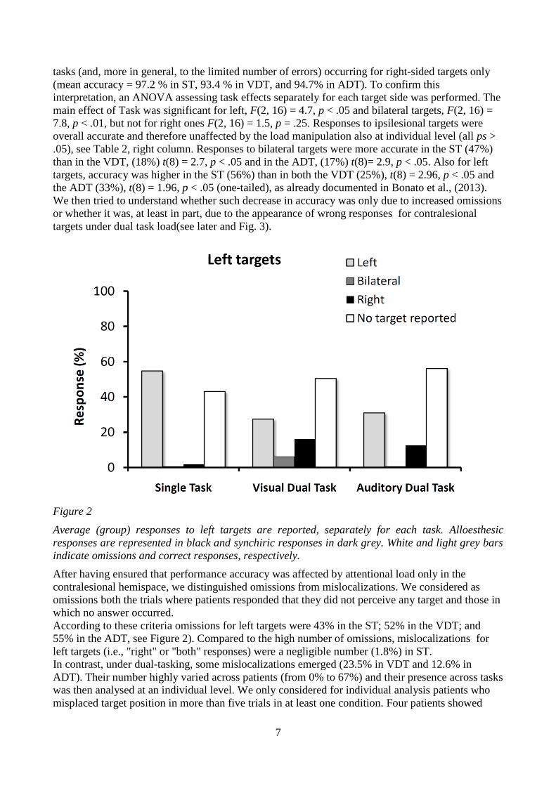
Figure 2
Average (group) responses to left targets are reported, separately for each task. Alloesthesic
responses are represented in black and synchiric responses in dark grey. White and light grey bars
indicate omissions and correct responses, respectively.
After having ensured that performance accuracy was affected by attentional load only in the
contralesional hemispace, we distinguished omissions from mislocalizations. We considered as
omissions both the trials where patients responded that they did not perceive any target and those in
which no answer occurred.
According to these criteria omissions for left targets were 43% in the ST; 52% in the VDT; and
55% in the ADT, see Figure 2). Compared to the high number of omissions, mislocalizations for
left targets (i.e., "right" or "both" responses) were a negligible number (1.8%) in ST.
In contrast, under dual-tasking, some mislocalizations emerged (23.5% in VDT and 12.6% in
ADT). Their number highly varied across patients (from 0% to 67%) and their presence across tasks
was then analysed at an individual level. We only considered for individual analysis patients who
misplaced target position in more than five trials in at least one condition. Four patients showed

8
mislocalizations, three of whom showed alloesthesia and one showed synchiria. These cases are
individually presented below.
--- Table 2 about here ---
Case 2 showed a significant increase in the number of left targets perceived as appearing on the
right (i.e. visual alloesthesia) from 0% (ST) to 67% (VDT), χ2 (1, N = 37) = 21.33 , p < .001,
(Fisher’s Exact Test, here and later). Performance in the ADT (10% of alloesthesic perceptions),
was not significantly different from ST, χ2 (1, N = 38) = 1.9, p = .28.
In Case 4 the number of left targets perceived as appearing on the right significantly increased from
3.1% (ST) to 65% in the VDT, χ2 (1, N = 16) = 9.9, p < .05, and to 34% in the ADT, χ
2 (1, N = 14)
= 8.56, p < .05. The difference between VDT and ADT was not significant, χ2 (1, N = 24) = 1, ns.
Case 8 showed 28% of alloesthesic responses in the ADT, significantly more than in the VDT, χ2
(1, N = 12) = 7.2 , p < .05 but not than in the ST. The difference between the ST (9 %) and the VDT
(3.1%) was not significant, either, χ2 (1, N = 6) = 3, p = .4.
We then tested whether alloesthesia under dual-task conditions was accompanied by a change in the
number of omissions with respect to the ST condition. Indeed, for Cases 2 and 4, that was the case.
Alloesthesia under dual tasking occurred with a concurrent, significant, reduction of contralesional
omissions with respect to ST (all ps < .05, see Table 2). In contrast, the remaining patients showed
more omissions in dual-tasking than in the ST.
One patient (Case 1) showed a different pattern of mislocalizations, called synchiria and consisting
in "bilateral" responses to left targets. In her case synchiric responses emerged selectively in the
VDT (54%), with a significant difference with respect to the ST (4%), χ2 (1, N = 47) = 15.4, p <
.001 and to ADT (3.7%), χ2 (1, N = 50) = 17.2 , p < .001. To assess whether synchiria occurred
selectively for contralesional targets and to exclude the impact of a potential response stereotypy,
the same analysis was performed after having subtracted the percentage of "bilateral" response to
ipsilesional targets (0% in the ST/ADT; 16% in VDT). The difference between the VDT and both
the ST and the ADT remained significant χ2 (1, N = 43) = 11.09, p < .001 and χ
2 (1, N = 46) =
12.49, p < .001 after the above mentioned correction. Case 1 did not show alloesthesia in ST (0%),
VDT (4 %) nor in ADT (0%). As opposed to the other patients she did not show neither omissions
nor extinction under load.
In summary: a significant number of mislocalizations occurred in four patients. Three patients
(Cases 2, 4 and 8) showed alloesthesia (reported left targets as right) and one (Case 1) synchiria
(reported left targets as appearing bilaterally). These mislocalizations were space-selective
(emerged for left targets) and load-selective (emerged under dual-task conditions only; See Figure 3
and Table 2)*.

9
Figure 3
Wrong responses for contralesional items are reported, separately for all patients presenting a
significant rate of mislocalizations and for each task. Cases 2, 4 and 8 presented alloesthesia
(black) whereas Case 1 presented synchiria (grey).
In Cases 2 and 8, two patients with alloesthesia, the “long duration” version gave rise to a
negligible number of alloesthesic responses (2.1% and 3.1%, for Cases 2 and 8, respectively). The
same target duration successfully induced severe extinction (> 95%) under dual-tasks across all
tasks (Case 2) or under dual-tasks only (Case 8). In contrast, patients' detection of contralesional
single targets was relatively spared (omissions < 20%). It is worth reminding that, with the shorter
target duration (50ms), omissions for contralesional single targets were, under single task, as high
as 54.8 % (Case 2) and 90.6% (Case 8) and that alloesthesia emerged under dual-tasks only.
Double simultaneous stimulation
No patient presented a significant number of alloesthesic/synchiric responses (all ps vs. 0 ns) at the
DSS.
Drawing/copying tests
Patients’ performance was also analysed in those paper-and-pencil tasks which might have revealed
spatial mislocalizations. Across all the drawing-from-memory (five) and copying (five) tests
administered to each patient (BIT + ENB) two examples of allochiria emerged in the ENB copy
subtest (see Figure 4). Allochiria is a physical transposition from the contralesional to the
ipsilesional space (Halligan, Marshall & Wade, 1992; Lepore, Conson, Grossi, & Trojano, 2003). It
occurred for Cases 3 and 5, two patients who did not present any mislocalization at the
computerized tasks.

10
Figure 4
The figure shows in its upper part the item (house) to be copied as part of the ENB screening. In the
lower part of the figure the allochiric performance of two patients is visible. Case 5 transposed the
fence from the left to the right (Fig. 4, panel A) whereas Case 3 symmetrically drew two fences, one
for each side of the door showing a sort of object(door) -based synchiria (Fig. 4, panel B).
Discussion
Here we have shown that, after right hemisphere damage, increased attentional load can not only
diminish the processing of contralesional information but also distort its representation. Under dual-
task conditions selective mislocalizations emerged, whereby contralesional targets were reported by
some patients to appear in the ipsilesional hemispace or bilaterally. We used a computer-based task
requiring patients to monitor visual space for target appearance either in isolation or while being
asked to also report an additional stimulus feature. The dual-task manipulation differently affected
contralesional spatial processing. It led four patients to misreport targets' spatial position, whereas it
led to unawareness in the remaining five patients. In contrast with the impairments found for
contralesional hemispace, performance for ipsilesional targets was almost errorless and unaffected
by task load across all patients.
When the neural circuitry subtending spatial processing is damaged, the veridical representation of
spatial location seems to be strongly dependent on the availability of unspecific attentional
resources. Indeed, mislocalizations emerged following purely top-down manipulations rather than
the manipulation of perceptual factors alone (e.g., reduced presentation time). Concurrent task
performance plausibly reduced the availability of attentional resources which otherwise would have
sufficed to effectively process the contralesional hemispace under single-task conditions ( Bonato et
al., 2010). A resource-based explanation can easily account for the good performance in the -less
demanding- DSS testing/single-task conditions, and it is also in line with the fact that patients
presented less severe impairments when targets were presented for a longer duration, customized to
the individual level of impairment (Bonato et al., 2012).

11
It is unlikely that mislocalizations were in fact a response bias, with patients responding "right" or
“bilateral” whenever they did not perceive the target. From a methodological point of view, verbal
responses without time-pressure (as opposed to key-presses, which are sensitive to motor neglect)
are considered an "unbiased" response index (Bisiach, Ricci, Lualdi, & Colombo, 1998; see also
Neppi-Modona, 1999) and are widely used to calculate the incidence of visual disorders after brain
damage (Baier, de Hann, Meller, et al., 2010). From an empirical point of view, this conjecture is at
odds with several aspects of the data. Under dual-task conditions, alloesthesic responses still
occurred together with residual omissions: an automatic “right” response would not have led to
omissions. Moreover, a stereotypic response would be hardly compatible with the good
neuropsychological profiles of the patient and it should have emerged also when target duration was
longer and under single-task conditions. Finally, it would have hardly dissociated across dual tasks
(as found for Cases 1, 2, and 8).
On top of that, spatial displacement from contralesional towards ipsilesional hemispace is thought
to index genuine distortions of contralesional space (see Pavani, Husain, Làdavas & Driver, 2004
for auditory and Liu et al., 2011 for tactile sensory modality). The same logic holds for synchiria.
Indeed, the fact that Case 1 presented several correct detections of contralesional targets and
presented synchiric responses only in one dual-task -and only for left but not for right targets-
allows ruling out the presence of a generalized response bias. If this was the case, "bilateral"
responses would have emerged under both dual-task conditions and also for single, unilateral, right
targets.
The "shift" of left items to the right here described is compatible with the pathological, automatic
orienting of spatial attention which, after right-hemisphere damage, is easily triggered towards
ipsilesional hemispace (Bonato, Priftis, Marenzi & Zorzi, 2009).
The binding of the sensory information into a coherent percept occurs through the coding of the
position of items in space and by linking objects' physical features with their spatial locations
(Cohen & Rafal, 1991; Robertson, 2003; Treisman, 1996). Since the processing of objects’ shape
and location occurs through separate cognitive systems and neural structures, only when the binding
is effective a coherent perception of features in space can emerge (Robertson, 2003).
Present findings support the view that the binding process requires spatial attention resources to
successfully occur (Marcel et al, 2004; Treisman, 1996; 1998). Indeed, we showed that under load
the representations of items are mislocated FROM a contralesional location -where spatial attention
can only in some contexts be allocated as if “no reliable space” would exist there- TOWARDS a
location where spatial attention is pathologically attracted.
Present data also suggest that the heterogeneous consequences of increased load across patients
might be, at least in part, related to the initial severity of the deficit. When contralesional awareness
was already low in the single-task, load increase more easily led to spatial transpositions. This
conjecture seems to be confirmed by the data: with the exception of the only non-vascular patient
(Case 7) no patient with a “baseline” extinction below 50% presented alloesthesia. In contrast,
three out of four patients with “baseline” omissions above 50% showed alloesthesia and all patients
with alloesthesia also presented an extinction rate > 90 % in the same conditions. In contrast, when
fewer omissions were present under single-task conditions, load increase more probably resulted in
omissions (rather than in transpositions).
These data do not support the -admittedly tempting- possibility to conceive alloesthesia as an
intermediate step in a gradient leading from neglect to extinction. It seems, in contrast, that
alloesthesia (under dual task) was only present in patients showing several omissions under ST. In
the two patients with severe alloesthesia (i.e., > 50%) more targets were omitted in the single than
in the dual-task condition. It therefore seems as if the same stimuli, which were not consciously
perceived under single-task conditions, "re-entered" awareness under load, albeit at a wrong
location. Thus, mislocalizing may be worse than omitting. The present findings seem to suggest that
alloesthesia might be due to a distortion of the contralesional spatial map, whilst neglect would

12
instead be present when a failure in the access to such map (Robertson, 2003) occurs. This
interpretation would be consistent with the body of evidence showing that contralesional stimuli
which are not consciously perceived can nonetheless influence performance of neglect patients.
Supporting evidence comes from priming (Marshall & Halligan, 1988), from redundant-target
effects (Marzi et al., 1996) and from the physiological activation of primary visual areas in the
absence of conscious perception (Rees, Wojciulik, Clarke, Husain, Frith & Driver, 2000).
Counterintuitively, alloesthesia might be then somehow considered a spatial impairment more
"disruptive" than neglect, rather than the opposite. It seems as if load would not allow creating a
representation of contralesional space rather than merely hampering its access as if the “only” space
available for representing spatial positions was the ipsilesional one.
In summary, computer-based tasks revealed load-dependent mislocalizations and the presence of a
gradient in the severity of unawareness for contralesional hemispace from extinction as less severe
deficit, to then go to neglect, and, finally, to alloesthesia.
According to BIT scores, most of the patients had no neglect. It might therefore be interesting to
speculate about the pattern of results which should be expected in a clinical sample characterized by
severe neglect. These hypothetical patients will no doubt omit many contralesional targets already
in the ST. The question then becomes whether they will present alloesthesic responses already in
the ST, and whether they will present, under dual-task, more mislocalizations than the group here
described. We speculate that, as in the sample we tested, mislocalizations would not affect all
patients and synchiria will be uncommon. It seems also plausible to predict, in the most affected
patients, a severe alloesthesic pattern under dual-tasking. Finally, the relatively high (16%)
percentage of synchiric responses to ipsilateral trials, as well as its presence in one patient and in
one condition only, calls for caution when the potential frequency of presentation of this disorder
has to be estimated.A second dissociation was found between synchiria and neglect/extinction, and
it is supported by the fact that Case 1 showed synchiria with no hint of neglect and extinction nor
alloesthesia, neither at clinical testing nor under computer-based tasks.
The data suggest that spatial processing is multicomponential and that its proper functioning
requires the integration of several different processes subtending awareness and spatial localization.
As a result of a disruption to the mechanisms subtending spatial processing, a target can be
consciously perceived but in a wrong spatial location. This impaired spatial coding in the presence
of preserved awareness complements recent evidence suggesting the possibility of preserved spatial
coding in the presence of impaired awareness (Treccani, Cubelli, Sellaro, Umiltà & Della Sala,
2012). Moreover, the fact that targets which were not consciously perceived in single task
conditions were mislocated under dual task conceptually replicates the Treccani et al. findings.
Patients with alloesthesia under visual load (Cases 2 and 4) might have presented, in the single task,
some representation for the presence of contralesional targets which could however not enter
awareness.
The dissociation between mislocalizations on the computerized tasks and allochiria on the drawing
copying (see Halligan et al., 1992) suggests that, in allochiria, transpositions might be due to the
modality of output. The potential presence of directional hypokinesia would then affect motor
(allochiria) but not verbal (alloesthesia/synchiria) responses. According to this speculation,
alloesthesia would then be a more veridical index of spatial distortions.
Crucially from a clinical perspective, the present results suggest that standard neurological (DSS)
approach is not sufficiently sensitive to induce and detect mislocalizational disorders. The ceiling
performance found at the DSS resembled the absence of mislocalizations found under computer-
based, single-task conditions, suggesting that the brief presentation of stimuli is a necessary but not
a sufficient condition for alloesthesia to occur. Moreover this suggests that low-demand tasks,
despite being very sensitive for the detection of omissions, are still not sufficiently demanding to
induce (nor sensitive to detect) mislocalizations, confirming the above proposed severity continuum
between extinction, neglect and alloesthesia.

13
Rather obviously, it is not the use of a computer in itself but rather the requirement to concurrently
process various stimuli which results in mislocalizations (Bonato & Deouell, 2013). On top of that,
it is possible that some alloesthesic responses might in fact go undetected in the presence of
extinction (Medina & Rapp, 2008).
The degree of initial neglect severity provides a potential explanation as to why in some patients the
load manipulation leads to omissions whereas in some others it led to mislocalizations. However,
this does not imply that all neglect patients with severe neglect must present mislocalizations under
load. In the presence of damage to the circuitry subtending spatial processing, the need to devote
attention to several aspects of the stimuli seems be crucial to induce mislocalizations. Evidence by
research in the multimodal domain supports this possibility (Ortigue et al., 2005). The patient
described by Ortigue et al., (2005) showed alloesthesia for contralesional task-relevant stimuli,
either auditory or tactile when task irrelevant, cross modality, distracters (auditory for tactile stimuli
and tactile for auditory ones) were presented within the ipsilesional hemispace.
A major limitation of the present study is the absence of lesion mapping due to the unavailability of
neuroradiological images for two patients. Even taking into account the scans we had available, the
lesions resembled the well-known heterogeneity of the areas potentially leading to neglect. As a
consequence of the limited sample and of the heterogeneity of the lesions, any anatomically-based
explanation for the presence of these dissociations at individual level would seem unjustified.
Another limitation is the presence of a large variability in the degree and characteristics of
mislocalizations and omissions. For instance, whereas for Cases 1, 2, and 4 performance was less
impaired in ADT than in VDT, for Case 8 the opposite was true.
Even focusing on omissions only, Case 6 was characterized by a larger number of them in the ADT
than in the VDT. This could be easily explained by assuming a “novelty effect” due to the auditory
presentation (Case 6 underwent the auditory number presentation in the ADT only). This
conjecture, however, falls short in explaining the reason why the same patient, under bilateral target
presentation, presented an identical rate of omissions (extinction) in both dual tasks. It therefore
remains currently unexplained the heterogeneity of the factors leading to omissions and
mislocalizations. It is possible that deficits emerged in those conditions which were individually
more difficult, but this is, for the moment, only a speculation.
Nonetheless, the current results clearly illustrate the deficits that can arise in the computation of the
spatial locations of stimuli and in the integration of position information into a coherent spatial
representation.
Appendix
The definitions of alloesthesia and allochiria remarkably vary across studies (Halligan et al., 1992;
Jones, 1907; Lepore et al., 2003; Marcel, Postma, Gillmeister, Cox, Rorden, Nimmo-Smith et al.,
2004; Meador, Allen, Adams & Loring, 1991; Medina & Rapp, 2008; Obersteiner, 1882; Ortigue et
al., 2005; Treccani et al., 2012). We have adopted the term “alloesthesia” (Ortigue et al., 2005) to
indicate the mislocalization of contralesional items within the ipsilesional hemispace in those
contexts (e.g. the computer-based task we administered) without motor component. We used the
term “allochiria” to indicate the occurrence of spatial transpositions from the contralesional to the
ipsilesional hemispace only within copying/drawing from memory tasks, that is when the target
item had to be “physically” reproduced (Halligan et al., 1992).
Even though, broadly speaking, extinction might also be considered a type of mislocalization (e.g.
an alloesthesic phenomenon), we preferred to classify it (together with neglect) as an awareness
deficit.
14
Acknowledgements
This study was supported by a FWO Pegasus/Marie Curie Intra European Fellowship within the 7th
European Community Framework Program to M.B. S.C. was supported by grant STPD11B8HM
from the University of Padova.
MB is grateful to K. Priftis and to two anonymous reviewers for helpful suggestions on a previous
version of this paper; to M. Zorzi, C. Umiltà, P. Bartolomeo and R. Cubelli for useful discussions,
and to J-P van Dijck for proofreading.
References
Ardila, A., Botero, M., & Gomez, J. (1987). Palinopsia and Visual Allesthesia. International
Journal of Neuroscience, 32, 775-782.
Baier, B., de Haan, B., Mueller, N., Thoemke, F., Birklein, F., Dieterich, M., & Karnath, H.-O.
(2010). Anatomical correlate of positive spontaneous visual phenomena: a voxelwise lesion study.
Neurology, 74, 218-222.
Ball, K., Owsley, C., Sloane, M.E., Roenker, D.L., & Bruni, J.R. (1993). Visual attention problems
as a predictor of vehicle crashes in older drivers. Investigative Ophthalmology Visual Science, 34,
3110-3123.
Bartolomeo, P. (2014) Attention disorders after right brain damage. Living in halved worlds. New
York: Springer.
Bartolomeo, P., Thiebaut de Schotten, M., & Chica, A. B. (2012). Brain networks of visuospatial
attention and their disruption in visual neglect. Frontiers in Human Neuroscience, 6:110.
Bellgrove, M.A., Eramudugolla, R., Newman, D.P., Vance, A., & Mattingley, J.B. (2013).
Influence of attentional load on spatial attention in acquired and developmental disorders of
attention. Neuropsychologia, 51, 1085-1093.
Bisiach, E., Cappa, S.F., & Vallar, G. (1983). Guida all’esame neuropsicologico. Milano, Italy:
Raffaello Cortina Editore.
Bisiach, E., Ricci, R., Lualdi, M., & Colombo, M.R. (1998). Perceptual and response bias in
unilateral neglect: Two modified versions of the milner landmark task. Brain and Cognition, 37,
369-386.
Bonato, M. (2012). Neglect and extinction depend greatly on task demands: a Review. Frontiers in
Human Neuroscience, 6:195.
Bonato, M. (2015). Unveiling residual, spontaneous recovery from subtle hemispatial neglect three
years after stroke. Frontiers in Human Neuroscience 9:413. doi:10.3389/fnhum.2015.00413
Bonato, M., Priftis, K., Marenzi, R., Umiltà, C., & Zorzi, M. (2010). Increased attentional demands
impair contralesional space awareness following stroke. Neuropsychologia, 48, 3934-3940.
Bonato, M., Priftis, K., Marenzi, R., Umiltà, C., & Zorzi, M. (2012). Deficits of contralesional
awareness: A case study on what paper-and-pencil tests neglect. Neuropsychology, 26, 20-36.
Bonato, M., Priftis, K., Umiltà C., & Zorzi, M. (2013). Computer-based testing unveils severe
neglect in apparently intact patients. Behavioural Neurology, 25, 1-3.
Brozzoli, C., Demattè, M.L., Pavani, F., Frassinetti, F., & Farnè, A. (2006) Neglect and extinction:
within and between sensory modalities. Restorative Neurology and Neuroscience, 24, 217-232.
Buxbaum, L.J., Dawson, A.M., & Linsley, D. (2012). Reliability and validity of the Virtual Reality
Lateralized Attention Test in assessing hemispatial neglect in right-hemisphere stroke.
Neuropsychology, 26, 430-441.
Dawson, A., Buxbaum, L.J., & Rizzo, A.A. (2008). The virtual reality lateralized attention test:
Sensitivity and validity of a new clinical tool for assessing hemispatial neglect. IEEE XPlore,
Virtual Rehabilitation 88-92. doi:10.1109/ICVR.2008.4625140
Dehaene, S., Kerszberg, M., & Changeux J. P. (1998). A neuronal model of a global workspace in
effortful cognitive tasks. Proceedings of National Academy of Sciences USA, 95, 14529-14534.
15
Driver, J., & Vuilleumier, P. (2001). Perceptual awareness and its loss in unilateral neglect and
extinction. Cognition, 79, 39-88.
Ferber, S., & Karnath, H.O. (2001). How to assess spatial neglect-line bisection or cancellation
tasks? Journal of Clinical and Experimental Neuropsychology, 23, 599-607.
Halligan, P.W., & Marshall, J.C., (1991). Left neglect for near but not far space in man. Nature,
350, 498-500.
Halligan, P.W., Marshall, J.C., & Wade, D. (1992). Left on the right: Allochiria in a case of left
visuo-spatial neglect. Journal of Neurology, Neurosurgery, and Psychiatry, 55, 717-719.
Jacobs, L. (1980). Visual allesthesia. Neurology, 30, 1059-1063.
Jacobs, S., Brozzoli, C., Hadj-Bouziane, F., Meunier, M., & Farnè, A. (2011). Studying
multisensory processing and its role in the representation of space through pathological and
physiological crossmodal extinction. Frontiers in Psychology 6, 2:89. doi:
10.3389/fpsyg.2011.00089. Jones, E. (1907). The precise diagnostic value of allochiria. Brain,
30,490-532.
Karnath, H.-O. (2015). Spatial attention systems in spatial neglect. Neuropsychologia, 75, 61-73.
Kinsbourne, M., & Warrington, E.K. (1962). A disorder of simultaneous form perception. Brain,
85, 461-486.
Lepore, M., Conson, M., Grossi, D., & Trojano, L. (2003). On the different mechanisms of spatial
transpositions: a case of representational allochiria in clock drawing. Neuropsychologia, 41, 1290-
1295.
Liu, C.C., Veldhuijzen, D.S., Ohara, S., Winberry, J., Greenspan, J.D., & Lenz, F.A. (2011). Spatial
attention to thermal pain stimuli in subjects with visual spatial hemi-neglect: extinction,
mislocalization and misidentification of stimulus modality. Pain, 152, 498-506.
Magni, E., Benedetti, G., Bianchetti, A., Rozzini, R., & Trabucchi, M. (1996). Mini-Mental state
examination: a normative study in italian elderly population. European Journal of Neurology 3: 1-5.
Maravita, A., Posteraro, L., Husain, M., Vuilleumier, P., Schwartz, S., & Driver, J. (2007). Looking
at human eyes affects contralesional stimulus processing after right hemispheric stroke. Neurology,
69, 1619-1621.
Marcel, A., Postma, P., Gillmeister, H., Cox, S., Rorden, C., Nimmo-Smith, I. & Mackintosh, B.J.
(2004). Migration and fusion of tactile sensation: premorbid susceptibility to Allochiria, neglect and
extinction? Neuropsychologia, 42, 1749-1767.
Marini, F., Chelazzi, L., & Maravita, A. (2013). The costly filtering of potential distraction:
evidence for a supramodal mechanism. Journal of Experimental Psychology: General, 142, 906-
922.
Marzi, C.A., Smania, N., Martini, M.C., Gambina, G., Tomelleri, G., Palamara, A., Alessandrini,
F., & Prior, M. (1996) Implicit redundant- targets effect in visual extinction Neuropsychologia, 34,
9-22.
Meador, K.J., Allen, M.E., Adams, R.J., & Loring, D.W. (1991). Allochiria vs Allesthesia. Is There
a Misperception? Archives of Neurology, 48, 546-549.
Medina, J., & Rapp, B. (2008). Phantom tactile sensations modulated by body position. Current
Biology, 18, 1937-1942.
Milner, A.D., Harvey, M., & Pritchard, C.L. (1998). Visual size processing in spatial neglect.
Experimental Brain Research, 123, 192-200.
Mondini, S., Mapelli, D., Vestri, A., & Bisiacchi, P. (2003). Esame Neuropsicologico Breve.
Milano, Italy: Raffaello Cortina Editore.
Neppi-Modona, M. (1999). Influence of response modality on awareness of contralesional tactile
stimuli in right brain damaged patients. Cortex, 35, 687-699.
Obersteiner, H. (1882). On allochiria. Brain, 4, 153-168.
Ortigue, S., Jabaudon, D., Landis, T., Michel, C. M., Maravita, A., & Blanke, O. (2005).
Preattentive interference between touch and audition: a case study on multisensory alloesthesia.
Neuroreport, 16, 865.

16
Pavani, F., Husain, M., Ládavas, E., & Driver, J. (2004). Auditory deficits in visuospatial neglect
patients. Cortex, 40, 347-365.
Peers, P.V., Cusack, R., & Duncan J. (2006). Modulation of spatial bias in the dual task paradigm:
evidence from patients with unilateral parietal lesions and controls. Neuropsychologia, 44, 1325-
1335.
Rees, G., Wojciulik, E., Clarke, K., Husain, M., Frith, C., & Driver, J. (2000). Unconscious
activation of visual cortex in the damaged right hemisphere of a parietal patient with extinction.
Brain, 123, 1624-1633.
Robertson, I.H., Manly, T., Andrade, J., Baddeley, B.T., & Yiend, J. (1997). 'Oops!': performance
correlates of everyday attentional failures in traumatic brain injured and normal subjects.
Neuropsychologia, 35, 747-758.
Robertson I.H., & -Frasca R. (1992). Attentional load and visual neglect. International Journal
Neuroscience, 62, 45-56.
Robertson, L.C. (2003). Binding, spatial attention and perceptual awareness. Nature Reviews
Neuroscience, 4, 93-102.
Ronchi, R., Posteraro, L., Fortis, P., Bricolo, E., & Vallar, G. (2009). Perseveration in left spatial
neglect: drawing and cancellation tasks. Cortex, 45, 300-312.
Russell, C., Husain, M., & Malhotra, P. (2004). Attention modulates the visual field in healthy
observers and parietal patients. Neuroreport, 15, 2189-2193.
Russell, C., Malhotra, P., Deidda, C., & Husain, M., (2013). Dynamic attentional modulation of
vision across space and time after right hemisphere stroke and in ageing. Cortex, 49, 1874-1883.
Sagiv, N. & Robertson, L.C. (2005). Synesthesia and the Binding Problem. In L.C. Robertson & N.
Sagiv (Eds.), Synesthesia: Perspectives from Cognitive Neuroscience (pp.90-107). New York:
Oxford University Press.
Sarri, M., Greenwood, R., Kalra, L., & Driver, J. (2009). Task-related modulation of visual neglect
in cancellation tasks. Neuropsychologia, 47, 91-103.
Schneider, W., Eschman, A., & Zuccolotto, A. (2002). E-Prime User's Guide. Pittsburgh:
Psychology Software Tools, Inc.
Spillane, J. (1938). Olfactory alloesthesia. Brain, 61, 393-401.
Treccani, B., Cubelli, R., Sellaro, R., Umiltà, C., & Della Sala, S. (2012). Dissociation between
awareness and spatial coding: evidence from unilateral neglect. Journal of Cognitive Neuroscience,
24, 854-867.
Treisman, A., & Schmidt, H. (1982). Illusory conjunctions in the perception of objects. Cognitive
Psychology, 14, 107-141.
Treisman, A. (1996). The binding problem. Current Opinion Neurobiology, 6, 171-178.
Treisman, A. (1998). Feature binding, attention and object perception. Philosophical Transactions
of the Royal Society of London: Series B, Biological Sciences, 353, 1295-1306.
van Kessel, M.E., van Nes, I.J., Geurts, A.C., Brouwer, W.H., & Fasotti, L. (2013). Visuospatial
asymmetry in dual-task performance after subacute stroke. Journal of Neuropsychology, 7, 72-90.
Vuilleumier, P. (2013). Mapping the functional neuroanatomy of spatial neglect and human parietal
lobe functions: progress and challenges. Annals New York Academy Science, 1296, 50-74.
Vuilleumier, P., Schwartz, S., Verdon, V., Maravita, A., Hutton, C., Husain, M., & Driver, J.
(2008). Abnormal attentional modulation of retinotopic cortex in parietal patients with spatial
neglect. Current Biology, 18, 1525-1529.
Wilson, B., Cockburn, J., & Halligan, P. W. (1987). The behavioural inattention test. Bury St.
Edmunds, UK: Thames Valley Test Company.

17
Table 1 Sample characteristics and neuropsychological profiling
Patient number
Age (yrs)
Sex
Education (yrs)
Days from onset
MMSE Lesion Ethiolo
gy
Lesion site
BIT
Lines L
Lines R
Letters L
Letters R
Stars L
Stars R
1 78 F 4 476 22 (21) Is T, P, SC
134 18 18 20 17 20 25
2 85 M 13 40 26.3 (26) He F, P, SC
131 18 18 17 19 26 26
3 64 F 5 34 26 Is F, I, P,
SC 138 18 18 20 15 27 27
4 77 M 5 76 25.7 (26) Is F, I, T,
SC 124 18 18 9 13 26 27
5 73 F 5 61 27.3 (28) He SC, C 135 18 18 20 18 22 24
6 61 M 13 31 28 Is I, T, SC 145 18 18 20 19 27 27
7 53 M 8 75 27 He SC 131 18 18 17 18 24 27
8 40 F 8 47 28 Ca P, SC 129 18 18 17 17 26 23
9 74 F 2 24 19.7 (19) He SC 136 18 18 15 19 25 26
Lesion site:
F = frontal; P = parietal; T = temporal; I = insula; C = capsular; SC = Subcortical
MMSE: For patients aged 65 or more, raw scores (between brackets) have been corrected according
to Magni et al. (1996).
Etiology:
Is = ischemic; He= hemorrhagic.
BIT: For each cancellation subtest (Lines, letters, stars), the number of detected items is shown,
separately for the left (L) and the right (R) half of the testing sheet. Scores ≤ 129 are to be
considered pathological.
Table 2: Response percentage according to target side and type of task.
Left Target Bilateral Target Right Target
Response (%)
Patient number
Task 0 n Left Bilat s Right a 0 Left Bilat Right e 0 Left Bilat Right
1 ST 4 92 4 0 0 0 93.8 6.3 0 0 0 100
VDT 0 41.7 54.2 4.2 0 0 96.3 3.7 0 0 16 84
ADT 0 96.3 3.7 0 0 6.7 83.3 10 0 3.4 0 96.6
2 ST 54.8 45.2 0 0 0 9.7 6.5 83.9 0 0 0 100
VDT 14.8^ 18.5 0 66.7 0 0 0 100 0 0 0 100
ADT 20* 70 0 10 3.2 0 3.2 93.5 3.2 0 0 96.8
3 ST 9.4 90.6 0 0 3.1 0 68.8 28.1 0 0 0 100
VDT 90.3* 0 0 9.7 0 0 0 100* 0 0 0 100
ADT 90.6* 0 0 9.4 0 0 0 100* 0 0 0 100

18
4 ST 90.6 6.3 0 3.1 0 0 0 100 0 0 0 100
VDT 35^ 0 0 65 4.5 0 0 95.5 0 0 0 100
ADT 65.6^ 0 0 34.4 6.3 0 0 93.8 0 0 0 100
5 ST 9.7 90.3 0 0 9.4 0 87.5 3.1 0 0 0 100
VDT 35.7* 60.7 3.6 0 0 3.4 48.3 48.3* 0 0 4 96
ADT 37.5* 50 0 12.5 0 0 48.4 51.6* 3.2 0 0 96.8
6 ST 0 100 0 0 0 0 93.8 6.3 0 0 0 100
VDT 6.5 93.5 0 0 0 0 18.8 81.3* 0 0 0 100
ADT 34.5* 65.5 0 0 0 0 19.4 80.6* 0 0 0 100
7 ST 84.4 12.5 0 3.1 0 0 6.3 93.8 6.3 0 0 93.8
VDT 95.2 0 0 4.8 8 0 0 92 5 0 0 95
ADT 84.4 3.1 0 12.5 3.3 0 0 96.7 6.9 0 0 93.1
8 ST 90.6 0 0 9.4 0 0 6.3 93.8 6.3 0 3.1 90.6
VDT 90.6 6.3 0 3.1 0 0 0 100 3.4 0 0 96.6
ADT 71.9 0 0 28.1 3.1 0 0 96.9 6.3 0 0 93.8
9 ST 33.3 66.7 0 0 9.4 0 62.5 28.1 6.3 3.1 0 90.6
VDT 100* 0 0 0 57.1 0 0 42.9* 30.8 0 0 69.2
ADT 87.5* 9.4 0 3.1 18.8 0 0 81.3* 25 0 0 75
ST: Single-Task
VDT: Visual Dual-Task
ADT: Auditory Dual-Task
n = neglect,
s = synchiria,
a = alloesthesia,
e = extinction
Bold: correct response
Shaded, light grey: synchiria = significant increase in “bilateral” responses to left targets with
respect to ST
Shaded, dark grey: alloesthesia = significant increase in “right” responses to left targets with respect
to ST
* = significant increase in omissions (unilateral left targets) or extinction (bilateral targets) with
respect to ST
^ = significant decrease in omissions (unilateral left targets) with respect to ST