Inceoglu AB, Clifton HL, Yang J, Hegedus C, Hammock BD
15
Inhibition of soluble epoxide hydrolase limits niacin-induced vasodilation in mice A. B. Inceoglu, Ph.D. 2 , H.L. Clifton, B.S. 1 , J. Yang, Ph.D. 2 , C. Hegedus, Ph.D. 2 , B. D. Hammock, Ph.D. 2 , and S. Schaefer, M.D. 1 1 Department of Internal Medicine University of California Davis 2 Department of Entomology University of California Davis Abstract Background—The use of niacin in the treatment of dyslipidemias is limited by the common side effect of cutaneous vasodilation, commonly termed flushing. Flushing is thought to be due to release of the vasodilatory prostanoids PGD 2 and PGE 2 from arachidonic acid metabolism through the cyclooxygenase (COX) pathway. Arachidonic acid is also metabolized by the cytochrome P450 system which is regulated, in part, by the enzyme soluble epoxide hydrolase (sEH). Methods: These experiments used an established murine model in which ear tissue perfusion was measured by laser Doppler to test the hypothesis that inhibition of sEH would limit niacin-induced flushing. Results: Niacin-induced flushing was reduced from 506 ± 126 to 213 ± 39 % in sEH knockout animals. Pharmacologic treatment with 3 structurally distinct sEH inhibitors similarly reduced flushing in a dose dependent manner, with maximal reduction to 143±15% of baseline flow using a concentration of 1 mg/kg TPAU (1-trifluoromethoxyphenyl-3-(1-acetylpiperidin-4- yl) urea). Systemically administered PGD 2 caused ear vasodilation which was not changed by either pharmacologic sEH inhibition or by sEH gene deletion. Conclusions: Inhibition of sEH markedly reduces niacin-induced flushing in this model without an apparent effect on the response to PGD 2 . sEH inhibition may be a new therapeutic approach to limit flushing in humans. Keywords nicotinic acid; prostaglandins; arachidonic acid; flushing; dyslipidemia; epoxide hydrolase Introduction Nicotinic acid, or niacin, is a water-soluble B vitamin which, in high doses, is effective in treating dyslipidemias [1, 2] and reduces both cardiovascular mortality and cardiac events [3]. However, the use of niacin is limited by a major side effect, cutaneous vasodilation, which is commonly described as flushing involving the head and upper torso [4]. Flushing occurs in up to 90% of patients, results in discontinuation in approximately 30% of patients [5] and is only marginally suppressed by interventions such as intake with food and pre- treatment with COX inhibitors such as aspirin [6]. Flushing is not a cosmetic side effect but often an intense, though transient, response and a major factor for patient non-compliance that greatly reduces the potential utility of niacin. Indeed, the approval in the European Address for reprints: Saul Schaefer, M.D. One Shields Avenue TB 172 Davis, CA 95616 tel: (530) 752-0718 [email protected]. This is a PDF file of an unedited manuscript that has been accepted for publication. As a service to our customers we are providing this early version of the manuscript. The manuscript will undergo copyediting, typesetting, and review of the resulting proof before it is published in its final citable form. Please note that during the production process errors may be discovered which could affect the content, and all legal disclaimers that apply to the journal pertain. NIH Public Access Author Manuscript J Cardiovasc Pharmacol. Author manuscript; available in PMC 2013 July 01. Published in final edited form as: J Cardiovasc Pharmacol. 2012 July ; 60(1): 70–75. doi:10.1097/FJC.0b013e3182580a5d. NIH-PA Author Manuscript NIH-PA Author Manuscript NIH-PA Author Manuscript
Transcript of Inceoglu AB, Clifton HL, Yang J, Hegedus C, Hammock BD

Inhibition of soluble epoxide hydrolase limits niacin-inducedvasodilation in mice
A. B. Inceoglu, Ph.D.2, H.L. Clifton, B.S.1, J. Yang, Ph.D.2, C. Hegedus, Ph.D.2, B. D.Hammock, Ph.D.2, and S. Schaefer, M.D.11Department of Internal Medicine University of California Davis2Department of Entomology University of California Davis
AbstractBackground—The use of niacin in the treatment of dyslipidemias is limited by the common sideeffect of cutaneous vasodilation, commonly termed flushing. Flushing is thought to be due torelease of the vasodilatory prostanoids PGD2 and PGE2 from arachidonic acid metabolism throughthe cyclooxygenase (COX) pathway. Arachidonic acid is also metabolized by the cytochromeP450 system which is regulated, in part, by the enzyme soluble epoxide hydrolase (sEH).Methods: These experiments used an established murine model in which ear tissue perfusion wasmeasured by laser Doppler to test the hypothesis that inhibition of sEH would limit niacin-inducedflushing. Results: Niacin-induced flushing was reduced from 506 ± 126 to 213 ± 39 % in sEHknockout animals. Pharmacologic treatment with 3 structurally distinct sEH inhibitors similarlyreduced flushing in a dose dependent manner, with maximal reduction to 143±15% of baselineflow using a concentration of 1 mg/kg TPAU (1-trifluoromethoxyphenyl-3-(1-acetylpiperidin-4-yl) urea). Systemically administered PGD2 caused ear vasodilation which was not changed byeither pharmacologic sEH inhibition or by sEH gene deletion. Conclusions: Inhibition of sEHmarkedly reduces niacin-induced flushing in this model without an apparent effect on the responseto PGD2. sEH inhibition may be a new therapeutic approach to limit flushing in humans.
Keywordsnicotinic acid; prostaglandins; arachidonic acid; flushing; dyslipidemia; epoxide hydrolase
IntroductionNicotinic acid, or niacin, is a water-soluble B vitamin which, in high doses, is effective intreating dyslipidemias [1, 2] and reduces both cardiovascular mortality and cardiac events[3]. However, the use of niacin is limited by a major side effect, cutaneous vasodilation,which is commonly described as flushing involving the head and upper torso [4]. Flushingoccurs in up to 90% of patients, results in discontinuation in approximately 30% of patients[5] and is only marginally suppressed by interventions such as intake with food and pre-treatment with COX inhibitors such as aspirin [6]. Flushing is not a cosmetic side effect butoften an intense, though transient, response and a major factor for patient non-compliancethat greatly reduces the potential utility of niacin. Indeed, the approval in the European
Address for reprints: Saul Schaefer, M.D. One Shields Avenue TB 172 Davis, CA 95616 tel: (530) 752-0718 [email protected].
This is a PDF file of an unedited manuscript that has been accepted for publication. As a service to our customers we are providingthis early version of the manuscript. The manuscript will undergo copyediting, typesetting, and review of the resulting proof before itis published in its final citable form. Please note that during the production process errors may be discovered which could affect thecontent, and all legal disclaimers that apply to the journal pertain.
NIH Public AccessAuthor ManuscriptJ Cardiovasc Pharmacol. Author manuscript; available in PMC 2013 July 01.
Published in final edited form as:J Cardiovasc Pharmacol. 2012 July ; 60(1): 70–75. doi:10.1097/FJC.0b013e3182580a5d.
NIH
-PA Author Manuscript
NIH
-PA Author Manuscript
NIH
-PA Author Manuscript
Union of the DP1 inhibitor laropiprant to limit flushing highlights the serious nature offlushing [7].
A prominent mechanism of niacin-induced flushing has been described using a mousemodel of niacin-induced flushing [8] as well as in cell lines and tissue samples [9]. Briefly,flushing is, in part, a result of niacin activating the GPR109A receptor, and, through apathway involving arachidonic acid (ARA), causing the release of the vasodilatoryprostanoids prostaglandin D2 (PGD2) and prostaglandin E2 (PGE2) acting on subepidermalblood vessels. The role of PGD2 as an important vasodilatory compound is supported by theefficacy of specific antagonists to the DP1 receptor to partially reduce vasodilation in miceand symptomatic flushing in man [8, 10]. However, since these inhibitory effects areincomplete, it is likely that this proposed mechanism is an oversimplification of the flushingprocess and other mechanisms of niacin-induced flushing have to be elucidated.
In addition to metabolism by the cyclooxygenase (COX) enzymes to prostanoids, ARA isalso metabolized by two other enzymatic routes, the lipoxygenase (LOX) and cytochromeP450 (cyp450) enzymes to leukotrienes and oxygenated fatty acids, respectively. One groupof products of the cytochrome P450 pathway are epoxygenated fatty acids, includingepoxyeicosatrienoic acids (EETs) [11] which are principally regulated by rapid degradationto dihydroxyeicosatrienoic acids (DHETs) by the enzyme soluble epoxide hydrolase (sEH)[12]. Inhibitors of sEH not only stabilize the levels of EETs, but also modulate themetabolism of ARA in a manner that is not governed by rules of mass action, principally byshifting ARA flow from the COX branch towards the other two branches [13].
Given the ability of sEH inhibitors to alter AA metabolism, we hypothesized that inhibitionof sEH would also reduce niacin-induced flushing. To test this hypothesis we used anestablished murine model of niacin-induced flushing, quantifying blood flow in the ear bylaser Doppler flowmetry, [8] with niacin and sEH inhibition. These data demonstrate thatgenetic or pharmacologic inhibition of sEH by structurally different sEH inhibitors is highlyeffective in attenuating niacin induced flushing; sEH inhibition, however, does not reducethe vasodilatory response to prostaglandin D2. Our findings strongly argue for the presenceof several mechanisms that mediate flushing in mice and possibly in humans [14] and theneed for further investigation of these mechanisms if the utility of niacin is to be expandedto a larger population of cardiovascular patients.
MethodsAll studies were approved by the UC Davis institutional animal care and use committee.Animals were housed under standard conditions with ad libitum food and water and a 12:12light:dark cycle at the UC Davis facilities.
Mouse ModelC57BL/6 male mice were obtained from Charles River Laboratories (Wilmington, MA),while the sEH targeted knockout mice were used from a UC Davis colony. [15]. For theexperiments, mice were anesthetized using Nembutal (50 mg/kg) given by I.P injection.Niacin was administered subcutaneously (s.c.) at a concentration of 30 mg/kg in physiologicsaline (equivalent to a human dose of ~ 2 grams). The sEH inhibitors and other compounds(e.g. aspirin, celecoxib) were administered subcutaneously over a range of relevant doses 45minutes before niacin. PGD2 (1 mg/kg) was administered subcutaneously to wild type micewith or without TPAU 1 mg/kg as well as sEH −/− mice.
Inceoglu et al. Page 2
J Cardiovasc Pharmacol. Author manuscript; available in PMC 2013 July 01.
NIH
-PA Author Manuscript
NIH
-PA Author Manuscript
NIH
-PA Author Manuscript

Laser Doppler ear blood flowThe change in ear flow was measured using a laser Doppler flowmeter (BLF 21, TransonicSystems, Inc., Ithaca, NY). The laser Doppler probe was fitted with a sleeve of 2 mm lengthplastic tubing and attached to a micromanipulator to standardize the depth of the tissue beingmeasured. The flow probe was placed against the ventral aspect of the right ear of theanesthetized mouse as described by Cheng et al [8]. Blood flow was measured at 30 secintervals before and during exposure to compounds. Baseline blood flow was established bythe average of measurements over 3-5 min prior to injection of drug or vehicle. Data wereanalyzed as % of baseline blood flow in tissue perfusion units [%BF (TPU)].
Inhibitors of soluble epoxide hydrolaseStructurally different sEH inhibitors were used to test the hypothesis that inhibition offlushing was a class effect of these compounds. Experiments were conducted using the sEHinhibitors TPAU (1-trifluoromethoxyphenyl-3-(1-acetylpiperidin-4-yl) urea, 0.01 – 1 mg/kg), t-AUCB (4mg/kg) and sorafenib (4 mg/kg). For comparison, identical experiments wereperformed using aspirin (4mg/kg) and celecoxib (4mg/kg) to inhibit COX-1 and COX-2pathways. The sEHI were synthesized in house [16], aspirin was from Fisher Scientific(Pittsburgh, PA) and celecoxib was from Tocris (Ellisville, MI).
LC-MS/MSAn LC-MS/MS–based method as described earlier with modifications was used to quantifyepoxyeicosatrienoic acids (EETs) and diydroepoxyeicosatrienoic acids (DHETs) as markersof the cytochrome P450 system and efficacy of sEH inhibition [17, 18]. Plasma sampleswere obtained from cardiac puncture at 5 min after administration of niacin or niacin +TPAU, as well as control. Blood was placed in a serum separator tube (BD Microtainer,Franklin Lakes, NJ), allowed to coagulate for 30 to 45 minutes, centrifuged at 10000 rpm for15 min, and then 250 ul of plasma was extracted and stored at −80°C.. Immediatelyfollowing cardiac puncture, the mouse was perfused with saline to remove any remainingblood and ears were cut, weighed, and also stored at −80°C.
Samples were spiked with a suite of deuterium labeled odd chain length analogs (surrogates)then solvent extracted and partially purified by passing through a solid phase (SP) extractioncolumn using Oasis HLB cartridges. The loaded column was washed with 2 mL 2.5 mMH2PO4 + 10% methanol and dried under vacuum. Analytes were eluted with 2 mL of ethylacetate. The collected extracts were evaporated to dryness under nitrogen, re-dissolved in afinal volume of 100 μL with methanol containing deuterated isomers of internal standardsincluding prostaglandins, and stored at −80°C until analysis. A 10 μL aliquot of each samplewas separated by reversed phase HPLC chromatography on a 150 mm × 2 mm I.D. 5 μmparticle size C18 column using a gradient of water - 0.1% acetic acid and 89:11 acetonitrile/methanol (v/v) - 0.1% acetic acid. The separated analytes were quantified using negativemode electrospray ionization and tandem mass spectrometry in multi-reaction monitoringmode (SRM, Waters Alliance 2795 LC system and Quattro Ultima tandem-quadrupole massspectrometer, Micromass). The system was calibrated with a minimum of five calibrationsolutions containing analytical targets at concentrations ranging from 1 to 1000 nM. Theanalysis of each sample was repeated three times. Calibration check solutions were analyzedat a minimum frequency of 10 hours to ensure stability of the analytical calibrationthroughout a given analysis.
Statistical AnalysesStatistical analysis of the results was done using SigmaPlot (SigmaStat Software, Inc,Chicago, IL). Data are expressed as mean +/− standard error. Differences in the flushing
Inceoglu et al. Page 3
J Cardiovasc Pharmacol. Author manuscript; available in PMC 2013 July 01.
NIH
-PA Author Manuscript
NIH
-PA Author Manuscript
NIH
-PA Author Manuscript
response were first analyzed for normality of residuals and homogeneity of variance at analpha level of 0.05. When these tests were not passed, the data was then analyzed by a one-way ANOVA based on ranks (Kruskal-Wallis test) followed by a comparison of groupmeans using Dunn’s method. Differences in EETs and DHETs levels were also firstanalyzed for normality of residuals and homogeneity of variance at an alpha level of 0.05.When these tests were passed, the data was analyzed by one-way ANOVA followed byDunnett’s test comparing group means to the vehicle control. Significance level w a s s e t atthe 0.05 probability level.
ResultsNiacin-induced flushing is decreased by genetic deletion or pharmacologic inhibition ofsEH
As previously described [8] niacin administration led to a significant increase in earperfusion that peaked approximately 4 minutes after injection, and returned to near controllevels by 15 min after injection (Fig. 1). This increase in perfusion was significantly limitedin sEH KO mice (ANOVA, p < 0.001), as reflected by a decrease in maximal blood flowfrom 506 ± 126 to 213 ± 39 % of baseline blood flow. Pre-treatment with the sEH inhibitorTPAU (1 mg/kg) led to a significant attenuation in ear blood flow following niacin to143±15% of baseline flow, whereas TPAU alone at the same dose was ineffective in alteringblood flow in the absence of niacin treatment (Fig. 2, open squares). Reduction in ear bloodflow by TPAU was dose dependent and significant at 0.4 and 0.1 mg/kg (Fig.3).
The sEHI TPAU (0.4 mg/kg) was as effective as aspirin or the selective COX-2 inhibitorcelecoxib in reducing vasodilation following niacin treatment (TPAU 159±30 vs. aspirin205±44 and celecoxib 222±64 % of baseline blood flow), though at a 10-fold lower dose(Fig. 4). The commercial raf-1 kinase inhibitor sorafenib (185±39% of baseline) [19] and t-AUCB (149±26% of baseline), both potent sEH inhibitors with structures different thanTPAU, similarly decreased niacin induced flushing (Fig. 5). Taken together, these datademonstrate that inhibition of sEH, either through genetic deletion of the sEH gene or theuse of structurally distinct sEH inhibitors, was responsible for the observed biologicaleffects.
Niacin reduces EETS while Inhibition of sEH restores EETs to baseline valuesEar tissue and plasma levels of EETS and DHETs were measured to validate the efficacy ofpharmacologic inhibition of sEH on CYP P450 metabolism. Niacin alone reduced the levelsof EETs in plasma from 28.2 ± 2.9 nM to 16.3 ± 1.2, nM while pre-treatment with TPAUrestored the levels of EETs to 25.3 ± 2.7 nM. There was no corresponding change in theoverall levels of DHETS, resulting in a niacin-induced reduction in the ratio of EETs/DHETS (Figure 6). However, when examining individual epoxyeicosatrienoic anddihydroxyeicosatrienoic acids, it was noted that while niacin did not significantly affect theplasma level of 14,15 DHET, pre-treatment with TPAU significantly reduced 14,15 DHET(control: 0.47 ± 0.04, niacin: 0.39 ± 0.06, niacin + TPAU: 0.158 ± 0.01 nM, P < 0.05).
Inhibition of sEH does not decrease vasodilation elicited by systemically administeredPGD2
One potential mechanism for the above observations is a reduced vasodilator response toPGD2 by a direct effect of sEH inhibition. To investigate whether sEH inhibition reducedthe vasodilator response to PGD2, the flushing response in mice produced by directsubcutaneous administration of PGD2 at 1 mg/kg was measured with and without sEHinhibition (TPAU 1 mg/kg) as well as in sEH −/− mice. Vasodilation produced by niacin andPGD2 displayed different qualitative properties (Fig. 7), with systemic administration of
Inceoglu et al. Page 4
J Cardiovasc Pharmacol. Author manuscript; available in PMC 2013 July 01.
NIH
-PA Author Manuscript
NIH
-PA Author Manuscript
NIH
-PA Author Manuscript

PGD2 resulting in a steady increase in cutaneous vasodilation over the course of 30 minutes,whereas niacin increased cutaneous vasodilation rapidly and transiently (Fig.1). Thevasodilatory response to PGD2 was not significantly altered in either the sEH −/− mice, norin the mice pretreated with TPAU. These findings indicate that sEHI, either pharmacologicor genetic, did not inhibit the vasodilator response to exogenous PGD2.
DiscussionThese experiments demonstrate that niacin-induced flushing can be blocked by genetic orpharmacologic inhibition of the key enzyme in the cytochrome P450 system of arachidonicacid metabolism, soluble epoxide hydrolase (sEH). The effect is qualitatively similar to thatof conventional agents inhibiting the cyclooxygenase pathway, albeit at 1/10 theconcentration. Furthermore, the effect of sEH inhibition does not appear to involve theresponse to PGD2.
Niacin has the potential to significantly improve cardiovascular health in a considerablenumber of patients. However the major obstacle in its widespread use, cutaneousvasodilation, is not well addressed. This side effect is serious enough that niacin is now usedas an aversive agent against opioid abuse in formulations of opioids [20]. One majorhypothesis of its mechanism of action in inducing flushing is a cascade of events initiated bythe binding of niacin to the GPR109A receptor leading to activation of phospholipase A2(PLA2) and the formation of arachidonic acid (ARA), which then is oxidized to thevasodilatory prostanoids, PGD2 and PGE2. Even though the GPR109A receptor is widelyexpressed, the flushing response is thought to be mediated by the epidermal Langerhanscells, and is therefore a local rather than systemic event. PGD2 and PGE2 produced in theepidermal Langerhans cells can act on subepidermal blood vessels to induce vasodilation viaactivation of DP1 and EP2/EP4 receptors [2]. Recently, experiments by Hanson et al [21]demonstrated that the biphasic vasodilatory response to niacin (as seen in Figure 1) could beexplained by an initial release of PGD2 in Langerhans cells, while the second phase was dueto PGE2 release in keratinocytes.
The hypothesis that PGD2 mediates the flushing effect of niacin has been tested in themurine model of niacin-induced flushing [8] as well as in cell lines and tissue samples [9].In support of this hypothesis, interventions which reduce prostanoid synthesis fromarachidonic acid (e.g. aspirin), and specific antagonists of the DP1 receptor, partiallyreduced vasodilation in mice and symptomatic flushing in man [8, 10]. However, since theseagents are not fully effective, other mechanisms of niacin-induced flushing, including niacininduced release of serotonin from platelets, have also been suggested [22].
The efficacy of sEH inhibition in the current experiments was demonstrated using sEHknockout mice, as well as pharmacologic inhibition with 3 structurally different inhibitors.Conventional predominantly COX1 and COX2 inhibitors, e.g. aspirin and celecoxib, werealso effective in reducing flushing in mice (Fig. 5), although equivalent efficacy of the sEHinhibitor TPAU was seen at one tenth the dose of the COX inhibitors. While the use of sEHinhibitors has not been reported in this flushing model, the data with COX inhibition isconsistent with prior animal studies [8], thereby validating the use of this model.
The efficacy of sEH inhibition was evaluated using measurements of EETs and DHETS. Anovel finding in this study was the significant reduction of EETs, and the ratio of EETs/DHETs, by niacin alone. Coupled with previous reports of increased prostaglandins inresponse to niacin [22], these data support a shift of AA metabolism from the cyp450pathway to the COX pathway and, potentially, the LOX pathway [23]. As expected,inhibition of sEH increased EETs [12] resulting in normalization of the ratio of EETs/
Inceoglu et al. Page 5
J Cardiovasc Pharmacol. Author manuscript; available in PMC 2013 July 01.
NIH
-PA Author Manuscript
NIH
-PA Author Manuscript
NIH
-PA Author Manuscript
DHETs. Since inhibitors of sEH not only stabilize the levels of EETs, but also modulate themetabolism of ARA by shifting flow from the COX branch towards the other two branches[13], it is possible that the mechanism of sEH inhibition was through reduced prostaglandinproduction and/or a change in lipoxygenase products [24]
Both EETs and DHETs have been shown to be direct non-endothelial dependentvasodilatory compounds, including coronary arteries [25] and capillary beds [26]. It isunclear why niacin, which results in vasodilation, would result in lower levels of EETs, bothin absolute levels and when compared to DHETs. However, it is possible that themechanism(s) of niacin-induced vasodilation are either independent of EETs and DHETS,or that the relative abundance of DHETS relative to EETs in the skin vascular bed followingniacin results in relative vasodilation. An intriguing possibility, since various EETs andDHETs may have different vasodilatory effects in different vascular beds [27] , thereduction in 14,15 DHET may be contributory to the results seen with TPAU. Confirmationof this would clearly require further experimentation.
LimitationsThese experiments were performed in an established mouse model of niacin-inducedflushing [8] using male animals and subcutaneous administration of both niacin and the sEHinhibitors. Therefore, it is unknown whether similar effects would be observed in otheranimal species (e.g. humans) with oral dosing. However, the successful translation of animalresults using laropiprant to humans suggests that this model is appropriate to determineefficacy [8, 28]. In addition, while it is clear that sEH inhibition reduced flushing withniacin, the mechanism of flushing reduction is unknown. However, the preserved responseto exogenous PGD2 indicates that the effect of sEH inhibition is upstream of the DP1receptor.
ConclusionsOverall, our findings indicate that niacin-induced flushing is, in part, dependent on the CYPP450 pathway of arachidonic acid metabolism since inhibition of this pathway, either usingsEH knockout mice or pharmacologic inhibition of sEH with structurally different sEHinhibitors, blocks the flushing response. Furthermore, the inhibitory effect of sEH inhibitionis not due decreased responsiveness to PGD2. Therefore, sEH inhibition may limit niacin-induced flushing by countering the shift of ARA metabolism from the COX pathway toothers such as the lipoxygenase pathways [24]. These findings strongly argue that inhibitionof sEH may be a new therapeutic strategy to attenuate the major side effect of niacin therapythereby increasing patient compliance.
Supplementary MaterialRefer to Web version on PubMed Central for supplementary material.
AcknowledgmentsSupported by a UC Davis Collaborative Research Grant (to S.S.), a National Institute of Environmental HealthSciences (NIEHS) Grant R01 ES002710 (to B.D.H.), and an NIEHS Superfund Basic Research Program P42ES004699.
References1. Canner PL, Berge KG, Wenger NK, Stamler J, Friedman L, Prineas RJ, Friedewald W. Fifteen year
mortality in Coronary Drug Project patients: long-term benefit with niacin. J Am Coll Cardiol.1986; 8:1245–55. [PubMed: 3782631]
Inceoglu et al. Page 6
J Cardiovasc Pharmacol. Author manuscript; available in PMC 2013 July 01.
NIH
-PA Author Manuscript
NIH
-PA Author Manuscript
NIH
-PA Author Manuscript

2. Gille A, Bodor ET, Ahmed K, Offermanns S. Nicotinic acid: pharmacological effects andmechanisms of action. Annu Rev Pharmacol Toxicol. 2008; 48:79–106. [PubMed: 17705685]
3. Berge KG, Canner PL. Coronary drug project: experience with niacin. Coronary Drug ProjectResearch Group. Eur J Clin Pharmacol. 1991; 40(Suppl 1):S49–51. [PubMed: 2044644]
4. Davidson MH. Niacin use and cutaneous flushing: mechanisms and strategies for prevention. Am JCardiol. 2008; 101:14B–9B.
5. Kamal-Bahl S, Watson DJ, Ambegaonkar BM. Patients’ experiences of niacin-induced flushing inclinical practice: a structured telephone interview. Clin Ther. 2009; 31:130–40. [PubMed:19243714]
6. Cefali EA, Simmons PD, Stanek EJ, McGovern ME, Kissling CJ. Aspirin reduces cutaneousflushing after administration of an optimized extended-release niacin formulation. Int J ClinPharmacol Ther. 2007; 45:78–88. [PubMed: 17323787]
7. Hussein AA, Nicholls SJ. Critical appraisal of laropiprant and extended-release niacin combinationin the management of mixed dyslipidemias and primary hypercholesterolemia. Ther Clin RiskManag. 2010; 6:183–90. [PubMed: 20421916]
8. Cheng K, Wu TJ, Wu KK, Sturino C, Metters K, Gottesdiener K, Wright SD, Wang Z, O’Neill G,Lai E, Waters MG. Antagonism of the prostaglandin D2 receptor 1 suppresses nicotinic acid-induced vasodilation in mice and humans. Proc Natl Acad Sci U S A. 2006; 103:6682–7. [PubMed:16617107]
9. Maciejewski-Lenoir D, Richman JG, Hakak Y, Gaidarov I, Behan DP, Connolly DT. Langerhanscells release prostaglandin D2 in response to nicotinic acid. J Invest Dermatol. 2006; 126:2637–46.[PubMed: 17008871]
10. Paolini JF, Bays HE, Ballantyne CM, Davidson M, Pasternak R, Maccubbin D, Norquist JM, LaiE, Waters MG, Kuznetsova O, Sisk CM, Mitchel YB. Extended-release niacin/laropiprant:reducing niacin-induced flushing to better realize the benefit of niacin in improving cardiovascularrisk factors. Cardiol Clin. 2008; 26:547–60. [PubMed: 19031552]
11. Spector AA, Norris AW. Action of epoxyeicosatrienoic acids on cellular function. Am J PhysiolCell Physiol. 2007; 292:C996–1012. [PubMed: 16987999]
12. Spector AA. Arachidonic acid cytochrome P450 epoxygenase pathway. J Lipid Res. 2009;50(Suppl):S52–6. [PubMed: 18952572]
13. Liu JY, Yang J, Inceoglu B, Qiu H, Ulu A, Hwang SH, Chiamvimonvat N, Hammock BD.Inhibition of soluble epoxide hydrolase enhances the anti-inflammatory effects of aspirin and 5-lipoxygenase activation protein inhibitor in a murine model. Biochem Pharmacol. 2010; 79:880–7.[PubMed: 19896470]
14. Iliff JJ, Wang R, Zeldin DC, Alkayed NJ. Epoxyeicosanoids as mediators of neurogenicvasodilation in cerebral vessels. Am J Physiol Heart Circ Physiol. 2009; 296:H1352–63. [PubMed:19304946]
15. Luria A, Weldon SM, Kabcenell AK, Ingraham RH, Matera D, Jiang H, Gill R, Morisseau C,Newman JW, Hammock BD. Compensatory mechanism for homeostatic blood pressure regulationin Ephx2 gene-disrupted mice. J Biol Chem. 2007; 282:2891–8. [PubMed: 17135253]
16. Jones PD, Tsai HJ, Do ZN, Morisseau C, Hammock BD. Synthesis and SAR of conformationallyrestricted inhibitors of soluble epoxide hydrolase. Bioorg Med Chem Lett. 2006; 16:5212–6.[PubMed: 16870439]
17. Morisseau C, Goodrow MH, Newman JW, Wheelock CE, Dowdy DL, Hammock BD. Structuralrefinement of inhibitors of urea-based soluble epoxide hydrolases. Biochem Pharmacol. 2002;63:1599–608. [PubMed: 12007563]
18. Yang J, Schmelzer K, Georgi K, Hammock BD. Quantitative profiling method for oxylipinmetabolome by liquid chromatography electrospray ionization tandem mass spectrometry. AnalChem. 2009; 81:8085–93. [PubMed: 19715299]
19. Liu JY, Park SH, Morisseau C, Hwang SH, Hammock BD, Weiss RH. Sorafenib has solubleepoxide hydrolase inhibitory activity, which contributes to its effect profile in vivo. Mol CancerTher. 2009; 8:2193–203. [PubMed: 19671760]
20. Raffa RB, Pergolizzi JV Jr. Opioid formulations designed to resist/deter abuse. Drugs. 70:1657–75.[PubMed: 20731474]
Inceoglu et al. Page 7
J Cardiovasc Pharmacol. Author manuscript; available in PMC 2013 July 01.
NIH
-PA Author Manuscript
NIH
-PA Author Manuscript
NIH
-PA Author Manuscript

21. Hanson J, Gille A, Zwykiel S, Lukasova M, Clausen BE, Ahmed K, Tunaru S, Wirth A,Offermanns S. Nicotinic acid- and monomethyl fumarate-induced flushing involves GPR109Aexpressed by keratinocytes and COX-2-dependent prostanoid formation in mice. J Clin Invest.2010; 120:2910–9. [PubMed: 20664170]
22. Papaliodis D, Boucher W, Kempuraj D, Michaelian M, Wolfberg A, House M, Theoharides TC.Niacin-induced “flush” involves release of prostaglandin D2 from mast cells and serotonin fromplatelets: evidence from human cells in vitro and an animal model. J Pharmacol Exp Ther. 2008;327:665–72. [PubMed: 18784348]
23. Saareks V, Mucha I, Sievi E, Riutta A. Nicotinic acid and pyridoxine modulate arachidonic acidmetabolism in vitro and ex vivo in man. Pharmacol Toxicol. 1999; 84:274–80. [PubMed:10401729]
24. Chawengsub Y, Aggarwal NT, Nithipatikom K, Gauthier KM, Anjaiah S, Hammock BD, FalckJR, Campbell WB. Identification of 15-hydroxy-11,12-epoxyeicosatrienoic acid as a vasoactive15-lipoxygenase metabolite in rabbit aorta. Am J Physiol Heart Circ Physiol. 2008; 294:H1348–56. [PubMed: 18192225]
25. Oltman CL, Weintraub NL, VanRollins M, Dellsperger KC. Epoxyeicosatrienoic acids anddihydroxyeicosatrienoic acids are potent vasodilators in the canine coronary microcirculation. CircRes. 1998; 83:932–9. [PubMed: 9797342]
26. Harder DR, Campbell WB, Roman RJ. Role of cytochrome P-450 enzymes and metabolites ofarachidonic acid in the control of vascular tone. J Vasc Res. 1995; 32:79–92. [PubMed: 7537544]
27. Larsen BT, Miura H, Hatoum OA, Campbell WB, Hammock BD, Zeldin DC, Falck JR, GuttermanDD. Epoxyeicosatrienoic and dihydroxyeicosatrienoic acids dilate human coronary arterioles viaBK(Ca) channels: implications for soluble epoxide hydrolase inhibition. Am J Physiol Heart CircPhysiol. 2006; 290:H491–9. [PubMed: 16258029]
28. Paolini JF, Mitchel YB, Reyes R, Kher U, Lai E, Watson DJ, Norquist JM, Meehan AG, Bays HE,Davidson M, Ballantyne CM. Effects of laropiprant on nicotinic acid-induced flushing in patientswith dyslipidemia. Am J Cardiol. 2008; 101:625–30. [PubMed: 18308010]
Inceoglu et al. Page 8
J Cardiovasc Pharmacol. Author manuscript; available in PMC 2013 July 01.
NIH
-PA Author Manuscript
NIH
-PA Author Manuscript
NIH
-PA Author Manuscript

Figure 1.Targeted deletion of the sEH gene attenuated niacin-induced skin vasodilation. Ear bloodflow (expressed as a % of baseline flow) in wild type (wt, n=8) and knockout (sEH −/−,n=6) mice given niacin. The normal niacin response was characterized by a 3-5 fold increasein ear blood flow which was significantly attenuated in the sEH knockout mice.%BF (TPU) denotes % increase in ear blood flow (tissue perfusion units)
Inceoglu et al. Page 9
J Cardiovasc Pharmacol. Author manuscript; available in PMC 2013 July 01.
NIH
-PA Author Manuscript
NIH
-PA Author Manuscript
NIH
-PA Author Manuscript

Figure 2.Pharmacological inhibition of the sEH enzyme attenuated niacin-induced skin vasodilation.The sEH inhibitor TPAU 1 mg/kg (n=4), when administered 45 min prior to niacin,significantly limited ear blood flow, whereas this TPAU + vehicle alone (n=3) did not alterskin blood flow (open squares) in the absence of niacin.See SI Fig. S1 for the structure of TPAU.Abbreviations same as Fig 1; data for WT mice given niacin are data from Figure 1.
Inceoglu et al. Page 10
J Cardiovasc Pharmacol. Author manuscript; available in PMC 2013 July 01.
NIH
-PA Author Manuscript
NIH
-PA Author Manuscript
NIH
-PA Author Manuscript

Figure 3.Ear blood flow 4 min following niacin administration in mice pretreated with increasingdoses of TPAU (n=4 for niacin without TPAU, and niacin with 0,04 and 0.4 mg/kg TPAU,n=3 for niacin with 0.01 and 1 mg/kg TPAU, and TPAU without niacin) displayed a dosedependent reduction in vasodilation. Due to potential niacin “tolerance”, each experimentalanimal was used only once, and at one dose of niacin (30 mg/kg) and sEHI. Abbreviationssame as Fig 1. * P < 0.05
Inceoglu et al. Page 11
J Cardiovasc Pharmacol. Author manuscript; available in PMC 2013 July 01.
NIH
-PA Author Manuscript
NIH
-PA Author Manuscript
NIH
-PA Author Manuscript

Figure 4.Time response of skin vasodilation of TPAU compared to celecoxib and aspirin. Inhibitionof sEH was more effective than inhibition of COX in reducing niacin induced skinvasodilation. All compounds significantly inhibited the flushing response to niacin, althoughTPAU was effective at an order of magnitude lower concentration than the COX-1 andCOX-2 selective inhibitors (niacin n=6, all other groups n=4 per group). Abbreviations sameas Fig 1; data for WT mice given niacin are data from Figure 1.
Inceoglu et al. Page 12
J Cardiovasc Pharmacol. Author manuscript; available in PMC 2013 July 01.
NIH
-PA Author Manuscript
NIH
-PA Author Manuscript
NIH
-PA Author Manuscript

Figure 5.Maximum ear blood flow 4 min following niacin administration in mice pretreated withthree sEH inhibitors (n=4 per group except for sorafenib, n=3) compared to aspirin andcelecoxib (* ANOVA, all groups p<0.05 vs. niacin alone).
Inceoglu et al. Page 13
J Cardiovasc Pharmacol. Author manuscript; available in PMC 2013 July 01.
NIH
-PA Author Manuscript
NIH
-PA Author Manuscript
NIH
-PA Author Manuscript
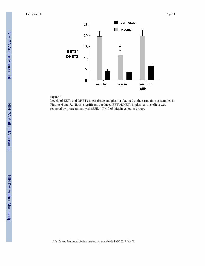
Figure 6.Levels of EETs and DHETs in ear tissue and plasma obtained at the same time as samples inFigures 6 and 7.. Niacin significantly reduced EETs/DHETs in plasma; this effect wasreversed by pretreatment with sEHI. * P < 0.05 niacin vs. other groups
Inceoglu et al. Page 14
J Cardiovasc Pharmacol. Author manuscript; available in PMC 2013 July 01.
NIH
-PA Author Manuscript
NIH
-PA Author Manuscript
NIH
-PA Author Manuscript

Figure 7.Ear blood flow induced by subcutaneous administration of PGD2 (1 mg/kg) was notdifferent in knockout (sEH −/−, n=4) mice or wild type (wt) mice pretreated with the sEHinhibitor TPAU (1 mg/kg, n=4) compared to wild type without TPAU pretreatment (n=5).This profile of vasodilation was markedly different than that produced by niacin (Fig.1).
Inceoglu et al. Page 15
J Cardiovasc Pharmacol. Author manuscript; available in PMC 2013 July 01.
NIH
-PA Author Manuscript
NIH
-PA Author Manuscript
NIH
-PA Author Manuscript