Inborn Errors of Metabolism involving Proteins -...
22
1 Inborn Errors of Metabolism involving Proteins Phenylketonuria Phenylketonuria (PKU) is an autosomal recessive metabolic genetic disorder characterized by a deficiency in the hepatic enzymephenylalanine hydroxylase (PAH). [1]:541 This enzyme is necessary to metabolize the amino acid phenylalanine ('Phe') to the amino acidtyrosine. When PAH is deficient, phenylalanine accumulates and is converted into phenylpyruvate (also known as phenylketone), which is detected in the urine. [2] Since its discovery, there have been many advances in its treatment. It can now be managed by the patient with little or no side-effects, just the inconvenience of managing the treatment. If, however, the condition is left untreated, it can cause problems with brain development, leading to progressive mental retardation, brain damage, and seizures. In the past, PKU was treated with a low-phenylalanine diet. Latter-day research now has shown that diet alone may not be enough to prevent the negative effects of phenylalanine levels. Optimal treatment involves lowering blood Phe levels to a safe range and monitoring diet and cognitive development. Lowering of phenylalanine levels to a safe range may be achieved by combining a low-phenylalanine diet with protein supplements. There is currently no cure for this disease; however, some treatments are available with varying success rates. In general, PKU is detected through newborn screening and diagnosed by a geneticist. PKU clinics around the world provide care for PKU patients to optimize phe levels, dietary intake, and cognitive outcomes. History Phenylketonuria was discovered by the Norwegian physician Ivar Asbjørn Følling in 1934 [3] when he noticed that hyperphenylalaninemia (HPA) was associated with mental retardation. In Norway, this disorder is known as Følling's disease, named after its discoverer. [4] Dr. Følling was one of the first physicians to apply detailed chemical analysis to the study of disease. His careful analysis of the urine of two affected siblings led him to request many physicians near Oslo to test the urine of other affected patients. This led to the discovery of the same substance that he had found in eight other patients. The substance found was subjected to much more basic and rudimentary chemical analysis (taste). He conducted tests and found reactions that gave rise to benzaldehyde and benzoic acid, which led him to conclude the compound contained a benzene ring. Further testing showed the melting point to be the same as phenylpyruvic acid,
Transcript of Inborn Errors of Metabolism involving Proteins -...
1
Inborn Errors of Metabolism involving Proteins
Phenylketonuria
Phenylketonuria (PKU) is an autosomal recessive metabolic genetic disorder characterized by a
deficiency in the hepatic enzymephenylalanine hydroxylase (PAH).[1]:541 This enzyme is necessary
to metabolize the amino acid phenylalanine ('Phe') to the amino acidtyrosine. When PAH is
deficient, phenylalanine accumulates and is converted into phenylpyruvate (also known as
phenylketone), which is detected in the urine.[2]
Since its discovery, there have been many advances in its treatment. It can now be managed by
the patient with little or no side-effects, just the inconvenience of managing the treatment. If,
however, the condition is left untreated, it can cause problems with brain development, leading to
progressive mental retardation, brain damage, and seizures. In the past, PKU was treated with a
low-phenylalanine diet. Latter-day research now has shown that diet alone may not be enough to
prevent the negative effects of phenylalanine levels. Optimal treatment involves lowering blood Phe
levels to a safe range and monitoring diet and cognitive development. Lowering of phenylalanine
levels to a safe range may be achieved by combining a low-phenylalanine diet with protein
supplements. There is currently no cure for this disease; however, some treatments are available
with varying success rates. In general, PKU is detected through newborn screening and diagnosed
by a geneticist. PKU clinics around the world provide care for PKU patients to optimize phe levels,
dietary intake, and cognitive outcomes.
History
Phenylketonuria was discovered by the Norwegian physician Ivar Asbjørn Følling in 1934[3] when
he noticed that hyperphenylalaninemia (HPA) was associated with mental retardation. In Norway,
this disorder is known as Følling's disease, named after its discoverer.[4] Dr. Følling was one of
the first physicians to apply detailed chemical analysis to the study of disease. His careful analysis
of the urine of two affected siblings led him to request many physicians near Oslo to test the urine
of other affected patients. This led to the discovery of the same substance that he had found in
eight other patients. The substance found was subjected to much more basic and rudimentary
chemical analysis (taste). He conducted tests and found reactions that gave rise
to benzaldehyde and benzoic acid, which led him to conclude the compound contained
a benzene ring. Further testing showed the melting point to be the same as phenylpyruvic acid,
2
which indicated that the substance was in the urine. His careful science inspired many to pursue
similar meticulous and painstaking research with other disorders.
Screening and presentation
PKU is normally detected using the HPLC test, but some clinics still use the Guthrie test, part of
national biochemical screening programs. Most babies in developed countries are screened for
PKU soon after birth.[5]
If a child is not screened during the routine newborn screening test (typically performed 6 -14 days
after birth, using samples drawn byNeonatal heel prick), the disease may present clinically
with seizures, albinism (excessively fair hair and skin), and a "musty odor" to the baby's sweat and
urine (due to phenylacetate, one of the ketones produced). In most cases, a repeat test should be
done at approximately 2 weeks of age to verify the initial test and uncover any phenylketonuria that
was initially missed.
Untreated children are normal at birth, but fail to attain early developmental milestones,
develop microcephaly, and demonstrate progressive impairment of cerebral
function. Hyperactivity, EEG abnormalities and seizures, and severe learning disabilities are major
clinical problems later in life. A "musty or mousy" odor of skin, hair, sweat and urine (due to
phenylacetate accumulation); and a tendency to hypopigmentationand eczema are also observed.
In contrast, affected children who are detected and treated are less likely to develop neurological
problems or have seizures and mental retardation, though such clinical disorders are still possible.
Pathophysiology
Classical PKU is caused by a mutated gene for the enzyme phenylalanine hydroxylase (PAH),
which converts the amino acid phenylalanine to other essential compounds in the body. Other non-
PAH mutations can also cause PKU. This is an example of genetic heterogeneity.
Classical PKU
The PAH gene is located on chromosome 12 in the bands 12q22-q24.1. More than four hundred
disease-causing mutations have been found in the PAH gene. PAH deficiency causes a spectrum
of disorders including classic phenylketonuria (PKU) and hyperphenylalaninemia (a less severe
accumulation of phenylalanine).[6]
3
PKU is known to be an autosomal recessive genetic disorder. This means that both parents must
have at least one mutated allele of the PAH gene. The child must inherit both mutated alleles, one
from each parent. Therefore, it is not impossible for a parent with the disease to have a child
without it if the other parent possesses one functional allele of the gene for PAH. Yet, a child from
two parents with PKU will inherit two mutated alleles every time, and therefore the disease.
Phenylketonuria can exist in mice, which have been extensively used in experiments into an
effective treatment for PKU.[7] The macaque monkey's genome was recently sequenced, and it was
found that the gene encoding phenylalanine hydroxylase has the same sequence that, in humans,
would be considered the PKU mutation.[8]
Tetrahydrobiopterin-deficient hyperphenylalaninemia
A rarer form of hyperphenilalaninemia occurs when PAH is normal but there is a defect in the
biosynthesis or recycling of the cofactor tetrahydrobiopterin (BH4) by the patient.[9] This cofactor is
necessary for proper activity of the enzyme.
Levels of dopamine can be used to distinguish between these two types. Tetrahydrobiopterin is
required to convert phenylalanine to tyrosine, but it is also required to convert tyrosine to
DOPA (via the enzyme tyrosine hydroxylase), which in turn is converted to dopamine. Low levels of
dopamine lead to high levels of prolactin. By contrast, in classical PKU, prolactin levels would be
relatively normal. Tetrahydrobiopterin deficiency can be caused by defects in four different genes.
These types are known as HPABH4A, HPABH4B, HPABH4C, and HPABH4D.
Metabolic pathways
The enzyme phenylalanine hydroxylase normally converts the amino acid phenylalanine into the
amino acid tyrosine. If this reaction does not take place, phenylalanine accumulates and tyrosine is
deficient. Excessive phenylalanine can be metabolized into phenylketones through the minor route,
a transaminase pathway with glutamate. Metabolites
includephenylacetate, phenylpyruvate and phenethylamine.[11] Elevated blood just because
phenylalanine and detection of phenylketones in the urine is diagnostic.
Phenylalanine is a large, neutral amino acid (LNAA). LNAAs compete for transport across
the blood-brain barrier (BBB) via the large neutral amino acid transporter (LNAAT). If phenylalanine
is in excess in the blood, it will saturate the transporter. Excessive levels of phenylalanine tend to
decrease the levels of other LNAAs in the brain. However, as these amino acids are necessary for
4
protein and neurotransmitter synthesis, phenylalanine buildup hinders the development of
the brain, causing mental retardation.[12]
Treatment
If PKU is diagnosed early enough, an affected newborn can grow up with normal brain
development, but only by managing and controlling phenylalanine (Phe) levels through diet, or a
combination of diet and medication. When phenylalanine cannot be metabolized by the body,
abnormally high levels accumulate in the blood and are toxic to the brain. When left untreated,
complications of PKU include severe mental retardation, brain function abnormalities,
microcephaly, mood disorders, irregular motor functioning, and behavioral problems such as
ADHD.
All PKU patients must adhere to a special diet low in phenylalanine for at least the first 16 years of
their lives. This requires severely restricting or eliminating foods high in phenylalanine, such
as meat, chicken, fish, eggs, nuts, cheese, legumes, cow milk and other dairy products. Starchy
foods such as potatoes, bread, pasta, and corn must be monitored. Infants may still be breastfed to
provide all of the benefits of breastmilk, but the quantity must also be monitored and
supplementation for missing nutrients will be required. Many diet foods and diet soft drinks that
contain the sweetener aspartame must also be avoided, as aspartame consists of two amino acids:
phenylalanine and aspartic acid.
Supplementary infant formulas are used in these patients to provide the amino acids and other
necessary nutrients that would otherwise be lacking in a low-phenylalanine diet. As the child grows
up, these can be replaced with pills, formulas, and specially formulated foods. (Since phenylalanine
is necessary for the synthesis of many proteins, it is required for appropriate growth but levels must
be strictly controlled in PKU patients). In addition, tyrosine, which is normally derived from
phenylalanine, must be supplemented.)
The oral administration of tetrahydrobiopterin (or BH4) (a cofactor for the oxidation of
phenylalanine) can reduce blood levels of this amino acid in certain patients.[13][14] The
companyBioMarin Pharmaceutical has produced a tablet preparation of the compound sapropterin
dihydrochloride (Kuvan),which is a form of tetrahydrobiopterin. Kuvan is the first drug that can help
BH4-responsive PKU patients (defined among clinicians as about 1/2 of the PKU population) lower
Phe levels to recommended ranges.[15] Working closely with a dietitian, some PKU patients who
respond to Kuvan may also be able to increase the amount of natural protein they can eat.[16] After
extensive clinical trials, Kuvan has been approved by the FDA for use in PKU therapy.
5
Researchers and clinicians working with PKU are finding Kuvan a safe and effective addition to
dietary treatment and beneficial to patients with PKU.[17][18]
There are several other therapies currently under investigation, including gene therapy, large
neutral amino acids, and enzyme substitution therapy with phenylalanine ammonia lyase (PAL). In
the past, PKU-affected people were allowed to go off diet after approximately 8, then 18 years of
age. Today most physicians recommend that PKU patients must manage their Phe levels
throughout life.
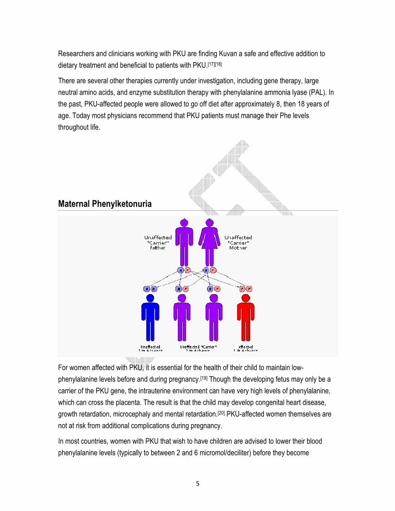
Maternal Phenylketonuria
For women affected with PKU, it is essential for the health of their child to maintain low-
phenylalanine levels before and during pregnancy.[19] Though the developing fetus may only be a
carrier of the PKU gene, the intrauterine environment can have very high levels of phenylalanine,
which can cross the placenta. The result is that the child may develop congenital heart disease,
growth retardation, microcephaly and mental retardation.[20] PKU-affected women themselves are
not at risk from additional complications during pregnancy.
In most countries, women with PKU that wish to have children are advised to lower their blood
phenylalanine levels (typically to between 2 and 6 micromol/deciliter) before they become
6
pregnant, and carefully control their phenylalanine levels throughout the pregnancy. This is
achieved by performing regular blood tests and adhering very strictly to a diet, in general monitored
on a day-to-day basis by a specialist metabolic dietitian. In many cases, as the fetus' liver begins to
develop and produce PAH normally, the mother's blood phenylalanine levels will drop, requiring an
increased phenylalanine intake to remain within the safe range of 2-6 micromol/dL. The mother's
daily phenylalanine intake may double or even triple by the end of the pregnancy, as a result.
When maternal blood phenylalanine levels fall below 2 micromol/dL, anecdotal reports indicate that
the mothers may suffer adverse effects including headaches, nausea, hair loss, and general
malaise. When low phenylalanine levels are maintained for the duration of pregnancy, there are no
elevated levels of risk of birth defects compared with a baby born to a non-PKU mother.[21] Babies
with PKU may drink breast milk, while also taking their special metabolic formula. Some research
has indicated that an exclusive diet of breast milk for PKU babies may alter the effects of the
deficiency, though during breastfeeding the mother must maintain a strict diet to keep their
phenylalanine levels low. More research is needed.
Incidence
The incidence of PKU is about 1 in 15,000 births, but the incidence varies widely in different human
populations from 1 in 4,500 births among the population of Ireland[22] to 1 in 13,000 births
in Norway[23] to fewer than one in 100,000 births among the population of Finland.[24] Turkey, at 1 in
2600, has the highest incidence rate in the world. The illness is also more common in Italy and
China, as well as in Yemeni populations.[25]
……………………………………………………………………………………………………………………………
…………
Maple syrup urine disease Maple syrup urine disease (MSUD), also called branched-chain ketoaciduria, is
an autosomal recessive[1] metabolic disorder affectingbranched-chain amino acids. It is one type
of organic acidemia.[2] The condition gets its name from the distinctive sweet odor of affected
infants' urine.[3]
Diagnosis and symptoms
MSUD is caused by a deficiency of the branched-chain alpha-keto acid dehydrogenase
complex (BCKDH), leading to a buildup of thebranched-chain amino acids (leucine, isoleucine,
and valine) and their toxic by-products in the blood and urine.
7
The disease is characterized in an infant by the presence of sweet-smelling urine, with an odor
similar to that of maple syrup. Infants with this disease seem healthy at birth but if left untreated
suffer severe brain damage, and eventually die.
From early infancy, symptoms of the condition include poor
feeding, vomiting, dehydration, lethargy, hypotonia, seizures, ketoacidosis,opisthotonus, pancreatiti
s, coma and neurological decline.
Classification
Maple syrup urine disease can be classified by its pattern of signs and symptoms, or by its genetic
cause. The most common and severe form of the disease is the classic type, which appears soon
after birth. Variant forms of the disorder may appear later in infancy or childhood and are typically
less severe, but still involve mental and physical problems if left untreated.
There are several variations of the disease:
� Classic Severe MSUD
� Intermediate MSUD
� Intermittent MSUD
� Thiamine-responsive MSUD
� E3-Deficient MSUD with Lactic Acidosis
Management
Keeping MSUD under control requires careful monitoring of blood chemistry and involves both a
special diet and frequent testing.
A diet with minimal levels of the amino acids leucine, isoleucine, and valine must be maintained in
order to prevent neurological damage. As these three amino acids are required for proper
metabolic function in all people, specialized protein preparations containing substitutes and
adjusted levels of the amino acids have been synthesized and tested, allowing MSUD patients to
meet normal nutritional requirements without causing harm.[4]
Usually, patients are also monitored by a dietitian. Their diet must be adhered to strictly and
permanently. However, with proper management those afflicted are able to live healthy, normal
lives and not suffer the severe neurological damage associated with the disease.
8
Genetic prevalence
Maple syrup urine disease affects approximately 1 out of 180,000 infants.[5] Due in part to
the founder effect,[6] however, MSUD has a much higher prevalence in children
of Amish, Mennonite, and Jewish descent.[7][5][8]
Mutations in the following genes cause maple syrup urine disease:
� BCKDHA (Online 'Mendelian Inheritance in Man' (OMIM) 608348)
� BCKDHB (Online 'Mendelian Inheritance in Man' (OMIM) 248611)
� DBT (Online 'Mendelian Inheritance in Man' (OMIM) 248610)
� DLD (Online 'Mendelian Inheritance in Man' (OMIM) 238331)
These four genes produce proteins that work together as the branched-chain alpha-keto acid
dehydrogenase complex. The complex is essential for breaking down the amino
acids leucine, isoleucine, and valine, which are present in many kinds of food (particularly protein-
rich foods such as milk, meat, and eggs). Mutations in any of these genes reduce or eliminate the
function of the enzyme complex, preventing the normal breakdown of isoleucine, leucine,
and valine. As a result, these amino acids and their by-products build up in the body. Because high
levels of these substances are toxic to the brain and other organs, this accumulation leads to the
serious medical problems associated with maple syrup urine disease.
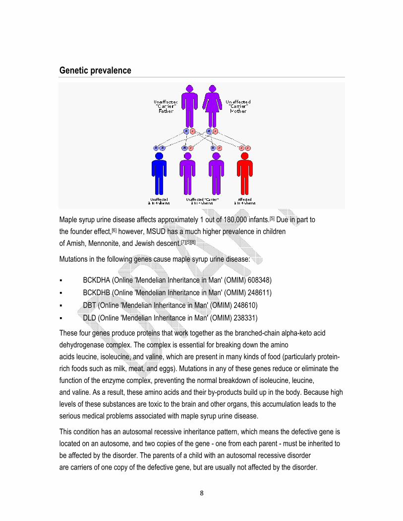
This condition has an autosomal recessive inheritance pattern, which means the defective gene is
located on an autosome, and two copies of the gene - one from each parent - must be inherited to
be affected by the disorder. The parents of a child with an autosomal recessive disorder
are carriers of one copy of the defective gene, but are usually not affected by the disorder.
9
……………………………………………………………………………………………………………………………
…………..
Glutaric aciduria type 1 Glutaric acidemia type 1 (or "Glutaric Aciduria", "GA1", or "GAT1") is an inherited disorder in
which the body is unable to break down completely the amino acids lysine, hydroxylysine
and tryptophan. Excessive levels of their intermediate breakdown products (glutaric acid,glutaryl-
CoA, 3-hydroxyglutaric acid, glutaconic acid) can accumulate and cause damage to the brain (and
also other organs[1]), but particularly the basal ganglia, which are regions that help regulate
movement. GA1 causes secondary carnitine deficiency, as glutaric acid, like other organic acids, is
detoxified by carnitine. Mental retardation may also occur.
Signs and symptoms
The severity of glutaric acidemia type 1 varies widely; some individuals are only mildly affected,
while others have severe problems. GA1 can be defined as two clinical entities: GA1 before the
encephalopathic crisis and GA1 after the encephalopathic crisis.
GA1 before the encephalopathic crisis
Macrocephaly
Babies with glutaric acidemia type 1 often are born with unusually large heads (macrocephaly).
Macrocephaly is amongst the earliest signs of GA1. It is thus important to investigate all cases of
macrocephaly of unknown origins for GCDH deficiency[2][3], given the importance of the early
diagnosis of GA1[4]. Macrocephaly is a "pivotal clinical sign" of many neurological diseases.
Physicians and parents should be aware of the benefits of investigating for an underlying
neurological disorder, particularly a neurometabolic one, in children with head circumferences in
the highest percentiles.
GA1 after the encephalopathic crisis
Neuromotor aspects
Affected individuals may have difficulty moving and may experience spasms, jerking, rigidity or
decreased muscle tone and muscle weakness (which may be the result of secondary carnitine
10
deficiency). Glutaric aciduria type 1, in many cases, can be defined as a cerebral palsy of genetic
origins.
Occupational therapy
A common way to manage striatal necrosis is to provide special seating. These special
wheelchairs are designed to limit abnormal movements. However, spasticity can be worsened by
constraint.
Parents and caregivers can provide a more interactive occupational therapy by enabling the child
to use his or her own excessive postural muscle tone to his ir her own advantage (see picture; note
the care with which minimal pressure is applied while ensuring safety).
The excessive tone can also be managed with "jolly jumpers" and other aids to the upright stance
that do not constrain the child but help him or her gradually tone down the rigidity.
Bleeding abnormalities
Some individuals with glutaric acidemia have developed bleeding in the brain or eyes that could be
mistaken for the effects of child abuse.
Treatment
Correction of secondary carnitine depletion
Like many other organic acidemias, GA1 causes carnitine depletion[5]. Whole-blood carnitine can
be raised by oral supplementation. However, this does not significantly change blood
concentrations of glutarylcarnitine or esterified carnitine,[4] suggesting that oral supplementation is
suboptimal in raising tissue levels of carnitine. In the field of clinical nutrition, researchers come to
the same conclusion, that oral carnitine raises plasma levels but doesn't affect muscle carnitine,
where most of it is stored and used.[6]
� In contrast, regular intravenous infusions of carnitine caused distinct clinical
improvements : "decreased frequency of decompensations, improved growth, improved
muscle strength and decreased reliance on medical foods with liberalization of protein
intake."[5]
� Choline increases carnitine uptake and retention[7]. Choline supplements are inexpensive,
safe (probably even in all children requiring anticholinergics) and can provide spectacular
11
evidence of the suboptimal efficiency of carnitine supplementation by increasing exercise
tolerance, truncal tone and general well-being.
Precursor restriction
Dietary control may help limit progression of the neurological damage.
Selective precursor restriction
Tryptophan
Formulas such as XLys, XTrp Analog, XLys, XTrp Maxamaid, XLys, XTrp Maxamum or Glutarex
1 are designed to provide amino acids other than lysine and tryptophan, in order to tentatively
prevent protein malnutrition.
The entry of tryptophan to the brain is crucial in the proper synthesis of the
neurotransmitter serotonin in the brain. One way to acutely cause depression or bulimia or anxiety
in humans, in order to assess an individual's vulnerability to those disorders, is to supplement with
a formula with all or most amino acids except tryptophan. The protein synthesis elicited by the
amino acids leads circulating amino acids, including tryptophan, to be incorporated into proteins.
Tryptophan thus lowers in the brain as a result of the protein synthesis enhancement (causing
circulating tryptophan to lower more than other amino acids),[8] and perhaps also competition of
large neutral amino acids for transport across the blood-brain barrier through the large neutral
amino acid transporter 1 (LNAA1). The consequence is acute tryptophan depletion (ATD) in the
brain and a consecutive lowering of serotonin synthesis. ATD, which is basically a diagnostic
procedure, is not a treatment for GA1.
In the Amish community, where GA1 is overrepresented (Morton, 2003), patients with GA1 did not
and still don't receive tryptophan-free formulas, neither as the sole source of amino acids, nor as a
supplement to protein restriction. Doctor D. Holmes Morton, the 1993 Albert Schweitzer Prize for
Humanitarianism laureate, is taking care of patients affected with GA1 and other metabolic
diseases in this community in his Clinic for Special Children.
5-hydroxytryptophan, the precursor of serotonin that is not metabolized to glutaryl-CoA, glutaric
acid and secondary metabolites, could be used as an adjunct to selective tryptophan restriction,
considering the risks associated with the procedure. However, the evidence in favour of selective
tryptophan restriction remains insufficient and the consensus evolves towards the restriction of
lysine only (Kolker & al. 2006).
12
Lysine
Lysine restriction, as well as carnitine supplementation, are considered the best predictors of a
good prognosis for GA1 (Kolker & al., 2006). This excludes, however, patients who already
suffered an encephalopathic crisis, for whom the prognosis is more related to the treatment of their
acquired disorder (striatal necrosis, frontotemporal atrophy).
Protein restriction
Vegetarian diets and, for younger children, breastfeeding[9]are common ways to limit protein intake
without endangering tryptophan transport to the brain.
Enhancement of precursor's anabolic pathway
Lysine and hydroxylysine anabolic pathway enhancement
A possible way to prevent the build-up of metabolites is to limit lysine and hydroxylysine
degradation, as lysine is one of the most abundant amino acids and tryptophan is one the least
abundant amino acids.
Interaction of GCDH deficiency with GLO deficiency
While GCDH deficiency is a rare disease, GLO deficiency is the most common of metabolic
diseases affecting Humanity, limiting ascorbic acid biosynthesis to a minute fraction of what other
non-primate species synthesize. It was thus called by OMIM (Online Mendeleian Inheritance in
Man) a "public" error of metabolism. Ascorbic acid (Vitamin C) is a necessary cofactor for the
utilization of lysine in collagen synthesis. Collagen, the most abundant protein in the human body,
requires great amounts of lysine, the most abundant amino acids in proteins. Ascorbic acid, the
main hydroxyl radical quencher, works as the cofactor providing the hydroxyl radical required to
collagen cross-linking; lysine thus becomes hydroxylysine.
GA1 worsens during stresses and catabolic episodes, such as fasts and infections. Endogenous
catabolism of proteins could be an important route for glutaric acid production. It thus follows that
collagen breakdown (and protein breakdown in general) should be prevented by all possible
means.
Ascorbic acid is used to prevent multiple organ failure and to lessen mortality and morbidity in
intensive care units.[10] It thus appears reasonable to include sufficient doses of ascorbic acid to the
13
treatment protocol during stresses and other challenges to growth in order to stimulate collagen
synthesis and thus prevent lysine breakdown.
Tryptophan anabolic pathway enhancement
The conversion of tryptophan to serotonin and other metabolites depends on vitamin B6.[11] If
tryptophan catabolism has any impact on brain glutaric acid and other catabolite levels, vitamin
B6 levels should be routinely assayed and normalized in the course of the treatment of GA1.
Management of intercurrent illnesses
Stress caused by infection, fever or other demands on the body may lead to worsening of the signs
and symptoms, with only partial recovery.
Genetics
The condition is inherited in an autosomal recessive pattern: mutated copies of the
gene GCDH must be provided by both parents to cause glutaric acidemia type 1. The GCDH gene
encodes the enzyme glutaryl-CoA dehydrogenase. This enzyme is involved in degrading the amino
acids lysine, hydroxylysine and tryptophan. Mutations in the GCDH' gene prevent production of the
enzyme or result in the production of a defective enzyme with very low residual activity, or an
enzyme with relatively high residual activity but still phenotypic consequences[12][13]. This enzyme
deficiency allows glutaric acid, 3-hydroxyglutaric acid and (to a lesser extent) glutaconic acid to
build up to abnormal levels, especially at times when the body is under stress. These intermediate
breakdown products are particularly prone to affect the basal ganglia, causing many of the signs
and symptoms of glutaric acidemia type 1.
Glutaric acidemia type 1 occurs in approximately 1 of every 30,000 to 40,000 births. It is much
more common in the Amish community and in the Ojibway population of Canada, where up to 1 in
300 newborns may be affected.
Relatives of children with GA1 can have very low GCDH activity: in an early study of GA1, GCDH
activity was found to be 38%, 42%, and 42% of controls in three of the four relatives tested[14].
Those levels are close to those found by Christensen & al[15] in some heavily symptomatic GA1-
affected children.
Epistemology
14
GA1 can be described as a metabolic disease, a neurometabolic disease, a cerebral palsy or
a basal ganglia disorder (it is also misdiagnosed as shaken baby syndrome). Depending on the
paradigm adopted, GA1 will mostly be managed with precursor restriction or with
neurorehabilitation (or with incarceration of the parents in the case of presumed shaken baby
syndrome).
So-called "orphan diseases", such as GA1, can be adopted into wider groups of diseases (such as
carnitine deficiency diseases, cerebral palsies of diverse origins, basal ganglia disorders, and
others); Morton at al. (2003b) emphacize that acute striatal necrosis is a distinctive pathologic
feature of at least 20 other disorders of very different etiologies (e.g. HIV encephalopathy-AIDS
dementia complex, pneumococcal meningitis, hypoadrenal crisis, methylmalonic
acidemia, propionic acidemia, middle cerebral artery occlusion, hypertensive vasculopathy,
acute mycoplasma pneumoniae infection, 3-nitropropionic acid intoxication, late onset
familial dystonia, cerebrovascular abrupt and severe neonatal asphyxia ("selective neuronal
necrosis")).
Amongst 279 patients who had been reported to have GA1, 185 were symptomatic (two thirds);
being symptomatic was seen as an indication of "low treatment efficacy". High risk screening,
neonatal screening and a diagnosis of macrocephaly were the ways to identify bearers of
the GCDH' defective gene who weren't frankly symptomatic. Macrocephaly remains the main sign
of GA1 for those who aren't related to GA1 in any way or benefit from no screening program. GA1
was considered as a "treatable disease"[16]. Two thirds of the patients who have GA1 will receive
little benefit from the treatment for GA1 but can benefit from treatments given to victims of middle
cerebral artery occlusion, AIDS dementia and other basal ganglia disorders : brain implants, stem
cell neurorestauration, growth factors, monoaminergic agents, and many other neurorehabilitation
strategies.
…………………………………………………………………………………………………………………
….
Urea cycle disorder An urea cycle disorder or urea cycle defect is a genetic disorder caused by a deficiency of one
of the enzymes in the urea cycle which is responsible for removing ammonia from
the blood stream. The urea cycle involves a series of biochemical steps in which nitrogen, a waste
product of protein metabolism, is removed from the blood and converted to urea. Normally, the
urea is transferred into the urine and removed from the body. In urea cycle disorders, the nitrogen
accumulates in the form of ammonia, a highly toxic substance, and is not removed from the body.
15
Urea cycle disorders are included in the category of inborn errors of metabolism. There is no cure.
Incidence
Inborn errors of metabolism are generally considered to be rare but represent a substantial cause
of brain damage and death among newborns and infants. Because many cases of urea cycle
disorders remain undiagnosed and/or infants born with the disorders die without a definitive
diagnosis, the exact incidence of these cases is unknown and underestimated. It is believed that
up to 20% of Sudden Infant Death Syndrome cases may be attributed to an undiagnosed inborn
error of metabolism such as urea cycle disorder. In April 2000, research experts at the Urea Cycle
Consensus Conference estimated the incidence of the disorders at 1 in 10000 births. This
represents a significant increase in case diagnosis in the last two years.
Symptoms
The neonatal period
Children with very severe urea cycle disorders typically show symptoms after the first 24 hours of
life. The baby may be irritable at first, followed by vomiting and increasing lethargy. Soon
after, seizures, hypotonia (poor muscle tone), respiratory distress, and coma may occur. If
untreated, the child will die. These symptoms are caused by rising ammonia levels in the blood.
Acute neonatal symptoms are most frequently seen in, but not limited to, boys with OTC
Deficiency.
Childhood
Children with mild or moderate urea cycle enzyme deficiencies may not show symptoms until early
childhood, or may be diagnosed subsequent to identification of the disorder in a more severely
affected relative or through newborn screening. Early symptoms may include hyperactive behavior,
sometimes accompanied by screaming and self-injurious behavior, and refusal to eat meat or other
high-protein foods. Later symptoms may include frequent episodes of vomiting, especially following
high-protein meals; lethargy and delirium; and finally, if the condition is undiagnosed and untreated,
coma and death. Children with this disorder may be referred to child psychologists because of their
behavior and eating problems. Childhood episodes of hyperammonemia (high ammonia levels in
the blood) may be brought on by viral illnesses including chicken pox, high-protein meals, or
16
even exhaustion. The condition is sometimes misdiagnosed as Reye’s Syndrome. Childhood onset
can be seen in both boys and girls.
Adulthood
Recently, the number of adult individuals being diagnosed with urea cycle disorders has increased
at an alarming rate. Recent evidence has indicated that these individuals have survived
undiagnosed to adulthood, probably due to less severe enzyme deficiencies. These individuals
exhibit stroke-like symptoms, episodes of lethargy, and delirium. These adults are likely to be
referred to neurologists or psychiatrists because of their psychiatric symptoms. However, without
proper diagnosis and treatment, these individuals are at risk for permanent brain damage, coma,
and death. Adult-onset symptoms have been observed following viral illnesses, childbirth, and use
of valproic acid (an anti-epileptic drug).
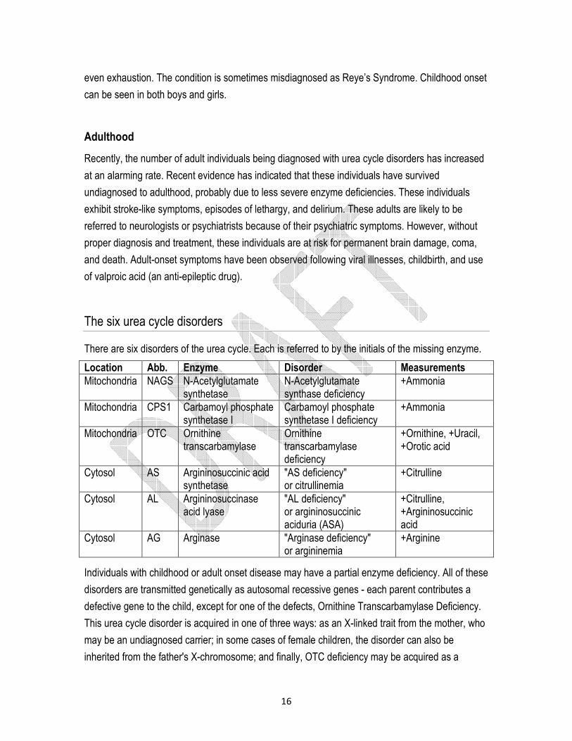
The six urea cycle disorders
There are six disorders of the urea cycle. Each is referred to by the initials of the missing enzyme.
Location Abb. Enzyme Disorder Measurements
Mitochondria NAGS N-Acetylglutamate synthetase
N-Acetylglutamate synthase deficiency
+Ammonia
Mitochondria CPS1 Carbamoyl phosphate synthetase I
Carbamoyl phosphate synthetase I deficiency
+Ammonia
Mitochondria OTC Ornithine transcarbamylase
Ornithine transcarbamylase deficiency
+Ornithine, +Uracil, +Orotic acid
Cytosol AS Argininosuccinic acid synthetase
"AS deficiency" or citrullinemia
+Citrulline
Cytosol AL Argininosuccinase acid lyase
"AL deficiency" or argininosuccinic aciduria (ASA)
+Citrulline, +Argininosuccinic acid
Cytosol AG Arginase "Arginase deficiency" or argininemia
+Arginine
Individuals with childhood or adult onset disease may have a partial enzyme deficiency. All of these
disorders are transmitted genetically as autosomal recessive genes - each parent contributes a
defective gene to the child, except for one of the defects, Ornithine Transcarbamylase Deficiency.
This urea cycle disorder is acquired in one of three ways: as an X-linked trait from the mother, who
may be an undiagnosed carrier; in some cases of female children, the disorder can also be
inherited from the father's X-chromosome; and finally, OTC deficiency may be acquired as a
17
"new" mutation occurring in the fetus uniquely. Recent research has shown that some female
carriers of the disease may become symptomatic with the disorder later in life, suffering high
ammonia levels. Several undiagnosed women have died during childbirth as a result of high
ammonia levels and on autopsy were determined to have been unknown carriers of the disorder.
Treatment
The treatment of urea cycle disorders consists of balancing dietary protein intake in order that the
body receive the essential amino acids responsible for cell growth and development, but not so
much protein that excessive ammonia is formed. This protein restriction is used in conjunction
with medications which provide alternative pathways for the removal of ammonia from the blood.
These medications are usually given by way of tube feedings, either via gastrostomy tube (a tube
surgically implanted in the stomach) or nasogastric tube through the nose into the stomach. The
treatment may also include supplementation with special amino acid formulas developed
specifically for urea cycle disorders, multiple vitamins and calciumsupplements. Frequent blood
tests are required to monitor the disorders and optimize treatment, and frequently hospitalizations
are necessary to control the disorder.
At the most extreme end of the spectrum, a few liver transplants have been done successfully as a
cure to the disorder. This treatment alternative must be carefully evaluated with medical
professionals to determine if potential of success as compared to the potential for new medical
concerns.
Inborn Errors of Metabolism Involving Carbohydrates
18
………………………………………………………………………………………………………………………………………………………………..
Galactosemia
Galactosemia (British Galactosaemia) is a rare genetic metabolic disorder that affects an
individual's ability to metabolize the sugargalactose properly. Galactosemia is not related to and
should not be confused with lactose intolerance. Galactosemia follows an autosomal
recessive mode of inheritance that confers a deficiency in an enzyme responsible for adequate
galactose degradation.
Goppert first described the disease in 1917,[1] with its cause as a defect in galactose metabolism
being identified by a group led by Herman Kalckar in 1956.[2]
Its incidence is about 1 per 60,000 births. It is much rarer in Japan and much more common
in Italy, specifically the traveler region. Galactosemia is also very common within the Irish
Traveller population. This is attributed to inbreeding within a relatively small gene pool.[citation needed]
Cause
Lactose in food (such as dairy products) is broken down by the enzyme lactase
into glucose and galactose. In individuals with galactosemia, the enzymes needed for further
metabolism of galactose are severely diminished or missing entirely, leading to toxic levels of
galactose in the blood, resulting in hepatomegaly (an enlarged liver), cirrhosis, renal
failure, cataracts,brain damage, and ovarian failure. Without treatment, mortality in infants with
galactosemia is about 75%.
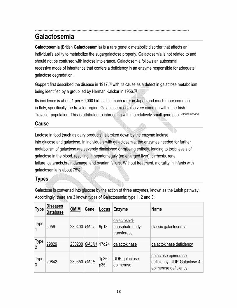
Types
Galactose is converted into glucose by the action of three enzymes, known as the Leloir pathway.
Accordingly, there are 3 known types of Galactosemia; type 1, 2 and 3:
Type Diseases
Database OMIM Gene Locus Enzyme Name
Type
1 5056 230400 GALT 9p13
galactose-1-
phosphate uridyl
transferase
classic galactosemia
Type
2 29829 230200 GALK1 17q24 galactokinase galactokinase deficiency
Type
3 29842 230350 GALE
1p36-
p35
UDP galactose
epimerase
galactose epimerase
deficiency, UDP-Galactose-4-
epimerase deficiency
19
The order of these three types is not the same as the order that the enzymes are encountered by
galactose on its metabolic path (which is closer to GALK, GALT, and then GALE, though many
variations can occur.)[citation needed]
Diagnosis
Infants are now routinely screened for galactosemia in the United States, and the diagnosis is
made while the person is still an infant. Infants affected by galactosemia typically present with
symptoms of lethargy, vomiting, diarrhea, failure to thrive, and jaundice. None of these symptoms
are specific to galactosemia, often leading to diagnostic delays. Luckily, most infants are diagnosed
on newborn screening. A galactosemia test is a blood test (from the heel of the infant) or urine test
that checks for three enzymes that are needed to change galactose sugar that is found in milk and
milk products-into glucose, a sugar that your body uses for energy. A person with galactosemia
doesn't have one of these enzymes. This causes high levels of galactose in the blood or urine.
Galactosemia is normally first detected through newborn screening, or NBS. Affected children can
have serious, irreversible effects or even die within days from birth. It is important that newborns be
screened for metabolic disorders without delay. Galactosemia can even be detected through NBS
before any ingestion of galactose-containing formula or breast milk.
Detection of the disorder through newborn screening (NBS) does not depend on protein or lactose
ingestion, and, therefore, it should be identified on the first specimen unless the infant has been
transfused. A specimen should be taken prior to transfusion. The enzyme is prone to damage if the
sample is delayed in the mail or exposed to high temperatures. The routine NBS is accurate for
detection of galactosemia. Two screening tests are used to screen infants affected with
galactosemia - the Beutler's test and the Hill test. In fact a third test, called the "Florida test", is also
normally performed on all galactosemia positives. The Beutler's test screens for galactosemia by
detecting the level of enzyme of the infant. Therefore, the ingestion of formula or breast milk does
not effect the outcome of this part of the NBS, and the NBS is accurate for detecting galactosemia
prior to any ingestion of galactose.
Treatment
The only treatment for classic galactosemia is eliminating lactose and galactose from the diet.
Even with an early diagnosis and a restricted diet, however, some individuals with galactosemia
experience long-term complications such as speech difficulties, learning disabilities, neurological
20
impairment (e.g. tremors, etc), symptoms have not been associated with Duarte galactosemia, and
many individuals with Duarte galactosemia do not need to restrict their diet at all. Infants with
classic galactosemia cannot be breast-fed due to lactose in human breast milk and are usually fed
a soy-based formula.[3]
Galactosemia is sometimes confused with lactose intolerance, but galactosemia is a more serious
condition. Lactose intolerant individuals have an acquired or inherited shortage of the
enzyme lactase, and experience abdominal pains after ingesting dairy products, but no long-term
effects. In contrast, a galactosemic individual who consumes galactose can cause permanent
damage to their bodies.
Long term complication of galactosemia includes:
� Speech deficits
� Ataxia
� Dysmetria
� Diminished bone density
� Premature ovarian failure
� Cataract ,,,,,,,,,,,,,,,,,,,,,,,,,,,,,,,,,,,,,,,,,,,,,,,,,,,,,,,,,,,,,,,,,,,,,,,,,,,,,,,,,,,,,,,,,,,,,,,,,,,,,,,,,,,,,,,,,,,,,,,,,,,,,,,,,,,,,,,,,,,,,,,,,,,,,,,,,,,,,,,,,,,,,,,,,,,,,,,,,,,,,,,,,,,,,,,,,,,,,,,,,,,,,,,,,,,,,,,,,,,,,,,,,,,,,,,,,,,,
,,,,,,,
Glycogen storage disease
Glycogen storage disease (GSD, also glycogenosis and dextrinosis) is the result of defects in
the processing of glycogen synthesis or breakdown within muscles, liver, and other cell
types.[1] GSD has two classes of cause: genetic and acquired. Genetic GSD is caused by
any inborn error of metabolism (genetically defective enzymes) involved in these processes. In
livestock, acquired GSD is caused byintoxication with the alkaloid castanospermine.[2]
Overall, according to a study in British Columbia, approximately 2.3 children per 100 000 births (1
in 43,000) have some form of glycogen storage disease.[3] In the United States, they are estimated
to occur in 1 per 20,000-25,000 births.[4] A Dutch study estimated it to be 1 in 40,000.[5]
Types
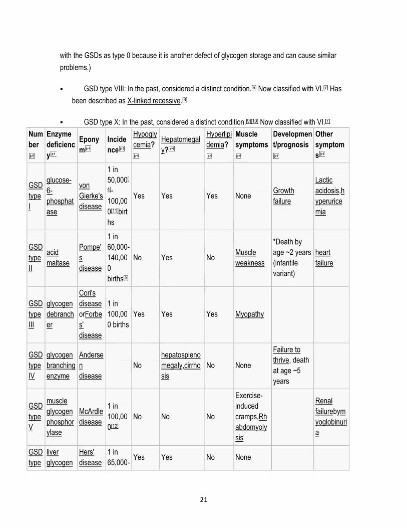
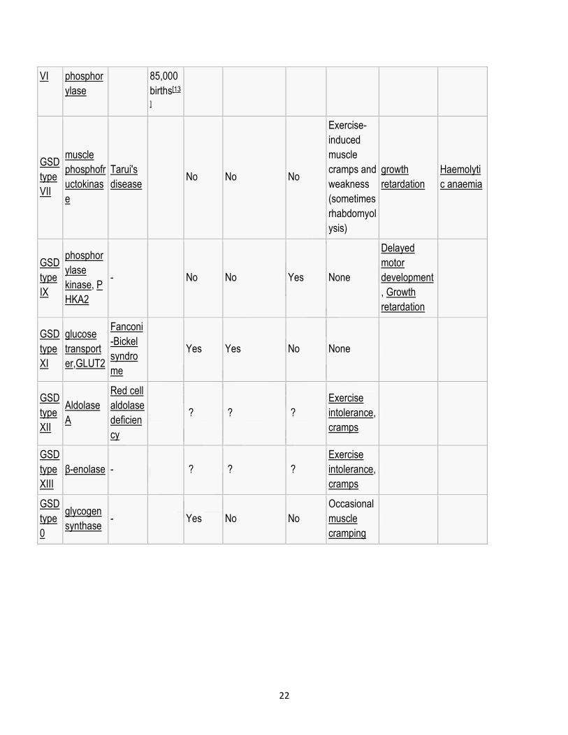
There are eleven distinct diseases that are commonly considered to be glycogen storage diseases
(some previously thought to be distinct have been reclassified). (Although glycogen
synthase deficiency does not result in storage of extra glycogen in the liver, it is often classified
21
with the GSDs as type 0 because it is another defect of glycogen storage and can cause similar
problems.)
� GSD type VIII: In the past, considered a distinct condition.[6] Now classified with VI.[7] Has
been described as X-linked recessive.[8]
� GSD type X: In the past, considered a distinct condition.[9][10] Now classified with VI.[7]
Num
ber
Enzyme
deficienc
y
Epony
m
Incide
nce
Hypogly
cemia?
Hepatomegal
y?
Hyperlipi
demia?
Muscle
symptoms
Developmen
t/prognosis
Other
symptom
s
GSD
type
I
glucose-
6-
phosphat
ase
von
Gierke's
disease
1 in
50,000[
4]-
100,00
0[11]birt
hs
Yes Yes Yes None Growth
failure
Lactic
acidosis,h
yperurice
mia
GSD
type
II
acid
maltase
Pompe'
s
disease
1 in
60,000-
140,00
0
births[5]
No Yes No Muscle
weakness
*Death by
age ~2 years
(infantile
variant)
heart
failure
GSD
type
III
glycogen
debranch
er
Cori's
disease
orForbe
s'
disease
1 in
100,00
0 births
Yes Yes Yes Myopathy
GSD
type
IV
glycogen
branching
enzyme
Anderse
n
disease
No
hepatospleno
megaly,cirrho
sis
No None
Failure to
thrive, death
at age ~5
years
GSD
type
V
muscle
glycogen
phosphor
ylase
McArdle
disease
1 in
100,00
0[12]
No No No
Exercise-
induced
cramps,Rh
abdomyoly
sis
Renal
failurebym
yoglobinuri
a
GSD
type
liver
glycogen
Hers'
disease
1 in
65,000-Yes Yes No None
22
VI phosphor
ylase
85,000
births[13
]
GSD
type
VII
muscle
phosphofr
uctokinas
e
Tarui's
disease No No No
Exercise-
induced
muscle
cramps and
weakness
(sometimes
rhabdomyol
ysis)
growth
retardation
Haemolyti
c anaemia
GSD
type
IX
phosphor
ylase
kinase, P
HKA2
- No No Yes None
Delayed
motor
development
, Growth
retardation
GSD
type
XI
glucose
transport
er,GLUT2
Fanconi
-Bickel
syndro
me
Yes Yes No None
GSD
type
XII
Aldolase
A
Red cell
aldolase
deficien
cy
? ? ?
Exercise
intolerance,
cramps
GSD
type
XIII
β-enolase - ? ? ?
Exercise
intolerance,
cramps
GSD
type
0
glycogen
synthase - Yes No No
Occasional
muscle
cramping