IN VIVO MOTION OF THE HUMAN CERVICAL SPINAL … VIVO MOTION OF THE HUMAN CERVICAL SPINAL CORD IN...
12
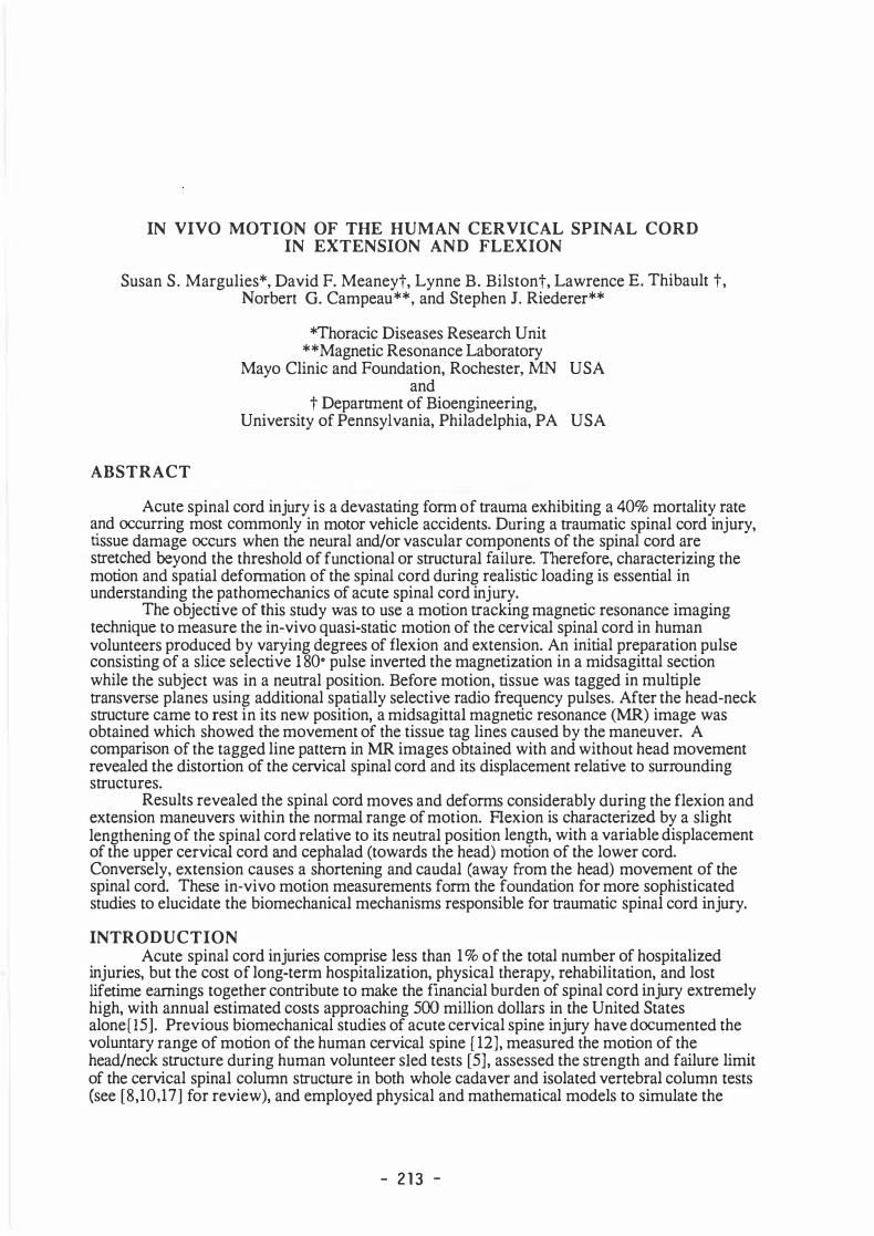
IN VIVO MOTION OF THE HUMAN CERVICAL SPINAL CORD IN EXTENSION AND FLEXION Susan S. Margulies*, David F. Meaneyt, Lynne B. Bilstont, Lawrence E. Thibault t, Norbert G. Campeau**, and Stephen J. Riederer** ABSTRACT *Thoracic Diseases Research Unit **Magnetic Resonance Laboratory Mayo Clinic and Foundation, Rochester, USA and t Deparent of Bioengineering, University of Pennsylvania, Philadelphia, PA USA Acute spinal cord injury is a devastang rm of auma exhibiting a 40% mortality rate and curri ng most commonly in motor vehicle accidents. During a aumatic spinal cord injury, ssue damage curs when the neural and/or vascular components of the spinal cord are setched yond the threshold of functional or suctural ilure. Therere, characterizing the moon and spatial defoaon of the spinal cord during realisc loading is essenal in understanding the pathomechics of acute spinal cord i njury. The objecve of this study was to use a moon acking magnec resonance imaging technique to measure the in-vivo quasi-stac moon of the cervical spinal cord in human volunteers produced by varying degrees of flexion and extension. An inial preparation pulse consisng of a slice selecve 1 80• pulse inverted the magnetization in a midsagittal secon while the subject was in a neual position. Bere motion, ssue was tagged in mulple ansverse planes using additional spaally selective radio equency pulses. After the head-neck sucture came to rest in its new position, a midsagittal magnec resonance () image was obtained which showed the movement of the tissue tag lines caused by the maneuver. A comparison of the tagged line pattem in images obtained with and without head movement revealed the distortion of the ceical spinal cord and its displacement relave to surrounding suctures. Results revealed the spinal cord moves and deforms considerably during the flexion and extension maneuvers within the normal range of motion. Aexion is characterized by a slight lengthening of the spinal cord relave to its neual posion length, with a variable displacement of the upper cervical cord d cephalad (towards the head) moon of the lower cord. Conversely, extension causes a shortening and caudal (away om the head) movement of the spinal cord. These in-vivo motion measurements f orm the foundaon r more sophisticated studies to elucidate the biomechanical mechanisms responsible r aumatic spinal cord injury. INTRODUCTION Acute spinal cord injuries comprise less than 1 % of the total number of hospitalized injuries, but the cost of long-term hospitalization, physical therapy, rehabilitaon, and lost lifet ime eings together conibute to make the financial burden of spinal cord i njury exemely high, with annual estimated costs approaching 5 million dollars in the United States alone[15]. Previous biomechanical studies of acute cervical spine injury have dumented the voluntary range of moon of the human cervical spine [ 12], measured the moon of the head/neck sucture during human volunteer sied tests [5], assessed the sength and failure limit of the cervical spinal column sucture in both whole cadaver and isolated vertebral column tests (see [8,10,17] r review), and employed physical and mathematical models to simulate the - 213 -
Transcript of IN VIVO MOTION OF THE HUMAN CERVICAL SPINAL … VIVO MOTION OF THE HUMAN CERVICAL SPINAL CORD IN...
IN VIVO MOTION OF THE HUMAN CERVICAL SPINAL CORD IN EXTENSION AND FLEXION
Susan S. Margulies*, David F. Meaneyt, Lynne B. Bilstont, Lawrence E. Thibault t, Norbert G. Campeau**, and Stephen J. Riederer**
ABSTRACT
*Thoracic Diseases Research Unit **Magnetic Resonance Laboratory
Mayo Clinic and Foundation, Rochester, MN USA and
t Department of Bioengineering, University of Pennsylvania, Philadelphia, PA USA
Acute spinal cord injury is a devastating form of trauma exhibiting a 40% mortality rate and occurring most commonly in motor vehicle accidents. During a traumatic spinal cord injury, tissue damage occurs when the neural and/or vascular components of the spinal cord are stretched beyond the threshold of functional or structural failure. Therefore, characterizing the motion and spatial deformation of the spinal cord during realistic loading is essential in understanding the pathomechanics of acute spinal cord injury.
The objective of this study was to use a motion tracking magnetic resonance imaging technique to measure the in-vivo quasi-static motion of the cervical spinal cord in human volunteers produced by varying degrees of flexion and extension. An initial preparation pulse consisting of a slice selective 1 80• pulse inverted the magnetization in a midsagittal section while the subject was in a neutral position. Before motion, tissue was tagged in multiple transverse planes using additional spatially selective radio frequency pulses. After the head-neck structure came to rest in its new position, a midsagittal magnetic resonance (MR) image was obtained which showed the movement of the tissue tag lines caused by the maneuver. A comparison of the tagged line pattem in MR images obtained with and without head movement revealed the distortion of the cervical spinal cord and its displacement relative to surrounding structures.
Results revealed the spinal cord moves and deforms considerably during the flexion and extension maneuvers within the normal range of motion. Aexion is characterized by a slight lengthening of the spinal cord relative to its neutral position length, with a variable displacement of the upper cervical cord and cephalad (towards the head) motion of the lower cord. Conversely, extension causes a shortening and caudal (away from the head) movement of the spinal cord. These in-vivo motion measurements form the foundation for more sophisticated studies to elucidate the biomechanical mechanisms responsible for traumatic spinal cord injury.
INTRODUCTION Acute spinal cord injuries comprise less than 1 % of the total number of hospitalized
injuries, but the cost of long-term hospitalization, physical therapy, rehabilitation, and lost lifetime earnings together contribute to make the financial burden of spinal cord injury extremely high, with annual estimated costs approaching 500 million dollars in the United States alone[15]. Previous biomechanical studies of acute cervical spine injury have documented the voluntary range of motion of the human cervical spine [ 12], measured the motion of the head/neck structure during human volunteer sied tests [5], assessed the strength and failure limit of the cervical spinal column structure in both whole cadaver and isolated vertebral column tests (see [8,10,17] for review), and employed physical and mathematical models to simulate the
- 2 1 3 -

articulated movement of the head-cervical spinal column structure to a prescribed mechanical input [3,4, 1 1 ,20]. These studies have provided inf ormation fundamental to establishing both the tolerance limits for vertebral fracture and ligament tears of the cervical spine and the associated injurious head/neck kinematics in both flexion and extension.
In contrast, much less is known regarding the tolerance of the human spinal cord to the various forms of injuries observed clinically. The primarily longitudinal orientation of neural and vascular elements within the spinal cord suggests that these clinically relevant spinal cord injuries (transient neuropraxia, cord concussion, and permanent paralysis) occur when the cord is stretched beyond its threshold of functional or structural failure. Recent experiments have demonstrated that the extent of injury sustained by isolated neural and vascular components of the central nervous system can be related to the level and rate of mechanical deformation applied to these components [ 19]. Characterizing the motion and spatial deformation of the spinal cord during realistic applied loads is essential to understanding the mechanical events associated with acute spinal cord injury.
Unfortunately, relatively little is known regarding the deformation and displacement of the cervical spinal cord within the vertebral canal during flexion (forward rotation of head towards the ehest) or extension (rearward rotation of head). Brieg [ 1 ] and Reid [ 14] measured the elongation and displacement of the spinal cord in human cadavers sectioned in half sagittally and placed in flexion and/or extension, and Smith [ 18] conducted similar experiments in rhesus monkey cadavers. However, no previous investigations measured the deformation or displacement of the spinal cord in vivo.
In this communication, we present measurements of in vivo cervical spinal cord displacement and distortion in human volunteers produced by a stepwise flexion and extension of the neck. The measurements were made non-invasively using a tagged snapshot motiontracking magnetic resonance (MR) imaging technique that has been described in detail previously [7, 13].
METHODOLOGY The tagged snapshot gradient echo MR imaging pulse sequence has three components:
a contrast preparation pulse, followed by a tagging phase, and finally a data acquisition phase. A non-selective 180° contrast preparation pulse initially inverts the magnetization throughout the midsagittal image section. Bright tag lines are produced by subsequently applying a series of selective 1 80° radio frequency (RF) pulses in a set of planes orthogonal to the selected image section. Each tagging pulse requires about 9 msec. Motion within the selected section under study may take place only during the time interval between application of the last tagging RF pulse and the first pulse of the image acquisition phase. After an appropriate recovery period, acquisition of a complete image data set is performed in 1. 7 sec, using a snapshot gradient echo pulse sequence described by Niitsu et al [ 1 3].
Experiments were performed on a whole-body MR imager (Signa; GE Medical Systems, Milwaukee, software version 4.6) operating at 1 .5 T with Standard, actively shielded gradients ( 10 mT/m, 0.6 ms rise time to full scale). Tagging pulse studies were performed on two normal volunteers using a posterior neck receiver surface coil. Images were obtained with a repetition time for data acquisition of 9 ms, echo time of 4.5 ms, and a 30° flip angle for the RF pulses of the data acquisition phase, and a 256x192 matrix. The imaged midsagittal section was 1 0 mm thick, with a 28cm x 28cm field of view. Typically 1 8 tagging bands, each 3mm wide, were applied to cover approximately 80% of the field of view. Each tag required 9 msec to produce, for a total tagging time of 1 62 msec. An 800 msec inversion time allowed adequate time for the prescribed motion to take place and cease prior to the start of the image acquisition, and provided good image contrast. Total scan time required per image for tagging and data acquisition was approximately 2.5 seconds.
- 2 1 4 -
Views of the neck/cervical spinal cord were obtained with varying degrees of flexion and extension of the head relative to the thorax. In both extension and flexion, it was difficult to move more than 20° and come to a complete stop prior to the Start of the image acquisition. Therefore, a stepwise approach was used to image the entire range of flexion and extension, such that the final position of one scan is also the starting position of the next scan. For example, in the flexion series shown in Figure 1, an initial reference scan (Row 1 , left) was obtained with the volunteer lying supine with her neck in a neutral starting position, and remaining motionless throughout. The next image started from this same initial position and tag lines were applied, then the subject rotated her head forward towards her ehest 6.5° relative to Tl after hearing the clearly audible hurst of tagging pulses. This flexed position was maintained during the data acquisition phase of the scan shown in Row 1 right, as weil as throughout the subsequent reference image (Row 2, left). Tag lines for the fourth scan in the series were applied with the head still flexed 6.5°. Before the acquisition phase of the fourth scan, the subject flexed an additional 15°, for a cumulative angular displacement of 21 .5°. This process of altemating reference and post-motion scans was repeated until the entire range of flex.ion (64.5° from the neutral starting position) was imaged. A similar approach was used for neck extension (Fig. 2), obtaining altemating reference and motion scans with the subject lying prone until the entire extension range was imaged. At least two large (>50°) flex.ion and extension series were obtained from each subject. A comparison of the tagged line pattem in each MR image pair obtained with and without head motion reveals the distortion of the cervical spinal cord and its displacement relative to surrounding structures for that step of the sequence.
DATA ANALYSIS Each MRI scan in a given sequence was enlarged to approx.imately 55% life-size in an
8"x10" photograph using a video printer and printed with a rule indicating the scale. Each photograph was placed on a digitizing tablet (Summagraphics Inc.) and three points along each of the 10-15 tag lines passing through the neck were digitized: two points located in the anterior portion of the spinal column (points A and B in Fig. 3) and one point where the tag line crossed the anterior surface of the spinal cord (point C in Fig. 3). Two points along the x and y rulers in the photograph were digitized and the coordinates were used to scale the results to life size. Points were digitized with an accuracy of ±0.2mm in the photograph, corresponding to approximately ±0.4mm in vivo.
The light-colored intervertebral disks that define the cephalad (upper) and caudal (lower) extent of each vertebral body were easily visible in each scan (Figs. 1 and 2). Using these anatomical landmarks, the approximate vertebral level of each tag line was determined by examining the reference scan of each motion step in a series. For example, a tag line at vertebral level C4.5 passed through the anterior surface of the cord at the midpoint of the fourth cervical vertebral body.
The angle of flex.ion or extension was defined as the rotation of the head relative to that at Tl, and was calculated for each step in a motion sequence. Using the change in the angle of inclination of tag lines passing through the brain, hard palette, or base of the skull. Rotation at Tl was defined as the change in orientation of a line tangent to the posterior edge of that vertebral body.
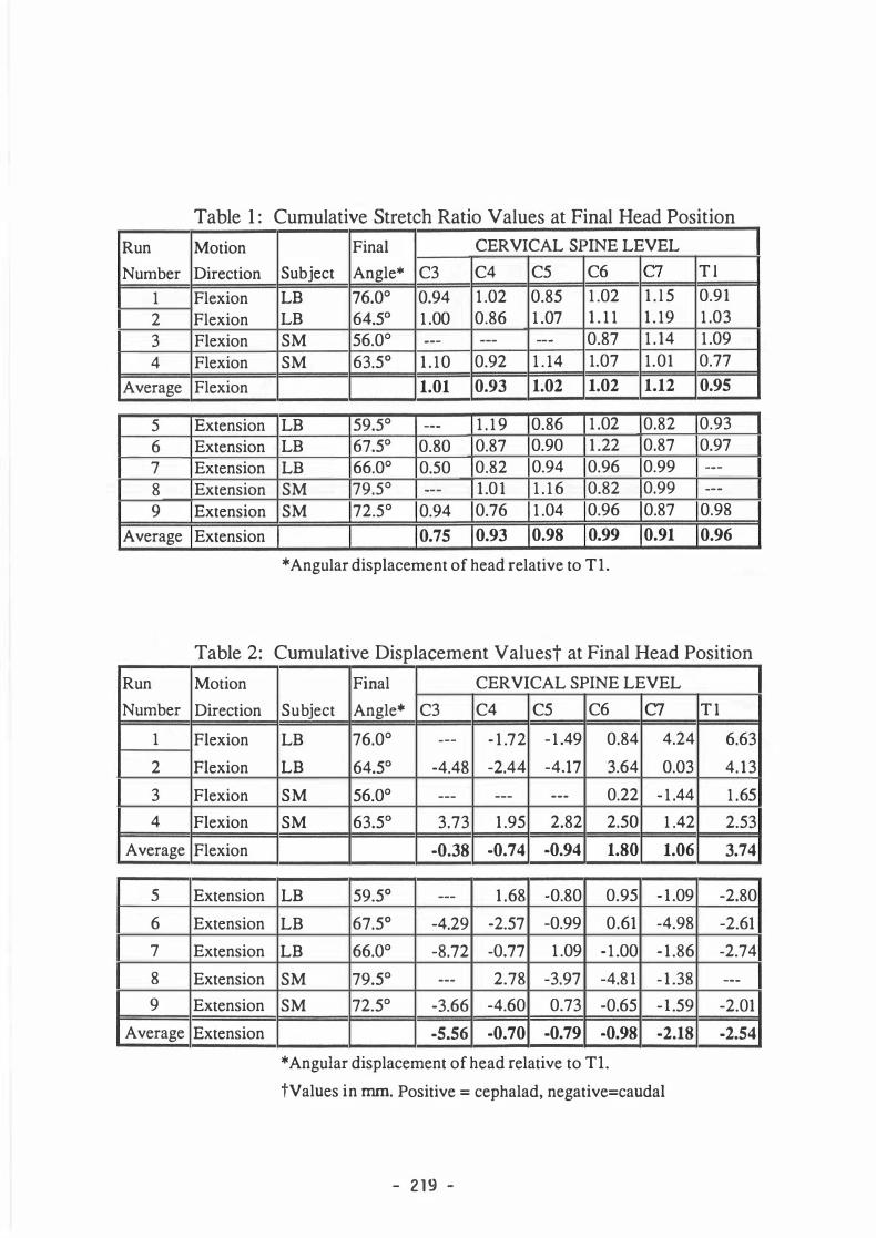
The coordinates of the three points digitized along each tag line were used to determine regional deformation or stretch ratio (Lfinal / Lreference) of the spinal cord and to calculate the local axial displacement of the spinal cord relative to its surrounding bony structures. The cumulative stretch ratio was calculated by multiplying together the stretch ratio for a given step with those from all previous steps. Cumulativ.e stretch ratlos greater than 1.0 indicate that the cord has elongated, values less than 1.0 indicate the cord has shortened relative to the neutral neck position.
- 21 5 -

- 2 1 5 -
Figure 1 : Flexion Series Run 2 in Tables 1 and 2 Cumulative angular displacement of the head is indicated on each post-motion scan. Total flexion of the head was 64.5° from the neutral starting position shown in the Row 1 , left scan.

Figure 2: Extension Series Run 9 in Tables 1 and 2. Cumulative angular displacement of
the head is indicated on each post-motion scan. Total extension of the head was 72.5° from the neutral starting position shown in the Row 1 , left scan.
- 2 1 7 -

Displacement of the cord relative to the vertebral canal was computed from absolute cord displacement by eliminating rigid body motion and rotation of the vertebra of interest (Points A and B, Fig. 3) between the reference and post-motion scans of the motion step. The cumulative cord displacement, or displacement relative to the initial neutral position at the vertebral level of interest, was calculated by summing the displacement values for that motion step with those from all previous steps.
RESULTS Nine stepwise motion series were analyzed with total flexion (N=4) or extension (N=5)
angles of the head greater than 50 degrees. Each series consisted of three to six motion steps. The cumulative stretch ratio and displacement values in the final head position for each series are presented in Tables 1 and 2 respectively. Data shown were computed at the level of the superior margin of each vertebral body; values at C3 represent information_ from tag lines located between C2.8 and C3.2. Information from adjacent tag lines were averaged if a tag line was not located appropriately. Displacement is given in mm, with positive and negative values indicating cephalad (towards the head) and caudad (away from the head) cord movement, respectively. In several instances stretch or displacement data is not reported due to blurry or absent tag lines. The stretch and displacement values at each vertebral level were averaged across all flexion observations, and similarly across all extension observations (Table 1 and 2).
Generally, flexion was associated with elongation of the cord (stretch ratios > 1.0) and extension with cord shortening (ratios < 1 .0). However, there was considerable variation in stretch response across cervical regions, both in any single position and over the entire motion sequence (Fig. 4a and 4b). In addition, because the motion of the head was unconstrained, two
Figure 3: Schematic Showing Points Digitized in Cervical Spine.
Three points along each tag line are digitized: Points A �nd B in the anterior cervical spine and Point C where the tag lme intersects the anterior surface of the spinal cord.
- 2 1 8 -
Run
Number
1 2 3 4
Average
5 6 7 8 9
Average
Run
Number
1
2
3
4
Average
s 6
7
8
9
Average
Table 1 : Cumulative Stretch Ratio Values at Final Head Position
Motion Final CERVICAL SPINE LEVEL
Direction Subject Angle* C3 C4 CS C6 C7 T l
Flexion LB 76.0° 0.94 1 .02 0.8S 1 .02 1 . l S 0.91
Flexion LB 64.S0 1 .00 0.86 1 .07 1 . 1 1 1 . 19 1 .03
Flexion SM S6.0° --- --- --- 0.87 1 . 14 1 .09
Flexion SM 63.S0 1 . 10 0.92 1 . 14 1.07 1.01 0.77
Flexion 1.01 0.93 1.02 1.02 1.12 0.95
Extension LB S9.5° --- 1 . 1 9 0.86 1 .02 0.82 0.93
Extension LB 67.S0 0.80 0.87 0.90 1 .22 0.87 0.97
Extension LB 66.0° 0.50 0.82 0.94 0.96 0.99 ---
Extension SM 79.5° --- 1 .01 1 . 1 6 0.82 0.99 ---Extension SM 72.5° 0.94 0.76 1 .04 0.96 0.87 0.98
Extension 0.75 0.93 0.98 0.99 0.91 0.96
*Angular displacement of head relative to Tl.
Table 2: Cumulative Displacement Valuest at Final Head Position
Motion
Direction
Flexion
Flexion
Flexion
Flexion
Flexion
Extension
Extension
Extension
Extension
Extension
Extension
Final CERVICAL SPINE LEVEL
Subject Angle* C3 C4 CS C6 C7 LB 76.0° --- - 1 .72 - 1 .49 0.84 4.24
LB 64.S0 -4.48 -2.44 -4.17 3.64 0.03
SM S6.0° --- --- --- 0.22 - 1 .44
SM 63.S0 3.73 1 .9S 2.82 2.SO 1 .42
-0.38 -0.74 -0.94 1.80 1.06
LB S9.S0 --- 1 .68 -0.80 0.9S - 1 .09
LB 67.S0 -4.29 -2.S7 -0.99 0.61 -4.98
LB 66.0° -8.72 -0.77 1 .09 - 1 .00 - 1 .86
SM 79.S0 --- 2.78 -3.97 -4.8 1 - 1 .38
SM 72.S0 -3.66 -4.60 0.73 -0.6S -l .S9
-5.56 -0.70 -0.79 -0.98 -2.18
*Angular displacement of head relative to Tl.
tValues in mm. Positive = cephalad, negative=caudal
- 2 1 9 -
T l
6.63
4. 13
1 .6S
2.S3
3.74
-2.80
-2.61
-2.74
---
-2.01
-2.54

Stretch � 1 .05� � : �! �: ! .95 -1 .04 .85 - .94 .75 - .84
Figure 4: Cumulative Stretch During Flexion and Extension
Representative flexion and extension sequences. Cumulative stretch computed for each step in the motion sequence. (a) Flexion series also shown in Fig. 1 ; (b) Extension series also shown in Fig. 2; (c) Flexion, Run 4 in Tables 1 and 2. Superimposed shaded blocks represent the cumulative stretch at that vertebral level. See legend for explanation of color scale.
- 220 -

maneuvers could produce a similar total rotation of the head relative to Tl, yet the head may follow very different paths. For example, in Fig. 4a subject LB flexed 64 degrees by first tucking in her chin (upper cervical flexion) and then brought her chin toward her ehest (midcervical flexion) (refer to Fig 1 , Row 1 , left for the initial neutral position scan). In contrast, in Fig 4c subject SM flexed 64° first with a rotation centered in the lower cervical spine, followed by midcervical flexion (refer to Fig 3 for the initial neutral position scan). As a consequence, we find a large variation in stretch ratios across maneuvers.
In flexion, the lower cervical cord displaced cephalad relative to the vertebrae in both subjects (Table 2). In subject SM the upper cervical cord also displaced cephalad, as if the entire cord were pulled upward by the rotating brain and brainstem. In subject LB the upper cervical cord moved caudad towards CS/C6, in contrast to the cephalad motion of the lower cervical cord. In extension, there was a general caudad motion of the entire cord as if the cord were forced downward relative to the vertebrae as the cervical spinal column shortens. Regional cord displacements associated with a representative extension series (Fig. 2) and flexion series (Fig. 1) are depicted pictorially in Figs. Sa and Sb, respectively. The final postmotion scan is shown for each series. In the regional cord displacement schematic shown below, arrow heads point in the direction of motion and originate at the superior margin of each vertebral body. Vector length is scaled to displacement magnitude.
DISCUSSION Our study represents the first report of in vivo measurements of human spinal cord
deformation and displacement relative to the vertebrae. We find that when the head/neck structure is in flexion the cervical spinal cord lengthens slightly and that in extension it shortens relative to its length in a neutral head/neck position. In flexion, the displacement of the upper cervical spinal cord was variable, while in the lower cord moved cephalad. In extension, the spinal cord moved caudad as the spinal column shortened.
Our study has three limitations: first, that only quasi-static deformation of the cord was measured. Although the snapshot MR imaging technique allows for remarkably short image acquisition times (typically 1 .7 sec) for a single, unrepeated event compared with other MR methods in common use, the acquisition time is far too long to capture any dynamic deformation occurring during normal or hazardous motions of the head. As new imaging modalities emerge, we hope to apply them to the study of spinal cord trauma. A second limitation of our study is that while the digitizing error associated with any single step is small, the measurement error is compounded when the digitized information from each step is combined. This limitation represents a trade-off between measurement accuracy at füll flexion or extension and intermediate information between the neutral and the final head/neck position. Third, we present results only for the anterior surface of the spinal cord. Brieg [1] observed that Stretch of the anterior surface of the cord was slightly smaller than that of the posterior surface during flexion and larger during extension. Because previous investigators [ 1 , 14, 18] did not specify the surface from which stretch and displacement measurements were taken, there may be some ambiguity when comparing our results with those of previous studies.
Previous studies of in situ spinal cord displacement and deformation were performed on human [ 1 , 14] or rhesus monkey [18] cadavers. After sectioning the cadaver just sagittal to the spinal cord, metal pins or nails were pushed into the cord and vertebral bodies at regular intervals. The displacement of these metallic markers relative to the vertebrae and the intermarker distances at füll extension and flexion were measured directly with rulers or calipers, or indirectly from lateral projection x-rays. For the purpose of comparison with our data, we present any previous results originally referenced to spinal cord root level now referenced to the appropriate vertebral level.
Earlier investigations have measured the stretch ratio of the entire cervical spinal cord between füll extension and füll flexion averaged 1.2 [l], with local stretch ratlos ranging from
- 221 -

J 1
•
(a)
1' = 3mm cephalad displacement
(b)
Figure 5: Cord Displacement During Flexion and Extension.
Cumulative axial spinal cord displacement at 64.5° flexion and 72.5° extension throughout the cervical spinal column. Displacement is relative vertebral motion, and is referenced to cord position in the initial neutral posture scan. See Figs. 1 and 2 for corresponding scan sequences. Arrowheads indicate direction of motion: up=cephalad, down=caudad. Line length is scaled to
- 222 -

1 . 1 at C3 to 1 .2 at C7 [ 1 8]. Reid [14] found stretch values of 1 . 1 in the upper and lower cervical spinal cord just between a neutral posture and füll flexion. Similarly, our results (Tables 1 and 2) show average stretch at C7 equal to 1 . 12 between flexion from neutral, and slight shortening (especially at C3) between the neutral posture and füll extension of the head/neck. These previous studies on cadavers do not report the angular head excursion, and it is not clear if the necks of our volunteer subjects underwent comparable degree of flexion and extension.
Breig [ 1] reported no axial displacement of the spinal cord between füll extension and füll flexion. In contrast, Smith [ 18] observed slight caudad displacement in the upper cervical spinal cord, little displacement at C4 and C5, and cephalad displacements of 1.7-4.7 mm in the lower cervical spinal cord between füll extension and füll flexion. Similarly, Reid [14] found little displacement at C4 and the largest displacements in the lower cervical spinal cord (4.5-6.8mm cephalad between neutral and flexion of head/neck, and approximately 5mm caudad between neutral and extension of the head/neck). These observations of cord displacement are comparable to our findings in the lower cervical spinal cord: significant upward displacements during flexion and downward displacements during extension from the neutral position. Our displacement results show considerable variation in the upper cervical spine, with some maneuvers agreeing with data from Smith [ 17] and some with Reid [ 13]. Reasons for the diff erences between studies may be due to differences in the kinematic movements of the head and variability between subjects and species.
Together, the in vivo and in vitro data clearly indicate that the cervical spinal cord moves and deforms during flexion and extension within the normal range. Cord movement suggests that the nerve roots and supporting dentate ligaments may tether the cord loosely within the spinal canal. Consequently, this loose tethering may reduce the cord deformation within the normal range of neck motion.
The quasistatic cord deformations reported in this communication were not associated with any injury. Previously, Hung et al. [9] found that slow uniaxial extension (strain rate = .002-.004 s- 1 ) of a preloaded (.04-.06 N) cat lumbar cord segment produced transient, recoverable changes in the motor function for relatively small extensions (e=O. l ), whereas larger extensions (e=0.3-0.5) caused severe and persistent changes in the motor and sensory functions. However, it is difficult to relate our results to those of Hung et al because it is unclear if a neutral head/neck posture is comparable to their preloaded reference state.
Brieg documented wavelike geometry of the white matter structures within the spinal cord during extension and in an intermediate (neutral) position, and straightening of the structures when the spine is placed in flexion. Deformation reported herein is referred to the neutral head/neck position and, thus, may overestimate the strain experienced by neural or vascular components of the spinal cord.
Studies by Galbraith[6] and Saatman [16] indicate that the functional and structural integrity of an isolated axon preparation changes appreciably when fairly rapid uniaxial loading rates (strain rate=l0-30 s·l) are applied For example, slow extension of a myelinated nerve fiber caused failure of the fiber at 40-45% of its original preloaded length, whereas more rapid elongations (strain rate=20 s·1) reduced the structural failure limit to only 20-30% of its original preloaded length. Taken together, we propose that damage to the spinal cord may be related to both the amount and rate of uniaxial deformation.
CONCLUSIONS This study presents novel information regarding the in vivo quasistatic motion of the
human cervical spinal cord during flexion and extension of the neck. Our findings include 1 ) during extension, the cord shortens and displaces caudad relative to the neutral head/neck posture, and 2) during flexion, the cord lengthens relative to the neutral position, the upper cord displacement is variable, and the lower cervical spinal cord displaces cephalad. Characterizing
- 223 -

cervical spinal cord distortion and displacement during normal maneuvers is an initial step towards identifying in vivo cord distortion associated with traumatic spinal cord injuries in man.
REFERENCES
1 .
2 .
3 .
4.
5 .
6 .
7 .
8 .
9.
1 0.
1 1 .
12 .
1 3 .
1 4.
1 5 .
16.
17 .
1 8 .
19.
20.
Brieg, A. Biomechanics of the Central Nervous System. Almqvist and Wiksell, Stockholm; Year Book, Chicago, 1960.
Bunegin L, T-K Hung, GL Chang. Biomechanics of spinal cord injury. Crit Care Clin 1987, 3 :453-470.
Deng, Y.C. and W. Goldsmith, Response of a human head/neck/upper torso replica to dynamic loading-1. Physical model. J. Biomech., 1987. 20: p. 471-486.
Deng, Y.C. and W. Goldsmith, Response of a human head/neck/upper torso replica to dynamic loading-II. Analytical/numerical model. J Biomech., 1987. 20: p. 487-497.
Ewing, C.L. and D.J. Thomas, Human head and neck response to impact acceleration. 1972, Pensacola, Fla.: NAMRL.
Galbraith JA. The effects of mechanical leading on the electrophysiology of the sguid giant axon. Ph.D. Dissertation, University of PA, 1988.
Holsinger AE, Riederer SJ. The importance of phase encoding order in ultra-short TR snapshot MR imaging. Magn Reson Med 1990; 16:48 1-488.
Huelke, D. and G. Nusholtz, Cervical Spine Biomechanics: A Review of the Literature. J. Orthop Res, 1986. 4: p. 232-245.
Hung, T.K., Chang, G.L., Chang, J.L., Albin, M.S., Stress-strain Relationship and Neurological Sequelae of Uniaxial Elongation of the Spinal Cord of Cats, Surg Neurol, v. 15, no. 6, 1981
McElhaney, J., et al. Combined Bending and Axial Loading Responses of the Human Cervical Spine. in Stapp Car Crash Conference. 1988. SAE.
Merrill, T., W. Goldsmith, and Y.C. Deng, Three-dimensional response of a lumped parameter head-neck model due to impact and impulsive loading. J. Biomech., 1984. 17: p. 8 1 -95.
Mertz, H. and L. Patrick. Strength and Response of the Human Neck. in 15th Stapp Car Crash Conference. 1971 . SAE.
Niitsu, M., NG Campeau, AE Holsinger-Bampton, SJ Riederer, RL Ehman. Tracking motion with tagged rapid gradient-echo magnetization-prepared MR imaging. JMRI, 1992, 2: 155-163.
Reid, JD. Effects of flexion-extension movements of the head and spine upon the spinal cord and nerve roots. J Neurol. Neurosurg. Psychiat., 1960, 23: 214-221
Rice, D., E. Mackenzie, and et al., Cost of Injmy in the US: A Report to Congress. 1989, Univ. of CA, Tue Johns Hopkins University:
Saatman K, LE Thibault Myelinated nerve fiber response to dynamic uniaxial stretch. Proc of IRCOBI, 1991 , 1 15- 125.
Sances, A., et al., Bioengineering Analysis of Head and Spine Injuries. CRC Crit Rev Bioeng, 198 1 . : p. 79- 1 1 8.
Smith, CG. Changes in length and position of the segments of the spinal cord with changes in posture in the monkey. Radiology, 1956, 66: 259-266.
Thibault, L., et al. The Strain Dependent Pathophysiological Consequences of lnertial Loading on the Central Nervous System. in International Conference on the Biokinetics of Impact. 1990. Lyon, France
Williams, J.L. and T.B. Belytschko, A three-dimensional model of the human cervical spine for impact simulation. J Biomech. Engng., 1983. 105: p. 321-331 .
- 224 -