In vitro study of aqueous leaf extract of Chenopodium ...
8
Full Length Article In vitro study of aqueous leaf extract of Chenopodium album for inhibition of calcium oxalate and brushite crystallization Deepti Sharma a , Yadu Nandan Dey a , Indu Sikarwar b , Richa Sijoria a , Manish M. Wanjari a, *, Ankush D. Jadhav a a National Research Institute for Ayurveda-Siddha Human Resource Development, Gwalior 474009, Madhya Pradesh, India b Shri Ram College of Pharmacy, Banmore 476444, Madhya Pradesh, India ARTICLE INFO Article history: Received 12 August 2015 Received in revised form 15 February 2016 Accepted 18 February 2016 Available online 2 March 2016 ABSTRACT The leaves of Chenopodium album Linn. are traditionally used for treatment of kidney dis- eases and urinary stones. The present work investigated the effect of aqueous extract of leaves of C. album (CAAE) on in-vitro crystallization of CaOx and brushite crystals. Crystal- lization was studied by using nucleation and aggregation assay of calcium oxalate (CaOx) crystals and growth assay of calcium oxalate monohydrate and brushite crystals.The effects of CAAE and cystone on slope of nucleation and aggregation as well as growth of calcium oxalate crystallization were evaluated spectrophotometrically.The densities of the formed crystals were compared under microscope. The effects of CAAE and citric acid on growth of brushite crystals were studied by using single diffusion gel growth technique, and the parameters evaluated were length, morphology and average size of the deposited crystals. CAAE significantly inhibited the slope of nucleation and aggregation of CaOx crystalliza- tion, and decreased the crystal density. It also inhibited the growth and caused the dissolution of brushite crystals. The standard drug cystone or citric acid also exhibited similar effects. The study reveals that the leaves of C. album were found effective in the prevention of the experimentally induced urinary stones and substantiate the traditional claim. It is con- cluded that the leaves of C. album have beneficial inhibitory effect on in-vitro crystallization of CaOx and CHPD (brushite) crystals. © 2016 Mansoura University. Production and hosting by Elsevier B.V.This is an open access article under the CC BY-NC-ND license (http://creativecommons.org/licenses/by- nc-nd/4.0/). Keywords: Bathua Urolithiasis Cystone Calcium oxalate Brushite Citric acid 1. Introduction Urolithiasis is defined as the presence of one or more calculi in any location within the urinary tract [1]. It is a common dis- order estimated to occur in approximately 12% of the population, with a recurrence rate of 70–80% in male and 47–60% in female [2]. Majority of the stones are calcium- containing stones, especially calcium oxalate (80%) and others are 20% [3]. * Corresponding author. Tel.: +91 9229205231. E-mail address: [email protected] (M.M. Wanjari). http://dx.doi.org/10.1016/j.ejbas.2016.02.001 2314-808X/© 2016 Mansoura University.Production and hosting by Elsevier B.V.This is an open access article under the CC BY-NC-ND license (http://creativecommons.org/licenses/by-nc-nd/4.0/). egyptian journal of basic and applied sciences 3 (2016) 164–171 Available online at www.sciencedirect.com journal homepage: http://ees.elsevier.com/ejbas/default.asp HOSTED BY ScienceDirect
Transcript of In vitro study of aqueous leaf extract of Chenopodium ...

Full Length Article
In vitro study of aqueous leaf extract ofChenopodium album for inhibition of calciumoxalate and brushite crystallization
Deepti Sharma a, Yadu Nandan Dey a, Indu Sikarwar b, Richa Sijoria a,Manish M. Wanjari a,*, Ankush D. Jadhav a
a National Research Institute for Ayurveda-Siddha Human Resource Development, Gwalior 474009, MadhyaPradesh, Indiab Shri Ram College of Pharmacy, Banmore 476444, Madhya Pradesh, India
A R T I C L E I N F O
Article history:
Received 12 August 2015
Received in revised form 15
February 2016
Accepted 18 February 2016
Available online 2 March 2016
A B S T R A C T
The leaves of Chenopodium album Linn. are traditionally used for treatment of kidney dis-
eases and urinary stones. The present work investigated the effect of aqueous extract of
leaves of C. album (CAAE) on in-vitro crystallization of CaOx and brushite crystals. Crystal-
lization was studied by using nucleation and aggregation assay of calcium oxalate (CaOx)
crystals and growth assay of calcium oxalate monohydrate and brushite crystals. The effects
of CAAE and cystone on slope of nucleation and aggregation as well as growth of calcium
oxalate crystallization were evaluated spectrophotometrically. The densities of the formed
crystals were compared under microscope. The effects of CAAE and citric acid on growth
of brushite crystals were studied by using single diffusion gel growth technique, and the
parameters evaluated were length, morphology and average size of the deposited crystals.
CAAE significantly inhibited the slope of nucleation and aggregation of CaOx crystalliza-
tion, and decreased the crystal density. It also inhibited the growth and caused the dissolution
of brushite crystals. The standard drug cystone or citric acid also exhibited similar effects.
The study reveals that the leaves of C. album were found effective in the prevention of the
experimentally induced urinary stones and substantiate the traditional claim. It is con-
cluded that the leaves of C. album have beneficial inhibitory effect on in-vitro crystallization
of CaOx and CHPD (brushite) crystals.
© 2016 Mansoura University. Production and hosting by Elsevier B.V. This is an open
access article under the CC BY-NC-ND license (http://creativecommons.org/licenses/by-
nc-nd/4.0/).
Keywords:
Bathua
Urolithiasis
Cystone
Calcium oxalate
Brushite
Citric acid
1. Introduction
Urolithiasis is defined as the presence of one or more calculiin any location within the urinary tract [1]. It is a common dis-
order estimated to occur in approximately 12% of thepopulation, with a recurrence rate of 70–80% in male and47–60% in female [2]. Majority of the stones are calcium-containing stones, especially calcium oxalate (80%) and othersare 20% [3].
* Corresponding author. Tel.: +91 9229205231.E-mail address: [email protected] (M.M. Wanjari).
http://dx.doi.org/10.1016/j.ejbas.2016.02.0012314-808X/© 2016 Mansoura University. Production and hosting by Elsevier B.V. This is an open access article under the CC BY-NC-NDlicense (http://creativecommons.org/licenses/by-nc-nd/4.0/).
e g y p t i an j o u rna l o f b a s i c and a p p l i e d s c i e n c e s 3 ( 2 0 1 6 ) 1 6 4 – 1 7 1
Available online at www.sciencedirect.com
journal homepage: ht tp : / /ees .e lsevier.com/ejbas/defaul t .asp
HOSTED BY
ScienceDirect

The medical management of urolithiasis involves drug treat-ment and extracorporeal shock wave lithotripsy (ESWL). Thevarious therapies including thiazide as diuretic and alkali-citrate are used to prevent the recurrence of hypercalciuriaand hyperoxaluria, which induce calculi formation, but evi-dence for their efficacy is less [4].The surgical endoscopic stoneremoval and extracorporeal shock wave lithotripsy have revo-lutionized the treatment of urolithiasis but does not preventthe likelihood of new stone formation [5]. Besides imposingthe high cost, shock waves in therapeutic doses may causeacute renal injury, decrease in renal function and an increasein stone recurrence. The recurrence of stone formation is alsovery high (50–80%). In addition, persistent residual stone frag-ments and the possibility of infection after ESWL represent aserious problem in the treatment of stones.Thus, medical man-agement of urolithiasis is either costly or poses serious sideeffects.
The crystallization of the stone begins with increased urinarysupersaturation, with the subsequent formation of the solidcrystalline particles within the urinary tract. This is followedby nucleation, by which stone-forming salts in supersatu-rated urinary solution coalesce into clusters that then increasein size by the addition of new constituents [6]. These crystalsthen grow and aggregate with other crystals in solution, andare ultimately retained and accumulated in the kidney [7].Therefore, if this progression of crystallization can be pre-vented, then lithiasis can also be prevented.
There is growing interest of public in herbal medicine,particularly in the treatment of urolithiasis partly because oflimited choice in the pharmacotherapy. Data from in-vitro,in-vivo and clinical trials reveal that phytotherapeutic agentscould be useful as either an alternative or an adjunctivetherapy in the management of urolithiasis. Many Indian plantsare useful as antilithiatic agents [5,8–13]. Hence, the Indianmedicinal plants are constantly being evaluated for possibleantilithiatic effects.
Chenopodium album L. (family: Chenopodiaceae) is a herba-ceous vegetable plant locally known as Bathua. It is cultivatedas pot-herb and usually grown in gardens, but can be foundin the corner of early grain fields in Bombay presidency andelsewhere in India (Kashmir and Sikkim). The medicinal prop-erty of this plant is mainly present in leaves and seeds. Theleaves of C. album are used in ethno-medicinal practices fortreatment of kidney diseases and urinary stones. Ethnobo-tanical studies of Aravalli region of Rajasthan (India) report thefolk medicinal uses of cooked leaves of C. album in kidney stonesand urinary tract troubles [14]. Cooked leaves of C. album areused as traditional medicine in the Shekhavati region of Ra-jasthan for treatment of urinary troubles and colic [15]. InLadakh, leaves are also used traditionally for controlling painfulurination [16]. C. album is an important medicinal weed of Mo-radabad useful in the treatment of urinary retention and kidneydiseases [17].
In view of traditional and ethno-medicinal use of leaves ofC. album in the treatment of kidney stones, the present workdemonstrated the effect of aqueous extract of the leaves ofC. album on in-vitro crystallization of CaOx and brushite crys-tals. As this plant is consumed as food substance by humanbeings and as weed fodder by cattle, its antilithiatic propertywould be good preventive option available.
2. Materials and methods
2.1. Plant material
The leaves of C. album were collected from the local marketof Gwalior in December 2012 and identified by Dr. N.K. Pandey,Research Officer (Botany), National Research Institute forAyurveda-Siddha Human Resource Development, Aamkho,Gwalior. A voucher specimen (Field Book No. 5-4/12–13/NRIASHRD/Tech/Survey/134) of the authenticated C. album hasbeen deposited in the herbarium of the institute.
2.2. Drugs and chemicals
Cystone (Himalaya Drug Company) and citric acid1-monohydrate (E. Merck (India) Ltd., Mumbai) were pur-chased from the local market. All remaining chemicals usedin the experiment were of the highest grade commerciallyavailable.
2.3. Preparation of aqueous extract of the leaves ofC. album (CAAE)
The leaves were separated from other extraneous matter andsubjected to shade drying. The dried leaves were subjected toa coarse powder by using dry grinder. The powder (100g) wassoaked (maceration) in 1 L purified water and kept in dark anddry place for 48 h at a temperature range of 20–26 °C. Chloro-form was added in quantity of 1% total mixture to avoidmicrobial growth. After 48 h, solutions were filtered by WhatmanFilter Paper No. 1. The filtered extracts were dried in a rotaryevaporator to obtain a dark brown powdery extract (13.4% w/w).
2.4. Preliminary phytochemical screening andquantitative estimation of phytoconstituents
Preliminary phytochemical screening [18] of CAAE was carriedout to detect the presence of sterols, alkaloids, saponins, ter-penes, tannins, phenolic substances, carbohydrates, volatile oiland mucilage. The total phenolic content of the extracts wasdetermined spectrometrically [19] and expressed as milli-gram of tannic acid equivalents (TAE) per gram of extract. Totalflavonoid content was measured by aluminum chloride colo-rimetric assay [20] and expressed as milligram of quercetinequivalent per gram of extract. Total saponins were deter-mined according to the previously described methods byObadoni and Ochuko (2002) with little modification [21].
2.5. Effect of CAAE on in-vitro crystallization
2.5.1. In-vitro crystallization of calcium oxalate
2.5.1.1. Nucleation and aggregation assay. Nucleation and ag-gregation assay were performed as per method previouslydescribed by Hess et al. [2000] with minor modifications [22].Briefly, freshly prepared solution of 10 mM calcium chloride di-hydrate and 1.0 mM sodium oxalate, containing 200 mM NaCland 10 mM sodium acetate trihydrate, was adjusted to pH 5.7.All experiments were performed at 37 °C, using a circulating
165e g y p t i an j o u rna l o f b a s i c and a p p l i e d s c i e n c e s 3 ( 2 0 1 6 ) 1 6 4 – 1 7 1

water bath. For crystallization experiments, 25 ml of sodiumoxalate solution was transferred into a beaker and placed inthe hot plate magnetic stirrer (Model 2MLH, REMI), which wasmaintained at 37 °C and constantly stirred at 800 rpm. An ad-ditional 1 ml of distilled water/standard (cystone)/extract wereadded and finally calcium chloride solution (25 ml) was added.The optical density was measured at 620 nm in spectropho-tometer (UV 1800, Shimadzu Corporation, Japan) after additionof calcium containing solution, on every 15 s over 5 min andthen every 1 min over 10 min. All the experiments were per-formed in triplicate. The final solutions were seen under a lightmicroscope to analyze the density of formed crystals in thesolution (Olympus, USA). Percent inhibition in the presence ofcystone or CAAE was compared with the control by the fol-lowing formula.
The percentage inhibition was calculated as:
1 100− ( )[ ] ×Tsi Tsc
where Tsc, the turbidity slope of the control; and Tsi, the tur-bidity slope in the presence of the inhibitor.
2.5.1.2. In vitro calcium oxalate crystal growth assay. Inhibitoryactivity of CAAE against CaOx crystal growth was measuredusing previously described methods [23,24]. Briefly, 20 ml eachof 4 mM calcium chloride and 4 mM sodium oxalate were addedto a 30 ml of solution, containing NaCl (90 mM) buffered withTris HCl (10 mM) pH 7.2.To this 600 μl of calcium oxalate mono-hydrate (COM) crystal slurry (1.5 mg/ml acetate buffer) wasadded. Consumption of oxalate begins immediately after COMslurry addition and was monitored for 600 s by disappear-ance of absorbance at 214 nm. One ml of CAAE (500) and CAAE(1000 μg/ml) was added separately into this solution.The deple-tion of free oxalate ions will decrease if CAAE inhibits calciumoxalate crystal growth. Rate of reduction of free oxalate wascalculated using the baseline value and the value after 30 s in-cubation with or without the extract. The relative inhibitoryactivity was calculated as follows:
% Relative inhibitory activity C S C= −( )( ) × 100
where C is the rate of reduction of free oxalate without anyextract and S is the rate of reduction of free oxalate with CAAE.
2.5.2. In-vitro crystallization of brushite crystals
2.5.2.1. Growth assay. Growth assay in single diffusion gelgrowth of brushite crystals was carried out according to themethods previously described by of Joshi et al. [2005b] with littlemodification [11]. Single diffusion gel growth method was fol-lowed to grow calcium hydrogen phosphate dihydrate (CHPD).When crystals achieved the maximum growth the inhibitiveeffects of citric acid solution having different concentrationswere studied by adding them into the supernatant solution.
Glass test tubes of 2.5 cm diameter and 15 cm length wereused for growing the crystals. Five milliliters of sodium meta-silicate solution of specific gravity 1.06 were acidified by addingappropriate amount (2.7 ml) of orthophosphoric acid so that5.0 pH could be obtained for the mixture, which was subse-quently transferred into different test tubes.After gelation took
place, 10 ml of 1 M aqueous solution of calcium chloride wascarefully poured on the set gels. Crystals were found growingvery rapidly within two days from pouring the supernatant so-lutions. Elongated platelet type and star shaped crystals weregrown in the gel. The apparent length of growing crystals wasmeasured under microscope at different time intervals. Theplot of apparent length of growing crystals versus time showedthat the crystals achieved maximum length on the 5th day ofpouring supernatant solution on set gel. Aqueous solution ofcitric acid as a standard and CAAE was added in the same volumeas the calcium chloride solution on the 5th day after acquir-ing the maximum length of CHPD crystals, and their effect wasstudied on the growth of CHPD crystals up to the 8th day. ForpH 5.0, many crystals having star and platelet star and plate-let type morphologies were observed. The photographs of thetest tube showing the growth of the crystals were recorded.
2.6. Statistical analysis
The data obtained were analyzed by one-way ANOVA, two-way ANOVA and linear regression analysis wherever necessary.A value of p < 0.05 was considered significant in all cases.
3. Results
3.1. Preliminary phytochemical screening andquantitative estimation of phytoconstituents
CAAE showed the presence of proteins, alkaloids, saponin gly-cosides, amino acids and flavonoids, while carbohydrates, sterolsand tannins were absent. The total phenolic and flavonoidcontent of CAAE were found to be 239.8 mg TAE/g of extractand 87.23 mg quercetin equivalents/g of extract, respectivelywhile the total saponins content was found to be 1.6 mgsaponins/100g of powder mass.
3.2. Effect of CAAE on in-vitro crystallization
3.2.1. In-vitro crystallization of calcium oxalate
3.2.1.1. Nucleation and aggregation assay. The changes in theturbidity or optical density of different solutions, viz control,cystone (1000 μg/ml), and CAAE (500 μg/ml and 1000 μg/ml), wereplotted at different time intervals. The turbidity increased lin-early up to 5 minutes, which indicated the nucleation processand then decreased linearly up to 15 minutes indicating theaggregation process. CAAE (500 μg/ml and 1000 μg/ml) andcystone (1000 μg/ml) inhibited both the rate of nucleation andthe rate of aggregation. The maximum optical density of thesolutions, viz control, cystone (1000 μg/ml) and CAAE (500 μg/ml) and CAAE (1000 μg/ml), recorded was 0.170, 0.072, 0.122 and0.095. The percent inhibition rates of nucleation of CaOx bycystone (1000 μg/ml), CAAE (500 μg/ml) and CAAE (1000 μg/ml) were found to be 58.33, 12.5 and 37.5 percent, respectively(Fig. 1). The percent inhibition rates of aggregation of CaOx bycystone (1000 μg/ml), CAAE (500 μg/ml) and CAAE (1000 μg/ml) were found to be 66.66, 66.66 and 33.33 respectively (Fig. 1).The photomicrographs of the CaOx crystals in solutions of
166 e g y p t i an j o u rna l o f b a s i c and a p p l i e d s c i e n c e s 3 ( 2 0 1 6 ) 1 6 4 – 1 7 1

control, cystone (1000 μg/ml) and CAAE (500 μg/ml and 1000 μg/ml) showed that CaOx crystals were less denser in cystone(1000 μg/ml), CAAE (500 μg/ml) and CAAE (1000 μg/ml) as com-pared to control (Fig. 2).
3.2.1.2. In vitro calcium oxalate crystal growth assay. In calciumoxalate growth assay, CAAE inhibited calcium oxalate mono-hydrate (COM) growth. The percentage inhibition of cystone(1000 μg/ml), CAAE (500 μg/ml) and CAAE (1000 μg/ml) were50.96, 22.05, and 39.57 respectively (Fig. 1).
3.2.2. In-vitro crystallization of brushite crystals
3.2.2.1. Growth assay. The growth of brushite crystals was mea-sured as length (thickness of crystal deposition). The crystalsacquired maximum length (approximately 0.35 cm) on day 3after gelation took place and then after the length of the crys-tals deposited became constant up to day 8.The average length
of the deposited CHPD crystals was decreased by citric acid(1 M), CAAE (500 μg/ml) and CAAE (1000 μg/ml) until day 8. Thelength of the crystals in control, CAAE (500 μg/ml) and CAAE(1000 μg/ml) were 0.35 cm, 0.21 cm and 0.133 cm, respec-tively; however, it could not be measured due to absence ofany measurable size crystals in citric acid (1 M) group. Figs. 3and 4 show the effects of citric acid and CAAE on the lengthof deposited CHPD crystals.
Fig. 5 shows the microphotographs of CHPD crystals in dif-ferent groups. Crystals in CAAE (500 and 1000 μg/ml) groupswere very small compared to the control, whereas in citric acidthere was no appearance of observable crystals. Citric acid andCAAE (500 and 1000 μg/ml) significantly decreased the size ofcrystals (p < 0.001) as measured by its thickness using stagemicrometer. The microphotographs of the CHPD crystals ofthe control showed the plate like crystals having an average
Fig. 1 – Effects of cystone and CAAE on CaOx nucleation,aggregation and growth.
Fig. 2 – Photomicrographs of CaOx crystal density in different solutions, viz control, cystone (1000 μg/ml), and CAAE (500 μg/ml and 1000 μg/ml) [A = control, B = cystone, C = CAAE500, D = CAAE1000], magnification 100×.
Fig. 3 – Effects of citric acid and CAAE on the length ofdeposited CHPD crystals. Values are mean ± SEM.
167e g y p t i an j o u rna l o f b a s i c and a p p l i e d s c i e n c e s 3 ( 2 0 1 6 ) 1 6 4 – 1 7 1
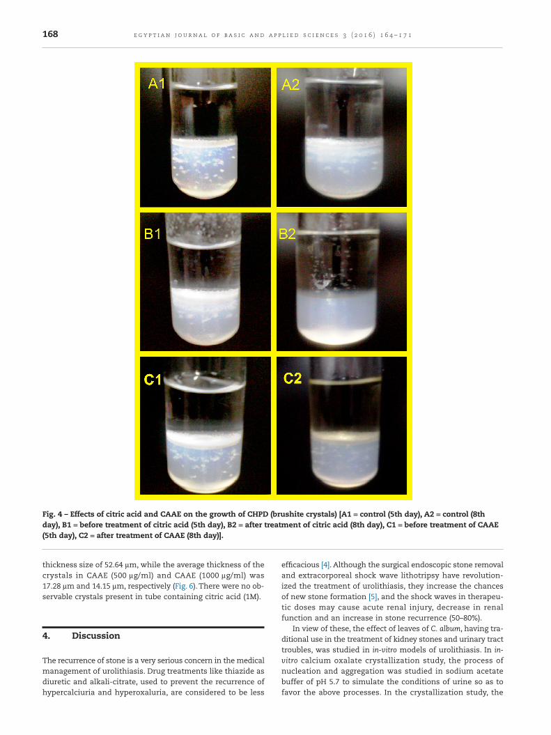
thickness size of 52.64 μm, while the average thickness of thecrystals in CAAE (500 μg/ml) and CAAE (1000 μg/ml) was17.28 μm and 14.15 μm, respectively (Fig. 6). There were no ob-servable crystals present in tube containing citric acid (1M).
4. Discussion
The recurrence of stone is a very serious concern in the medicalmanagement of urolithiasis. Drug treatments like thiazide asdiuretic and alkali-citrate, used to prevent the recurrence ofhypercalciuria and hyperoxaluria, are considered to be less
efficacious [4]. Although the surgical endoscopic stone removaland extracorporeal shock wave lithotripsy have revolution-ized the treatment of urolithiasis, they increase the chancesof new stone formation [5], and the shock waves in therapeu-tic doses may cause acute renal injury, decrease in renalfunction and an increase in stone recurrence (50–80%).
In view of these, the effect of leaves of C. album, having tra-ditional use in the treatment of kidney stones and urinary tracttroubles, was studied in in-vitro models of urolithiasis. In in-vitro calcium oxalate crystallization study, the process ofnucleation and aggregation was studied in sodium acetatebuffer of pH 5.7 to simulate the conditions of urine so as tofavor the above processes. In the crystallization study, the
Fig. 4 – Effects of citric acid and CAAE on the growth of CHPD (brushite crystals) [A1 = control (5th day), A2 = control (8thday), B1 = before treatment of citric acid (5th day), B2 = after treatment of citric acid (8th day), C1 = before treatment of CAAE(5th day), C2 = after treatment of CAAE (8th day)].
168 e g y p t i an j o u rna l o f b a s i c and a p p l i e d s c i e n c e s 3 ( 2 0 1 6 ) 1 6 4 – 1 7 1

turbidity increased linearly up to 5 min and then decreased lin-early up to 15 min after the addition of calcium chloridedihydrate. Earlier increase in the turbidity was suggestive ofthe nucleation phenomenon, while the decrease in the laterpart indicated the aggregation. These two phenomena repre-sented the complete process of in-vitro crystallization asobserved previously by Hess et al. [2000] [22]. Simultaneous ad-dition of CAAE (500 and 1000 μg/ml) and cystone (1000 μg/ml)along with calcium chloride dihydrate inhibited the nucle-ation as well as aggregation process of CaOx crystallizationas indicated by dose-dependent decrease in turbidity of thesolution in both phenomena. The inhibition of in-vitro crys-tallization of CaOx suggests that CAAE has influence on theformation of crystals from sodium oxalate and calcium chlo-ride and/or their aggregation. Most of the previous papers [25,26]stated that the test drug or extract inhibited the crystalliza-tion by favoring the formation of calcium oxalate dihydrate
(COD) crystals instead of calcium oxalate monohydrate (COM).The study needs a use of polarized light to differentiate betweenCOM and COD crystals, which is a limitation of the presentstudy and thus could not state whether the CAAE favored theformation of COD and less of COM, but the possibility of thiscannot be ignored. The non-significant decrease in turbidityas observed in the process of aggregation may be due to thepossibility of more amounts of COD crystals in the solution.Moreover, growth assay showed that CAAE also inhibited thegrowth of calcium oxalate monohydrate crystals with maximuminhibition at 1000 μg/ml concentration.This indicates that CAAEhas inhibitory influence on nucleation/aggregation and growthof calcium oxalate crystals.
Similarly, CAAE also showed significant inhibition of growthof brushite (CHPD) crystals and caused their dissolution, in-dicated by the small length of the crystal deposition thicknessin the test tube (See Figs. 3 and 4) and decreased microscopicthickness of the crystals (P < 0.001) as compared to control (seeFigs. 5 and 6). CAAE exhibited effects comparable to standarddrug citric acid, which showed the complete dissolution of theCHPD crystals. The effect of citric acid corroborates with pre-vious finding [27].The results indicated that CAAE has significantinfluence on the formation, growth and dissolution of crys-tals, and further suggest that the extract has beneficial effectin preventing the formation of crystals and their growth.
Literature of the previous studies is silent on the exactmechanism involved in the inhibition of in-vitro crystalliza-tion and stated that the extracts contained some chemicalcomponents that inhibited the crystallization.There are reportsthat flavonoids inhibit calcium oxalate crystallization in humanurine as well as in animal models [28] and crystal deposition[29]. Saponins showed anti-crystallization properties by
Fig. 5 – Effects of citric acid and CAAE on the size of crystals [A = control, B = citric acid, C = CAAE500, D = CAAE1000],magnification 400×.
Fig. 6 – Effects of citric acid and CAAE on microscopicthickness of crystals. Values are expressed as mean ± SEM;*p<0.001 when compared to control.
169e g y p t i an j o u rna l o f b a s i c and a p p l i e d s c i e n c e s 3 ( 2 0 1 6 ) 1 6 4 – 1 7 1
disaggregating the suspension of mucoproteins, the promot-ers of crystallization [30]. Phytochemical screening andestimation of important constituents revealed that CAAE con-tains flavonoids and saponins. It has total flavonoid contentof 87.23 mg quercetin equivalents/g of extract and total sa-ponins content of 1.6 mg saponins/100g of powder mass. Thus,the flavonoids and saponins present may be playing a con-tributing role in anti-crystallization action of CAAE. Fouda et al.[2006] studied the effect of in-vitro and in-vivo antilithiatic effectsof saponins rich fraction of Herniaria hirsuta and stated thatfraction contained a substance that promoted the nucleationof COD crystals [25]. Finally, the results of the present inves-tigations suggest that the leaves of the C. album have in-vitroanti-crystallization effect on CaOx and brushite crystals. Thesefindings substantiate the traditional use of the leaves in thetreatment of urinary stones and kidney problems. In order tosubstantiate its in-vitro effect, the in-vivo studies need to becarried out in experimental animals.
5. Conclusion
The leaves of C. album have beneficial inhibitory effect on in-vitro crystallization of CaOx and CHPD (brushite) crystals.
Acknowledgment
The authors acknowledge the Director General, Central Councilfor Research in Ayurvedic Sciences, New Delhi, India, for pro-viding necessary facilities to carry out the research work.
R E F E R E N C E S
[1] Thomas B, Hall J. Urolithiasis. Surgery (Oxford) 2005;23:129–33.
[2] Smith CL, Guay DRP. Nephrolithiasis. In: Di Piro JT, TalbertRL, Hayes PE, Yee GC, Matzke GR, Posey LM, editors.Pharmacotherapy and pathophysiologic approach, vol. 2.Elsevier; 1992. p. 720–36.
[3] Moe OW. Kidney stones: pathophysiology and medicalmanagement. Lancet 2006;367:333–44.
[4] Pak CY. Prevention and treatment of kidney stones. Role ofmedical prevention. J Urol 1989;141(3 Pt 2):798–801.
[5] Kalyan SB, Christina AJM, Syama SB, Selvakumar S, SundaraSK. Urolithiatic activity of Hibiscus sabdariffa Linn. onethylene glycol-induced lithiasis in rats. Nat Prod Rad2009;8:43–7.
[6] Basavaraj DR, Biyani CS, Browning AJ, Cartledge JJ. The roleof urinary kidney stone inhibitors and promoters in thepathogenesis of calcium containing renal stones. EAU-EBUUpdate Ser 2007;5:126–36.
[7] Kok DJ, Papapolous SE, Bijovet OL. Crystal agglomeration is amajor element in calcium oxalate urinary stone formation.Kidney Int 1990;37:51–6.
[8] Karadi RV, Gadge NB, Alagawadi KR, Savadi RV. Effect ofMoringa oleifera Lam. root-wood on ethylene glycol inducedurolithiasis in rats. J Ethnopharmacol 2006;105:306–11.
[9] Garimella TS, Jolly CI, Narayanan S. In-vitro studies onantilithiatic activity of seeds of Dolichos biflorus Linn. and
rhizomes of Bergenia ligulata Wall. Phytother Res 2001;15:351–5.
[10] Joshi VS, Parekh BB, Joshi MJ, Vaidya AB. Herbal extracts ofTribulus terrestris and Bergenia ligulata inhibit growth ofcalcium oxalate monohydrate crystals in-vitro. J CrystGrowth 2005a;275:e1403–8.
[11] Joshi VS, Parekh BB, Joshi MJ, Vaidya ADB. Inhibition of thegrowth of urinary calcium hydrogen phosphate dihydratecrystals with aqueous extracts of Tribulus terrestris andBergenia ligulata. Urol Res 2005b;33:80–6.
[12] Barros ME, Schor N, Boim MA. Effects of an aqueous extractfrom Phyllanthus niruri on calcium oxalate crystallization in-vitro. Urol Res 2003;30:374–9.
[13] Sangeeta D, Sidhu H, Thind SK, Vaidyanathan S, Nath R.Therapeutic response of Tribulus terrestris (Gokhru) aqueousextract on hyperoxaluria in male adult rats. Phytother Res1993;6:116–19.
[14] Sharma N, Tanwar BS, Vijayavergia R. Study of medicinalplants in Aravalli regions of Rajasthan for treatment ofkidney stone and urinary tract troubles. Int J Pharm Tech Res2011;3(1):110–13.
[15] Kateva SS, Galav PK. Traditional herbal medicines fromShekhawati regions of Rajasthan. Indian J Trad Know2005;4(3):237–45.
[16] Ballabh B, Chaurasia OP, Ahmed Z, Singh SB. Traditionalmedicinal plants of cold desert Ladakh – used againstkidney and urinary disorders. J Ethnopharmacol2008;118:331–9.
[17] Kumar A, Agarwal S, Singh A, Deepak D. Medico-botanicalstudy of some weeds growing in Moradabad district ofwestern Uttar Pradesh in India. Indian J Sci Res2012;3(1):107–11.
[18] Khandelwal KR. Practical pharmacognosy. 15th ed. Pune:Nirali Prakashan; 2006. p. 149–56.
[19] Singleton VL, Rossi JA Jr. Colorimetry of total phenolics withphosphomolybdic-phosphotungstic acid reagents. Am J EnolViticult 1965;16:144–58.
[20] Marinova D, Ribarova F, Atanasova M. Total phenolics andflavonoids in Bulgarian fruits and vegetables. J Univ ChemTechnol Metall 2005;40(3):255–60.
[21] Obadoni BO, Ochuko PO. Phytochemical studies andcomparative efficacy of the crude extracts of somehomeostatic plants in Edo and Delta States of Nigeria.Global J Pure Appl Sci 2002;8:203–8.
[22] Hess B, Jordi S, Zipperle L, Ettinger E, Giovanoli R. Citratedetermines calcium oxalate crystallization kinetics andcrystal morphology-studies in the presence of Tamm-Horsfall protein of a healthy subject and a severely recurrentcalcium stone former. Nephrol Dial Transplant 2000;15:366–74.
[23] Chaudhary A, Singla SK, Tandon C. In vitro evaluation ofTerminalia arjuna on calcium phosphate and calcium oxalatecrystallization. Indian J Pharm Sci 2010;72(3):340–5.
[24] Aggarwal A, Tandon S, Singla SK, Tandon C. Reduction ofoxalate-induced renal tubular epithelial (NRK-52E) cellinjury and inhibition of calcium oxalate crystallisation invitro by aqueous extract of Achyranthes aspera. Int J GreenPharm 2010;4(3):159–64.
[25] Fouda A, Yamina S, Nait MA, Mohammed B, Abdlekrim R.In-vitro and in-vivo antilithiatic effect of saponin richfraction isolated from Herniaria hirsuta. J Bras Nefrol2006;28:199–203.
[26] Saha S, Verma RJ. Inhibition of calcium oxalatecrystallization in-vitro by an extract of Bergenia ciliate. Arab JUrol 2013;11:187–92.
[27] Parekh BB, Joshi MJ. Crystal growth and dissolution ofbrushite crystals by different concentration of citric acidsolutions. Indian J Pure Appl Phys 2005;43:675–8.
170 e g y p t i an j o u rna l o f b a s i c and a p p l i e d s c i e n c e s 3 ( 2 0 1 6 ) 1 6 4 – 1 7 1

[28] Zhong YS, Yu CH, Ying HZ, Wang ZY, Cai HF. Prophylacticeffects of Orthosiphon stamineus Benth. extracts onexperimental induction of calcium oxalate nephrolithiasisin rats. J Ethnopharmacol 2012;144:761–7.
[29] Noorafshan A, Doust SK, Karimi F. Diosmin reduces calciumoxalate deposition and tissue degeneration in
nephrolithiasis in rats: a stereological study. Korean J Urol2013;54:252–7.
[30] Gurocak S, Kupeli B. Consumption of historical and currentphytotherapeutic agents for urolithiasis: a critical review.J Urol 2006;176:450–5.
171e g y p t i an j o u rna l o f b a s i c and a p p l i e d s c i e n c e s 3 ( 2 0 1 6 ) 1 6 4 – 1 7 1