Laser Dental Treatment In Bangalore | Best Dental Clinic In India
Research ArticleIn Vitro Laser Treatment Platform Constructionwith Dental Implant Thread Surface onBacterial Adhesion for Peri-Implantitis
Hsien-Nan Kuo,1 Hsiang-I Mei,2 Tung-Kuan Liu,1 Tse-Ying Liu,2
Lun-Jou Lo,3 and Chun-Li Lin2
1 Institute of Engineering Science and Technology, National Kaohsiung First University of Science and Technology, Kaohsiung, Taiwan2Department of Biomedical Engineering, National Yang-Ming University, Taipei, Taiwan3Department of Surgery, Chang Gung Memorial Hospital, Taoyuan, Taiwan
Correspondence should be addressed to Chun-Li Lin; [email protected]
Received 3 January 2017; Revised 9 February 2017; Accepted 7 June 2017; Published 16 July 2017
Academic Editor: Wei-Jen Chang
Copyright © 2017 Hsien-Nan Kuo et al.This is an open access article distributed under the Creative CommonsAttribution License,which permits unrestricted use, distribution, and reproduction in any medium, provided the original work is properly cited.
This study constructs a standard in vitro laser treatment platform with dental implant thread surface on bacterial adhesion forperi-implantitis at different tooth positions.The standard clinical adult tooth jawmodel was scanned to construct the digital modelwith 6mm bone loss depth on behalf of serious peri-implantitis at the incisor, first premolar, and first molar. A cylindrical suiteconnected to the implant and each tooth root in the jaw model was designed as one experimental unit set to allow the suite tobe replaced for individual bacterial adhesion. The digital peri-implantitis and suite models were exported to fulfill the physicalmodel using ABS material in a 3D printer. A 3mm diameter specimen implant on bacterial adhesion against Escherichia coli wasperformed for gram-negative bacteria. An Er:YAG laser, working with a chisel type glass tip, was moved from the buccal acrossthe implant thread to the lingual for about 30 seconds per sample to verify the in vitro laser treatment platform.The result showedthat the sterilization rate can reach 99.3% and the jaw model was not damaged after laser irradiation testing. This study concludedthat using integrated image processing, reverse engineering, CAD system, and a 3D printer to construct a peri-implantitis modelreplacing the implant on bacterial adhesion and acceptable sterilization rate proved the feasibility of the proposed laser treatmentplatform.
1. Introduction
Dental implants have become a common treatment formissing teeth owing to the advantages in reconstructingthe missing tooth form and function without having toprepare the adjacent teeth [1]. The successful dental implantconcerns at the early stage are related to osseointegration atthe implant-bone interface, while avoiding bone resorptionover the long term. The dental implant success rate canreach 90% at the early stage [2]. However, there are stillcomplications such as abutment screw fracture, soft tissuepenetration, mucosal inflammation, implant loosening, andbone resorption in long-term treatment [3]. One reasonfor these complications is peri-implantitis, which is causedby dental calculus attaching to the implant surface leading
to periodontal immune reaction, subsequently causing peri-odontal inflammation and bone loss [4, 5]. The peri-implantitis is usually treated using conservative mechanicaldebridement, air abrasives, antiseptic treatment, laser, andso on. However, all of these methods have no consistentclinical standard [6]. Muthukuru et al. compared nonsurgicalmethods such as topical antibiotics, air abrasives, and Er:YAG(Erbium doped: Yttrium-Aluminum-Gamet) laser treatmentand found that these three methods can all be remitted forperi-implantitis [6].
The Er:YAG laser was first proposed by Zharikov in1974 [7]. It is a 2940 nm wave length laser with the closestconnection to water among the existing lasers. It can beapplied to both hard and soft tissues, cut and treated underwater conditions. This dental laser has the highest potential
HindawiBioMed Research InternationalVolume 2017, Article ID 4732302, 7 pageshttps://doi.org/10.1155/2017/4732302

2 BioMed Research International
for peri-implantitis treatment. Many studies focused onperiodontal diseases suggested that using the Er:YAG laserwith the chisel type tip [8, 9] and the angle with tooth surfaceshould be 20∘–40∘ to meet the cleaning dental calculusrequirement, while avoiding hurting the cementum. It wasalso found that laser treatment efficiency is related to param-eters such as power, treatment time, tip type, and tooth posi-tion. However, a smooth titanium plate is usually used as thetest specimen to investigate the parameter influence in lasertreatment inmost current researches [10, 11].The smooth tita-nium plate surface is quite different from the dental implantthread surface and the thread shape on the implant surfacemay have a covering or reflection effect to the laser light. Thelaser illumination angle limitation caused by the interferenceof nearby teeth and surrounding bone resorption can makethe clinical situation different from in vitrometal plate exper-iments and bring doubt to the result. Unfortunately, untilnow there has been no in vitro laser experiment platformthat considers different tooth positions with the levels of peri-odontal diseases using a real dental implant with threadedsurface.
The bacteria description in Er:YAG laser treatment forperi-implantitis in the current literature is relatively scarce. Itis now known that periodontal disease or peri-implantitis iscaused by gram-negative bacteria and negative lipopolysac-charide body (LPS) produced by anaerobic bacteria [12–14]. Common periodontal bacteria include Porphyromonasgingivalis, Prevotella intermedia, Prevotella denticola; theseperiodontal bacteria can cause inflammation and immunitychanges in periodontal tissues, inducing the osteophageexcitation for osseointegration failure and bone resorption[15, 16]. In 2016, Chen et al. used three kinds of intervention-plastic curettage, air-powder abrasive system, and Er:YAGlaser to verify the sterilization effect on a titanium surfacefor dental implant Escherichia coli bacteria adhesion [17].The results showed that the Er:YAG laser can kill bacteriaeffectively and does not hurt the implant surface.
This study constructed a standard in vitro laser treatmentplatform that complies with real clinical situations, includingdifferent teeth positions and levels of periodontal diseases.This platform permits laser tip treatment at different teethpositions with varying angles. The implant with bacterialadhesion was designed using an extraction suite that canreplace the bacterial adhesion implant extraction. Er:YAGlaser treatment for peri-implantitis was performed to verifythe test platform and sterilization feasibility.
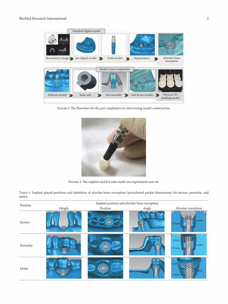
2. Materials and MethodsPeri-Implantitis Model
Figure 1 is the flowchart for the peri-implantitis in vitrotesting model construction.The standard clinical adult toothjaw model (PRO2002-UL-SP-FEM-28, Nissin Dental Prod-ucts Inc., Japan) and three 11.5mm length dental implantswith diameters of 3mm, 4mm, and 5mm, used for theincisor, premolar, andmolar, respectively, were selected as theexperimental samples (AnkerSB, Alliance Global TechnologyCo., Ltd., Taiwan). The i-CAT Dental CT (Imaging Sciences
International, PA, USA) were used to scan the tooth jawmodel and dental implants. All DICOM CT cross sectionimage data were processed on personal PC using commer-cially available image-processing software (Mimics� v. 10.01;Materialize Co., Leuven, Belgium) to identify the contoursof the different materials. Those contours were extracted andconverted into mathematical entities. A 3D digital tooth jawmodel and three dental implant solid models were recon-structed in a CAD system (Creo Parametric 2.0, PTC Inc.,Needham, MA, USA). Three implants with different diame-ters assembled in the relative middle incisor, premolar, andmolar tooth jawmodel positions and implant directions werereferenced to the major axis angle of the near teeth.
According to theCIST (Cumulative Interceptive Support-ive Therapy) peri-implantitis [18], mechanical debridementand surgical operation classification were needed when thebone loss depth was greater than 5mm. Following thisstandard, 6mm bone loss depth was defined in our modelon behalf of serious peri-implantitis, which usually requiresa flap in clinical surgery.
The periodontal pocket dimensions were defined as themaximum limitation with the root of the near teeth based oncommon alveolar bone loss appearance in X-ray images andwere as 1.8mm around the implant for the incisor, 2mm inthe upper part (occlusal direction), and 1.8mm in the lowerpart (root direction) for the first premolar and 3mm in theupper part (occlusal direction) and 2mm in the lower part(root direction) for the first molar (Table 1). Structural solidmodels of these three periodontal pockets were constructedand edited in the CAD system.
A cylindrical suite connected to the implant and eachtooth root in the jaw model was designed to permit thesuite to be replaced for individual bacterial adhesion. Thedental implant and the suite made one experimental unit setin which the bacterial culture can be performed (Figure 2).The digital peri-implantitis and suite models were exportedas a stereolithographic (STL) file that can be loaded into afused deposition modeling (FDM) 3D printer (3DP) with0.254mm slicing additive manufacturing (Dimension 1200esSST, Stratasys, Ltd., Minnesota, USA) to duplicate the ABSmaterial (ABS-P430, Stratasys, Ltd., Minnesota, USA).
3. Bacterial Culture and Adhesion
The jaw model at the incisor was used to verify our plat-form feasibility in bacterial adhesion and laser sterilizationexperiments. Three samples were performed in both exper-iments. The bacterial adhesion to the specimens againstEscherichia coli (ATCC�25922� KWIK-STIK, Microbiolog-ics, Inc., USA) was investigated as a model for gram-negativebacteria. Bacteria were cultured onto the solidmedium (Agarbase, Oxoid Ltd., Basingstoke, Hampshire, UK) and placedinto an incubator (37∘C) for 24 hours for agar cultivation(Figure 3(a)). Broth medium cultivation was then performedin a spectrophotometer (DU800, Beckman Coulter, Fuller-ton, CA, USA) operated in the wavelength 600 nm with readaverage time 0.5 sec (at three circulation) tomeasure its liquidOD (Optical Density) for quantized correction (Figure 3(b)).
BioMed Research International 3
Reconstruct image Jaw digital model Solid model Alveolar boneresorption
Model of peri-implantitis
Pathosis model Suite unit Suit assembly Suit & jaw models Physical 3Dprinting model
Standard digital model
Implantation
Figure 1: The flowchart for the peri-implantitis in vitro testing model construction.
Figure 2: The implant and the suite made one experiment unit set.
Table 1: Implant placed positions and definition of alveolar bone resorption (periodontal pocket dimensions) for incisor, premolar, andmolar.
Position Implant position and alveolar bone resorptionHeight Position Angle Alveolar resorption
Incisor 6mm1.8mm
Premolar2mm
1.8mm6mm
Molar3mm
2mm6mm

4 BioMed Research International
(a) (b)
(c) (d)
Figure 3: Bacterial culture. (a) Agar culture. (b) Broth culture. (c) Installed implant specimen. (d) Bacteria (Escherichia coli) culture withimplant.
A 3mm diameter implant and corresponding suite wereused to fulfill a set for the bacterial adhesion experiment(Figure 3(c)). The set was placed into medium at 37∘Ctemperature incubator for 24 hours (Figure 3(d)) and 2mlmedium added to centrifuge tubes. The liquid OD wasmeasured with a spectrophotometer and diluted 10 : 1. Platecount was performed to test the adhesion effect after 5minutes shaking using an ultra-sonicator.
Three dental set samples were assembled into the 3DPjaw model and irradiated with the Er:YAG laser (Er:YAGlaser, LightMed Dental Technology Corp., Taiwan) workingat 2940 nm with pulse energy at the tip∗85mJ/pulse. Aperiodontal hand piece was used with a chisel type glass tip(Figure 4). The application tip was moved from the buccalacross the implant thread to the lingual and occlusal to theroot directions for about 30 seconds per sample. The abut-ment was then removed and the implant with healing cap wasdisassembled to form the model set. The implant was placedinto a centrifuge tube for the bacteria count (Figures 4(c) and4(d)).The bacteria number was counted by plate count after 5minutes shaking using an ultrasonicator (Elmasonic P, ElmaGroup Inc., Pforzheim, Germany) with frequency 37 kHZ at
21∘C for continuity. The 𝑇-test method was performed tounderstand the variations between different groups.
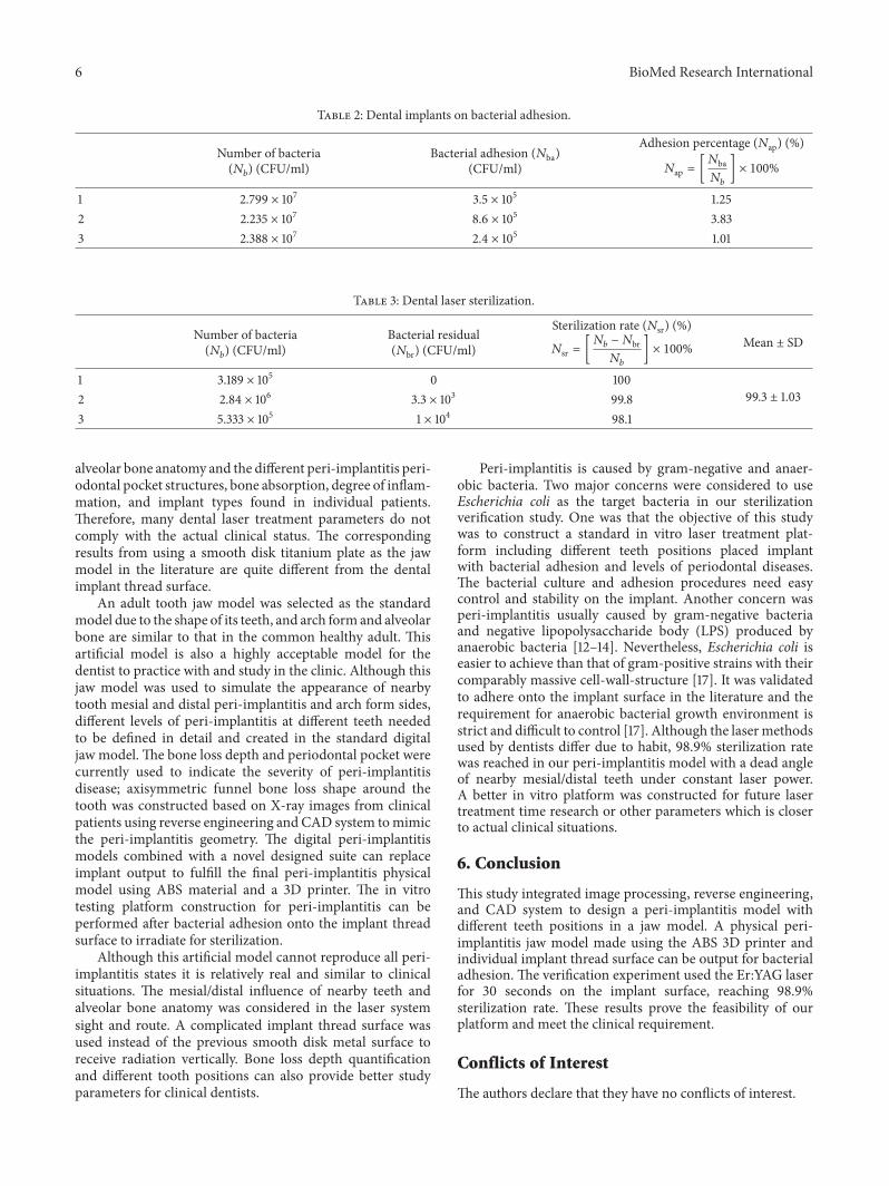
4. Results
Figure 5(a) shows the peri-implantitis in vitro testing modelwith 3DP jaw ABS model with different teeth positions suchas incisor, premolar, andmolar. Different dental implants canbe assembled and replaced in the 3DP jaw model based onthe clinical requirement (Figure 5(b)).
The results indicated that gram-negative bacteriumEscherichia coli can be cultured to find the calibration curve.The number of bacterial colonies was 107 when the OD valuewas 0.1. The bacteria adhered onto the implant successfullyabout 1.01% to 3.83% of the adhesion rate after 24 hours ofculture owing tomicropores on the implant surface (Table 2).The Er:YAG dental laser sterilization result showed thatthe sterilization rate can reach 99.3% (standard deviationis ±1.03) (Table 3). Noncontact video measurement system(SVP-2010, ARCS Co., Ltd., Taichung, Taiwan) observationswere performed in evaluating jaw model defects. The imageswere obtained using 37.5 times magnification with a color

BioMed Research International 5
(a) (b)
(c) (d)
Figure 4: Laser experiment process. (a) Implantation model. (b) Laser treatment. (c) Removal of implant abutment. (d) Removal of implant.
First molar
IncisorFirst premolar
(a)
First molarIncisor
First premolar
(b)
Figure 5: Physical ABS 3D printing model of peri-implantitis for (a) jaw tooth model; (b) jaw tooth and three implants.
CCD camera and transferred into an imaging programto evaluate whether the jaw model fractured/melted. Nodamage was found on the jaw model after laser irradiatingtesting.
5. Discussion
No standardmodel exists that represents all clinical situationsbecause of the large variation in tooth, dental arch, and
6 BioMed Research International
Table 2: Dental implants on bacterial adhesion.
Number of bacteria(𝑁𝑏) (CFU/ml)
Bacterial adhesion (𝑁ba)(CFU/ml)
Adhesion percentage (𝑁ap) (%)
𝑁ap = [𝑁ba𝑁𝑏 ] × 100%1 2.799 × 10⁷ 3.5 × 10⁵ 1.252 2.235 × 10⁷ 8.6 × 10⁵ 3.833 2.388 × 10⁷ 2.4 × 10⁵ 1.01
Table 3: Dental laser sterilization.
Number of bacteria(𝑁𝑏) (CFU/ml)
Bacterial residual(𝑁br) (CFU/ml)
Sterilization rate (𝑁sr) (%)
𝑁sr = [𝑁𝑏 − 𝑁br𝑁𝑏
] × 100% Mean ± SD
1 3.189 × 10⁵ 0 10099.3 ± 1.032 2.84 × 10⁶ 3.3 × 10³ 99.8
3 5.333 × 10⁵ 1 × 10⁴ 98.1
alveolar bone anatomy and the different peri-implantitis peri-odontal pocket structures, bone absorption, degree of inflam-mation, and implant types found in individual patients.Therefore, many dental laser treatment parameters do notcomply with the actual clinical status. The correspondingresults from using a smooth disk titanium plate as the jawmodel in the literature are quite different from the dentalimplant thread surface.
An adult tooth jaw model was selected as the standardmodel due to the shape of its teeth, and arch form and alveolarbone are similar to that in the common healthy adult. Thisartificial model is also a highly acceptable model for thedentist to practice with and study in the clinic. Although thisjaw model was used to simulate the appearance of nearbytooth mesial and distal peri-implantitis and arch form sides,different levels of peri-implantitis at different teeth neededto be defined in detail and created in the standard digitaljaw model. The bone loss depth and periodontal pocket werecurrently used to indicate the severity of peri-implantitisdisease; axisymmetric funnel bone loss shape around thetooth was constructed based on X-ray images from clinicalpatients using reverse engineering and CAD system tomimicthe peri-implantitis geometry. The digital peri-implantitismodels combined with a novel designed suite can replaceimplant output to fulfill the final peri-implantitis physicalmodel using ABS material and a 3D printer. The in vitrotesting platform construction for peri-implantitis can beperformed after bacterial adhesion onto the implant threadsurface to irradiate for sterilization.
Although this artificial model cannot reproduce all peri-implantitis states it is relatively real and similar to clinicalsituations. The mesial/distal influence of nearby teeth andalveolar bone anatomy was considered in the laser systemsight and route. A complicated implant thread surface wasused instead of the previous smooth disk metal surface toreceive radiation vertically. Bone loss depth quantificationand different tooth positions can also provide better studyparameters for clinical dentists.
Peri-implantitis is caused by gram-negative and anaer-obic bacteria. Two major concerns were considered to useEscherichia coli as the target bacteria in our sterilizationverification study. One was that the objective of this studywas to construct a standard in vitro laser treatment plat-form including different teeth positions placed implantwith bacterial adhesion and levels of periodontal diseases.The bacterial culture and adhesion procedures need easycontrol and stability on the implant. Another concern wasperi-implantitis usually caused by gram-negative bacteriaand negative lipopolysaccharide body (LPS) produced byanaerobic bacteria [12–14]. Nevertheless, Escherichia coli iseasier to achieve than that of gram-positive strains with theircomparably massive cell-wall-structure [17]. It was validatedto adhere onto the implant surface in the literature and therequirement for anaerobic bacterial growth environment isstrict and difficult to control [17]. Although the lasermethodsused by dentists differ due to habit, 98.9% sterilization ratewas reached in our peri-implantitis model with a dead angleof nearby mesial/distal teeth under constant laser power.A better in vitro platform was constructed for future lasertreatment time research or other parameters which is closerto actual clinical situations.
6. Conclusion
This study integrated image processing, reverse engineering,and CAD system to design a peri-implantitis model withdifferent teeth positions in a jaw model. A physical peri-implantitis jaw model made using the ABS 3D printer andindividual implant thread surface can be output for bacterialadhesion. The verification experiment used the Er:YAG laserfor 30 seconds on the implant surface, reaching 98.9%sterilization rate. These results prove the feasibility of ourplatform and meet the clinical requirement.
Conflicts of Interest
The authors declare that they have no conflicts of interest.
BioMed Research International 7
Acknowledgments
This study is supported in part by BY-12-05-28-105 of theSouthern Taiwan Science Park Bureau, Ministry of Sci-ence and Technology, Taiwan. The authors would like toacknowledge Alliance Global Technology Co., Ltd., Taiwan,for providing the implants for experimentation.
References
[1] E.-S. Kim and S.-Y. Shin, “Influence of the implant abutmenttypes and the dynamic loading on initial screw loosening,”Journal of Advanced Prosthodontics, vol. 5, no. 1, pp. 21–28, 2013.
[2] T. Albrektsson and N. Donos, “Implant survival and complica-tions.TheThird EAO consensus conference 2012,” Clinical OralImplants Research, vol. 23, supplement 6, pp. 63–65, 2012.
[3] J. Lindhe and S. Nyman, “The effect of plaque control and surgi-cal pocket elimination on the establishment andmaintenance ofperiodontal health. A longitudinal study of periodontal therapyin cases of advanced disease,” Journal of Clinical Periodontology,vol. 2, no. 2, pp. 67–79, 1975.
[4] A. Mombelli and N. P. Lang, “Microbial aspects of implantdentistry,” Periodontology 2000, vol. 4, no. 1, pp. 74–80, 1994.
[5] A.Mombelli,M.A.C. vanOosten, E. Schurch Jr., andN. P. Lang,“Themicrobiota associated with successful or failing osseointe-grated titanium implants,” Oral Microbiology and Immunology,vol. 2, no. 4, pp. 145–151, 1987.
[6] M. Muthukuru, A. Zainvi, E. O. Esplugues, and T. F. Flemmig,“Non-surgical therapy for the management of peri-implantitis:a systematic review,” Clinical Oral Implants Research, vol. 23,supplement 6, pp. 77–83, 2012.
[7] A. Mombelli and N. P. Lang, “Antimicrobial treatment of peri-implant infections,” Clinical Oral Implants Research, vol. 3, no.4, pp. 162–168, 1992.
[8] F. Schwarz, A. Sculean, M. Berakdar, L. Szathmari, T. Georg,and J. Becker, “In vivo and in vitro effects of an Er:YAG laser, aGaAlAs diode laser, and scaling and root planing on periodon-tally diseased root surfaces: a comparative histologic study,”Lasers in Surgery and Medicine, vol. 32, no. 5, pp. 359–366,2003.
[9] U. Keller, K. Stock, and R. Hibst, “Morphology of Er:YAG laser-treated root surfaces,” in Proceedings of the Medical Applica-tions of Lasers in Dermatology, Ophthalmology, Dentistry andEndoscopy, pp. 24–31, September 1997.
[10] T. Matsuyama, A. Aoki, S. Oda, T. Yoneyama, and I. Ishikawa,“Effects of the Er:YAG laser irradiation on titanium implantmaterials and contaminated implant abutment surfaces,” Jour-nal of Clinical LaserMedicine and Surgery, vol. 21, no. 1, pp. 7–17,2003.
[11] M. Folwaczny, G. George, L. Thiele, A. Mehl, and R. Hickel,“Root surface roughness following Er:YAG laser irradiation atdifferent radiation energies and working tip angulations,” Jour-nal of Clinical Periodontology, vol. 29, no. 7, pp. 598–603, 2002.
[12] R. J. Genco, “Assessment of risk of periodontal disease,” Com-pendium (Newtown, Pa.). Supplement, vol. 18, pp. S678–S683,quiz S714-7, 1994.
[13] R. C. Page, “The role of inflammatory mediators in the patho-genesis of periodontal disease,” Journal of Periodontal Research,vol. 26, no. 3, part 2, pp. 230–242, 1991.
[14] M. A. Listgarten and C.-H. Lai, “Comparative microbiologicalcharacteristics of failing implants and periodontally diseasedteeth,” Journal of Periodontology, vol. 70, no. 4, pp. 431–437, 1999.
[15] T. G. Wilson Jr and F. L. Higginbottom, “Periodontal Diseasesand Dental Implants in Older Adults,” Journal of Esthetic andRestorative Dentistry, vol. 10, no. 5, pp. 265–271, 1998.
[16] M. HP, PeriodontalMicrobiology. Periodontology:The Essentials,New YorkThieme, New York, NY, USA, 2004.
[17] C.-J. Chen, S.-J. Ding, and C.-C. Chen, “Effects of surfaceconditions of titanium dental implants on bacterial adhesion,”Photomedicine and Laser Surgery, vol. 34, no. 9, pp. 379–388,2016.
[18] N. D. Shumaker, “Periodontal and periimplant maintenance: acritical factor in long-term treatment success,” Compendium ofContinuing Education in Dentistry, vol. 30, no. 7, pp. 388–390,2009, quiz 407, 418.

Submit your manuscripts athttps://www.hindawi.com
Stem CellsInternational
Hindawi Publishing Corporationhttp://www.hindawi.com Volume 2014
Hindawi Publishing Corporationhttp://www.hindawi.com Volume 2014
MEDIATORSINFLAMMATION
of
Hindawi Publishing Corporationhttp://www.hindawi.com Volume 2014
Behavioural Neurology
EndocrinologyInternational Journal of
Hindawi Publishing Corporationhttp://www.hindawi.com Volume 2014
Hindawi Publishing Corporationhttp://www.hindawi.com Volume 2014
Disease Markers
Hindawi Publishing Corporationhttp://www.hindawi.com Volume 2014
BioMed Research International
OncologyJournal of
Hindawi Publishing Corporationhttp://www.hindawi.com Volume 2014
Hindawi Publishing Corporationhttp://www.hindawi.com Volume 2014
Oxidative Medicine and Cellular Longevity
Hindawi Publishing Corporationhttp://www.hindawi.com Volume 2014
PPAR Research
The Scientific World JournalHindawi Publishing Corporation http://www.hindawi.com Volume 2014
Immunology ResearchHindawi Publishing Corporationhttp://www.hindawi.com Volume 2014
Journal of
ObesityJournal of
Hindawi Publishing Corporationhttp://www.hindawi.com Volume 2014
Hindawi Publishing Corporationhttp://www.hindawi.com Volume 2014
Computational and Mathematical Methods in Medicine
OphthalmologyJournal of
Hindawi Publishing Corporationhttp://www.hindawi.com Volume 2014
Diabetes ResearchJournal of
Hindawi Publishing Corporationhttp://www.hindawi.com Volume 2014
Hindawi Publishing Corporationhttp://www.hindawi.com Volume 2014
Research and TreatmentAIDS
Hindawi Publishing Corporationhttp://www.hindawi.com Volume 2014
Gastroenterology Research and Practice
Hindawi Publishing Corporationhttp://www.hindawi.com Volume 2014
Parkinson’s Disease
Evidence-Based Complementary and Alternative Medicine
Volume 2014Hindawi Publishing Corporationhttp://www.hindawi.com