In vitro embryo production from prepubertal goat oocytes ... · Dolors Izquierdo i Tugas Dra. Roser...
108
UNIVERSITAT AUTÒNOMA DE BARCELONA In vitro embryo production from prepubertal goat oocytes in different culture media Sondes Hammami Facultat de Veterinària Departament de Ciència Animal i dels Aliments Tesis Doctoral Bellaterra, Junio del 2014
Transcript of In vitro embryo production from prepubertal goat oocytes ... · Dolors Izquierdo i Tugas Dra. Roser...

UNIVERSITAT AUTÒNOMA DE BARCELONA
In vitro embryo production from prepubertal
goat oocytes in different culture media
Sondes Hammami
Facultat de Veterinària
Departament de Ciència Animal i dels Aliments
Tesis Doctoral
Bellaterra, Junio del 2014


La Dra. Dolors Izquierdo Tugas, professora agregada del Departament de Ciència
Animal i dels Aliments de la Universitat Autònoma de Barcelona,
I la Dra. Roser Morató Molet, investigadora Juan de la Cierva, amb destí al
Departament de Biologia de la Universitat de Girona, Biotecnologia de la Reproducció
Animal i Humana de la Universitat de Girona,
CERTIFIQUEN
Que Sondes Hammami ha realitzat sota la seva direcció el present treball
d’investigació que porta per títol “In vitro embryo production from prepubertal goat
oocytes in different culture media ” per optar al grau de Doctor per la Universitat
Autònoma de Barcelona. Aquest treball ha estat finançat pel Ministeri de Ciència i
Innovació, a través del projecte AGL 2011-23784, el grup de recerca consolidat
SGR2009SGR0621 i una beca atorgada pel Ministeri d’Assumptes Exteriors i de
Cooperació.
I per a que així consti, firmen el present certificat
Dra. Dolors Izquierdo i Tugas Dra. Roser Morató Molet
Bellaterra, Maig del 2014

Aquest treball ha estat finançat pel Ministerio de Ciencia e Innovación, a través del
projecte AGL2011-23784 i el grup de recerca consolidat SGR2009SGR0621.

À ma mère, À mon frère
À mes soeurs


Abstract
The prepubertal goat ovary shows a large number of small oocytes with a compromised
competence to develop up to blastocyst stage following IVM, IVF and IVC. Culture
conditions used to support in vitro embryo production appears to be an important factor
to suit the requirements of these oocytes and to increase the blastocyst yield. In the
present thesis work, we have carried out two studies to evaluate the effects of addition
of different supplements to in vitro maturation and culture media on embryo
development and the quality of resultant blastocyst in prepubertal goat oocytes.
In oocytes from prepubertal females, different types and concentrations of substances
including gonadotrophic hormones, growth factors, protein sources and antioxidants,
such as insulin-transferrin-selenium (ITS) and L-ascorbic acid (AA), have been shown
to improve oocyte cytoplasmic maturation and embryo development. The aim of the
first study was to assess the effects of adding low level of hormones and/or ITS plus AA
to IVM medium through the developmental competence and embryo quality of small
oocytes from prepubertal goats. Specifically, we tested four maturation media:
conventional IVM medium (CM), CM+ITS+AA and low level of hormones (named as
Growth Medium; GM), CM with low level of hormones (modified CM; mCM) and
CM+ITS+AA and normal level of hormones (modified GM; mGM). Cumulus-Oocyte
Complexes (COCs) were classified into two categories according to oocyte diameter:
small (<125 μm) and large (≥125 μm) oocytes. Large oocytes were matured in CM and
small oocytes were matured in different combination of CM, GM, mCM or mGM. After
IVM, oocytes were fertilized and after 24h presumptive zygotes were cultured for 8
days. Results of this study showed that using different combinations of CM, GM, mCM,
and mGM for IVM of prepubertal goat small oocytes did not have a beneficial effect on
the percentage of blastocysts. The high blastocyst rate was only related to oocyte
diameter. However, the culture of small oocytes in GM improved the quality of
blastocysts and enhanced their survival post-warming compared to CM (53.3% vs
30.8%, respectively; P<0.05).
The second study examined the effect of activin-A during IVM and embryo culture on
the blastocyst rate of prepubertal goat oocytes. Activin is a member of the transforming
growth factor β superfamily that plays a functional role in the process of cellular
proliferation and differentiation. In experiment 1, with a view to search for optimal

concentration of recombinant human activin-A during IVM, three concentrations (0, 10
and 100 ng/mL) were tested on oocytes meiotic maturation and subsequent in vitro
embryo development. The experiment 2 was performed to evaluate the effect of adding
10 ng/mL activin-A at the IVM and/or IVC media on embryo development and
blastocyst quality. Results of this study demonstrated that the addition of activin-A to
IVM yielded similar percentages of maturation and blastocyst. Moreover, the addition
of 10 ng/mL of activin-A throughout the whole embryo culture significantly increased
rates of development to the blastocyst stage as compared with the control group (19.5 ±
2.21% vs 13.1 ± 2.37%, respectively; P<0.05). Blastocyst quality was not improved in
the presence of activin-A neither during IVM evaluated after Hoechst 33342 staining,
nor during IVM and/ or IVC evaluated by differential staining.
In conclusion, IVM of prepubertal goat small oocytes in GM would be useful to
improve the quality of blastocysts produced in vitro and the presence of activin-A
during IVC enhances embryo development of prepubertal goat oocytes.

Resumen
Los ovarios de cabras prepúberes proporcionan una población de ovocitos de pequeño
diámetro con una menor competencia para el desarrollo hasta blastocisto después de la
maduración, fecundación y posterior cultivo in vitro (MIV, FIV y CIV). Las
condiciones del cultivo in vitro aportan los requerimientos necesarios a los ovocitos e
influyen sobre su posterior desarrollo embrionario. En esta tesis hemos llevado a cabo
dos estudios para evaluar los efectos de la adición de diferentes suplementos a los
medios de MIV y CIV sobre el desarrollo embrionario y la calidad de los blastocistos
obtenidos a partir de ovocitos de cabras prepúberes.
En ovocitos de hembras prepúberes, la adición de sustancias, incluyendo las hormonas
gonadotrópicas, factores de crecimiento, fuentes de proteínas y antioxidantes, como son
el complejo insulina-transferrina-selenio (ITS) y el L-ácido ascórbico (AA), han
demostrado producir una mejora en la maduración citoplasmática de los ovocitos y su
subsiguiente desarrollo embrionario in vitro. El objetivo del primer estudio fue evaluar
los efectos de la adición de AA, ITS y un bajo nivel hormonal en el medio de MIV
sobre el desarrollo embrionario y la calidad de los blastocistos obtenidos. En concreto,
hemos utilizado 4 medios maduración: Medio Tradicional (MT), Medio de Crecimiento
(MC): MT + ITS + AA + bajo nivel hormonal, Medio Tradicional modificado (MTm):
MT + bajo nivel hormonal y Medio de Crecimiento modificado (MCm): MT + ITS +
AA + nivel hormonal normal. Los complejos–ovocito-cúmulus (COCs) se clasificaron
en dos grupos según el diámetro ovocitario: pequeños (<125 μm) y grandes (≥125 μm).
Los ovocitos ≥125 μm fueron madurados en MT mientras que los ovocitos <125 μm
fueron madurados en combinaciones de MT, MC, MTm y MCm. Después de la MIV,
los ovocitos fueron fecundados in vitro y posteriormente los embriones fueron CIV
durante 8 días. En este estudio se observó que el desarrollo embrionario de los ovocitos
<125 μm no mejoró en las diferentes combinaciones de medios de MIV estudiadas (MT,
MC, MTm y MCm). El mayor desarrollo hasta blastocisto se obtuvo en el grupo de
embriones procedentes de ovocitos ≥125 μm. Sin embargo, cuando los ovocitos de
menor diámetro se maduraron en MC, se observó una mejoría significativa en la calidad
de los blastocistos obtenidos, optimizando la supervivencia de los embriones tras la
descongelación comparado con el MT (53.3% vs 30.8%, respectivamente; P<0.05).

En el segundo estudio se analizó el efecto de la suplementación de los medios de MIV y
CIV con activina-A sobre el desarrollo embrionario. La activina-A es un miembro de la
superfamilia del factor de crecimiento transformante β (TGF-β) y cumple un papel
importante en la proliferación y diferenciación celular. Con el fin de determinar el
efecto de la activina-A sobre los ovocitos de cabras prepúberes, se realizaron 2
experimentos, analizando la maduración nuclear de los ovocitos, su posterior desarrollo
embrionario y calidad de los blastocistos obtenidos. En el primer experimento se estudió
la suplementación del medio de MIV con varias concentraciones de activina-A (0, 10 y
100 ng/mL) y en el segundo experimento se evaluó la adición de 10 ng/mL de activina-
A a los medios de MIV y CIV. Los resultados de este estudio demostraron que la
suplementación del medio de MIV con diferentes concentraciones de activina-A
proporciona similares porcentajes de maduración nuclear y de blastocistos comparado
con la MIV sin activina-A. Además, la adición de 10 ng/mL de activina-A durante el
CIV aumentó significativamente las tasas de desarrollo hasta blastocisto en
comparación con el grupo control (19.5 ± 2.21% vs 13.1 ± 2.37%, respectivamente;
P<0.05). Sin embargo, no se observó una mejora en la calidad de los blastocistos
cuando se añadió activina-A en el medio de MIV (evaluada por la tinción de Hoechst
33342) y/o en el de CIV (evaluado por la tinción diferencial).
En conclusión, la maduración in vitro de los ovocitos de cabras prepúberes de diámetro
<125 μm en medio de crecimiento sería una alternativa útil para mejorar la calidad de
los blastocistos producidos in vitro, mientras que la presencia de activina-A durante el
cultivo in vitro incrementa el porcentaje de embriones desarrollados hasta el estadío de
blastocisto.
11
Index Chapter 1: Introduction ............................................................................................... 15
Chapter 2: Literature Review ..................................................................................... 23
2.1. In vitro maturation (IVM)........................................................................................ 25
2.1.1. Nuclear maturation ............................................................................................... 25
2.1.2. Cytoplasmic maturation ....................................................................................... 26
2.1.3. Factors affecting oocyte quality .......................................................................... 26
2.1.3.1. Effect of follicle and oocyte size ....................................................................... 27
2.1.3.2. Age of the donor ................................................................................................ 29
2.1.3.3. Media and supplements used for IVM ............................................................. 30
2.1.3.3.1. Hormones ....................................................................................................... 31
2.1.3.3.2. Antioxidants ................................................................................................... 32
2.1.3.3.3. Insulin-Transferrin-Selenium ......................................................................... 34
2.1.3.3.4. Activin-A ........................................................................................................ 35
2.1.3.3.5. Serum and other substances ............................................................................ 37
2.2. In vitro fertilization (IVF)........................................................................................ 39
2.3. In vitro culture (IVC) ............................................................................................... 40
2.4. Assessment of embryo quality ................................................................................ 43
Chapter 3: Objectives ................................................................................................... 47
Chapter 4: Developmental competence and embryo quality of small oocytes from
pre-pubertal goats cultured in IVM medium supplemented with low level of
hormones, insulin-transferrin-selenium and ascorbic acid ...................................... 51
Chapter 5: In vitro developmental competence of prepubertal goat oocytes
cultured with recombinant activin-A .......................................................................... 59
Chapter 6: General Discussion .................................................................................... 69
Chapter 7: General Conclusions ................................................................................. 77
Chapter 8: References .................................................................................................. 81
Chapter 9: Aknowledgments ..................................................................................... 107
12
List of abbreviations μg
μm
AA
AI
ARTs
AI-TI
BCB
CM
COCs
DNA
E2-17β
EGF
ET
FCS
FF
FSH
GDF9
GH
GM
GnRH
GV
GVBD
ICM
ICSI
IGF-I
ITS
IU
IVC
IVEP
IVF
IVM
IVP
Microgram
Micrometer
Ascorbic Acid
Artificial Insemination
Assisted Reproductive Technologies
Anaphase I- Telophase I
Brilliant Cresyl Blue
Conventional Medium
Cumulus–Oocyte Complexes
Desoxyribonucleic Acid
Estradiol-17β
Epidermal Growth Factor
Embryo Transfer
Fetal Calf Serum
Folicular Fluid
Follicle Stimulating Hormone
Growth-Differentiation Factor 9
Growth Hormone
Growth Medium
Gonadotrophin-Releasing Hormone
Germinal Vesicle
Germinal Vesicle Breakdown
Inner Cell Mass
Intracytoplasmic Sperm Injection
Insulin-Like Growth Factor-I
Insulin-Transferrin-Selenium
International Unit
In Vitro Culture
In Vitro Embryo Production
In Vitro Fertilization
In Vitro Maturation
In Vitro Production
13
JIVET
LH
LOPU
MI
MII
mL
mm
mM
MOET
MPF
ng
OPU
PN
PUFAs
RNA
ROS
SR
TE
TGFβ
ZP
Juvenile In Vitro Embryo Technology
Luteinizing Hormone
Laparoscopic Ovum Pick-Up
Metaphase I
Metaphase II
Milliliter
Millimeter
Millimolar
Multiple Ovulation and Embryo Transfer
Maturation Promoting Factor
Nanogram
Ovum Pick-Up
Pronuclei
Polyunsaturated Fatty Acids
Ribonucleic Acid
Reactive Oxygen Species
Serum Replacer
Trophectoderm
Transforming Growth Factor β
Zona Pellucida

14

15
INTRODUCTION
Chapter 1

16

Introduction
17
Introduction
In recent years, demand for goat products has increased in both developing and
developed countries; the population of goats in the world has increased by nearly 100%
over the past four decades. In 2012 there were more than 921 million goats around the
world: Asia had the greatest number, followed by Africa, South America, Europe, North
America and Oceania (http://www.fao.org/). Goats have always had a major role to play
in developing countries due to their adaptability under harsh and marginal rural
conditions, predominantly in the tropical and subtropical zones of the world.
A growing interest in assisted reproductive technologies (ARTs) has occurred. ARTs
are being used for enhancement of reproductive performance and genetic improvement
in sheep and goats. In addition, they can have substantial contribution in preservation of
endangered species or breeds (Cognié et al., 2004). While their applications are
widespread in cattle, in small ruminants it is almost restricted to artificial insemination.
Nonetheless, during the last 30 years, considerable progress has been made in sheep and
goat embryo technologies.
Furthermore, in vitro embryo production (IVEP) in small ruminants provides an
excellent source of low cost embryos for basic research on developmental biology and
physiology and for commercial application of the emerging biotechnologies such as
nuclear transfer, transgenesis, embryo sexing and stem cells. Thus, goats offer a good
model for the development of these technologies (reviewed by Cognié et al., 2003).
In the laboratory, embryos can be routinely produced and developed up to the blastocyst
stage using three subsequent techniques: in vitro maturation (IVM) of oocytes recovered
from ovaries, followed by sperm capacitation and in vitro fertilization (IVF) of matured
oocytes and then in vitro culture (IVC) of the fertilized oocytes up to the blastocyst
stage that can be transferred to recipient females or cryopreserved for future use.
However, in vitro embryo production still is not very efficient due to several limiting
factors affecting the outcome of each step of the process.
The use of prepubertal females as oocyte donors for in vitro embryo production
(juvenile in vitro embryo technology or JIVET) offers considerable potential for
accelerating genetic gain in domestic livestock through reduced generation interval as
compared to classical approach (Armstrong et al., 1997). However, reduced

Introduction
18
developmental competence in oocytes of juvenile animals as compared to their adult
counterpart when matured in vitro, was addressed in numerous studies on farm animals
including bovine (Presicce et al., 1997), ovine (Ledda et al., 1997), caprine (Leoni et
al., 2009) and porcine (Marchal et al., 2001).
Evidence suggests that the low number of embryos obtained from prepubertal females is
mainly due to the low competence of these oocytes, and this competence is defined by
Sirard et al., (2006) as the ability to: 1) resume meiosis; 2) cleave following
fertilization; 3) develop to the blastocyst stage; 4) induce pregnancy; and 5) bring
offspring to term in good health, and it is acquired progressively during oocyte growth
(Sorensen and Wassarman, 1976; Eppig, 1996).
Embryo development is influenced by events occurring during oocyte maturation. For
successful IVM, oocytes must undergo nuclear and cytoplasmic maturation. Therefore
the efficiency of embryo development from these oocytes is mainly attributed to
limitations induced by two factors: failure to select the most competent oocytes for in
vitro production and an inadequate understanding of oocyte maturation and culture
conditions to provide an optimized environment, necessary to support the physiological
changes associated with oocytes maturation and required for attaining developmental
competence (Rizos et al., 2002a; Rizos et al., 2002b; Morton et al., 2008).
To overcome the reduced maturational competence of oocytes from juvenile females,
several factors and strategies have been shown to affect the oocyte competence such as:
selection follicles and oocytes diameters (Anguita et al., 2007; Romaguera et al.,
2010a), donor’s age (Ledda et al., 1997; Baldassarre and Karatzas, 2004), hormonal
stimulation (Koeman et al., 2003), in vitro maturation protocol (Wu et al., 2006) and
supplementation to oocyte maturation media (Mertens et al., 2005; Romaguera et al.,
2010b; Córdova et al., 2011).
Numerous research groups have reported a positive relationship between follicle size
and the ability of its oocyte to develop to blastocyst stage in cows (Lonergan et al.,
1994), ewes (Cognié et al., 1998), sows (Motlik et al., 1984), buffalos (Raghu et al.,
2002a) and goats (Martino et al., 1994; Crozet et al., 1995). The developmental
competence of an oocyte is obtained progressively as follicular diameter increases to
maximum size, suggesting that the follicle size and oocyte diameter are closely related,
and as both increase oocyte competence is gradually acquired within the ovary.
Introduction
19
According to oocyte diameter, previous studies in our laboratory concluded that a
higher blastocyst rate is obtained in oocytes larger than 135 μm (12.5%) compared to
oocytes of 125 to 135 μm diameter (1.95%) after IVF (Anguita et al., 2007). However,
when ICSI was used to fertilize oocytes, Jiménez-Macedo et al., (2006) did not find
differences between these two oocytes categories.
A range of oocyte maturation media and additives have been tried to accomplish with
the in vitro maturation of oocytes, and their developmental competence. Reports in the
literature have described the effect of maturing oocytes in vitro in culture medium
supplemented with different types and concentrations of substances including
gonadotrophic hormones, antioxidants, growth factors and protein sources. The addition
of gonadotropins (FSH and LH) and estradiol-17β (E2-17β), to the maturation medium
significantly has improved maturation rates (nuclear, cytoplasmic maturation and
cumulus expansion) in mammalian oocytes (Sanbuissho and Threlfall, 1990; Mattioli et
al., 1991; Singh et al., 1993; Guler et al., 2000; Abdoon et al., 2001; Roberts et al.,
2005; Alvarez et al., 2009), has promoted the ability of the oocytes to form a male
pronucleus after in vitro fertilization (Thibault et al., 1975; Funahashi and Day, 1993)
and their developmental competence up to blastocyst stage (Men et al., 2002). The
concentration of gonadotropins used during the process of IVM of oocytes has been
showed to determine the results of oocyte maturation and developmental competence. In
bovine, Liu et al., (2011) reported that extremely high concentrations of gonadotropins
have detrimental effects on oocyte nuclear maturation and embryo development and
increase apoptosis in cumulus cells. In porcine, Bing et al., (2001) have reported that
using a low concentration of FSH (1 μg/mL) is not sufficient to induce full nuclear
maturation of oocytes, compared with 10 μg/mL FSH. On the other hand, some studies
have shown that lower concentrations of hormones are enough for IVM in mice (Peluso,
1988; Byskov et al., 1997). Also, in prepubertal gilts Wu et al., (2006) have observed a
significant increase in blastocyst yield derived from oocytes obtained from small
follicles by reducing 250-fold the concentration of the hormones in a protocol of IVM
medium (2-step culture system). Results of those studies suggest the importance of
judicious use of gonadotropins to enhance in vitro oocyte maturation and embryonic
development, depending on the species and the donor age.
Introduction
20
During in vitro culture, cells are exposed to higher concentrations of oxygen than those
that occur in vivo and this causes the constant production of free radicals. High levels of
free radicals cause damage to cell components by lipid peroxidation, protein
modification, and DNA damage resulting in impaired cell function and subsequently
can affect oocyte maturation and further embryonic development (Cetica et al., 2001).
Thus, to optimize embryo production, oocytes need to be protected against oxidative
stress during in vitro culture. Several antioxidants have been added to maturation media
(Ali et al., 2003). Insulin-transferrin-selenium (ITS) combination is routinely used in
culture systems to promote the development of oocytes because of its protecting effects
from oxidative damage by reducing free radical production and inhibiting lipid
peroxidation, and the promoting effect of the uptake of glucose and amino acids
(Tatemoto et al., 2004; Ebert et al., 2006). Studies performed in several species during
in vitro maturation, showed that ITS addition during IVM, improved cytoplasmic
maturation and decreased polyspermic fertilization rate in porcine oocytes (Jeong et al.,
2008). Similarly, in bovine, supplementation with ITS (Jaakma et al., 1997; Bowles and
Lishman, 1998) showed a higher blastocyst rate. In mice, an increase on blastocyst rate
(Zhang and Armstrong, 1990) as well as their quality (Jeong et al., 2008) was observed
when IVM medium was supplemented with ITS. Moreover, the ascorbic acid is an anti-
oxidant that has been shown to support cytoplasmic maturation of porcine oocytes
relating to developmental competence after fertilization by alleviating oxidative stress
(Tatemoto et al., 2001). In addition, the presence of ascorbic acid in the culture medium
has been shown to significantly reduce the level of apoptosis in mouse oocyte-
associated granulosa cells although no improvement of oocyte developmental
competence was detected (Eppig et al., 2000). Ascorbic acid addition plus the ITS
complex to the culture medium also enhances the blastocyst production of gilts (Wu et
al., 2006) and bovine oocytes (Córdova et al., 2010).
A second approach to improve the developmental competence of oocytes after their
recovery from the follicle has been the addition of growth promoting substances to in
vitro maturation and culture media, such as epidermal growth factor (EGF), growth
hormone (GH), inhibin, activin and follistatin. Those growth factors are generally added
to the basal medium to increase cell proliferation and to stimulate specific cell functions
(Van der Valk et al., 2010) despite modest improvements in development have been
achieved in this way. Activin-A is an important member of the transforming growth

Introduction
21
factor β (TGFβ) superfamily. In the ovaries of vertebrates, activin-A is expressed
predominantly in the granulosa cell layer of follicles, suggesting important roles in
processes such as folliculogenesis, steroid hormone production, and oocyte maturation
as paracrine or autocrine factors (Peng and Mukai, 2000). Studies in vitro showed that
exposure of oocytes during maturation and/or culture to different concentrations of
exogenous activin-A, stimulates the resumption of meiosis of oocytes in several species
(mice (Itoh et al., 1990); cattle (Stock et al., 1997; Park et al., 2010; Trigal et al., 2011);
rhesus monkey (Alak et al., 1996) and human (Alak et al., 1998)). Moreover, a positive
effect of activin-A on embryo development were shown when the protein was added at
the earliest stages of bovine (Yoshioka and Kamomae, 1996, Yoshioka et al., 1998a,
Lee et al., 2009) embryo culture.
The basal components of the TGFβ superfamily signalling pathways are the type II and
type I transmembrane serine and threonine kinase receptors and the cytoplasmic Smad
proteins (Heldin et al., 1997). Activins signal through type I and type II receptor
proteins, both of which are serine/threonine kinases. Subsequently, signals such as
Smad proteins are phosphorylated (Peng and Mukai, 2000). Activin receptors are
represented by two isoforms: activin receptors type IA (ActR-IA), IB (ActR-IB), IIA
(ActR-IIA) and IIB (ActR-IIB). Both protein and mRNA for activin-A and activin
receptors type I and II have been localized in both oocyte and granulosa cells of follicles
at various developmental stages in a variety of species, including human (Eramaa et al.,
1995; Mather et al., 1997; Sidis et al., 1998), rodent (Zhao et al., 2001), porcine (Van
den Hurk and Van de Pavert, 2001), and bovine (Hulshof et al., 1997) follicles and
caprine ovary and follicles (Silva et al., 2004; 2006). These facts suggest that activin-A
may play a role in development of prepubertal goat oocytes, and investigation about the
mechanism of action of this protein in oocytes from prepubertal goat during in vitro
maturation and during embryo in vitro culture is required because, to date, no studies on
the effects of activin-A on in vitro developmental potential of prepubertal goat oocytes
have been reported.
In summary, juvenile donors will have an important place in livestock-improvement as
well as in biotechnology programs. However, further investigations on molecular and
cellular approaches will increase our understanding of cytoplasmic maturation in
prepubertal oocytes allowing a more efficient in vitro production of embryos by
IVM/IVF, ICSI and nuclear transfer, and the production of offspring from juveniles to
Introduction
22
reduce the generation interval and increase the rate of genetic progress in breeding
schemes.
All these points above mentioned need to be addressed in future studies to find the most
efficient in vitro embryo production to achieve high rates of oocyte maturation and
embryo development from prepubertal goats.
Continuing efforts of previous research performed in our laboratory with the aim to
improve the in vitro embryo yield of prepubertal goat oocytes, the present thesis work is
designed to evaluate the effects of addition of different supplements to in vitro
maturation and culture media on embryo development.

23
LITERATURE REVIEW
Chapter 2

24

Literature Review
25
Literature Review
Over recent years, considerable research into in vitro embryo production (IVEP)
technology has been undertaken in an attempt to determine which conditions are needed
during the three subsequent steps: in vitro maturation (IVM), in vitro fertilization (IVF)
and in vitro culture (IVC). These in vitro processes are intended to mimic the processes
in vivo, for improving animal breeding programs and biomedical field. These steps will
be discussed in more detail in subsequent sections of this thesis.
2.1. In vitro maturation (IVM)
Full maturation involves both nuclear and cytoplasmic events that confer on the oocyte
the capacity for supporting normal fertilization and early embryonic development
(Thibault and Gerard, 1973; Moor and Trounson, 1977). Mammalian oocytes arrested at
the germinal vesicle (GV) stage acquire the capacity to resume meiosis during growth
of the ovarian follicle from the primary to the preovulatory stage. As removed from
antral follicles of females ovaries, cumulus–oocyte complexes (COCs) can be induced
to undergo in vitro the sequence of events found during in vivo maturation. Matured
oocytes reach metaphase II stage (nuclear maturation), accompanying by the molecular
and structural changes that allow them to support normal fertilization and embryonic
development (cytoplasmic maturation) (Bevers et al., 1997; Watson, 2006).
2.1.1. Nuclear maturation
Oocytes that are collected from growing follicles for IVEP are blocked at the prophase
of the first meiotic division. As soon as they are removed from follicles and transferred
to a culture medium, the oocytes can spontaneously resume the meiotic division
(Edwards, 1965). The resumption of meiosis is characterized by dissolution of the
nuclear membrane and a condensation of the chromatin, a nuclear stage referred as
germinal vesicle breakdown (GVBD). Chromosomes are then arranged in metaphase I
(MI) stage which is followed by anaphase I to telophase I (AI-TI) transition and oocytes
progress to metaphase II (MII) where they remain until their fertilization. Nuclear
maturation can be visualized by the extrusion of the first polar body. Studies in vitro
(Samake et al., 2000; Bormann et al., 2003), showed that under specific conditions, pre-
Literature Review
26
selected oocytes can resume the nuclear maturation reaching 70–90% of the oocytes the
MII stage. Prepubertal goat oocytes recovered from follicles larger than 3 mm (Martino
et al., 1994), were able to reach the MII stage at a rate of 72 % after IVM.
2.1.2. Cytoplasmic maturation
Embryo development is strongly influenced by events occurring during oocyte
maturation. IVM seems to be the limiting factor, because even after selection of a
homogenous population of cumulus-oocyte complexes, only 35% will attain full
cytoplasmic maturation and possess the competence to produce a viable, transferable
blastocyst (Blondin and Sirard, 1995). Cytoplasmic maturation covers all morphologic
and molecular events accompanying nuclear maturation after LH surge in preovulatory
follicles and preparing oocyte cytoplasm to successful fertilization and embryo
development. As reviewed by Mermillod et al., (2006), cytoplasmic maturation includes
well known morphological modifications, such as the migration of cortical granules in
the cortical region of the ooplasm. These granules are stored during oocyte growth and
release their enzymatic content in the perivitellin space after fertilization. Cytoplasmic
maturation also includes the accumulation of mRNA, proteins, alterations to Golgi
complexes, mitochondrial accumulation in the ooplasm, and cumulus expansion (Sirard,
2001). Cumulus expansion can importantly be used to microscopically assess the in
vitro maturation rate of oocytes (Gupta et al., 2005). This store of maternal RNA instill
upon the oocyte a capacity to decondense and remodel the sperm head, to form the
pronuclei (PN), zygote formation, early embryogenesis and normal fetal development
(Mermillod et al., 1999; Watson, 2006).
2.1.3. Factors affecting oocyte quality
Good quality oocyte maturation is an essential prerequisite for embryo development.
Naturally the oocyte quality is determined by the oocyte’s ability to mature, be fertilized
and give rise to normal offspring after gestation (Sirard et al., 2006). This capacity is
acquired during folliculogenesis, as the oocyte grows and during the period of oocyte
maturation (Krisher, 2004). Proper oocyte selection in the laboratory is crucial for
successful embryo production. A study performed in bovine (Blondin and Sirard, 1995)

Literature Review
27
showed that the best indicators of an immature oocyte ability to undergo maturation and
embryonic development were the presence of an intact complement of cumulus cell
layers surrounding the oocyte and a homogenous appearing cytoplasm. Also, the
follicular and oocyte sizes, as well as several factors, such as age of the donor animal,
and the media used for maturing the oocytes are some factors that have been linked to
the maturational competence of oocytes and have therefore been proposed as selection
criteria for oocyte quality, as a key factor in determining the development of embryos to
the blastocyst stage (Lonergan et al., 2003).
2.1.3.1. Effect of follicle and oocyte size
The size of the follicle seems to be an important factor in the selection of potential
oocytes (reviewed by Sirard et al., 2006), involving RNA or protein stores as factors
involved in oocyte competence. Increased developmental competence of oocytes has
been associated with increased follicular diameter as reported in several studies in
various species. In cattle, Lonergan et al., (1994) reported a higher proportion of
blastocysts obtained from follicles > 6 mm compared to 2–6 mm follicles. Similarly, in
calves, Kauffold et al., (2005) showed an increase in blastocyst production in oocytes
coming from follicles with diameter > 8 mm than from follicles of < 8 mm. It has been
suggested that the reason for the differences between the follicular diameters on oocyte
quality is due to their content. The follicular fluid (FF) constitutes the
microenvironment of the oocyte during follicular maturation and contains molecules
involved in nuclear and cytoplasmic maturation, ovulation and fertilization (Yoshida et
al., 1992). Thereby, Ali et al., (2004) illustrated the effect of follicle diameter by the use
of bovine follicular fluid obtained from large (> 8 mm) and small follicles (2–5 mm) as
a supplement of in vitro maturation media of bovine oocytes. Results of this study
showed that following fertilization and embryo culture, more oocytes reached the
blastocyst stage when oocytes were cultured with FF from large follicles compared with
FF derived from small follicles. Similarly, in buffalo (Raghu et al., 2002a), sheep (adult:
Cognié et al., 1998; prepubertal: Ledda et al., 1999b), goats (adult: De Smedt et al.,
1994; Crozet et al., 1995; prepubertal: Romaguera et al., 2010a) and pigs (Marchal et
al., 2002) has been reported that the acquisition of meiotic competence as well as the
ability to develop up to the blastocyst stage is acquired sequentially as the follicle
Literature Review
28
enlarges. In prepubertal goats, Romaguera et al,. (2010a) demonstrated that oocytes
from follicles of ≥ 3 mm showed greater mean oocyte diameter, higher percentages of
fragmented DNA cells, higher cleavage rates and greater developmental competence to
the blastocyst stage than oocytes from follicles of < 3 mm.
The comparison of the oocyte diameter is often used as a marker for oocyte maturity or
meiotic competence, able to attain their full developmental competence to blastocysts in
vitro, as there is an intensive synthesis of RNA during this phase that causes an increase
in size. According to Brevini et al., (2007), during the oocytes growth, messenger RNAs
and proteins of maternal origin are accumulated into the oocyte throughout its growth in
the ovary, upon fertilization, several mechanisms are activated that control the
appropriate use of such material and prepare for the synthesis of new products
supporting fertilization and initiating embryo development. The association between the
oocyte diameter and its ability to resume and complete meiotic maturation in vitro has
been described in several farm animals. In bovine, Otoi et al., (1997) classified oocytes
in six categories according to oocyte diameter (< 110 μm, 110 to < 115 μm, 115 to <
120 μm, 120 to < 125 μm, 125 to < 130 μm and ≥ 130 μm), and concluded that bovine
oocytes have acquired full meiotic competence at a diameter of 115 μm but not yet
attained full developmental competence to blastocysts, and that oocytes have acquired
full developmental competence at a diameter of 120 μm. In ovine, Shirazi and Sadeghi
(2007) reported no significant differences in the percentage of oocytes that reached the
MII stage (81, 82, and 84%) with diameters of < 110 μm, 110–150 μm, > 150 μm,
respectively. In buffalo, the rate of in vitro blastocyst production was significantly
higher in oocytes with diameters greater than 145 μm (Raghu et al., 2002a). In
prepubertal goats, oocytes were classified by Anguita et al., (2007) and Jiménez-
Macedo et al., (2006), into 4 categories of diameter: < 110 μm; 110 to 125 μm; 125 to
135 μm and > de 135 μm, results of those studies showed that oocytes smaller than 125
μm fertilized by IVF (Anguita et al., 2007) and ICSI (Jiménez-Macedo et al., 2006),
were unable to develop up to blastocyst stage. Anguita et al., (2007) observed that the
oocyte diameter was positively related to the percentage of oocytes at MII after IVM (0,
21, 58 and 78%, respectively) and the percentage of blastocysts obtained at 8 days
postinsemination (0, 0, 1.95 and 12.5%, respectively). However, using ICSI to fertilize
these oocytes categories, the percentage of ICSI derived blastocysts
(blastocysts/injected oocytes) obtained from oocytes of 125–135 μm diameter had

Literature Review
29
similar blastocyst development to oocytes larger than 135 μm (15.9 and 11.1%,
respectively) (Jiménez-Macedo et al., 2006). This difference between oocyte categories
after IVF and ICSI protocols could be explained by the fact that oocytes selected to
perform ICSI have completed their nuclear maturation. Also, the lack of polyspermic
zygotes by the microinjection could be one other explanation.
2.1.3.2. Age of the donor
There is a general agreement upon the fact that the use of prepubertal females as
oocytes donors plays an important role on in vitro embryo production programs, by the
reduction of the generation interval and consequently increasing the intensity of
breeding. However, oocytes derived from juvenile females show a reduced
developmental competence as reported in numerous studies on farm species including
bovine (Revel et al., 1995; Damiani et al., 1996; Khatir et al., 1998), ovine (Ledda et
al., 1997; O'Brien et al., 1997b), and porcine (Marchal et al., 2002). In caprine, using in
vitro produced zygotes from laparoscopic ovum pick-up (LOPU), both cleavage and
blastocyst development rates of embryos from adult donors have been higher than those
from prepubertal donors (90 and 16% vs. 82 and 6%, respectively) (Wang et al., 2002a).
Many factors may reduce the development competence of oocytes collected from
prepubertal animals. In bovine, the lower developmental ability of oocytes from 3-
month-old calves compared with that of cyclic cow oocytes may depend on some
defective endocrine environment encountered in vivo before the onset of puberty (Revel
et al., 1995). Gandolfi et al., (1998) observed differences in size between calf and cow
oocytes (118 μm and 123 μm, respectively) and showed that the difference in the
developmental competence may be induced because of difference in gene expression
abundance between adult and the prepubertal oocytes, showing a reduced protein
synthesis in oocytes and cumulus cells from calves. It has been also reported that in
oocytes from prepubertal donors, structural changes are delayed and incomplete and
may contribute to failures of appropriate zona pellucida (ZP) changes (reviewed by
Slavik et al., 2005). In prepubertal goats, it has been revealed functional deficiencies in
cytoplasmic maturation of oocytes, such as altered distribution of cortical granules
(Velilla et al., 2004) and mitochondria (Velilla et al., 2006), disorganization of
microtubule and microfilament (Velilla et al., 2005) and alteration in total RNA content,
Literature Review
30
p34 (cdc2) and cyclin B1 expression as well as maturation promoting factor (MPF)
activity (Anguita et al., 2007; 2008). After in vitro fertilization, a low incidence of
sperm head decondensation at fertilization (Martino et al., 1995; Mogas et al., 1997b), a
high percentage of haploid embryos (Villamediana et al., 2001) and, consequently, a
low rate of blastocyst production (Izquierdo et al., 2002) has been also observed.
However, other reports suggest that donor age may not be the only important criterion,
since oocytes coming from follicles (> 3mm diameter) of 45 days old goats have the
capacity to in vitro develop to blastocyst stage as well as oocytes derived from adult
goats (Romaguera et al., 2011). Thus, the low embryo development of prepubertal
females oocytes could be related to the small number of large follicules in their ovaries,
as reported by Martino et al., (1994) since only 1.1% of follicles were larger than 3 mm.
Studies in ovine (Earl et al., 1994; O'Brien et al., 1997a; Ledda et al., 1999a) prove a
better effectiveness of using this source of oocytes applying a previous stimulation of
prepubertal females with exogeneous gonadotropins. A positive effect of gonadotropins
on follicular and oocyte size could be the cause of the lack of differences in embryo
development between prepubertal and adult oocytes, which allow the oocytes to develop
up to blastocyst stage similarly to that obtained when oocytes from adult sheep. Besides,
these oocytes showed higher diameter than oocytes from unstimulated lambs. Also in
caprine, following ovarian stimulation and laparoscopic recovery, prepubertal and adult
goat oocytes cultured in semi-defined media presented a similar developmental
competence to blastocyst stage (Koeman et al., 2003).
For all above mentioned and in order to make use of these prepubertal oocytes more
efficiently, it is important to develop culture systems that permit oocytes to acquire the
competence for undergoing maturation, fertilization, and development up to blastocyst
stage in vitro in a similar way than their adult counterparts and those coming from in
vivo.
2.1.3.3. Media and supplements used for IVM
In vitro maturation is the most critical part of the whole process of in vitro embryo
production. Due to the heterogeneous nature of immature oocytes (oocytes from a range
of follicle stages) used for IVEP, the in vitro maturation of oocyte can be influenced by
Literature Review
31
culture media components and culture conditions used for IVM (Cognié et al., 2004).
Several culture media have been proposed for IVM (MEM, Waymouth, Ham-F12, etc.).
However, the most widely used seems to be the TCM199 medium, bicarbonate buffered
and containing minerals, carbon and energy sources (glucose, glutamine) as well as
vitamins and amino acids (reviewed by Mermillod et al., 2006), supplemented with L-
glutamine, pyruvate, FSH, LH, estradiol-17β (E2-17β), plus complex fluids (heat-treated
serum, follicular fluid) (reviewed by Cognié et al., 2003; Tibary et al., 2005; Paramio,
2010). Nevertheless, there are some research teams using synthetic oviductal fluid
(SOF) medium instead of TCM199 in sheep (Shabankareh and Akhondi, 2012) and goat
(Ongeri et al., 2001; Bormann et al., 2003; Herrick et al., 2004) oocytes.
2.1.3.3.1. Hormones
During in vivo follicular development, the mammalian follicular oocyte is arrested at
diplotene stage of meiosis I, and resumption from this arrest depends upon the
preovulatory surge of gonadotropins (Eppig, 1996). Gonadotropin hormones and E2-
17β are generally used in in vitro maturation protocols. It has been shown to improve
nuclear and cytoplasmic oocyte maturation as well as expansion of the surrounding
cumulus cells of several species including porcine (Mattioli et al., 1991; Singh et al.,
1993; Schoevers et al., 2003; Alvarez et al., 2009), ovine (Guler et al., 2000), murine
(Roberts et al., 2005), bovine (Sanbuissho and Threlfall, 1990), equine (Tremoleda et
al., 2003), buffalo (Abdoon et al., 2001) and caprine (Pawshe et al., 1996). Moreover,
gonadotropins improve the ability of mammalian oocytes to form the male pronucleus
after IVF (Thibault et al., 1975; Funahashi and Day, 1993) and their developmental
competence up to blastocyst stage (Men et al., 2002). In goats, blastocyst formation is
also improved by media containing 1 IU/ml FSH reporting a rate of 19.4% to 22.6%
compared to a 12% blastocyst formation rate in media without FSH (Wang et al., 2007).
Other effects of the presence of gonadotropins during IVM have been observed on gene
profile expression of granulosa cells from bovine (Endo et al., 2013) and maternal
mRNA levels in oocytes from buffalo (Nath et al., 2013), that have been shown to be
gonadotropin (FSH and LH) dependents.
The concentration on gonadotropins used during the process of IVM of oocytes has
been showed to determine the results of oocyte maturation and developmental

Literature Review
32
competence. In bovine, Liu et al,. (2011) reported that extremely high concentrations of
gonadotropins have detrimental effects on oocyte nuclear maturation and embryo
development and increase apoptosis in cumulus cells. In porcine, the use of a low
concentration of FSH (1 μg/mL) seems not be sufficient to induce full nuclear
maturation of oocytes, compared with 10 μg/mL FSH (Bing et al., 2001). However, and
in contrast, some studies have shown that lower concentrations of hormones are enough
for IVM of mice oocytes (Peluso, 1988; Byskov et al., 1997). Also in gilt, Wu et al.,
(2006) have observed a significant increase in blastocyst yield derived from oocytes
obtained from small follicles by reducing 250-fold the concentration of the hormones in
the protocol of IVM (2-step culture system).
Estradiol-17β is also involved in in vitro oocyte maturation in numerous species,
including cattle (Sirotkin, 1992), sheep (Guler et al., 2000) and goats (Pawshe et al.,
1996). In bovine, the supplementing of IVM medium with 1 μg/mL E2-17β enhanced
oocyte maturational success in some cases (Sirotkin, 1992), but was deleterious in
others (Beker-van Woudenberg et al., 2004). In prepubertal goats, supplementation with
high concentrations of E2-17β (10 and 100 μg/mL) during IVM was found to be
inhibitory on the progression to metaphase II of oocytes collected from prepubertal
goats as compared with 1 μg/mL (Lv et al., 2010).
Results of those studies suggest the importance of judicious use of gonadotropins to
enhance in vitro oocyte maturation and embryonic development, depending on the dose,
specie and the donor age. Gonadotropic concentration commonly added to oocyte
maturation of prepubertal and adult goats IVM medium is 10 μg/mL LH, 10 μg/mL
FSH and 1 μg/mL E2-17β (Mogas et al., 1997a; Cognié et al., 2004).
2.1.3.3.2. Antioxidants
The manipulation of gametes and embryos in an in vitro environment when performing
assisted reproductive techniques carries the risk of exposure of these cells to
supraphysiological levels of reactive oxygen species (ROS) (Agarwal et al., 2006),
especially when the oxygen concentration employed in most IVM protocols is
approximately 20%. When the physiological balance between ROS production and
antioxidant defences is lost during exposure to stressful stimuli, oxidative stress
Literature Review
33
subsequently results in damage to nucleic acids, proteins and lipids despite that they are
produced endogenously and derived from external sources (Liu et al., 2003). Studies
performed in vitro showed that oxidative stress during in vitro culture provokes
alterations in bovine (Fatehi et al., 2005), murine (Choi et al., 2007), porcine (Tatemoto
et al., 2000) and goat (Rodriguez-Gonzalez et al., 2003) oocytes, that impair their
maturation and developmental competence.
Within cells, one of the means to control excessive ROS formation is their degradation
by antioxidant enzymes. For the past few years, the regulatory role of antioxidants,
during oocyte maturation and embryonic development has been a subject of great
research interest for their potential use to improve oocyte nuclear and cytoplasmic
competence and quality of in vitro produced embryos. An antioxidant is any substance
that, when present at low concentrations compared to that of an oxidizable substrate can
significantly delay or prevent the oxidation process (Halliwell and Gutteridge, 1989).
Several previous studies have reported that the presence of non-enzymatic antioxidants,
such as -mercaptoethanol (Choe et al., 2010), vitamin A (Rajesh et al., 2010), vitamin
C (Tao et al., 2010), vitamin E (Tareq et al., 2012), cysteine/cysteamine (Zhou et al.,
2008), hypotaurine (Mizushima and Fukui, 2001), taurine (Manjunatha et al., 2009) or
melatonin (Cebrian-Serrano et al., 2013), enhance embryo development ability or
quality after IVC. For instance, improvements in oocyte maturation, embryo
development and quality, and embryo survival ability have been observed when oocytes
and embryos are treated with these antioxidants in pig (Kitagawa et al., 2004; Hossein
et al., 2007), cattle (Olson and Seidel, 2000; Takahashi et al., 2002; Wongsrikeao et al.,
2007; Hosseini et al., 2009; Rooke et al., 2012, Korhonen et al., 2012), mouse (Lane et
al., 2002; Wang et al., 2002b) and sheep (Miclea et al., 2012). In addition, gene
expression profile has been shown to be altered when porcine embryos are cultured with
vitamin C (Huang et al., 2011). Likewise, the presence of flavonoids or melatonin in the
culture medium has also reported to affect the gene expression profile in bovine (Lee et
al., 2011) and mouse (Wang et al., 2013a) embryos. Thiol compounds, such as
cysteamine, 2-mercaptoethanol, cysteine, cystin and glutathione (GSH), added to IVM
media protect the oocytes from ROS (De Matos et al., 2002). These thiols also increase
intracytoplasmic GSH concentration which has a positive effect on fertilization and
male pronucleus formation in oocytes from adult (De Matos et al., 2002) and
prepubertal (Bai et al., 2008; Rodriguez-Gonzalez et al., 2003) females. In prepubertal

Literature Review
34
goats, Rodriguez-Gonzalez et al., (2003) showed that the addition of 100 μM of
cysteamine to the maturation medium improves embryo development. The relationship
between glutathione and development competence has also been previously
demonstrated in bovine and sheep oocytes (De Matos et al., 1995; 2002).
One of the most important antioxidant in extracellular fluids, ascorbic acid (vitamin C),
plays a key role in many biological processes such as the protection of lipid structures
against peroxidation and the biosynthesis of collagen and other components of the
extracellular matrix (Buettner, 1993; Rose and Bode, 1993). The antioxidant properties
of ascorbic acid are attributed to its capacity to significantly reduce the damage from
ROS, forming ascorbate as a stable free radical (Buettner, 1993). Although it is
reasonable to hypothesise that ascorbic acid mediates this ROS-decreasing through
altering the expression of genes related with antioxidant response. Several works
emphasize the antioxidant effect of ascorbic acid in different species. In this way,
studies in porcine (Tatemoto et al., 2001) showed that oocytes treated with 250 μM
ascorbic acid during IVM displayed enhanced abilities to undergo the male pronucleus
formation and to develop to the blastocyst stage after in vitro fertilization by prevention
of oxidative stress. Similarly, after parthenogenetic activation, ascorbic acid promoted
the subsequent development of porcine cumulus-denuded oocytes (Tao et al., 2010),
and prevented cumulus cell DNA fragmentation (Tao et al., 2004). Also in mice,
ascorbic acid has been shown to exhibit anti-apoptotic effects, when addition of
ascorbic acid (0.5 mM) into the culture medium with no serum supplementation
significantly reduced the level of apoptosis in the oocyte-associated granulosa cells
(Eppig et al., 2000).
2.1.3.3.3. Insulin-Transferrin-Selenium
Insulin is a polypeptide hormone that promotes the uptake of glucose and amino acids
and may have mitogenic effects (Spicer and Echternkamp, 1995). Selenium (Se) is an
essential trace element. The predominant biochemical action of Se in both humans and
animals is to serve as an anti-oxidant, via the Se-dependent enzyme glutathione
peroxidase, and thus protect cellular membranes and organelles from peroxidative
damage (Moslemi and Tavanbakhsh, 2011). Iron-mediated toxicity has been attributed
to iron (Fe), which reacts with oxygen to generate free radicals that damage

Literature Review
35
macromolecules and cause cell death (Chamnongpol et al., 2002). Transferrin binds
iron, making the Fe ions less toxic, but still available to the cell. Thus, transferrin
exercises its supportive effect on in vitro embryo production via its chelating effects
(reviewed by Bowles and Lishman, 1998).
Studies performed in vitro reported that the combination of Insulin–transferrin–
selenium (ITS) had a positive effect on nuclear and cytoplasmic maturation that make
an improvement of the post-fertilization and embryonic development in pig oocytes (Hu
et al., 2011). In buffalo, the yield of blastocysts was higher in IVM media containing
ITS as compared to control group (Raghu et al., 2002b). In bovine, Córdova et al.,
(2010) showed that the presence of ITS and ascorbic acid during the first 12h of IVM
rendered significantly higher blastocyst rates. Whereas in lamb oocytes the
supplementation of maturation medium with ITS and ascorbic acid have shown no
effects on the percentage of in vitro embryos produced (Catalá et al., 2013). Moreover,
the combination of L-ascobic acid with insulin-transferrin-selenium (ITS) in a growth-
supporting maturation medium of gilt oocytes (Wu et al., 2006) has showed to improve
the competence of oocytes isolated from small follicles.
2.1.3.3.4. Activin-A
The transforming growth factor-beta (TGF-β) superfamily is a large group of peptide
growth and differentiation factors that have important functions in many physiological
processes, including reproduction (Bilezikjian et al., 2006). The activins and inhibins
are members of the TGF-β superfamily and, along with follistatin, a high affinity
binding protein of activin, form a group of interrelated factors originally isolated for
their role in regulating the release of follicle-stimulating hormone (FSH) (Phillips,
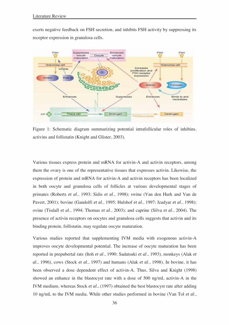
2005). As described by Knight and Glister (2003) (Figure 1), all three proteins are
synthesized and secreted by granulosa cells in an FSH-responsive manner. Local actions
of activins include promotion of granulosa cell proliferation, FSH receptor expression,
higher levels of P450 aromatase expression and estrogen production, and inhibition of
LH-induced androgen production by theca cells. Thus, granulosa cells are likely to be
the main source of paracrine factors, and are crucial for oocyte maturation. Follistatin is
structurally unrelated to the activins and inhibins, but binds with high affinity to the β-
subunits and neutralize the activity of inhibin (Phillips, 2005). The inhibin α-subunit
Literature Review
36
exerts negative feedback on FSH secretion, and inhibits FSH activity by suppressing its
receptor expression in granulosa cells.
Figure 1: Schematic diagram summarizing potential intrafollicular roles of inhibins,
activins and follistatin (Knight and Glister, 2003).
Various tissues express protein and mRNA for activin-A and activin receptors, among
them the ovary is one of the representative tissues that expresses activin. Likewise, the
expression of protein and mRNA for activin-A and activin receptors has been localized
in both oocyte and granulosa cells of follicles at various developmental stages of
primates (Roberts et al., 1993; Sidis et al., 1998); swine (Van den Hurk and Van de
Pavert, 2001); bovine (Gandolfi et al., 1995; Hulshof et al., 1997; Izadyar et al., 1998);
ovine (Tisdall et al., 1994; Thomas et al., 2003); and caprine (Silva et al., 2004). The
presence of activin receptors on oocytes and granulosa cells suggests that activin and its
binding protein, follistatin, may regulate oocyte maturation.
Various studies reported that supplementing IVM media with exogenous activin-A
improves oocyte developmental potential. The increase of oocyte maturation has been
reported in prepubertal rats (Itoh et al., 1990; Sadatsuki et al., 1993), monkeys (Alak et
al., 1996), cows (Stock et al., 1997) and humans (Alak et al., 1998). In bovine, it has
been observed a dose dependent effect of activin-A. Thus, Silva and Knight (1998)
showed an enhance in the blastocyst rate with a dose of 500 ng/mL activin-A in the
IVM medium, whereas Stock et al., (1997) obtained the best blastocyst rate after adding
10 ng/mL to the IVM media. While other studies performed in bovine (Van Tol et al.,
Literature Review
37
1994) and porcine (Coskun and Lin, 1994) oocytes reported no effect of activin-A on
oocyte maturation. Also in bovine, IVM in the presence of 10 ng/mL activin-A did not
change the proportion of cleaved oocytes that reached the blastocyst stage (Izadyar et
al., 1996). The reasons for these differences are likely related to a fundamental
difference between culture conditions and size and maturation stage of COCs.
2.1.3.3.5. Serum and other substances
In vitro oocyte maturation (IVM) procedures frequently include supplements of animal
origin, such as serum and follicular fluid, and it is considered as non-defined culture
medium because of its unknown composition. Their actions are not fully understood,
but it is believed that they provide proteins and/or some growth factors that contribute to
the success of in vitro maturation and subsequent development. Despite the undefined
and variable nature of serum composition, the supplementation of maturation media
with serum is practiced widely. In goat, maturation media are generally supplemented
with 10–20% heat-treated serum, among them: fetal bovine or calf serum (FCS) (Rho et
al., 2001; Cognié et al., 2004; Katska-Ksiazkiewicz et al., 2007; Khatun et al., 2011),
steer serum (SS) (Jiménez-Macedo et al., 2006; Romaguera et al., 2010b), estrous goat
serum (EGS) (Keskintepe et al., 1994; Teotia et al., 2001; Baldassarre et al., 2002;
Izquierdo et al., 2002), estrous sheep serum (ESS) (Tajik and Esfandabadi, 2003),
serum of peritoneal fluid from rabbit and goat (Malik and Lohan, 1999), and bovine
serum albumin (BSA)+EGS (Rajikin et al., 1994). The follicular fluid is also used as a
supplement in the IVM media in goats and sheep (Cognié et al., 2004). The
supplementation of IVM media with follicular fluid from non-atretic or gonadotropin
stimulated large follicles (>4 mm) resulted in beneficial effects in both sheep and goat
oocytes (reviewed by Tibary et al., 2005). In goat, Mogas et al., (1997a) tested EGS (at
different times of estrus), FCS and SS and did not find any significant differences on
maturation and embryo production. However, the presence of serum or follicular fluid
in culture media introduces a variation from batch to batch thus contributing to the lack
of reproducibility often observed in IVEP laboratories. Furthermore, serum and
follicular fluid contain many components including hormones, trace elements, and
growth factors.
Literature Review
38
Culture media supplemented with different growth factors have been used to study their
potential role on maturation and embryonic development in a growing number of
studies. Among the latter, epidermal growth factor (EGF) has been implicated in oocyte
maturation and subsequent development in several species. In sheep, in vitro matured
oocytes in the presence of EGF had greater cumulus cell expansion and higher
fertilization rates (Guler et al., 2000) and increases blastocyst formation in FSH-treated
ewes (Grazul-Bilska et al., 2003). In goat, the expression of EGF-R both in oocytes and
in follicular cells in goat cumulus-oocyte complexes (Gall et al., 2004), suggested that
EGF may regulate the oocyte growth and may be involved in nuclear and cytoplasmic
maturation. In prepubertal gilt, addition of 10 ng/mL EGF to IVM medium increased
the percentage of meiotically matured oocytes (88% vs. 70%) and also the embryos
presented a higher number of cells per blastocyst compared with those of control
blastocysts (51.1±5.1 % vs 36.0±3.1 %) (Grupen et al., 1997), showing that EGF plays
an important role in both the meiotic and cytoplasmic in vitro maturation of porcine
oocytes. Another factor to consider is insulin-like growth factor-I (IGF-I) that has also
been known to stimulate oocyte maturation and promoting blastocyst development in
several species, such as bovine (Matsui et al., 1995), ovine (Guler et al., 2000), porcine
(Grupen et al., 1997) and caprine (Magalhães-Padilha et al., 2012).
Also the positive influence of growth hormone (GH) on oocyte maturation has been
well reported. In ovine, Shirazi et al., (2010) showed a positive effect of GH added to in
vitro maturation medium that resulted in 73% of hatched blastocyst. In bovine, it was
observed that GH induced cumulus expansion and promoted subsequent embryonic
development in terms of enhancement of the number of cleaved embryos and
blastocysts (Izadyar et al., 1996).
A part of hormones and growth factors that are found in the follicular fluid,
polyunsaturated fatty acids (PUFAs) constitute the major portion of the fatty acid
content of the follicular fluid in small and large follicles (Homa and Brown, 1992).
PUFAs concentration in the follicular fluid is associated with the content of the diet and
may alter the oocyte maturation process and affect its further development. Actually,
studies performed in cattle have shown that in vitro supplementation of bovine oocytes
with physiological concentrations of α-linolenic acid (ALA; 18:3n-3) or linoleic acid
(LA; 18:2n-6) during in vitro maturation had diverse effects on maturation and

Literature Review
39
subsequent embryo development; stimulatory effects were achieved by ALA
supplementation (an increased maturation rate and a higher blastocyst yield and
production of better quality blastocysts) whereas LA was inhibitory compared with
untreated controls (Marei et al., 2009; 2010). However, to our knowledge, no studies of
the effect of PUFAs on in vitro embryo development in goats have been reported.
2.2. In vitro fertilization (IVF)
In vitro fertilization is a complex procedure whose success requires appropriate oocyte
maturation, sperm selection, sperm capacitation, and IVF media. In goat, freshly
ejaculated semen is usually used for IVF (Cox et al., 1994; Keskintepe et al., 1994;
Crozet et al., 1995; Mogas et al., 1997a; Izquierdo et al., 1998; Anguita et al., 2007;
Romaguera et al., 2011). Few trials have been described where IVF was carried out
using frozen–thawed sperm (Keskintepe et al., 1998; Rho et al., 2001; Bormann et al.,
2003).
Under in vivo conditions, potentially fertile spermatozoa are separated from immotile
spermatozoa, debris and seminal plasma in the female genital tract by active migration
through the cervical mucus. So, prior to in vitro fertilization, spermatozoa need to be
selected and prepared to inseminate the oocytes. The ejaculate comprises of a mixture of
seminal plasma, mature and immature spermatozoa, non-reproductive cells, various
microorganisms and non-specific debris. In bucks, the most common methods used for
separating the sample into motile and non-motile fractions from fresh ejaculate or from
the frozen-thawed sperm are the swim-up (Keskintepe et al., 1994; Izquierdo et al.,
1998; Katska-Ksiazkiewicz et al., 2004; Jiménez-Macedo et al., 2005; Anguita et al.,
2007; Romaguera et al., 2010b) and discontinuous density gradient centrifugation
(Pawshe et al., 1996; Rho et al., 2001; Wang et al., 2002a; Bormann et al., 2003), or
sephadex filtration (Rho et al., 2001) among others. Greater yields of highly motile
spermatozoa can be obtained by swim-up, compared to density gradient centrifugation,
but no differences were observed in terms of oocyte penetration and cleavage rate after
IVF with fresh goat semen (Palomo et al., 1999).
Once the most viable and motile spermatozoa are selected, sperm capacitation is carried
out in vitro and using defined media. Capacitation is a crucial process that mammalian
Literature Review
40
sperm must undergo in order to achieve fertilizing ability. It is defined as the
phenomenon leads to "acrosome reaction" causing the release of proteolytic enzymes
that may assist sperm penetration into the oocyte. In goat, several capacitating agents
have been used to capacitate spermatozoa and to yield good fertilization and cleavage
rates, such as heat-inactivated estrous from sheep (De Smedt et al., 1992), goat
serum (Koeman et al., 2003; Katska-Ksiazkiewicz et al., 2004), heparin using fresh
semen (Izquierdo et al., 1998; Jiménez-Macedo et al., 2005) or frozen-thawed sperm
(De Souza et al., 2013), heparin and ionomycin (Wang et al., 2002a; Urdaneta et al.,
2004), calcium ionophore (Pereira et al., 2000) and heparin and caffeine (Younis et al.,
1991).
Regarding the fertilization media used in goats, these include: modified Defined Media
(mDM) (Crozet et al., 1995) Tyrode’s Albumin Lactate Pyruvate (TALP) medium
(Parrish et al., 1986) supplemented with hypotaurine (Mogas et al., 1997b; Izquierdo et
al., 1998), and Synthetic Oviductal Fluid (SOF) medium (Rho et al., 2001). Incubation
of sperm selected by swim-up in TALP with heparin (50 mg/mL) for 45 min resulted in
excellent fertilization rates (Palomo et al., 1999; Katska et al., 2002; Katska-
Ksiazkiewicz et al., 2004). Similarly, mDM plus heparin for sperm capacitation and
TALP medium with hypotaurine for oocyte fertilization provided the highest proportion
of penetrated oocytes (Izquierdo et al., 1998).
2.3. In vitro culture (IVC)
The last stage of in vitro embryo production is the culture of the presumptive zygotes in
culture media where they undergo a number of divisions until the blastocyst stage 6-7 days
after in vitro fertilization in ruminant species (Gardner et al., 1994). This period of
postfertilization culture is the period having the greatest impact on the blastocyst quality
(Rizos et al., 2002a). Several major developmental events take place, including the first
cleavage division; the switching on of the embryonic genome; the compaction of the
morula and the blastocyst formation. The blastocyst stage involves the differentiation of
two types of cells, the inner cell mass (ICM), which after further differentiation gives
rise to the fetus, and the trophectoderm (TE), which ultimately contributes to the
placenta formation (Watson, 1992). Clearly, any modifications of the culture
environment, which could affect any or all of these processes, could have a major effect

Literature Review
41
on the quality of the embryo. Some of these factors that could influence on the rate of
embryo development and the quality of the embryo are described below.
The in vitro oxygen concentration under which embryo development occurs has been
found to modify the development up to the blastocyst stage. The oxygen tension that
most mammalian embryos encounter in the reproductive tract, range from 3.5 to 8%
(Fischer and Bavister, 1993). The positive effect of low oxygen tension on embryo
culture has been reported for many species. For instance, the culture of in vitro fertilized
pig embryos in low oxygen increased the number of cells (Booth et al., 2005) and
embryo production rate (Karja et al., 2004) to greater than those cultured in 20%
oxygen. In bovine, embryo development and survival rates after cryopreservation were
higher when 5% oxygen was applied during IVC compared to 20% oxygen in air (Rizos
et al., 2001). Those results suggested that greater O2 tension during culture is
detrimental for embryo development, probably due to the accumulation of reactive
oxygen species (ROS).
Different culture systems have been studied to support pre-implantation development of
embryos up to the blastocyst stage. Given that female reproductive tract secretions have
several amino acids that can be used as energetic substrate by the embryo, the use of
amino acids in culture media improves embryo development probably through an
antioxidant action and reducing the stress and cell fragmentation caused by in vitro
embryo culture. On the other hand, in the undefined culture systems, serum is one of the
main components. It can provide many beneficial factors to the embryo such as amino
acids, vitamins, growth factors, and energetic substrates; however, it may also
contaminate the culture media with embryotoxic factors (Bavister, 1995).
Diverse culture media have been successfully used for small ruminant embryo
development such as TCM199 (Wani et al., 2012), B2 (Katska-Ksianzkiewicz et al.,
2007) and “Sydney IVF Blastocyst” medium (Beilby et al., 2011). However, the most
widely used medium is the synthetic oviduct fluid (SOF). Tervit et al., (1972; 1974)
were among the first to report successful culture of ruminant zygotes to the blastocyst
stage in vitro using SOF medium, which was based on the composition of ovine oviduct
fluid. Subsequently, changes to the original composition have been made with some
modification (Takahashi and First, 1992). Some laboratories routinely supplement SOF
medium with serum. Studies in bovine have shown that serum has a biphasic effect; the

Literature Review
42
presence of serum can inhibit the early cleavage divisions, while it can have an
accelerating effect later in development, resulting in the appearance of blastocysts
earlier in culture (Lonergan et al., 1999; Gutiérrez-Adán et al., 2001). Thus, some
researchers supplement IVC medium with 5-10% FCS (Cognié et al., 2003; Jiménez-
Macedo et al., 2005; 2006; Anguita et al., 2007) at 2-3 days post-insemination to
promote a higher viability after transfer of such IVP embryos (Cognié, 1999). Other
studies only add BSA to SOF media (Leoni et al., 2007; Shabankareh and Akhondi,
2012; Wang et al., 2013b).
Currently, several animal and human studies attest a beneficial effect of addition of
growth factors to culture media. However, there is still ambiguity regarding the exact
role of growth factors in embryonic development, the optimal dose of growth factors to
be added to the culture media, and the combinatorial effect of growth factors in
embryonic development (Hegde and Behr, 2012).
Previous research has shown that growth factors such as EGF and IGF-I induced a
positive effect on preimplantation development by stimulating metabolism and growth
of embryos. In bovine, Palma et al., (1997) demonstrated that culture media containing
high concentrations of IGF-I improved the development of embryos produced in vitro,
and significantly reduced apoptosis (Makarevich and Markkula, 2002). Furthermore,
combination of EGF and IGF-I showed an additive effect, and higher rates of embryos
developed into blastocysts (Sakagami et al., 2012). In murine embryos, it has been
reported that a high dose of EGF resulted in an improvement on blastocyst rate and cell
number (Głabowski et al., 2005). Similarly, Mtango et al., (2003) demonstrated that the
addition of growth factors and growth hormone (GH) to the culture media of bovine
embryos had favorable effects on in vitro embryo development, freezing sensitivity and
post-thawing survival, hatching rate and total cell number of blastocysts. Other growth
factors may also have beneficial effects on embryo development. Regarding the use of
hormones, mouse blastocysts that have been cultured with growth hormone showed a
higher chance for implantation after transfer to the recipient (Fukaya et al., 1998).
Moreover, ghrelin is a widespread hormone that several studies have linked with
reproductive physiology (Garcia-Garcia et al., 2007; Tena-Sempere, 2008). In sheep
(Wang et al., 2013b), the blastocyst rate, total cell number of blastocysts and the
expression levels of the GLUT1 and IFNT genes were increased when 50 ng/mL ghrelin
was added during IVC to the SOF medium.

Literature Review
43
Since activin-A is produced by oviduct epithelial cells (Gandolfi et al., 1995), the
addition of activin-A to embryo in vitro culture may reproduce the environment of the
cleavage-stage embryos in the oviduct in vivo. Activin-A and activin receptors have
been detected in mouse (Lu et al., 1993) and bovine (Yoshioka et al., 1998b) embryos
from zygote to the morula stage, suggesting that the protein might play a role in
embryogenesis. According to studies performed in vitro, activin-A has been shown to
have an effect in regulation of the development of bovine embryos (Yoshioka and
Kamomae, 1996; Yoshioka et al., 1998a). However, the effect of activin-A on in vitro
embryo culture is still controversial. That depending on the timing of its addition to the
culture medium, exogenous activin-A may promote the morula and blastocyst
development when the protein was added at the earliest stages of bovine (Yoshioka and
Kamomae, 1996; Yoshioka et al., 1998a; Lee et al., 2009) embryo culture. Similar
beneficial effects of activin, when added to the second half of the embryo culture, were
reported as increased expanding and hatching rates (Park et al., 2010; Trigal et al.,
2011). As described by Park et al., (2010), activin-A, during the later stage of in vitro
bovine embryo development, can enhance in vitro development of embryos by affecting
expression levels of genes related to hatching and implantation in defined culture
medium, and the development-enhancing effect of activin-A may be associated with
timing of activation of appropriate genes within the embryonic genome (Trigal et al.,
2011). Also, maternal levels of activin protein in the mouse embryo decline during early
cleavage stages but increase, presumably due to embryo transcription, in the compacted
morula (Albano et al., 1993). It has been also shown a dose effect when activin-A was
supplemented to in vitro culture media of IVM/IVF bovine oocytes. Yoshioka et al.,
(1998a) observed that the percentages of zygotes that developed to the morula and
blastocyst stages increased dose-dependently with addition of activin-A, similar to the
percentages of expanded blastocysts. Therefore, it may be advisable to test what culture
system or culture medium will provide better results for a given lab.
2.4. Assessment of embryo quality
A proper evaluation of blastocyst quality remains an important challenge for every
researcher involved in embryology to select the best embryos for transfer, chance to
obtain pregnancies and to define its sanitary condition.
Literature Review
44
The noninvasive methods used for evaluating the embryo quality are the timing of early
cleavage (Van Soom et al., 1992), understanding that good quality embryos must
exhibit appropriate kinetics and synchrony of division, the evaluation of the timing of
blastocyst formation (Van Soom et al., 1997) and the blastocyst morphology (Van
Soom et al., 2003). All of those methods are practical to perform and without any
consequence for the embryo’s viability.
The cryopreservation of embryos is an essential method in assisted reproduction
technology (ART) for world-wide embryo transfer (ET) technology that also allows to
select the most robust and highest quality embryos will be able to support the freeze-
thaw process. Therefore, the ability of embryos to survive cryopreservation is
commonly used as an indicator of embryo quality and viability (Rizos et al., 2008).
Studies performed in several species reported differences on survival rates and hatching
percentages after the cryopreservation of blastocysts depending on the origin of
embryos (in vivo versus in vitro), the stage of embryo development, the age of the donor
female, and media for IVEP.
Additionally, research on animal models has demonstrated that stress, and the resultant
adaptation to conditions during pre-implantation stages, can affect pregnancy loss and
fetal growth. It is therefore important to understand the role of each medium component
and to identify possible sources of cellular stress to the embryo that will ultimately
affect the function and viability of the fetus.
Serum supplementation has been associated with accumulation of cytoplasmic lipid
droplets in IVP embryos (Abe et al., 1999) and can lead to poor cryosurvival (Leibo and
Loskutoff, 1993). In ovine, Shirazi et al., (2012) showed that a significantly lower
survival rate of vitrified-warmed blastocysts derived from oocytes matured in presence
of FCS, might be due to the excess accumulation of cytoplasmic lipid droplets in
resulting blastocysts compared to the other group. In bovine, Moore et al., (2007) used a
serum replacer (SR) for maturation of oocytes and results showed an improved embryo
development in vitro as well as better cryotolerance and survival when replacing serum
in culture. In the same way, the presence of somatic cells during the IVC was shown
to affect the freezability of in vitro produced bovine embryos (Gómez et al., 2008).
Shehab-El-Deen et al., (2009) demonstrated that the addition of elevated concentrations
of saturated fatty acids (palmitic and stearic acid) during IVM of bovine oocyte have

Literature Review
45
deleterious on embryo quality, featured by a significantly reduced post-warming
survival of vitrified blastocysts.
In research, the quality of embryos obtained can be also assessed with invasive
techniques. Invasive assessment of embryo quality mostly involves a kind of fixation
and staining of the embryo. The assessment of embryo quality is carried out under a
stereomicroscope on grounds of morphological criteria. Many measures can be
investigated to determine embryo quality such as total cell number, the ICM/TE ratio of
blastocysts, the degree of cell fragmentation, and determination of genes related with
embryo quality and ability to achieve pregnancy after transfer. A simplified technique
for differential staining of inner cell mass and trophectoderm cells of mouse and bovine
blastocysts (Thouas et al., 2001) permits the differentiation of the two cell populations
as the blue ICM cell nuclei (stained only with Hoechst) that can be clearly distinguished
from the pink TE cell nuclei (stained both with propidium iodide and Hoechst). This
technique has been applied to embryos from different species, including bovine
(Iwasaki et al., 1990; Fouladi-Nashta et al., 2005), ovine (Catalá et al., 2011), porcine
(Dang-Nguyen et al., 2011) and buffalo (Selokar et al., 2012) among others. Several
factors have been reported to affect the proportion of cell number in each blastocyst
compartment. If compare in vivo versus in vitro produced embryos, it has been reported
that the in vitro production systems affect the blastocyst quality in terms of total cell
number, incidence of cell apoptosis, oxidative stress and up and down regulation of
gene expression. In bovine, Knijn et al., (2003) reported a significantly lower total cell
number for those embryos produced completely in vitro compared to that of embryos
developed entirely in vivo, as well as a higher incidence of DNA fragmentation.
Until the blastocyst stage of development is attained, the embryo is somewhat
autonomous (i.e., does not need contact with the maternal reproductive tract) as
evidenced by the fact that blastocysts can be successfully developed in vitro in large
number using IVF technology and transferred to reproductive synchronized recipients
(Mamo et al., 2012). However, the ability to undergo implantation and pregnancies
depends on blastocyst quality. Thus, cell number and apoptosis levels are also important
parameters that are emerging as useful indicators of embryo development and health
(Brison and Schultz, 1997; De la Fuente and King, 1997). This was further supported by
Fouladi-Nashta et al., (2005) who demonstrated that counting the total number of
blastomeres and the percentage of apoptotic nuclei provides more detailed information
Literature Review
46
on embryo quality and developmental potential. These authors investigated the effect of
serum supplementation on total cell number, ICM/TE ratio and apoptosis index after in
vitro production of bovine embryos. Results of this study showed that supplementation
of serum will possibly increase the number of TE cells leading a reduced ratio of
ICM/TE. The high number of TE cells in the serum treated embryos may cause the
increased placenta size in the large offspring syndrome.
Another parameter used to assess the quality of embryos is the ratio of programmed cell
death or DNA fragmentation. Cell death is present in mammalian embryos, which might
be involved in the elimination of abnormal cells and its normal pattern is crucial for
further development necessary to maintain the morphogenesis and to allow cell
renovation. Cell death was confirmed in goat blastocysts using the terminal
deoxynucleotidyl transferase (TdT) mediated dUTP nick-end labeling (TUNEL) assay
(Jiménez-Macedo et al., 2007). TUNEL allows in situ assessment of DNA breaks in the
nuclei (reviewed by Neuber et al., 2002) and it has been observed a negative relation
with the implantation rates.
Likewise, oxygen free radicals can cause DNA strand breaks and other forms of cellular
damage (lipid peroxidation) which are known to result in cell cycle arrest or death.
Although apoptosis can be considered to be a normal process in preimplantation
embryos to eliminate deviating cells, a high incidence of apoptotic cells is correlated
with abnormal morphology of the embryo. Moreover, embryonic cell apoptosis has
received increasing attention, mostly due to its suggested role in the cellular response to
suboptimal developmental conditions (Betts and King, 2001). Thus, increased apoptosis
is an important indicator of inadequate in vitro conditions for eggs.
Ending, environmentally induced changes in oocyte and embryo biology have both
immediate effects on the viability and development of these structures, but can also
have pronounced and often persistent effects on the resultant offspring. For all above
mentioned and keeping on mind that the ultimate test of the quality of an embryo is its
ability to produce live and healthy offspring after transfer to a recipient, it is really
important consider and optimize the conditions of the in vitro culture systems in order to
get embryos able to undergo pregnancies.

47
OBJECTIVES
Chapter 3

48
Objectives
49
Objectives of the study
The overall aim of this thesis work was to improve the in vitro embryo production from
oocytes of prepubertal goats. More specifically, the aims were:
I: To examine the effect of supplementing in vitro maturation medium with low
level of hormones and/or insulin-transferrin-selenium plus ascorbic acid in the
developmental competence and embryo quality of small oocytes from prepubertal goats.
II: To evaluate the effect of adding activin-A at the in vitro oocytes maturation and
in vitro embryo culture media on meiotic maturation, embryo development and embryo
quality of in vitro-produced blastocysts.

50
51
Developmental competence and embryo quality of small oocytes from pre-pubertal goats cultured in IVM medium supplemented with low level of hormones, insulin-
transferrin-selenium and ascorbic acid
Hammami S1, Morató R2, Romaguera R1, Roura M1, Catalá MG1, Paramio MT1, Mogas T2, Izquierdo D1.
1 Departament de Ciència Animal i dels Aliments, Universitat Autònoma de Barcelona, Bellaterra, Spain
2 Departament de Medicina i Cirurgia Animals, Universitat Autònoma de Barcelona, Bellaterra, Spain
Reprod Domest Anim. 48 (2013) 339-344.
Chapter 4

52

53

54

55

56
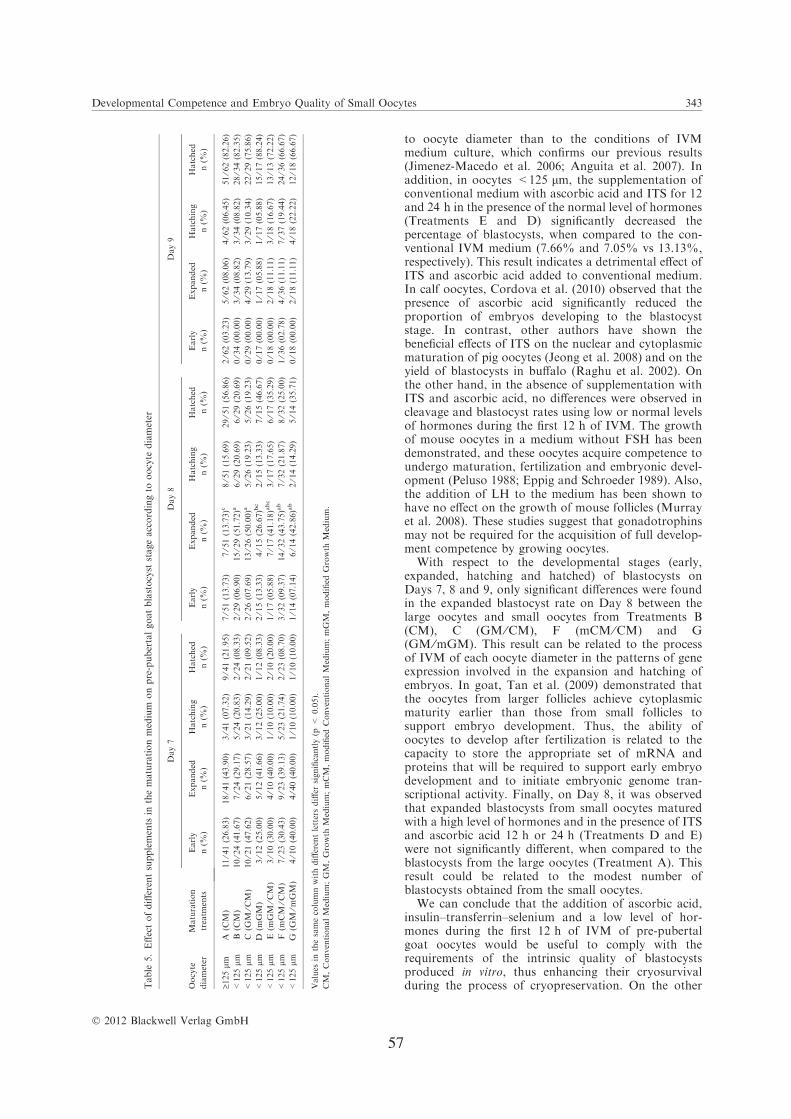
57

58

59
In vitro developmental competence of prepubertal goat oocytes cultured with recombinant activin-A
Hammami S1, Izquierdo D1, Catalá MG1, Paramio MT1, Morató R2.
1Departament de Ciència Animal i dels Aliments, Universitat Autònoma de Barcelona, Bellaterra, Spain
2Departament de Biologia i Biotecnologia de la Reproducció Animal i Humana (TechnoSperm), Institut de Tecnologia Agroalimentaria, Universitat de Girona,
Girona, Spain
Animal 8 (2014) 94-101
Chapter 5

60

61

62
63

64
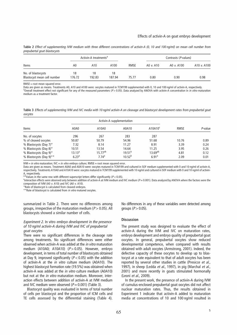
65
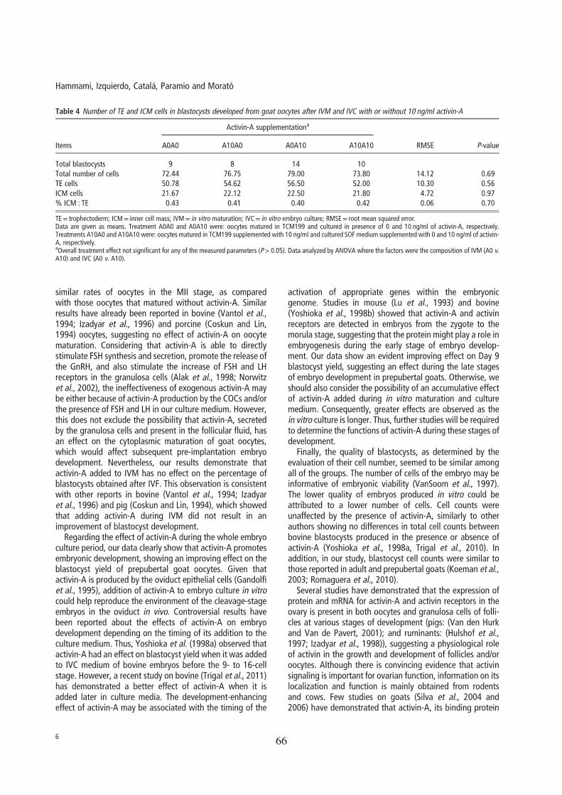
66

67
68

69
GENERAL DISCUSSION
Chapter 6

70

General Discussion
71
General Discussion
This thesis is focused on improvement of the in vitro embryo production from oocytes
of prepubertal goats. For several years, in vitro embryo production from prepubertal
goats has been considered a promising option for providing an excellent source of
embryos for basic research on development biology and for commercial applications of
transgenic and cloning technologies. However, despite successful results and reports of
numerous attempts, the effectiveness of in vitro embryo production protocols for
prepubertal goat oocytes has never reached a satisfactory level. Oocyte quality is the
main factor for embryos reaching blastocyst stage from IVM/IVF/IVC oocytes. Until
now, attention has been focused on selection of criteria to assess the most competent
oocytes to develop up to blastocyst stage. In our laboratory, several studies have been
reported describing the best criteria to select the larger and more competent oocytes for
in vitro blastocyst production: Brilliant Cresyl Blue (BCB) stain (Rodriguez-Gonzalez
et al., 2002; Catalá et al., 2011), oocyte (Jiménez-Macedo et al., 2006; Anguita et al.,
2007) and follicle (Romaguera et al., 2010a; 2011) diameters.
People working in this field has to combine the most adequate oocyte selection criteria
and the in vitro culture protocol to became high rates of blastocyst development. For
this reason, the two studies included in the present thesis work were designed to
optimize in vitro culture system of prepubertal goat oocytes collected from ovaries of
slaughtered goat females, using supplements added during IVM and/or IVC, in order to
increase both rates of development and quality of in vitro-produced blastocysts at a rate
equivalent to that of adult oocytes.
The first study was conducted to examine the effect of supplementing in vitro
maturation medium with low level of hormones and/or insulin-transferrin-selenium plus
ascorbic acid in the developmental competence and embryo quality of small oocytes
from prepubertal goats.
Previous studies in our laboratory using prepubertal goat oocytes showed that not only
the age of the oocyte donor is crucial for embryo production, but also the size of oocyte
plays an important role in embryo production. Firstly, we hypothesized that
supplementing the IVM medium of small oocytes with low hormone levels, ITS and L-
General Discussion
72
ascorbic acid called growth medium, could significantly improve maturation along with
developmental competency compared to the conventional goat oocyte maturation
system. However, our results have showed that the culture of small prepubertal goat
oocytes in the growth medium during IVM did not improve the blastocyst percentage
when compared to those matured in conventional IVM medium (Hammami et al.,
2013). Similarly, in ovine, Catalá et al., (2013) also observed that growth medium failed
to increase embryo production of lamb oocytes. These discrepancies with the findings
of Wu et al., (2006) on gilt oocytes and Córdova et al., (2010) on calf oocytes may be
related to variability among biological requirements of immature oocytes depending on
species used.
In these initial studies it was possible to duplicate the performance of blastocyst
production with the use of oocyte diameter as selection criteria. The rate of blastocysts
production from prepubertal goat oocytes ≥ 125 μm was 20.65% compared to 10.40 %
from oocytes < 125 μm diameter. Thus, the capacity of those oocytes, once fertilized, to
develop to the blastocyst stage in vitro seems to be more related to oocyte diameter than
to the conditions of in vitro maturation system. Several studies concluded that oocyte
diameter is correlated to follicle diameter and as both increase the developmental
capability of the oocytes improves (revised by Gandolfi et al., 2005). It is noteworthy
that COCs coming from a wide range of follicle diameters and selected based on their
morphology alone produced higher cleavage and blastocyst rates than oocytes from
small follicles. One possibility could be, as Romaguera et al., (2010a) discussed, that
oocytes from follicles of ≥ 3 mm could have enough time to be submitted to
prematuration processes and to acquire a complete developmental competence to
blastocyst stage. Anguita et al., (2007) confirmed this hypothesis previously in our
laboratory showing that prepubertal goat oocyte diameter and, therefore, oocyte
developmental competence seems to be related to the amount of cyclin B, cdc2 kinase
protein and MPF activity. Maturation promoting factor (MPF) is a heterodimer
composed of a p34cdc2 catalytic subunit, with serine-threonine kinase activity, and a
cyclin B1 regulatory subunit. MPF activity appears just before germinal vesicle break
down (GVBD) increasing until metaphase I; its activity is decreased in anaphase-
telophase while its maximum level is reached at metaphase II (MII). In prepubertal
goats, Anguita et al., (2007) showed a higher MPF activity in oocytes with a diameter
larger than 135 μm and, after IVM, p34cdc2 protein expression increased with the
General Discussion
73
oocyte diameter. Possibly, the p34cdc2 RNA accumulation in the oocyte takes place
during the last growth stage and the translation begins when the oocyte reaches its
maximum size. MPF activity seems to confer higher developmental competence to
oocytes through promotion of cytoplasmic maturation. Hence, enough time to be
submitted to prematuration processes and to acquire a complete oocyte competence is
important for successful embryo development.
When analysing the effects of different growth medium components combinations (low
level of hormones, ITS and ascorbic acid with the conventional medium during IVM, on
the cleavage and developmental capacity of prepubertal goat oocytes, our findings
indicated that the supplementation of conventional medium with ascorbic acid and ITS
during 12h and 24h in presence of normal level of hormones provided a significant
decrease in the percentage of blastocysts obtained from <125 μm oocytes diameter,
compared to the conventional IVM medium (7.65% and 7.05% vs 13.12%,
respectively). According to our data, in calf oocytes, Córdova et al., (2010) observed
that the presence of ascorbic acid significantly reduced the proportion of embryos
developing to the blastocyst stage. These data are in disagreement with observations in
pig and buffalo oocytes in that the presence of ITS during in vitro maturation had a
beneficial effect on the nuclear and cytoplasmic maturation (Jeong et al., 2008) and on
the yield of blastocysts (Raghu et al., 2002a). On the other hand, in absence of
supplementation with ITS and ascorbic acid, no differences were observed on cleavage
and blastocyst rates using low or normal level of hormones during the first 12h of IVM.
The growth of mouse oocytes in a medium without FSH has been demonstrated, and
these oocytes acquire competence to undergo maturation, fertilization and embryonic
development (Peluso, 1988; Eppig and Schroeder, 1989; Wu et al., 2000; Murray et al.,
2008). Also, when mouse follicles have been cultured in presence of LH, their growth
was not affected (Murray et al., 2008). These results suggest that gonadotrophins may
not be required for the acquisition of full development competence by growing oocytes.
Aside from adding antioxidants, vitamins and/or different hormone levels during IVM,
in the second study we investigated whether activin-A supplementation to the IVM
medium of prepubertal goat oocytes, could affect their nuclear maturation and
competence to develop up to blastocyst stage. Results of this study (Hammami et al.,
2014) showed no effect of activin-A on oocyte maturation and subsequent embryo
General Discussion
74
development, similarly to results reported in bovine (Van Tol et al., 1994; Izadyar et al.,
1996) and porcine (Coskun and Lin, 1994) oocytes. Considering that activin-A is able to
directly stimulate FSH synthesis and secretion, promote the release of the
gonadotrophin-releasing hormone (GnRH), and also stimulate the increase of FSH and
LH receptors in granulosa cells (Alak et al., 1998; Norwitz et al., 2002), the
ineffectiveness of exogenous activin-A may be either due to activin-A production by the
COCs and/or the presence of FSH and LH in our culture medium.
The two studies suggest that in our conditions no effectiveness on the subsequent
development to the blastocyst stage of prepubertal goat oocytes is obtained, neither
when different maturation media are combined (ascorbic acid, insulin–transferrin–
selenium, and a low level of hormones) with the conventional IVM medium using small
oocytes diameter, nor when activin-A is added to the conventional IVM medium.
Given that activin-A is produced by oviduct epithelial cells (Gandolfi et al., 1995),
addition of activin-A to in vitro embryo culture could help to reproduce the environment
of the in vivo embryonic development in the oviduct. In bovine, activin-A was reported
a benefit effect depending on the timing of its addition to the culture medium (Yoshioka
et al., 1998a; Trigal et al., 2011). Our study also sought to determine the suitability of a
culture medium supplemented with activin-A for the in vitro culture of in vitro matured
and fertilized prepubertal goat oocytes. The main finding of this study was that the
addition of 10 ng/mL of activin-A throughout the whole embryo culture promotes
embryonic development, improving the blastocyst yield (Hammami et al., 2014).
Otherwise, we should also consider the possibility of an accumulative effect of activin-
A added during in vitro maturation and culture medium. Consequently, greater effects
are observed as the in vitro culture is longer. Thus, further studies will be required to
determine the functions of activin-A during these stages of development. The
development-enhancing effect of activin-A may be associated with the timing of the
activation of appropriate genes within the embryonic genome. Studies in mouse (Lu et
al., 1993) and bovine (Yoshioka et al., 1998b) showed that activin-A and activin
receptors are detected in embryos from the zygote to the morula stage, suggesting that
the protein might play a role in embryogenesis during the early stage of embryo
development. As far as we know, no studies on detection of the activin receptors in goat
oocytes and embryos have been previously reported, though studies using other species
confirm the presence of them in different stages of development. A detailed profiling of

General Discussion
75
the activin-A effects on embryonic development of prepubertal goat oocytes and
determination of gene expression and protein synthesis for activin-A require further
experiments.
Finally, the effect of growth medium during IVM on blastocyst quality was measured
by post-warming survival following vitrification method and blastocyst cell count. The
ability of blastocysts derived from big oocytes to survive the vitrification/warming
process at 3h was lower than that of blastocysts obtained from small oocytes. According
to previous data in bovine (Morató et al., 2010), these differences in survival rates of in
vitro-produced blastocysts depend on their developmental stage. So, the decrease in the
cryosurvival rate may be explained by the developmental stage of blastocysts derived
from large oocytes (hatching and hatched) as compared to the stage of development of
the blastocysts obtained from small oocytes (early and expanded). Otherwise, at 72h the
re-expansion rate was significantly higher in the blastocysts obtained from small
oocytes matured in growth medium than in those blastocysts from oocytes matured in
conventional medium, reflecting a higher oocyte quality. Previous studies in bovine
showed that the culture medium supplementation (Rizos et al., 2001; Gómez et al.,
2008) affects intrinsic embryo quality in terms of cryosurvival. Thus, a higher oocyte
quality was showed after the oocyte culture in growth medium. However, our results
showed that the use of growth medium did not improve the blastocyst cell counts.
Similarly, cell number was unaffected by the presence of activin-A in the IVM media.
So, in summary, we can conclude that the addition of ascorbic acid, insulin-transferrin-
selenium and a low level of hormones during the first 12h of IVM of prepubertal goat
oocytes would be useful to comply with the requirements of the intrinsic quality of
blastocysts produced in vitro. Notwithstanding, the capacity of those oocytes to develop
to the blastocyst stage in vitro seems to be more related to oocyte diameter than to the
conditions of the IVM system. Finally, according to the results obtained in the second
study, the addition of 10 ng/mL of activin-A throughout the whole embryo culture
improves blastocyst yield.

76
77
GENERAL CONCLUSIONS
Chapter 7

78
General Conclusions
79
General Conclusions
The results of this PhD thesis let conclude that:
1. The addition of ascorbic acid, insulin-transferrin-selenium and a low level of
hormones during the first 12h of IVM of prepubertal goat oocytes would be useful
to comply with the requirements of the intrinsic quality of blastocysts produced in
vitro.
2. The capacity of prepubertal goat oocytes to develop to the blastocyst stage in vitro
seems to be more related to oocyte diameter than to the conditions of the IVM
system.
3. Activin-A added to IVM did not result in an improvement of blastocyst
development.
4. The addition of 10 ng/mL of activin-A throughout the whole embryo culture period
promotes embryonic development, showing an improving effect on the blastocyst
yield of prepubertal goat oocytes.

80

81
REFERENCES
Chapter 8

References
82

References
83
References
Abdoon A, Kandil O, Otoi T, Suzuki T, 2001: Influence of oocyte quality, culture media and gonadotropins on cleavage rate and development of in vitro fertilized buffalo embryos. Animal Reproduction Science 65 215-223.
Abe H, Yamashita S, Itoh T, Satoh T, Hoshi H, 1999: Ultrastructure of bovine embryos developed from in vitro-matured and -fertilized oocytes: comparative morphological evaluation of embryos cultured either in serum-free medium or in serum-supplemented medium. Molecular Reproduction and Development 53 325-35.
Agarwal A, Said T, Bedaiwy M, Banerjee J, Alvarez J, 2006: Oxidative stress in an assisted reproductive techniques setting. Fertility and Sterility 86 503-512.
Alak B, Smith G, Woodruff T, Stouffer R, Wolf D, 1996: Enhancement of primate oocyte maturation and fertilization in vitro by inhibin A and activin A. Fertility and Sterility 66 646-653.
Alak B, Coskun S, Friedman C, Kennard E, Kim M, Seifer D, 1998: Activin A stimulates meiotic maturation of human oocytes and modulates granulosa cell steroidogenesis in vitro. Fertility and Sterility 70 1126-1130.
Albano RM, Groome N, Smith JC, 1993: Activins are expressed in preimplantation mouse embryos and in ES and EC cells and are regulated on their differentiation. Development 117 711-23.
Ali A, Bilodeau J, Sirard M, 2003: Antioxidant requirements for bovine oocytes varies during in vitro maturation, fertilization and development. Theriogenology 59 939-949.
Ali A, Coenen K, Bousquet D, Sirard MA, 2004: Origin of bovine follicular fluid and its effect during in vitro maturation on the developmental competence of bovine oocytes. Theriogenology 62 1596-1606.
Alvarez G, Dalvit G, Achi M, Miguez M, Cetica P, 2009: Immature oocyte quality and maturational competence of porcine cumulus-oocyte complexes subpopulations. Biocell 33 167-177.
Anguita B, Jiménez-Macedo A, Izquierdo D, Mogas T, Paramio M, 2007: Effect of oocyte diameter on meiotic competence, embryo development, p34 (cdc2) expression and MPF activity in prepubertal goat oocytes. Theriogenology 67 526-536.
Anguita B, Paramio M, Jiménez-Macedo A, Morató R, Mogas T, Izquierdo D, 2008: Total RNA and protein content, Cyclin B 1 expression and developmental competence of prepubertal goat oocytes. Animal Reproduction Science 103 290-303.
Armstrong D, Kotaras P, Earl C, 1997: Advances in production of embryos in vitro from juvenile and prepubertal oocytes from the calf and lamb. Reproduction Fertility and Development 9 333-339.

References
84
Bai J, Hou J, Guan H, Yan F, Cui X, Liu L, Wang S, An X, 2008: Effect of 2-mercaptoethanol and cysteine supplementation during in vitro maturation on the developmental competence of oocytes from hormone-stimulated lambs. Theriogenology 70 758-764.
Baldassarre H, Wang B, Kafidi N, Keefer C, Lazaris A, Karatzas C, 2002: Advances in the production and propagation of transgenic goats using laparoscopic ovum pick-up and in vitro embryo production technologies. Theriogenology 57 275-284.
Baldassarre H, Karatzas C, 2004: Advanced assisted reproduction technologies (ART) in goats. Animal Reproduction Science 82-83 255-266.
Bavister BD, 1995. Culture of preimplantation embryos: facts and artifacts. Human Reproduction 1 91-148.
Beilby KH, de Graaf SP, Evans G, Maxwell WMC, Wilkening S, Wrenzycki C, Grupen CG, 2011: Quantitative mRNA expression in ovine blastocysts produced from X- and Y-chromosome bearing sperm, both in vitro and in vivo. Theriogenology 76 471-481.
Beker-van Woudenberg A, van Tol H, Roelen B, Colenbrander B, Bevers M, 2004: Estradiol and its membrane-impermeable conjugate (estradiol-bovine serum albumin) during in vitro maturation of bovine oocytes: Effects on nuclear and cytoplasmic maturation, cytoskeleton, and embryo quality. Biology of Reproduction 70 1465-1474.
Betts DH, King WA, 2001: Genetic regulation of embryo death and senescence. Theriogenology 55 171–191
Bevers M, Dieleman S, vandenHurk R, Izadyar F, 1997: Regulation and modulation of oocyte maturation in the bovine. Theriogenology 47 13-22.
Bilezikjian L, Blount A, Donaldson C, Vale W, 2006: Pituitary actions of ligands of the TGF-beta family: activins and inhibins. Reproduction 132 207-215.
Bing Y, Nagai T, Rodriguez-Martinez H, 2001: Effects of cysteamine, FSH and estradiol-17 beta on in vitro maturation of porcine oocytes. Theriogenology 55 867-876.
Blondin P, Sirard MA, 1995: Oocyte and follicular morphology as determining characteristics for developmental competence in bovine oocytes. Molecular Reproduction and Development 41 54–62.
Booth PJ, Holm P, Callesen H, 2005: The effect of oxygen tension on porcine embryonic development is dependent on embryo type. Theriogenology 63 2040-2052.
Bormann C, Ongeri E, Krisher R, 2003: The effect of vitamins during maturation of caprine oocytes on subsequent developmental potential in vitro. Theriogenology 59 1373-1380.

References
85
Bowles C, Lishman A, 1998: Attempts to improve the yield of bovine blastocysts by incorporating insulin, selenium and transferrin in the in vitro system. South African Journal of Animal Science 28 30-37.
Brevini T, Cillo F, Antonini S, Tosetti V, Gandolfi F, 2007: Temporal and spatial control of gene expression in early embryos of farm animals. Reproduction Fertility and Development 19 35-42.
Brison D, Schultz R, 1997: Apoptosis during mouse blastocyst formation: Evidence for a role for survival factors including transforming growth factor alpha. Biology of Reproduction 56 1088-1096.
Buettner G, 1993: The pecking order of free-radicals and antioxidants: lipid-peroxidation, alpha-tocopherol, and ascorbate. Archives of Biochemistry and Biophysics 300 535-543.
Byskov A, Andersen C, Hossaini A, Xia G, 1997: Cumulus cells of oocyte-cumulus complexes secrete a meiosis-activating substance when stimulated with FSH. Molecular Reproduction and Development 46 296-305.
Catalá M, Izquierdo D, Uzbekova S, Morató R, Roura M, Romaguera R, Papillier P, Paramio M, 2011: Brilliant cresyl blue stain selects largest oocytes with highest mitochondrial activity, maturation-promoting factor activity and embryo developmental competence in prepubertal sheep. Reproduction 142 517-527.
Catalá M, Montserrat R, Izquierdo D, Morató R, Hammami S, Paramio MT, 2013: Blastocyst development, MPF activity and ATP content of lamb oocytes supplemented with insulin-transferrin-selenium (ITS) and ascorbic acid at IVM. Small Ruminant Research 112 103-107.
Cebrian-Serrano A, Salvador I, Raga E, Dinnyes A, Silvestre MA, 2013: Beneficial effect of melatonin on blastocyst in vitro production from heat-stressed bovine oocytes. Reproduction in Domestic Animals 48 738-746.
Cetica P, Pintos L, Dalvit G, Beconi M, 2001: Antioxidant enzyme activity and oxidative stress in bovine oocyte in vitro maturation. IUBMB Life 51 57-64.
Chamnongpol S, Dodson W, Cromie MJ, Harris ZL, Groisman EA, 2002: Fe(III)-mediated cellular toxicity. Molecular Microbiology 45 711-719.
Choe C, Shin YW, Kim EJ, Cho SR, Kim HJ, Choi SH, Han MH, Han J, Son DS, Kang D, 2010: Synergistic effects of glutathione and β-mercaptoethanol treatment during in vitro maturation of porcine oocytes on early embryonic development in a culture system supplemented with L-cysteine. Journal of Reproduction and Development 56 575-582.
Choi W, Banerjee J, Falcone T, Bena J, Agarwal A, Sharma R, 2007: Oxidative stress and tumor necrosis factor-alpha-induced alterations in metaphase II mouse oocyte spindle structure. Fertility and Sterility 88 1220-1231.
References
86
Cognié Y, Benoit F, Poulin N, Khatir H, Driancourt MA, 1998: Effect of follicle size and of the Fec (B) Booroola gene on oocyte function in sheep. Journals of Reproduction and Fertility 112 379-386.
Cognié Y, 1999: State of the art in sheep-goat embryo transfer. Theriogenology 51 105-116.
Cognié Y, Baril G, Poulin N, Mermillod P, 2003: Current status of embryo technologies in sheep and goat. Theriogenology 59 171-188.
Cognié Y, Poulin N, Locatelli Y, Mermillod P, 2004: State-of-the-art production, conservation and transfer of in-vitro-produced embryos in small ruminants. Reproduction Fertility and Development 16 437-445.
Córdova B, Morató R, Izquierdo D, Paramio T, Mogas T, 2010: Effect of the addition of insulin-transferrin-selenium and/or L-ascorbic acid to the in vitro maturation of prepubertal bovine oocytes on cytoplasmic maturation and embryo development. Theriogenology 74 1341-1348.
Córdova B, Morató R, De Frutos C, Bermejo-Alvarez P, Paramio T, Gutierrez-Adan A, Mogas T, 2011: Effect of leptin during in vitro maturation of prepubertal calf oocytes: Embryonic development and relative mRNA abundances of genes involved in apoptosis and oocyte competence. Theriogenology 76 1706-1715.
Coskun S, Lin Y, 1994: Effects of Transforming Growth-Factors and activin A on in-vitro porcine oocyte maturation. Molecular Reproduction and Development 38 153-159.
Cox J, Avila J, Saravia F, Maria A, 1994: Assessment of fertilizing ability of goat spermatozoa by in-vitro fertilization of cattle and sheep intact oocytes. Theriogenology 41 1621-1629.
Crozet N, Ahmedali M, Dubos MP, 1995: Developmental competence of goat oocytes from follicles of different size categories following maturation, fertilization and culture in vitro. Journals of Reproduction and Fertility 103 293-298.
Damiani P, Fissore R, Cibelli J, Long C, Balise J, Robl J, Duby R, 1996: Evaluation of developmental competence, nuclear and ooplasmic maturation of calf oocytes. Molecular Reproduction and Development 45 521-534.
Dang-Nguyen T, Kaneda M, Somfai T, Haraguchi S, Matsukawa K, Akagi S, Kikuchi K, Nakai M, Nguyen B, Tajima A, Kanai Y, Nagai T, 2011: Development of single blastomeres derived from two-cell embryos produced in vitro in pigs. Theriogenology 76 88-96.
De la Fuente R, King W, 1997: Use of a chemically defined system for the direct comparison of inner cell mass and trophectoderm distribution in murine, porcine and bovine embryos. Zygote 5 309-320.

References
87
De Matos DG, Furnus CC, Moses DF, Baldassarre H, 1995: Effect of cysteamine on glutathione level and developmental capacity of bovine oocyte matured in vitro. Molecular Reproduction and Development 42 432-436.
De Matos DG, Herrera C, Cortvrindt R, Smitz J, Van Soom A, Nogueira D, Pasqualini RS, 2002: Cysteamine supplementation during in vitro maturation and embryo culture: a useful tool for increasing the efficiency of bovine in vitro embryo production. Molecular Reproduction and Development 62 203-209.
De Smedt V, Crozet N, Ahmedali M, Martino A, Cognié Y, 1992: In vitro maturation and fertilization of goat oocytes. Theriogenology 37 1049-1060.
De Smedt V, Crozet N, Gall L, 1994: Morphological and functional-changes accompanying the acquisition of meiotic competence in ovarian goat oocyte. Journal of Experimental Zoology 269 128-139.
De Souza J, Duffard N, Bertoldo M, Locatelli Y, Corbin E, Fatet A, Freitas V, Vicente J., Mermillod P, 2013: Influence of heparin or the presence of cumulus cells during fertilization on the in vitro production of goat embryos. Animal Reproduction Science 138 82-89.
Earl C, Irvine B, Armstrong D, 1994: Development of techniques for the production of viable embryos from 6 to 7 week old lambs. Animal Production in Australia 20 428.
Ebert RM, Ulmer S, Zeck J, Meissner-Weigl D, Schneider H, Stopper N, Schupp M, Kassem F, 2006: Selenium supplementation restores the antioxidative capacity and prevents cell damage in bone marrow stromal cells in vitro. Stem Cells 24 1226 –1235.
Edwards R, 1965: Maturation in vitro of mouse, sheep, cow, pig, rhesus monkey and human ovarian oocytes. Nature 208 349-351.
Endo M, Kawahara-Miki R, Cao F, Kimura K, Kuwayama T, Monji Y, Iwata H, 2013: Estradiol supports in vitro development of bovine early antral follicles. Reproduction 145 85-96.
Eppig J, Schroeder A, 1989: Capacity of mouse oocytes from preantral follicles to undergo embryogenesis and development to live young after growth, maturation, and fertilization in vitro. Biology of Reproduction 41 268-276.
Eppig J, 1996: Coordination of nuclear and cytoplasmic oocyte maturation in eutherian mammals. Reproduction, Fertility and Development 8 485-489.
Eppig J, Hosoe M, O'Brien M, Pendola F, Requena A, Watanabe S, 2000: Conditions that affect acquisition of developmental competence by mouse oocytes in vitro: FSH, insulin, glucose and ascorbic acid. Molecular and Cellular Endocrinology 163 109-116.
Eramaa M, Hilden K, Tuuri T, Ritvos O, 1995: Regulation of inhibin activin sunbunit messenger ribonucleic-acids (messenger-rnas) by Activin-a and expression of Activin

References
88
receptor messenger-rnas in cultured human granulosa-luteal cells. Endocrinology 136 4382-4389.
Fatehi A, Roelen B, Colenbrander B, Schoevers E, Gadella B, Bevers M, van den Hurk R, 2005: Presence of cumulus cells during in vitro fertilization protects the bovine oocyte against oxidative stress and improves first cleavage but does not affect further development. Zygote 13 177-185.
Fischer B, Bavister BD, 1993: Oxygen tension in the oviduct and uterus of rhesus monkeys, hamsters and rabbits. Journal of Reproduction and Fertility 99 673-679.
Fouladi-Nashta A, Alberio R, Kafi M, Nicholas B, Campbell K, Webb R, 2005: Differential staining combined with TUNEL labelling to detect apoptosis in preimplantation bovine embryos. Reproductive BioMedicine Online 10 497-502.
Fukaya T, Yamanaka T, Terada Y, Murakami T, Yajima A, 1998: Growth hormone improves mouse embryo development in vitro, and the effect is neutralized by growth hormone receptor antibody. The Tohoku Journal of Experimental Medicine 184 113-122.
Funahashi H, Day B, 1993: Effects of the duration of exposure to hormone supplements on cytoplasmic maturation of pig oocytes in vitro. Journals of Reproduction and Fertility 98 179-185.
Gall L, Chene N, Dahirel M, Ruffini S, Boulesteix C, 2004: Expression of epidermal growth factor receptor in the goat cumulus-oocyte complex. Molecular Reproduction and Development 67 439-445.
Gandolfi F, Modina S, Brevini T, Passoni L, Artini P, Petraglia F, Lauria A, 1995: Activin beta (A) subunit is expressed in bovine oviduct. Molecular Reproduction and Development 40 286-291.
Gandolfi F, Milanesi E, Pocar P, Luciano A, Brevini T, Acocella F, Lauria A, Armstrong D, 1998: Comparative analysis of calf and cow oocytes during in vitro maturation. Molecular Reproduction and Development 49 168-175.
Gandolfi F, Brevini TA, Cillo F, Antonini S, 2005: Cellular and molecular mechanisms regulating oocyte quality and the relevance for farm animal reproductive efficiency. Revue scientifique et technique 24 413-423.
Garcia-Garcia RM, Ward F, Fair S, O’Meara CM, Wad M, Duffy P, Lonergan P, 2007: Development and quality of sheep embryos cultured in commercial G1.3/G2.3 sequential media. Animal Reproduction Science 98 233-240.
Gardner DK, Lane M, Spitzer A, Batt PA, 1994: Enhanced rates of cleavage and development for sheep zygotes cultured to the blastocyst stage in vitro in the absence of serum and somatic cells: amino acids, vitamins, and culturing embryos in groups stimulate development. Biology of Reproduction 50 390-400.
References
89
Głabowski W, Kurzawa R, Wiszniewska B, Baczkowski T, Marchlewicz M, Brelik P, 2005: Growth factors effects on preimplantation development of mouse embryos exposed to tumor necrosis factor alpha. Reproductive Biology 5 83-99.
Gómez E, Rodriguez A, Munoz M, Caamano JN, Hidalgo CO, Moran E, Facal N, Diez C, 2008: Serum free embryo culture medium improves in vitro survival of bovine blastocysts to vitrification. Theriogenology 69 1013-1021.
Grazul-Bilska AT, Choi JT, Bilski JJ, Weigl RM, Kirsch JD, Kraft KC, Reynolds LP, Redmer DA, 2003: Effects of epidermal growth factor on early embryonic development after in vitro fertilization of oocytes collected from ewes treated with follicle stimulating hormone. Theriogenology 59 1449-1457.
Grupen CG, Nagashima H, Nottle MB, 1997: Role of epidermal growth factor and insulin-like growth factor-I on porcine oocyte maturation and embryonic development in vitro. Reproduction Fertility and Development 9 571-575.
Guler A, Poulin N, Mermillod P, Terqui M, Cognié Y, 2000: Effect of growth factors, EGF and IGF-I, and estradiol on in vitro maturation of sheep oocytes. Theriogenology 54 209-218.
Gupta, PSP, Ravindra, JP, Girish Kumar V, Raghu HM, Nandi S, 2005: Stimulation of in vitro oocyte maturation with a novel peptide isolated from follicular fluid of the buffalo (Bubalus bubalis). Small Ruminant Research 59 33-40.
Gutiérrez-Adán A, Lonergan P, Rizos D, Ward F, Boland M, Pintado B, de la Fuente J, 2001: Effect of the in vitro culture system on the kinetics of blastocyst development and sex ratio of bovine embryos. Theriogenology 55 1117-1126.
Halliwell B, Gutteridge JMC,1989: Free Radicals in Biology and Medicine, Ed 2. Clarendon Press, Oxford.
Hammami S, Morató R, Romaguera R, Roura M, Catalá M, Paramio M, Mogas T, Izquierdo D, 2013: Developmental competence and embryo quality of small oocytes from pre-pubertal goats cultured in IVM medium supplemented with low level of hormones, insulin–transferrin selenium and ascorbic acid. Reproduction in Domestic Animals 48 339-344.
Hammami S, Izquierdo D, Catalá M, Paramio M, Morató R, 2014: In vitro developmental competence of prepubertal goat oocytes cultured with recombinant activin-A. Animal 8 94-101.
Hegde A, Behr B, 2012: Media composition: growth factors. Methods in Molecular Biology 912 177-198.
Heldin C, Miyazono K, tenDijke P, 1997: TGF-beta signalling from cell membrane to nucleus through SMAD proteins. Nature 390 465-471.

References
90
Herrick JR, Behboodi E, Memili E, Blash S, Echelard Y, Krisher RL, 2004: Effect of macromolecule supplementation during in vitro maturation of goat oocytes on developmental potential. Molecular Reproduction and Development 69 338-346.
Homa ST, Brown CA, 1992: Changes in linoleic acid during follicular development and inhibition of spontaneous breakdown of germinal vesicles in cumulus-free bovine oocytes. Journal of Reproduction and Fertility 94 153–160.
Hossein MS, Hashem MA, Jeong YW, Lee MS, Kim S, Kim JH, Koo OJ, Park SM, Lee EG, Park SW, Kang SK, Lee BC, Hwang WS, 2007: Temporal effects of alpha-tocopherol and L-ascorbic acid on in vitro fertilized porcine embryo development. Animal Reproduction Science 100 107-117.
Hosseini SM, Forouzanfar M, Hajian M, Asgari V, Abedi P, Hosseini L, Ostadhosseini S, Moulavi F, Safahani Langrroodi M, Sadeghi H, Bahramian H, Eghbalsaied Sh, Nasr-Esfahani MH, 2009: Antioxidant supplementation of culture medium during embryo development and/or after vitrification-warming; which is the most important? Journal of Assisted Reproduction and Genetics 26 355-364.
Hu J, Ma X, Bao J, Li W, Cheng D, Gao Z, Lei A, Yang C, Wang H, 2011: Insulin-transferrin-selenium (ITS) improves maturation of porcine oocytes in vitro. Zygote 19 191-197.
Huang Y, Tang X, Xie W, Zhou Y, Li D, Zhou Y, Zhu J, Yuan T, Lai L, Pang D, Ouyang H, 2011: Vitamin C enhances in vitro and in vivo development of porcine somatic cell nuclear transfer embryos. Biochem Biophys Res Commun 411 397-401.
Hulshof S, Figueiredo J, Beckers J, Bevers M, Vanderstichele H, VandenHurk R, 1997: Bovine preantral follicles and activin: Immunohistochemistry for activin and activin receptor and the effect of bovine activin A in vitro. Theriogenology 48 133-142.
Itoh M, Igarashi M, Yamada K, Hasegawa Y, Seki M, Eto Y, Shibai H, 1990: Activin A stimulates meiotic maturation of the rat oocyte in vitro. Biochemical and Biophysical Research Communications 166 1479-1484.
Iwasaki S, Yoshiba N, Ushijima H, Watanabe S, Nakahara T, 1990: Morphology and proportion of inner cell mass of bovine blastocysts fertilized in vitro and in vivo. Journals of Reproduction and Fertility 90 279-284.
Izadyar F, Zeinstra E, Colenbrander B, Vanderstichele H, Bevers M, 1996: In vitro maturation of bovine oocytes in the presence of bovine activin A does not affect the number of embryos. Animal Reproduction Science 45 37-45.
Izadyar F, Dijkstra G, Van Tol H, van de Eljnden-Van Raaij A, Van den Hurk R, Colenbrander B, Bevers M, 1998: Immunohistochemical localization and mRNA expression of activin, inhibin, follistatin, and activin receptor in bovine cumulus-oocyte
References
91
complexes during in vitro maturation. Molecular Reproduction and Development 49 186-195.
Izquierdo D, Villamediana P, Palomo M, Mogas T, Paramio M, 1998: Effect of sperm capacitation and fertilization media on IVF and early embryo development of prepubertal goat oocytes. Theriogenology 49 1501-1513.
Izquierdo D, Villamediana P, Lopez-Bejar M, Paramio M, 2002: Effect of in vitro and in vivo culture on embryo development from prepubertal goat IVM-IVF oocytes. Theriogenology 57 1431-1441.
Jaakma U, Zhang B, Larsson B, Niwa K, RodriguezMartinez H, 1997: Effects of sperm treatments on the in vitro development of bovine oocytes in semidefined and defined media. Theriogenology 48 711-720.
Jeong Y, Hossein M, Bhandari D, Kim YW, Kim J, Park S, Lee E, Park S, Jeong Y, Lee J, Kim S, Hwang W, 2008: Effects of insulin-transferrin-selenium in defined and porcine follicular fluid supplemented IVM media on porcine IVF and SCNT embryo production. Animal Reproduction Science 106 13-24.
Jiménez-Macedo A, Izquiredo D, Anguita B, Paramio M, 2005: Comparison between intracytoplasmic sperm injection and in vitro fertilisation employing oocytes derived from prepubertal goats. Theriogenology 64 1249-1262.
Jiménez-Macedo A, Anguita B, Izquierdo D, Mogas T, Paramio M, 2006: Embryo development of prepubertal goat oocytes fertilised by intracytoplasmic sperm injection (ICSI) according to oocyte diameter. Theriogenology 66 1065-1072.
Jiménez-Macedo AR, Paramio MT, Anguita B, Morató R, Romaguera R, Mogas T, Izquierdo D, 2007: Effect of ICSI and embryo biopsy on embryo development and apoptosis according to oocyte diameter in prepubertal goats. Theriogenology 67 1399-1408.
Karja NW, Wongsrikeao P, Murakami M, Agung B, Fahrudin M, Nagai T, Otoi T, 2004: Effects of oxygen tension on the development and quality of porcine in vitro fertilized embryos. Theriogenology 62 1585-1595.
Katska L, Kania G, Rynska B, Gajda B, Smorag Z, 2002: Offspring after transferring goat embryos originating from in vitro matured and fertilised oocytes. Med.Weter. 58 462-463.
Katska-Ksiazkiewicz L, Rynska B, Gajda B, Smorag Z, 2004: Effect of donor stimulation, frozen semen and heparin treatment on the efficiency of in vitro embryo production in goats. Theriogenology 62 576-586.
Katska-Ksiazkiewicz L, Opiela J, Rynska B, 2007: Effects of oocyte quality, semen donor and embryo co-culture system on the efficiency of blastocyst production in goats. Theriogenology 68 736-744.

References
92
Kauffold J, Amer HAH, Bergfeld U, Weber W, Sobiraj A, 2005: The in vitro developmental competence of oocytes from juvenile calves is related to follicular diameter. Journal of Reproduction and Development 51 325-332.
Keskintepe L, Darwish G, Younis A, Brackett B, 1994: In vitro development of morulae from immature caprine oocytes. Zygote 2 97-102.
Keskintepe L, Simplicio A, Brackett B, 1998: Caprine blastocyst development after in vitro fertilization with spermatozoa frozen in different extenders. Theriogenology 49 1265-1274.
Khatir H, Lonergan P, Mermillod P, 1998: Kinetics of nuclear maturation and protein profiles of oocytes from prepubertal and adult cattle during in vitro maturation. Theriogenology 50 917-929.
Khatun M, Bhuiyan M, Ahmed J, Haque A, Rahman M, Shamsuddin M, 2011: In vitro maturation and fertilization of prepubertal and pubertal black Bengal goat oocytes. Journal of Veterinary Science 12 75-82.
Kitagawa Y, Suzuki K, Yoneda A, Watanabe T, 2004: Effects of oxygen concentration and antioxidants on the in vitro developmental ability, production of reactive oxygen species (ROS), and DNA fragmentation in porcine embryos. Theriogenology 62 1186-1197.
Knight P, Glister C, 2003: Local roles of TGF-beta superfamily members in the control of ovarian follicle development. Animal Reproduction Science 78 165-183.
Knijn H, Gjorret J, Vos P, Hendriksen P, Van der Weijden B, Maddox-Hyttel P, Dieleman S, 2003: Consequences of in vivo development and subsequent culture on apoptosis, cell number, and blastocyst formation in bovine embryos. Biology of Reproduction 69 1371-1378.
Koeman J, Keefer C, Baldassarre H, Downey B, 2003: Developmental competence of prepubertal and adult goat oocytes cultured in semi-defined media following laparoscopic recovery. Theriogenology 60 879-889.
Korhonen K, Julkunen H, Kananen K, Bredbacka P, Tiirikka T, Räty M, Vartia K, Kaimio I, Kontinen A, Halmekytö M, Vilkki J, Peippo J, Lindeberg H, 2012: The effect of ascorbic acid during biopsy and cryopreservation on viability of bovine embryos produced in vivo. Theriogenology 77 201-205.
Krisher RL, 2004: The effect of oocyte quality on development. Journal of Animal Science 82 14-23.
Lane M, Maybach JM, Gardner DK, 2002: Addition of ascorbate during cryopreservation stimulates subsequent embryo development. Human Reproduction 17 2686-2693.
References
93
Ledda S, Bogliolo L, Calvia P, Leoni G, Naitana S, 1997: Meiotic progression and developmental competence of oocytes collected from juvenile and adult ewes. Journals of Reproduction and Fertility 109 73-78.
Ledda S, Bogliolo L, Leoni G, Naitana S, 1999a: Production and lambing rate of blastocysts derived from in vitro matured oocytes after gonadotropin treatment of prepubertal ewes. Journal of Animal Science 77 2234-2239.
Ledda S, Bogliolo L, Leoni G, Naitana S, 1999b: Follicular size affects the meiotic competence of in vitro matured prepubertal and adult oocytes in sheep. Reproduction Nutrition Development 39 503-508.
Lee K, Bettegowda A, Wee G, Ireland J, Smith G, 2009: Molecular determinants of oocyte competence: Potential functional role for maternal (Oocyte-Derived) follistatin in promoting bovine early embryogenesis. Endocrinology 150 2463-2471.
Lee KS, Kim EY, Jeon K, Cho SG, Han YJ, Yang BC, Lee SS, Ko MS, Riu KJ, Lee HT, Park SP, 2011: 3,4-Dihydroxyflavone acts as an antioxidant and antiapoptotic agent to support bovine embryo development in vitro. Journal of Reproduction and Development 57 127-134.
Leibo SP, Loskutoff N.M, 1993: Cryobiology of in vitro-derived bovine embryos. Theriogenology 39 81–94
Leoni GG, Rosati I, Succu S, Bogliolo L, Bebbere D, Berlinguer F, Ledda S, Naitana S, 2007: A low oxygen atmosphere during IVF accelerates the kinetic of formation of in vitro produced ovine blastocysts. Reproduction in Domestic Animals 42 299-304.
Leoni GG, Succu S, Satta V, Paolo M, Bogliolo L, Bebbere D, Spezzigu A, Madeddu M, Berlinguer F, Ledda S, Naitana S, 2009: In vitro production and cryotolerance of prepubertal and adult goat blastocysts obtained from oocytes collected by laparoscopic oocyte-pick-up (LOPU) after FSH treatment. Reproduction Fertility and Development 21 901-908.
Liu S, Feng H, Marchesi D, Chen Z, Hershlag A, 2011: Dose-dependent effects of gonadotropin on oocyte developmental competence and apoptosis. Reproduction Fertility and Development 23 990-996.
Liu Y, Sakaeda T, Takara K, Nakamura T, Ohmoto N, Komoto C, Kobayashi H, Yagami T, Okamura N, Okumura K, 2003: Effects of reactive oxygen species on cell proliferation and death in HeLa cells and its MDR1-overexpressing derivative cell line. Biological and Pharmaceutical Bulletin. 26 278-281.
Lonergan P, Monaghan P, Rizos D, Boland M, Gordon I, 1994: Effect of follicle size on bovine oocyte quality and developmental competence following maturation, fertilization, and culture in-vitro. Molecular Reproduction and Development 37 48-53.

References
94
Lonergan P, O'Kearney-Flynn M, Boland M, 1999: Effect of protein supplementation and presence of an antioxidant on the development of bovine zygotes in synthetic oviduct fluid medium under high or low oxygen tension. Theriogenology 51 1565-1576.
Lonergan P, Rizos D, Gutierrez-Adan A, Fair T, Boland M, 2003: Oocyte and embryo quality: Effect of origin, culture conditions and gene expression patterns. Reproduction in Domestic Animals 38 259-267.
Lu R, Matsuyama S, Nishihara M, Takahashi M, 1993: Developmental expression of activin/inhibin beta A, beta B, and alpha subunits, and activin receptor-IIB genes in preimplantation mouse embryos. Biology of Reproduction 49 1163-1169.
Lv L, Yue W, Liu W, Ren Y, Li F, Lee K, Smith G, 2010: Effect of oocyte selection, estradiol and antioxidant treatment on in vitro maturation of oocytes collected from prepubertal Boer goats. Italian Journal of Animal Science 9 50-54.
Magalhães-Padilha DM, Duarte AB, Araújo VR, Saraiva MV, Almeida AP, Rodrigues GQ, Matos MH, Campello CC, Silva JR, Gastal MO, Gastal EL, Figueiredo JR, 2012: Steady-state level of insulin-like growth factor-I (IGF-I) receptor mRNA and the effect of IGF-I on the in vitro culture of caprine preantral follicles. Theriogenology 77 206-213.
Makarevich AV, Markkula M, 2002: Apoptosis and cell proliferation potential of bovine embryos stimulated with insulin-like growth factor I during in vitro maturation and culture. Biology of Reproduction 66 386-392.
Malik R, Lohan I, 1999: Peritoneal fluid from rabbits or goats as media for in vitro maturation, fertilization and initial culture of caprine oocytes. Animal Reproduction Science 54 195-201.
Mamo S, Rizos D, Lonergan P, 2012: Transcriptomic changes in the bovine conceptus between the blastocyst stage and initiation of implantation. Animal Reproduction Science 134 56-63.
Manjunatha BM, Devaraj M, Gupta PS, Ravindra JP, Nandi S, 2009: Effect of taurine and melatonin in the culture medium on buffalo in vitro embryo development. Reproduction in Domestic Animals 44 12-16.
Marchal R, Feugang J, Perreau C, Venturi E, Terqui M, Mermillod P, 2001: Meiotic and developmental competence of prepubertal and adult swine oocytes. Theriogenology 56 17-29.
Marchal R, Vigneron C, Perreau C, Bali-Papp A, Mermillod P, 2002: Effect of follicular size on meiotic and developmental competence of porcine oocytes. Theriogenology 57 1523-1532.
Marei WF, Wathes DC, Fouladi-Nashta AA, 2009: The effect of linolenic acid on bovine oocyte maturation and development. Biology osuring follicular development and

References
95
inhibition of spontaneous breakdown of germinal vesicles in cumulus-free bovine oocytes. Journal of Reproduction and Fertility 94 153– 160.
Marei WF, Wathes DC, Fouladi-Nashta AA, 2010. Impact of linoleic acid on bovine oocyte maturation and embryo development. Biology of Reproduction,139 979-988.
Martino A, Palomo M, Mogas T, Paramio M, 1994: Influence of the collection technique of prepubertal goat oocytes on in vitro maturation and fertilization. Theriogenology 42 859-873.
Martino A, Mogas T, Palomo M, Paramio M, 1995: In vitro maturation and fertilization of prepubertal goat oocytes. Theriogenology 43 473-485.
Mather J, Moore A, Li R, 1997: Activins, inhibins, and follistatins: Further thoughts on a growing family of regulators. Proceedings of the Society for Experimental Biology and Medicine 215.
Matsui M, Takahashi Y, Hishinuma M and Kanagawa H 1995: Insulin and insulin-like growth factor-I (IGF-I) stimulate the development of bovine embryos fertilized in vitro. Journal of Veterinary Medical Science 57 1109–1111.
Mattioli M, Bacci M, Galeati G, Seren E, 1991: Effects of LH and FSH on the maturation of pig oocytes In vitro. Theriogenology 36 95-105.
Men H, Monson R, Rutledge J, 2002: Effect of meiotic stages and maturation protocols on bovine oocyte's resistance to cryopreservation. Theriogenology 57 1095-1103.
Mermillod P, Oussaid B, Cognié Y, 1999: Aspects of follicular and oocyte maturation that affect the developmental potential of embryos. Journal of Reproduction and Fertility.Supplement 54 449-460.
Mermillod P, Locatelli Y, Dalbiès-Tran R, Uzbekova S, Baril G, Guignot F, Perreau C, Poulin N, Touzé JL, Pennetier S, Schmaltz B, Cognié Y, 2006: In vitro production of ruminant embryos: results, limits and perspectives. Symposium COA/INRA Scientific Cooperation in Agriculture 59-78.
Mertens M, Lopez-Bejar M, Paramio M, 2005: Intracytoplasmic glutathione levels in heifer oocytes cultured in different maturation media and its effect on embryo development. Reproduction in Domestic Animals 40 126-130.
Miclea I, Pacală N, Hettig A, Zăhan M, Miclea V, 2012: Alpha-tocopherol and Ascorbic Acid Combinations Influence the Maturation of Sheep Oocytes. Scientific Papers: Animal Science and Biotechnologies 45 310-313.
Mizushima S, Fukui Y, 2001: Fertilizability and developmental capacity of bovine oocytes cultured individually in a chemically defined maturation medium. Theriogenology 55 1431-1445.

References
96
Mogas T, Palomo M, Izquierdo M, Paramio M, 1997a: Developmental capacity of in vitro matured and fertilized oocytes from prepubertal and adult goats. Theriogenology 47 1189-1203.
Mogas T, Palomo M, Izquierdo M, Paramio M, 1997b: Morphological events during in vitro fertilization of prepubertal goat oocytes matured in vitro. Theriogenology 48 815-829.
Moor R, Trounson A, 1977: Hormonal and follicular factors affecting maturation of sheep oocytes in vitro and their subsequent developmental capacity. Journal of Reproduction and Fertility 49 101-109.
Moore K, Rodríguez-Sallaberry CJ, Kramer JM, Johnson S, Wroclawska E, Goicoa S, Niasari-Naslaji A, 2007: In vitro production of bovine embryos in medium supplemented with a serum replacer: effects on blastocyst development, cryotolerance and survival to term. Theriogenology 68 1316-1325.
Morató R, Izquierdo D, Paramio MT, Mogas T, 2010: Survival and apoptosis rates after vitrification in cryotop devices of in vitro-produced calf and cow blastocysts at different developmental stages. Fertility and Development 22 1141-1147.
Morton KM, Maxwell WMC, Evans G, 2008: Effect of aspiration pressure during oocyte harvesting on oocyte recovery and in vitro development of ovine oocytes. Reproduction in Domestic Animals 43 106-110.
Moslemi M, Tavanbakhsh S, 2011: Selenium-vitamin E supplementation in infertile men: effects on semen parameters and pregnancy rate. International Journal of General Medicine 4 99-104.
Motlik J, Crozet N, Fulka J, 1984: Meiotic competence in vitro of pig oocytes isolated from early antral follicles. Journal of Reproduction and Fertility 72 323-328.
Mtango N, Varisanga M, Dong Y, Rajamahendran R, Suzuki T, 2003: Growth factors and growth hormone enhance in vitro embryo production and post-thaw survival of vitrified bovine blastocysts. Theriogenology 59 1393-1402.
Murray A, Swales A, Smith R, Molinek M, Hillier S, Spears N, 2008: Follicular growth and oocyte competence in the in vitro cultured mouse follicle: effects of gonadotrophins and steroids. Molecular Human Reproduction 14 75-83.
Nath A, Sharma V, Dubey P, Pratheesh M, Gade N, Saikumar G, Sharma G, 2013: Impact of gonadotropin supplementation on the expression of germ cell marker genes (MATER, ZAR1, GDF9, and BMP15) during in vitro maturation of buffalo (Bubalus bubalis) oocyte. In vitro Cellular and Developmental Biology-Animal 49 34-41.
Neuber E, Luetjens CM, Chan AW, Schatten GP, 2002: Analysis of DNA fragmentation of in vitro cultured bovine blastocysts using TUNEL. Theriogenology 57 2193-2202.
References
97
Norwitz E, Xu S, Jeong K, Bedecarrats G, Winebrenner L, Chin W, Kaiser U, 2002: Activin A augments GnRH-mediated transcriptional activation of the mouse GnRH receptor gene. Endocrinology 143 985-997.
O'Brien J, Beck N, Maxwell W, Evans G, 1997a: Effect of hormone pre-treatment of prepubertal sheep on the production and developmental capacity of oocytes in vitro and in vivo. Reproduction Fertility and Development 9 625-631.
O'Brien J, Catt S, Ireland K, Maxwell W, Evans G, 1997b: In vitro and in vivo developmental capacity of oocytes from prepubertal and adult sheep. Theriogenology 47 1433-1443.
Olson SE, Seidel GE Jr, 2000: Culture of in vitro-produced bovine embryos with vitamin E improves development in vitro and after transfer to recipients. Biology of Reproduction 62 248-252.
Ongeri EM, Bormann CL, Butler RE, Melican D, Gavin WG, Echelard Y, Krisher RL, Behboodi E, 2001: Development of goat embryos after in vitro fertilization and parthenogenetic activation by different methods. Theriogenology 55 1933-1945
Otoi T, Yamamoto K, Koyama N, Tachikawa S, Suzuki T, 1997: Bovine oocyte diameter in relation to developmental competence. Theriogenology 48 769-774.
Palma GA, Müller M, Brem G. 1997: Effect of insulin-like growth factor I (IGF-I) at high concentrations on blastocyst development of bovine embryos produced in vitro. Journal of Reproduction and Fertility 110 347-353.
Palomo M, Izquierdo D, Mogas T, Paramio M, 1999: Effect of semen preparation on IVF of prepubertal goat oocytes. Theriogenology 51 927-940.
Paramio MT, 2010: In vivo and in vitro embryo production in goats. Small Ruminant Research 89 144–148.
Park J, Oh H, Hong S, Jang G, Kim M, Lee B, 2010: Effects of Activin A on the in vitro development and mRNA expression of bovine embryos cultured in chemically-defined two-step culture medium. Reproduction in Domestic Animals 45 585-593.
Parrish JJ, Suskoparrish JL, Leibfriedrutledge ML, Critser ES, Eyestone WH, First NL, 1986: Bovine in vitro fertilization with frozen-thawed semen. Theriogenology 25 591-600.
Pawshe C, Palanisamy A, Taneja M, Jain S, Totey S, 1996: Comparison of various maturation treatments on in vitro maturation of goat oocytes and their early embryonic development and cell numbers. Theriogenology 46 971-982.
Peluso J, 1988: Effect of the peripubertal pattern of LH and FSH secretion on in vitro oestradiol-17-Beta secretion and follicular growth within juvenile rat ovaries. Journals of Reproduction and Fertility 84 239-245.

References
98
Peng C, Mukai S, 2000: Activins and their receptors in female reproduction. Biochemistry and Cell Biology-Biochimie Et Biologie Cellulaire 78 261-279.
Pereira R, Tuli R, Wallenhorst S, Holtz W, 2000: The effect of heparin, caffeine and calcium ionophore A 23187 on in vitro induction of the acrosome reaction in frozen-thawed bovine and caprine spermatozoa. Theriogenology 54 185-192.
Phillips D, 2005: Activins, inhibins and follistatins in the large domestic species. Domestic Animal Endocrinology 28 1-16.
Presicce G, Jiang S, Simkin M, Zhang L, Looney C, Godke R, Yang X, 1997: Age and hormonal dependence of acquisition of oocyte competence for embryogenesis in prepubertal calves. Biology of Reproduction 56 386-392.
Raghu H, Nandi S, Reddy S, 2002a: Follicle size and oocyte diameter in relation to developmental competence of buffalo oocytes in vitro. Reproduction Fertility and Development 14 55-61.
Raghu H, Nandi S, Reddy S, 2002b: Effect of insulin, transferrin and selenium and epidermal growth factor on development of buffalo oocytes to the blastocyst stage in vitro in serum-free, semidefined media. Veterinary Record 151 260-265.
Rajesh N, Shankar MB, Deecaraman M, 2010: Effect of vitamin A supplementation at different gaseous environments on in vitro development of pre-implantation sheep embryos to the blastocyst stage. Animal 4 1884-1890.
Rajikin M, Yusoff M, Abdullah R, 1994: Ultrastructural studies of developing goat oocytes in-vitro. Theriogenology 42 1003-1016.
Revel F, Mermillod P, Peynot N, Renard J, Heyman Y, 1995: Low Developmental Capacity of In-Vitro Matured and Fertilized Oocytes from Calves Compared with that of Cows. Journal of Reproduction and Fertility 103 115-120.
Rho G, Hahnel A, Betteridge K, 2001: Comparisons of oocyte maturation times and of three methods of sperm preparation for their effects on the production of goat embryos in vitro. Theriogenology 56 503-516.
Rizos D, Ward F, Boland MP, Lonergan P, 2001: Effect of culture system on the yield and quality of bovine blastocysts as assessed by survival after vitrification. Theriogenology 56 1-16.
Rizos D, Ward F, Duffy P, Boland M, Lonergan P, 2002a: Consequences of bovine oocyte maturation, fertilization or early embryo development in vitro versus in vivo: Implications for blastocyst yield and blastocyst quality. Molecular Reproduction and Development 61 234-248.
Rizos D, Lonergan P, Boland MP, Arroyo-Garcia R, Pintado B, de la Fuente J, Gutierrez-Adan A, 2002b: Analysis of differential messenger RNA expression between

References
99
bovine blastocysts produced in different culture systems: implications for blastocyst quality. Biology of Reproduction 66 589-595
Rizos D, Clemente M, Bermejo-Alvarez P, de La Fuente J, Lonergan P, Gutierrez-Adan A, 2008: Consequences of in vitro culture conditions on embryo development and quality. Reproduction in Domestic Animals 43 44-50.
Roberts R, Iatropoulou A, Ciantar D, Stark J, Becker D, Franks S, Hardy K, 2005: Follicle-stimulating hormone affects metaphase I chromosome alignment and increases aneuploidy in mouse oocytes matured in vitro. Biology of Reproduction 72 107-118.
Roberts V, Barth S, Elroeiy A, Yen S, 1993: Expression of Inhibin/activin subunits and follistatin messenger ribonucleic-acids and proteins in ovarian follicles and the corpus-luteum during the human menstrual-cycle. Journal of Clinical Endocrinology & Metabolism 77 1402-1410.
Rodriguez-Gonzalez E, Lopez-Bejar M, Velilla E, Paramio M, 2002: Selection of prepubertal goat oocytes using the brilliant cresyl blue test. Theriogenology 57 1397-1409.
Rodriguez-Gonzalez E, Lopez-Bejar M, Mertens M, Paramio M, 2003: Effects on in vitro embryo development and intracellular glutathione content of the presence of thiol compounds during maturation of prepubertal goat oocytes. Molecular Reproduction and Development 65 446-453
Romaguera R, Casanovas A, Morató R, Izquierdo D, Catalá M, Jiménez-Macedo A, Mogas T, Paramio M, 2010a: Effect of follicle diameter on oocyte apoptosis, embryo development and chromosomal ploidy in prepubertal goats. Theriogenology 74 364-373.
Romaguera R, Morató R, Jiménez-Macedo A, Catalá M, Roura M, Paramio M, Palomo M, Mogas T, Izquierdo D, 2010b: Oocyte secreted factors improve embryo developmental competence of COCs from small follicles in prepubertal goats. Theriogenology 74 1050-1059.
Romaguera R, Moll X, Morató R, Roura M, Palomo MJ, Catalá MG, Jiménez-Macedo A, Hammami S, Izquierdo D, Mogas T, Paramio M, 2011: Prepubertal goat oocytes from large follicles result in similar blastocyst production and embryo ploidy than those from adult goats. Theriogenology 76 1-11.
Rooke JA, Watt RG, Ashworth CJ, McEvoy TG, 2012: Inclusion of bovine lipoproteins and the vitamin E analogue, Trolox, during in vitro culture of bovine embryos changes both embryo and fetal development. Journal of Reproduction and Development 24 309-316.
Rose RC, Bode AM, 1993: Biology of free radical scavengers: an evaluation of ascorbate. The FASEB Journal 7 1135-1142.
References
100
Sadatsuki M, Tsutsumi O, Yamama R, Muramatsu M, Taketani Y, 1993: Local regulatory effects of activin-A and follistatin on meiotic maturation of rat oocytes. Biochem.Biophys.Res.Commun 196 388-395.
Sakagami N, Umeki H, Nishino O, Uchiyama H, Ichikawa K, Takeshita K, Kaneko E, Akiyama K, Kobayashi S, Tamada H, 2012: Normal calves produced after transfer of embryos cultured in a chemically defined medium supplemented with epidermal growth factor and insulin-like growth factor I following ovum pick up and in vitro fertilization in Japanese black cows. Journal of Reproduction and Development 58 140-146.
Samake S, Amoah E, Mobini S, Gazal O, Gelaye S, 2000: In vitro fertilization of goat oocytes during the non-breeding season. Small Ruminant Research 35 49-54.
Sanbuissho A, Threlfall W, 1990: The influence of serum and gonadotropins on in vitro maturation and fertilization of bovine oocytes. Theriogenology 34 341-348.
Schoevers E, Kidson A, Verheijden J, Bevers M, 2003: Effect of follicle-stimulating hormone on nuclear and cytoplasmic maturation of sow oocytes in vitro. Theriogenology 59 2017-2028.
Selokar N, Saha A, Saini M, Muzaffar M, Chauhan M, Manik R, Palta P, Singla S, 2012: A protocol for differential staining of inner cell mass and trophectoderm of embryos for evaluation of health status. Curr.Sci 102 1256-1257.
Shabankareh HK, Akhondi M, 2012: Effect of glucose levels and MEM vitamins during the first step of IVC in synthetic oviduct fluid medium on in vitro development of ovine zygotes. Small Ruminant Research 106 54-58.
Shehab-El-Deen MA, Leroy JL, Maes D, Van Soom A, 2009: Cryotolerance of bovine blastocysts is affected by oocyte maturation in media containing palmitic or stearic acid. Reproduction in Domestic Animal 44(1) 140-142.
Shirazi A, Sadeghi N, 2007: The effect of ovine oocyte diameter on nuclear maturation. Small Ruminant Research 69 103-107.
Shirazi A, Shams-Esfandabadi N, Ahmadi E, Heidari B, 2010: Effects of growth hormone on nuclear maturation of ovine oocytes and subsequent embryo development. Reproduction in Domestic Animals 45 530-536.
Shirazi A, Ardali MA, Ahmadi E, Nazari H, Mamuee M, Heidari B, 2012: The effect of macromolecule source and type of media during in vitro maturation of sheep oocytes on subsequent embryo development. Journal of Reproduction and Infertility 13 13-19.
Sidis Y, Fujiwara T, Leykin L, Isaacson K, Toth T, Schneyer A, 1998: Characterization of inhibin/activin subunit, activin receptor, and follistatin messenger ribonucleic acid in human and mouse oocytes: Evidence for activin's paracrine signaling from granulosa cells to oocytes. Biology of Reproduction 59 807-812.

References
101
Silva C, Knight P, 1998: Modulatory actions of activin-A and follistatin on the developmental competence of in vitro-matured bovine oocytes. Biology of Reproduction 58 558-565.
Silva J, van den Hurk R, van Tol H, Roelen B, Figueiredo J, 2004: Gene expression and protein localisation for activin-A, follistatin and activin receptors in goat ovaries. Journal of Endocrinology 183 405-415.
Silva J, Tharasanit T, Taverne M, Van der Weijden G, Santos R, Figueiredo J, Van den Hurk R, 2006: The activin-follistatin system and in vitro early follicle development in goats. Journal of Endocrinology 189 113-125.
Singh B, Barbe G, Armstrong D, 1993: Factors influencing resumption of meiotic maturation and cumulus expansion of porcine oocyte-cumulus cell complexes in-vitro. Molecular Reproduction and Development 36 113-119.
Sirard MA, 2001: Resumption of meiosis: Mechanism involved in meiotic progression and its relation with developmental competence. Theriogenology 55 1241-1254.
Sirard MA, Richard F, Blondin P, Robert C, 2006: Contribution of the oocyte to embryo quality. Theriogenology 65 126-136.
Sirotkin A, 1992: Involvement of steroid-hormones in bovine oocytes maturation in vitro. The Journal of Steroid Biochemistry and Molecular Biology 41 855-858.
Slavik T, Libik M, Wierzchos E, Fulka J, 2005: An attempt to reduce polyspermic penetration in lamb oocytes. Folia Biologica 51 34-39.
Sorensen R, Wassarman P, 1976: Relationship between growth and meiotic maturation of mouse oocyte. Developmental Biology 50 531-536.
Spicer L, Echternkamp S, 1995: The ovarian insulin and insulin-like growth-factor system with an emphasis on domestic-animals. Domestic Animal Endocrinology 12 223-245.
Stock A, Woodruff T, Smith L, 1997: Effects of inhibin A and activin A during in vitro maturation of bovine oocytes in hormone- and serum-free medium. Biology of Reproduction 56 1559-1564.
Tajik P, Esfandabadi N, 2003: In vitro maturation of caprine oocytes in different culture media. Small Ruminant Research 47 155-158.
Takahashi Y, First NL, 1992: In vitro development of bovine one cell embryos influence of glucose, lactate, pyruvate, amino acids and vitamins. Theriogenology 37 963-978.
Takahashi M, Nagai T, Okamura N, Takahashi H, Okano A, 2002: Promoting effect of beta-mercaptoethanol on in vitro development under oxidative stress and cystine uptake of bovine embryos. Biology of Reproduction 66 562-567.

References
102
Tao Y, Zhou B, Xia G, Wang F, Wu Z, Fu M, 2004: Exposure to L-ascorbic acid or alpha-tocopherol facilitates the development of porcine denuded oocytes from metaphase I to metaphase II and prevents cumulus cells from fragmentation. Reproduction in Domestic Animals 39 52-57.
Tao Y, Chen H, Tian NN, Huo DT, Li G, Zhang YH, Liu Y, Fang FG, Ding JP, Zhang XR, 2010: Effects of L-ascorbic acid, alpha-tocopherol and co-culture on in vitro developmental potential of porcine cumulus cells free oocytes. Reproduction in Domestic Animal 45 19-25.
Tareq KM, Akter QS, Khandoker MA, Tsujii H, 2012: Selenium and vitamin E improve the in vitro maturation, fertilization and culture to blastocyst of porcine oocytes. Journal of Reproduction and Development 58 621-628.
Tatemoto H, Sakurai N, Muto N, 2000: Protection of porcine oocytes against apoptotic cell death caused by oxidative stress during in vitro maturation: Role of cumulus cells. Biology of Reproduction 63 805-810.
Tatemoto H, Ootaki K, Shigeta K, Muto N, 2001: Enhancement of developmental competence after in vitro fertilization of porcine oocytes by treatment with ascorbic acid 2-O-alpha-glucoside during in vitro maturation. Biology of Reproduction 65 1800-1806.
Tatemoto H, Muto N, Sunagawa I, Shinjo A, Nakada T, 2004: Protection of porcine oocytes against cell damage caused by oxidative stress during in vitro maturation: role of superoxide dismutase activity in porcine follicular fluid. Biology of Reproduction 71 1150-1157.
Tena-Sempere M, 2008: Ghrelin as a pleotrophic modulator of gonadal function and reproduction. Nature Reviews Endocrinology 4 666-674.
Teotia A, Sharma G, Majumdar A, 2001: Fertilization and development of caprine oocytes matured over granulosa cell monolayers. Small Ruminant Research 40 165-177.
Tervit HR, Whittingham DG, Rowson LE, 1972: Successful culture in vitro of sheep and cattle ova. Journal of Reproduction and Fertility 30 493-497.
Tervit HR, Rowson LE, 1974: Birth of lambs after culture of sheep ova in vitro for up to 6 days. Journal of Reproduction and Fertility 38 177-179.
Thibault C, Gerard M, 1973: Cytoplasmic and nuclear maturation of rabbit oocytes in-vitro. Annales De Biologie Animale Biochimie Biophysique 13 145-156.
Thibault C, Gerard M, Menezo Y, 1975: Preovulatory and ovulatory mechanisms in oocyte maturation. Journals of Reproduction and Fertility 45 605-610.
Thomas F, Armstrong D, Telfer E, 2003: Activin promotes oocyte development in ovine preantral follicles in vitro. Reproductive Biology and Endocrinology 1 76.
References
103
Thouas G, Korfiatis N, French A, Jones G, Trounson A, 2001: Simplified technique for differential staining of inner cell mass and trophectoderm cells of mouse and bovine blastocysts. Reproductive BioMedicine Online 3 25-29.
Tibary A, Anouassi A, Khatir H, 2005: Update on reproductive biotechnologies in small ruminants and camelids. Theriogenology 64 618-638.
Tisdall D, Hudson N, Smith P, Mcnatt K, 1994: Localization of Ovine Follistatin and Alpha-Inhibin and Beta(a)-Inhibin Messenger-Rna in the Sheep Ovary during the Estrous-Cycle. Journal of Molecular Endocrinology 12 181-193.
Tremoleda J, Tharasanit T, Van Tol H, Stout T, Colenbrander B, Bevers M, 2003: Effects of follicular cells and FSH on the resumption of meiosis in equine oocytes matured in vitro. Reproduction 125 565-577.
Trigal B, Gómez E, Diez C, Caamano J, Martin D, Carrocera S, Munoz M, 2011: In vitro development of bovine embryos cultured with activin A. Theriogenology 75 584-588.
Urdaneta A, Jimenez A, Paramio M, Izquierdo D, 2004: Cysteamine, glutathione and ionomycin treatments improve in vitro fertilization of prepubertal goat oocytes. Zygote 12 277-284.
Van den Hurk R, Van de Pavert S, 2001: Localization of an activin/activin receptor system in the porcine ovary. Molecular Reproduction and Development 60 463-471.
Van der Valk J, Brunner D, De Smet K, Svenningsen AF, Honegger P, Knudsen L, Lindl T, Noraberg J, Price A, Scarino ML, Gstraunthaler G, 2010: Optimization of chemically defined cell culture media - Replacing fetal bovine serum in mammalian in vitro methods. Toxicology in Vitro 24 1053-1063.
Van Soom A, Vanvlaenderen I, Mahmoudzadeh A, Deluyker H, Dekruif A, 1992: Compaction rate of in vitro fertilized bovine embryos related to the interval from insemination to 1st cleavage. Theriogenology 38 905-919.
Van Soom A, Boerjan M, Bols P, Vanroose G, Lein A, Coryn M, deKruif A, 1997: Timing of compaction and inner cell allocation in bovine embryos produced in vivo after superovulation. Biology of Reproduction 57 1041-1049.
Van Soom A, Mateusen B, Leroy J, De Kruif A, 2003: Assessment of mammalian embryo quality: what can we learn from embryo morphology? Reproductive Biomedicine Online 7 664-670.
Van Tol H, Deloos F, Vanderstichele H, Bevers M, 1994: Bovine activin A does not affect the in vitro maturation of bovine oocytes. Theriogenology 41 673-679.
Velilla E, Izquierdo D, Rodriguez-Gonzalez E, Lopez-Bejar M, Vidal F, Paramio M, 2004: Distribution of prepubertal and adult goat oocyte cortical granules during meiotic
References
104
maturation and fertilisation: Ultrastructural and cytochemical study. Molecular Reproduction and Development 68 507-514.
Velilla E, Rodriguez-Gonzalez E, Vidal F, Paramio M, 2005: Microtubule and microfilament organization in immature, in vitro matured and in vitro fertilized prepubertal goat oocytes. Zygote 13 155-165.
Velilla E, Rodriguez-Gonzalez E, Vidal F, Izquierdo D, Paramio M, 2006: Mitochondrial organization in prepubertal goat oocytes during in vitro maturation and fertilization. Molecular Reproduction and Development 73 617-626.
Villamediana P, Vidal F, Paramio M, 2001: Cytogenetic analysis of caprine 2-to 4-cell embryos produced in vitro. Zygote 9 193-199.
Wang B, Baldassarre H, Tao T, Gauthier M, Neveu N, Zhou J, Leduc M, Duguay F, Bilodeau A, Lazaris A, Keefer C, Karatzas C, 2002a: Transgenic goats produced by DNA pronuclear microinjection of in vitro derived zygotes. Molecular Reproduction and Development 63 437-443.
Wang F, Tian X, Zhang L, Tan D, Reiter RJ, Liu G. 2013a: Melatonin promotes the in vitro development of pronuclear embryos and increases the efficiency of blastocyst implantation in murine. Journal of Pineal Research 55 267-274.
Wang X, Falcone T, Attaran M, Goldberg JM, Agarwal A, Sharma RK, 2002b: Vitamin C and vitamin E supplementation reduce oxidative stress-induced embryo toxicity and improve the blastocyst development rate. Fertility and Sterility 78 1272-1277.
Wang Z, Xu Z, Yu S, 2007: Effects of oocyte collection techniques and maturation media on in vitro maturation and subsequent embryo development in Boer goat. Czech Journal of Animal Science 52 21-25.
Wang Z, Lin P, Yu S, 2013b: Effects of ghrelin on developmental competence and gene expression of in vitro fertilized ovine embryos. Theriogenology 79 695-701.
Wani AR, Khan MZ, Sofi KA, Lone FA, Malik AA, Bhat FA, 2012: Effect of cysteamine and epidermal growth factor (EGF) supplementation in maturation medium on in vitro maturation, fertilization and culturing of embryos in sheep Small Ruminant Research 106 160-164.
Watson AJ, 1992: The cell biology of blastocyst development. Molecular Reproduction and Development 33 492-504.
Watson AJ, 2006: Oocyte cytoplasmic maturation: A key mediator of both oocyte and embryo developmental competence. Journal of Animal Science 84 1.
Wongsrikeao P, Nagai T, Agung B, Taniguchi M, Kunishi M, Suto S, Otoi T, 2007: Improvement of transgenic cloning efficiencies by culturing recipient oocytes and donor
References
105
cells with antioxidant vitamins in cattle. Molecular Reproduction and Development 74 694-702.
Wu D, Cheung QCK, Wen L, Li J, 2006: A growth-maturation system that enhances the meiotic and developmental competence of porcine oocytes isolated from small follicles. Biology of Reproduction 75 547-554.
Wu J, Nayudu PL, Kiesel PS, Michelmann HW, 2000: Luteinizing hormone has a stage-limited effect on preantral follicle development in vitro. Biology of Reproduction 63 320-327.
Yoshida M, Ishizaki Y, Kawagishi H, Bamba K, Kojima Y, 1992: Effects of pig follicular fluid on maturation of pig oocytes in vitro and on their subsequent fertilizing and developmental capacity in vitro. Journal of Reproduction and Fertility 95 481-488.
Yoshioka K, Kamomae H, 1996: Recombinant human activin-A stimulates development of bovine one-cell embryos matured and fertilized in vitro. Molecular Reproduction and Development 45 151-156.
Yoshioka K, Suzuki C, Iwamura S, 1998a: Activin A and follistatin regulate developmental competence of in vitro-produced bovine embryos. Biology of Reproduction 59 1017-1022.
Yoshioka K, Takata M, Taniguchi T, Yamanaka H, Sekikawa K, 1998b: Differential expression of activin subunits, activin receptors and follistatin genes in bovine oocytes and preimplantation embryos. Reproduction Fertility and Development 10 293-298.
Younis A, Zuelke K, Happer K, Oliveira M, Brackett B, 1991: In vitro fertilization of goat oocytes. Biology of Reproduction 44 1177-1182.
Zhang X, Armstrong D, 1990: Presence of amino-acids and insulin in a chemically defined medium improves development of 8-cell rat embryos in vitro and subsequent implantation in vivo. Biology of Reproduction 42 662-668.
Zhao J, Taverne M, van der Weijden G, Bevers M, van den Hurk R, 2001: Effect of activin A on in vitro development of rat preantral follicles and localization of activin A and activin receptor II. Biology of Reproduction 65 967-977.
Zhou P, Wu YG, Li Q, Lan GC, Wang G, Gao D, Tan JH, 2008: The interactions between cysteamine, cystine and cumulus cells increase the intracellular glutathione level and developmental capacity of goat cumulus-denuded oocytes. Reproduction. 135 605-611.

106

Aknowledgements
107
Agradecimientos
Sin lugar a duda, esta tesis no podría haberse realizado sin las aportaciones de un conjunto de personas e instituciones a quien quiero manifestar mis agradecimientos, entre ellas:
A mis directoras de tesis, Dra. Dolors Izquierdo i Tugas y Dra. Roser Morató Molet por sus consejos, seguimiento, supervisión continua a lo largo de estos años y sobre todo las revisiones del presente trabajo. Siempre con un gran criterio científico, pero también por todo el apoyo recibido y sus cualidades humanas. A ambas les agradezco sinceramente, no sólo sus aportaciones, sino también su paciencia.
A la Dra María Teresa Paramio, por aceptarme en su grupo de investigación y por el afecto demostrado durante todos estos años, este trabajo no hubiese sido posible sin su apoyo durante toda la investigación.
Agradezco de manera muy especial a mis compañeras del laboratorio María Catalá, Montse Roura y Roser Romaguera por su colaboración en los trabajos de esta tesis, por estar siempre dispuestas a solucionar cualquier problema que me pudiera surgir, por todas las horas que compartimos juntas, sobre todo por la amistad que me brindaron y los ánimos recibidos.
A la Dra María Rodríguez Prado por haber compartido conmigo sus conocimientos estadísticos, su colaboración en el análisis de los resultados y sobre todo su amistad.
A todas ellas, por haberme permitido aprender tantas cosas, por su ejemplo de profesionalidad y por haber confiado en mí, por todo muchas gracias!!
Mi agradecimiento a todo el personal del departamento de ciència animal i dels aliments: investigadores, profesores, también a todo el personal administrativo y técnico, tanto del departamento como de la granja experimental de la facultat, y en especial a Júlia, Carmen, Blas y Cristóbal. Todos ellos siempre han puesto a mi disposición los medios necesarios para llevar a cabo cualquier asunto y por el buen trato que me han dispensado durante el transcurso de esta tesis.
Quisiera dar las gracias a los amigos y compañeros de la facultat que han estado en cada uno de los instantes vividos en esta etapa: Dra Pipi, Furius, María R, Roser R, Cavrini, Abi, Rosa, Núria A, Ghizlène, Cristina, Manel, Leidy, Ridha, Adel, Arturo y Carlos. Con ellos he compartido momentos inolvidables, diversión, viajes, conversaciones, cumpleaños, platos típicos de nuestros países, fiestas (como la boda sin sofá), risas, libro de excusas,..... Sin olvidarme de los amigos que hice en Barcelona, Fedwa, Tugba, Núria F, Roger, Joan C, Miguel, Najet, Mahrez… A todos gracias por haberme acompañado, por hacerme reír, por brindarme su ayuda y amistad desde el primer momento y siempre.
Al personal del matadero Castellbisbal por facilitarnos la obtención de los ovarios, también a Xinxeta (DEP) y los que hicieron posible todos estos experimentos: Panxona y los machos cabríos de la granja.
Aknowledgements
108
A la Agencia Española de Cooperación Internacional para el Desarrollo, por la beca entregada durante gran parte del desarrollo de la presente investigación.
¡¡Gracias a todos!!
**************
Mes remerciements les plus sincères vont également à ma famille: ma mère, mon frère et mes sœurs qui m'ont toujours soutenus et encouragés sans faille, pour leur générosité y compréhension. Qu'ils trouvent dans ce travail le résultat de leur confiance et bienveillance!!!
À mes chers petits neveux et nièces, aucune dédicace ne saurait exprimer tout l’amour que j’ai pour vous, votre joie et votre gaieté me comblent de bonheur, que Dieu, vous protège et vous garde!! De même je remercie ma grand-mère, mes oncles, tantes, cousins et toute ma famille.
J’adresse également mes remerciements à tous mes professeurs en Tunisie, spécialement Dr. Fethi Mensi, pour ses précieux conseils au cours de ces années.
Ces remerciements ne seraient pas complets sans une pensée pour mes ami(e)s de longue date, Hajer, Wafa, Nadhem, Mohamed, Chokri…. qui n’ont jamais cessé de me témoigner leur sincère amitié, confiance et encouragements.
Un immense merci à ma chère et meilleure amie Esma, qui a su m’encourager et me soutenir tout au long de ces années, merci de m’avoir montré que l’on peut toujours rire face aux difficultés, de m’avoir soutenue dans les moments où j’avais vraiment besoin de bons conseils.
Mes derniers remerciements vont à la personne qui me manque terriblement aujourd’hui mon très cher et adoré grand-père «Ahmed», pour son soutien, sa tendresse, son amour, sa générosité…..et la compréhension dont il a toujours fait preuve à notre égard!!!
Merci à tous!!