IN VITRO ANTIBACTERIAL ACTIVITY OF MEDICINAL Lucilia...
22
IN VITRO ANTIBACTERIAL ACTIVITY OF MEDICINAL Lucilia cuprina LARVAE (DIPTERA: CALLIPHORIDAE) AGAINST SELECTED PATHOGENIC BACTERIA TEH CHIEN HUEY FACULTY OF SCIENCE UNIVERSITY OF MALAYA KUALA LUMPUR 2012
Transcript of IN VITRO ANTIBACTERIAL ACTIVITY OF MEDICINAL Lucilia...
IN VITRO ANTIBACTERIAL ACTIVITY OF MEDICINAL Lucilia
cuprina LARVAE (DIPTERA: CALLIPHORIDAE) AGAINST
SELECTED PATHOGENIC BACTERIA
TEH CHIEN HUEY
FACULTY OF SCIENCE
UNIVERSITY OF MALAYA
KUALA LUMPUR
2012
IN VITRO ANTIBACTERIAL ACTIVITY OF MEDICINAL Lucilia
cuprina LARVAE (DIPTERA: CALLIPHORIDAE) AGAINST
SELECTED PATHOGENIC BACTERIA
TEH CHIEN HUEY
DISSERTATION SUBMITTED IN FULFILLMENT OF THE
REQUIREMENTS FOR THE DEGREE OF
MASTER OF SCIENCE
INSTITUTE OF BIOLOGICAL SCIENCES
FACULTY OF SCIENCE
UNIVERSITY OF MALAYA
KUALA LUMPUR
2012
i
UNIVERSITY OF MALAYA
ORIGINAL LITERARY WORK DECLARATION
Name of Candidate: Teh Chien Huey (I.C/Passport No.: 851022-14-5936)
Matric No.: SGR080141
Name of Degree: Master of Science (MSc.)
Title of Dissertation (“this Work”):
In Vitro Antibacterial Activity of Medicinal Lucilia Cuprina Larvae (Diptera:
Calliphoridae) Against Selected Pathogenic Bacteria
Field of Study: Medical Entomology
I do solemnly and sincerely declare that:
(1) I am the sole author/writer of this Work;
(2) This Work is original;
(3) Any use of any work in which copyright exists was done by way of fair dealing
and for permitted purposes and any excerpt or extract from, or reference to or
reproduction of any copyright work has been disclosed expressly and
sufficiently and the title of the Work and its authorship have been acknowledged
in this Work;
(4) I do not have any actual knowledge nor do I ought reasonably to know that the
making of this work constitutes an infringement of any copyright work;
(5) I hereby assign all and every rights in the copyright to this Work to the
University of Malaya (“UM”), who henceforth shall be owner of the copyright
in this Work and that any reproduction or use in any form or by any means
whatsoever is prohibited without the written consent of UM having been first
had and obtain;
(6) I am fully aware that if in the course of making this Work I have infringed any
copyright whether intentionally or otherwise, I may be subject to legal action or
any other action as may be determined by UM.
Candidate’s Signature Date:
Subscribed and solemnly declared before,
Witness’s Signature Date:
Name: Prof Dato Dr Mohd Sofian Azirun
Designation: Penyelia
ii
ABSTRACT
Maggot Debridement Therapy (MDT) is a type of biosurgery involving the intentional
application of live, disinfected fly larvae into the chronic non-healing wounds of human
or animal to debride the necrotic tissue and disinfect the infected wounds. Many studies
have demonstrated the potent antibacterial activity of Lucilia sericata larval
excretions/secretions against bacteria, however, the antibacterial activity of the local
strain of blowfly, L. cuprina (Wiedeman) larval extract against bacteria has never been
determined, although MDT using L. cuprina larvae was successfully conducted. In view
of this, the objectives of this study are to develop a procedure for the production of
sterile L. cuprina larval extract as well as to study the in vitro antibacterial activity of L.
cuprina larval extract against seven selected potentially pathogenic wound bacteria:
Staphylococcus aureus, Methicillin-resistant Staphylococcus aureus (MRSA),
Staphylococcus epidermidis, Streptococcus pyogenes, Klebsiella pneumoniae,
Pseudomonas aeruginosa and Escherichia coli. Larvae were sterilized using established
procedures and sterile larval extract was produced successfully via subsequent
methanol-homogenization of larvae, centrifugation of homogenate and vacuum-
concentration of the resultant supernatant. The vacuum-concentrated product (larval
extract) was kept at -70°C and re-suspended in sterile distilled water prior to use.
Turbidometric (TB), Colony-Forming Units (CFU), Agar Well-Diffusion and Minimum
Inhibitory Concentration (MIC) assays were adopted to determine the in vitro
antibacterial activity and properties (bactericidal and/or bacteriostatic) of larval extract
against the seven selected bacteria. TB Assay has demonstrated significant growth
inhibition of all bacteria tested (p<0.001). However, both CFU and well-diffusion
assays have demonstrated the significant potent inhibitory effect of L. cuprina larval
extract towards P. aeruginosa and these results were substantiated by the MIC assay
iii
that as little as 0.78 mg/ml of larval extract was able to inhibit at least 50% of the
growth of P. aeruginosa. L. cuprina larval extract has proven to withstand long-term
storage (13 months) and was thermally stable. In conclusion, the highly robust L.
cuprina larval extract exhibited broad-spectrum antibacterial activity and was
particularly potent against the Gram-negative bacteria.
iv
ABSTRAK
Terapi Ulat merupakan sejenis bio-terapi yang melibatkan aplikasi ulat lalat hidup dan
steril dalam luka kronik manusia atau binatang untuk membersihkan tisu nekrotik dan
menyah-infeksikan luka terinfeksi. Kajian-kajian lepas telah membuktikan
keberkesanan aktiviti anti-bakteria yang ditunjukkan oleh ulat lalat Lucilia sericata.
Walau bagaimanapun, aktiviti anti-bakteria bagi ulat strain tempatan, iaitu L. cuprina
tidak pernah dikaji sedangkan terapi ulat yang menggunakan ulat L. cuprina telah
dilaksanakan dengan berjayanya. Oleh itu, objektif kajian ini adalah untuk mewujudkan
prosedur penghasilan ekstrak ulat L. cuprina serta mengaji secara in vitro aktiviti anti-
backteria ekstrak ulat L. cuprina terhadap tujuh jenis bacteria pathogenik yang kerap
menginfeksi luka, iaitu Staphylococcus aureus, Methicillin-resistant Staphylococcus
aureus (MRSA), Staphylococcus epidermidis, Streptococcus pyogenes, Klebsiella
pneumoniae, Pseudomonas aeruginosa dan Escherichia coli. Ulat lalat dinyah-
infeksikan dengan prosedur pembersihan tertentu dan kemudian di-homogenisasi dalam
methanol, di-sentrifugasi dan akhirnya supernatant yang didapati dipekatkan melalui
pengvakuman untuk menghasilkan ekstrak ulat yang steril. Produk akhir yang didapati
(ekstrak ulat) disimpankan pada -70°C dan dilarutkan dalam air suling steril sebelum
digunakan. Asai turbidometrik (TB), unit pembentukan-koloni (CFU) dan Agar Well-
Diffusion telah digunakan untuk menentukan secara in vitro aktiviti dan ciri
(bakterisidal dan/atau bakteriostatik) anti-bakteria ekstrak ulat terhadap tujuh spesis
bacteria yang terpilih. Asai TB telah menunjukkan bahawa ekstrak ulat merencatkan
pertumbuhan semua bacteria yang dikaji secara signifikan (p<0.001). Bagaimanapun,
asai CFU dan Agar Well-Diffusion menunjukkan bahawa kesan perencatan ekstrak ulat
adalah lebih signifikan dan berkesan terhadap P. aeruginosa dan keputusan ini pula
disokong oleh data dari asai Minimum Inhibitory Concentration yang membuktikan
v hanya sebanyak 0.78 mg/ml ekstrak ulat adalah mencukupi untuk merencatkan
sekurang-kurangnya 50% pertumbuhan P. aeruginosa. Selain itu, ekstrak ulat L.
cuprina telah dibukti dapat menahan masa penyimpanan yang panjang (13 bulan) dan
amat stabil terhadap haba. Secara kesimpulannya, ekstrak ulat L. cuprina yang tahan
lazak ini menunjukkan spektrum aktiviti anti-bakteria yang luas dan adalah secara
khususnya berkesan terhadap bakteria Gram-negatif.

vi
ACKNOWLEDGEMENT
First and foremost, I would like to take this opportunity to express my upmost gratitude
to my supervisors, Professor Dato’ Dr. Sofian Mohd Azirun and co-supervisor, Dr.
Nazni Wasi Ahmad (Institute for Medical Research, IMR) for their continuous
guidance, support, encouragement, patience and crucial contribution from the
preliminary stage of the proposed project to the writing of this dissertation.
Besides, it is my pleasure to convey my earnest thanks again to Dr. Lee Han Lim
and Dr Fairuz Amran from IMR for their valuable technical advice and experienced ears
for my doubts throughout the implementation of this project, particularly in the
development of methodology for this study. I am most thankful to them for the
provision of well-equipped laboratories and insectarium for my research work. They
have also been my inspiration as I hurdle the obstacles in the completion of this study.
In addition, I am heartily thankful to Dr. Norazah Ahmad and her staff from the
Bacteriology Unit, IMR for their kind cooperation in supplying the American Type
Culture Collection (ATCC) bacterial samples to me, with consistent quality.
Nevertheless, I would like to express my appreciation to the friendly and cheerful group
of staff from the Medical Entomology Unit, IMR for the willingness and generosity in
sharing their experience in the field of medical entomology with me.
I gratefully acknowledge the Director of IMR, Dr Shahnaz Murad for her
support while I was conducting the research in IMR as well as the Director of the
Institute for Public Health (IKU), Dr. Tahir Aris for approving and encouraging me to
pursue my Master Degree despite my commitment to the institute. His enthusiasm in
research has been my motivation to proceed till the completion of this dissertation.
vii
Furthermore, it is a pleasure to pay tribute to the Head of Disease Control Division
(IKU), Dr. Fadzilah Kamaludin and the Head of Burden of Disease Unit (IKU), Dr.
Noor Azah Daud for their exceptional consideration in allowing me to complete my
laboratory work during working hours.
Above all and the most important, I convey my special thanks and
acknowledgement to the Ministry of Health Malaysia and the Malaysian Technology
Development Corporation (MTDC) for granting this research project. Without the
financial support from the ministry, this dissertation would not have been completed or
written.
Words fail me to express my deepest appreciation to my beloved grandparents,
Mr. Teh Eng Peng and Mdm. Lee Ah Lek, my adoring and supportive parents, Mr. Teh
Swee Kate and Mdm. Tan Swee Beng as well as my caring siblings, Teh Ming Woey
and Teh Yit Sen for providing me a family of bliss in which I have grown up as well as
completed my writing up. Thank you so much and I love you all!
viii
TABLE OF CONTENTS
Page Numbers
DECLARATION i
ABSTRACT ii
ABSTRAK iv
ACKNOWLEDGEMENT vi
CONTENTS viii
LISTS OF FIGURES xii
LISTS OF TABLES xv
LISTS OF SYMBOLS AND ABBRVIATIONS xvii
LIST OF APPENDICES xx
CHAPTER I INTRODUCTION
1.1 Background 1
1.2 Research Question 3
1.3 General Objective 3
1.4 Specific Objectives 4
1.5 Hypotheses 4
ix CHAPTER II LITERATURE REVIEW
2.1 Maggot Therapy 5
2.1.1 History of Maggot Therapy 6
2.1.2 Maggot Therapy in Malaysia 7
2.1.3 Lucilia cuprina Larvae - The Candidate for Maggot 8
Therapy
2.1.4 Synergistic Therapeutic Mechanisms of Maggot 12
Therapy
2.2 Bacteria 14
2.2.1 Symbiosis of Bacteria with Other Organisms 16
2.2.2 Pathogenic Bacteria 17
2.3 Wounds 18
2.3.1 Wound Types 19
2.3.2 Wound Healing 19
2.3.3 Wound Microflora 21
2.4 Antibiotic-Resistant Bacteria 23
x CHAPTER III MATERIALS AND METHODS
3.1 Materials
3.1.1 Larvae 27
3.1.2 Bacteria 29
3.1.3 Chemicals 29
3.2 Methods
3.2.1 Production of Larval Extract 29
3.2.2 Preparation of the 0.5 McFarland Standard 30
3.2.3 Preparation of Bacterial Suspension 33
3.2.4 Turbidometric (TB) Assay 33
3.2.5 Colony-Forming Unit (CFU) Assay 34
3.2.6 Well-Diffusion Assay 34
3.2.7 Minimum Inhibitory Concentration (MIC) Assay 35
3.2.8 Robustness of Larval Extract 36
3.2.9 Heat Stability of Larval Extract 36
3.2.10 Freeze-Thaw Stability of Larval Extract 36
3.2.11 Statistical Analysis 36
xi CHAPTER IV RESULTS AND DISCUSSION
4.1 Production of Sterile Larval Extract for Antibacterial Assays 38
4.2 Antibacterial Assays
4.2.1 Turbidometric Assay 42
4.2.2 Colony-Forming Unit Assay 53
4.2.3 Agar Well Diffusion Assay 61
4.2.4 Minimum Inhibitory Concentrations Assay 70
4.3 Physiochemical Properties of Larval Extract 74
4.3.1 Robustness 75
4.3.2 Heat Stability 80
4.3.3 Freeze-Thaw Stability 85
CHAPTER V CONCLUSION 89
BIBLIOGRAPHY 91
APPENDICES 99
xii
LIST OF FIGURES
Figure Page Numbers
Fig. 2.1.2 Condition of a 52-year-old diabetic woman’s
wound before, during and after undergoing
maggot therapy
9
Fig. 2.1.3a Two-day old Lucilia curpina larvae fed on fresh,
raw cow liver and mouse pellet
11
Fig. 2.1.3b Cross-sectional view of the vertically feeding L.
cuprina larvae
11
Fig. 3.1.1 Fly colonies of L. cuprina at the insectariums of
Medical Entomology Unit, IMR
28
Fig. 3.2.1a 15-ml glass Dounce homogenizer used to
homogenize L. cuprina late second-instar larvae
31
Fig. 3.2.1b Centrifuged L. cuprina larval homogenate 31
Fig. 3.2.1c Vacuum-concentrator used for concentrating the
larval extract and removing methanol
32
Fig. 4.1a Enlarged view of a Dounce homogenizer
39
Fig. 4.1b Vacuum- concentrated larval extract of L. cuprina 41
Fig. 4.1c Suspension of L. cuprina larval extract (200
mg/ml) for antibacterial assays
41
Fig. 4.2.1a Effect of Lucilia cuprina larval extract on bacterial
growth
44
Fig. 4.2.1b Potency of Lucilia cuprina larval extract against
the seven bacteria tested
50
xiii Fig. 4.2.2a Effect of Lucilia cuprina larval extract on bacterial
viability using CFU assay
55
Fig. 4.2.2b Potent bactericidal effect of L. cuprina larval
extract on P. aeruginosa cultures
58
Fig. 4.2.2c Potent bactericidal effect of L. cuprina larval
extract on E. coli cultures
59
Fig.4.2.3a Antibacterial activity of L. cuprina larval extract
against bacteria using agar well diffusion assay
63
Fig.4.2.3b Inactivity of L. cuprina larval extract against S.
aureus in agar well diffusion assay
64
Fig.4.2.3c Inactivity of L. cuprina larval extract against
MRSA in agar well diffusion assay
64
Fig.4.2.3d Inactivity of L. cuprina larval extract against S.
epidermidis in agar well diffusion assay
65
Fig.4.2.3e Inactivity of L. cuprina larval extract against S.
pyogenes in agar well diffusion assay
65
Fig.4.2.3f Inactivity of L. cuprina larval extract against K.
pneumoniae in agar well diffusion assay
66
Fig.4.2.3g Inactivity of L. cuprina larval extract against E.
coli in agar well diffusion assay
66
Fig. 4.2.3h Inhibition zone of P. aeruginosa against L.
cuprina larval extract in agar well diffusion assay
68
Fig. 4.3.1a Potency of 13-month-old L. cuprina larval extract
against bacteria in comparison to the controls
(freshly prepared larval extract)
76
Fig. 4.3.1b Change of colour in the 13-month-old L. cuprina
larval extract (right) as compared to the freshly
prepared larval extract (left)
79
xiv Fig. 4.3.2 Potency of heat-treated L. cuprina larval extract
against bacteria
81
Fig. 4.3.3 Potency of freeze-thawed L. cuprina larval extract
against bacteria
86
xv
LIST OF TABLES
Table Page Numbers
Table 4.2.1a Comparison of mean OD ratio of controls and test
samples at 630 nm for seven bacteria tested
45
Table 4.2.1b Mean OD ratios for controls between the Gram-
positive and Gram-negative bacteria
48
Table 4.2.1c Mean OD ratios for test samples between the
Gram-positive and Gram-negative bacteria
49
Table 4.2.1d Mean potency of L. cuprina larval extract against
bacteria
51
Table 4.2.2a Comparison of mean CFU/ml of control and test
sample plates for seven bacteria tested
54
Table 4.2.2b Percentage of viable bacterial cells in the test
sample plates after overnight incubation with L.
cuprina larval extract
57
Table 4.2.3 Diameter of inhibition zones produced in BHIA
plates after overnight incubation
67
Table 4.2.4 Broth microdilution MICs of L. cuprina larval
extract against bacteria
72
Table 4.3.1a Comparison of mean potency of freshly prepared
L. cuprina larval extract and 13-month-old L.
cuprina larval extract against bacteria
77
Table 4.3.1b Mean potency of 13-month-old L. cuprina larval
extract against bacteria
78
Table 4.3.2a Comparison of mean potency of freshly prepared
L. cuprina larval extract and boiled L. cuprina
larval extract against bacteria
82
xvi
Table 4.3.2b Comparison of mean potency of freshly prepared
L. cuprina larval extract and autoclaved L. cuprina
larval extract against bacteria
83
Table 4.3.2c Mean potency of boiled and autoclaved L. cuprina
larval extract against bacteria
84
Table 4.3.3a Comparison of mean potency of freshly prepared
L. cuprina larval extract and freeze-thawed L.
cuprina larval extract against bacteria
87
Table 4.3.3b Mean potency freeze-thawed L. cuprina larval
extract against bacteria
88
xvii
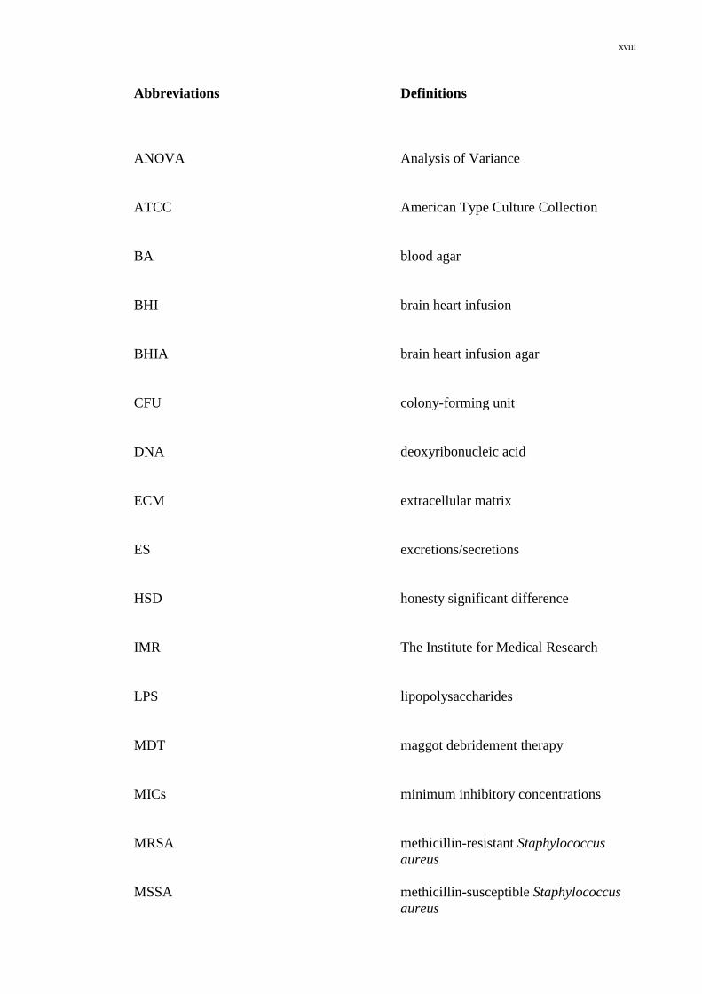
LIST OF SYMBOLS AND ABBREVIATIONS
Symbols Definitions
%
percent
˚C
degree Celsius
µl
microliter
µm
micrometer
BaCl2.2H2O
barium chloride dihydrate
g
gram
H2SO4
sulphuric acid
M
molar
mg
milligram
mg/ml
milligram/mililiter
ml
mililiter
w/v
weight/volume
xviii
Abbreviations Definitions
ANOVA
Analysis of Variance
ATCC
American Type Culture Collection
BA
blood agar
BHI
brain heart infusion
BHIA
brain heart infusion agar
CFU
colony-forming unit
DNA
deoxyribonucleic acid
ECM
extracellular matrix
ES
excretions/secretions
HSD
honesty significant difference
IMR
The Institute for Medical Research
LPS
lipopolysaccharides
MDT
maggot debridement therapy
MICs
minimum inhibitory concentrations
MRSA
MSSA
methicillin-resistant Staphylococcus
aureus
methicillin-susceptible Staphylococcus
aureus
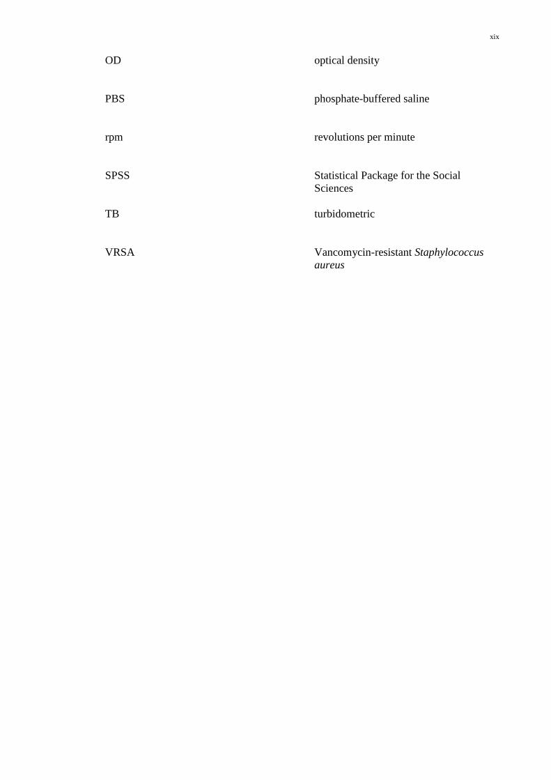
xix
OD
PBS
optical density
phosphate-buffered saline
rpm
revolutions per minute
SPSS
Statistical Package for the Social
Sciences
TB
turbidometric
VRSA
Vancomycin-resistant Staphylococcus
aureus
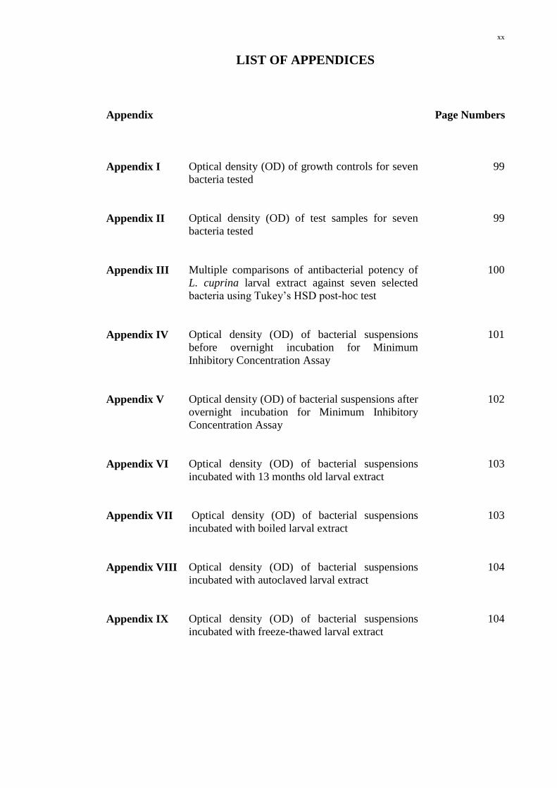
xx
LIST OF APPENDICES
Appendix Page Numbers
Appendix I Optical density (OD) of growth controls for seven
bacteria tested
99
Appendix II Optical density (OD) of test samples for seven
bacteria tested
99
Appendix III Multiple comparisons of antibacterial potency of
L. cuprina larval extract against seven selected
bacteria using Tukey’s HSD post-hoc test
100
Appendix IV Optical density (OD) of bacterial suspensions
before overnight incubation for Minimum
Inhibitory Concentration Assay
101
Appendix V Optical density (OD) of bacterial suspensions after
overnight incubation for Minimum Inhibitory
Concentration Assay
102
Appendix VI Optical density (OD) of bacterial suspensions
incubated with 13 months old larval extract
103
Appendix VII Optical density (OD) of bacterial suspensions
incubated with boiled larval extract
103
Appendix VIII Optical density (OD) of bacterial suspensions
incubated with autoclaved larval extract
104
Appendix IX Optical density (OD) of bacterial suspensions
incubated with freeze-thawed larval extract
104