
In the name of God By: Dr. S. S. Khoramrooz Department of Microbiology, Faculty of Medicine, Yasuj...
191
In the name of God By: Dr. S. S. Khoramrooz Department of Microbiology, Faculty of Medicine, Yasuj University of Medical Sciences, Yasuj, Iran uj University of Medical Sciences Department of Microbiology TYPES OF CULTURE MEDIA IN MEDICAL MICROBIOLOGY
-
Upload
rahul-garfield -
Category
Documents
-
view
214 -
download
0
Transcript of In the name of God By: Dr. S. S. Khoramrooz Department of Microbiology, Faculty of Medicine, Yasuj...

In the name of God
By: Dr. S. S. KhoramroozDepartment of Microbiology, Faculty of Medicine,
Yasuj University of Medical Sciences, Yasuj, Iran
Yasuj University of Medical Sciences
Department of Microbiology
TYPES OF CULTURE MEDIAIN MEDICAL MICROBIOLOGY
Dr. S. S. Khoramrooz 2
BILE ESCULIN AGARIntended Use
Bile Esculin Agar is used to differentiate enterococci and the Streptococcus bovis group from other streptococci.
Principles of the Procedure
Enterococci and certain streptococci hydrolyze the glycoside esculin to esculetin and dextrose.
Esculetin reacts with an iron salt to form a dark brown or black complex.
Ferric citrate is incorporated into the medium as an indicator of esculin hydrolysis and resulting esculetin formation.
Oxgall is used to inhibit gram-positive bacteria other than enterococci.

Dr. S. S. Khoramrooz 3
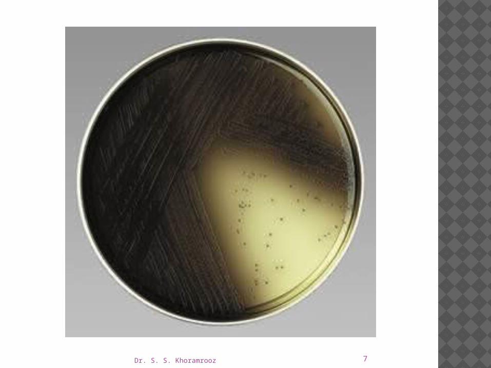
Expected results
Any blackening of the plated medium indicates a positive result; if no blackening occurs, the test is negative.
For slants, if more than half of the slant is blackened within 24-48 hours, the test is positive; if less than half is blackened or no blackening occurs within 24-48 hours, the test is negative.

Dr. S. S. Khoramrooz 4
+

Dr. S. S. Khoramrooz 5

Dr. S. S. Khoramrooz 6
Dr. S. S. Khoramrooz 7

Dr. S. S. Khoramrooz 8
DNASE TEST AGAR
Intended Use DNase Test Agar, DNase Test Agar with Methyl
Green and DNase Test Agar with Toluidine Blue are differential media used for the detection of deoxyribonuclease activity to aid in the identification of bacteria isolated from clinical specimens.
Dr. S. S. Khoramrooz 9
Summary and Explanation
The DNase test is used to detect the degradation of deoxyribonucleic acid (DNA).
The test is useful for differentiating Serratia from Enterobacter, Staphylococcus aureus from coagulase-negative staphylococci, and Moraxella catarrhalis from Neisseria species.
DNase Test Agar with Toluidine Blue contains a metachromatic dye to eliminate the necessity of reagent addition to the agar following incubation.
Toluidine blue may be toxic to some gram-positive cocci and, therefore, should be used primarily with Enterobacteriaceae.

Dr. S. S. Khoramrooz 10
Principles of the Procedure
DNA is the substrate for DNase activity.
DNase is an extracellular enzyme that breaks the DNA down into subunits composed of nucleotides.
The depolymerization of the DNA may be detected by flooding the surface of the medium with 1 N HCl and observing for clear zones in the medium surrounding growth.

Dr. S. S. Khoramrooz 11
In the absence of DNase activity, the reagent reacts with the intact nucleic acid, resulting in the formation of a cloudy precipitate.
The HCl reagent is not needed to detect DNase activity on DNase Agar with Methyl Green.
Methyl green forms a complex with intact (polymerized) DNA to form the green color of the medium.
Dr. S. S. Khoramrooz 12
DNase activity depolymerizes the DNA, breaking down the methyl green-DNA complex, which results in the formation of colorless zones around colonies of the test organism.
A negative test is indicated by the absence of a colorless zone around the colonies.

Dr. S. S. Khoramrooz 13
DNASE TEST AGAR W/TOLUIDINE BLUE
For detection of deoxyribonuclease activity in microorganisms including staphylococci
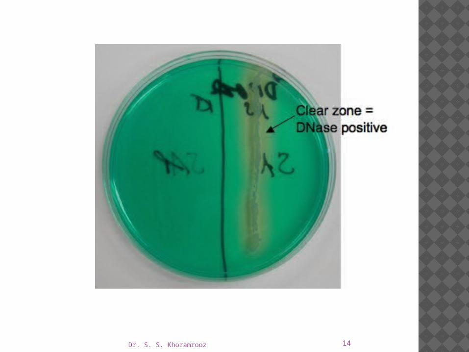
Dr. S. S. Khoramrooz 14
Dr. S. S. Khoramrooz 15
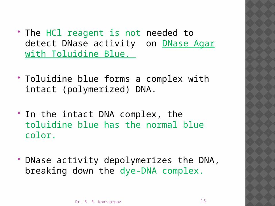
The HCl reagent is not needed to detect DNase activity on DNase Agar with Toluidine Blue.
Toluidine blue forms a complex with intact (polymerized) DNA.
In the intact DNA complex, the toluidine blue has the normal blue color.
DNase activity depolymerizes the DNA, breaking down the dye-DNA complex.
Dr. S. S. Khoramrooz 16
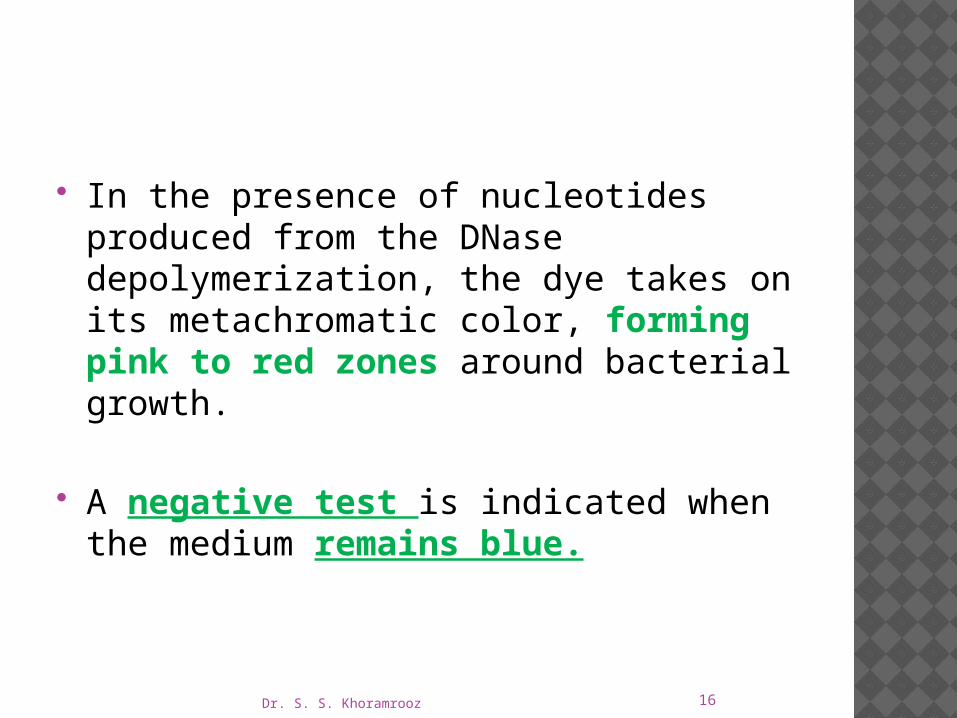
In the presence of nucleotides produced from the DNase depolymerization, the dye takes on its metachromatic color, forming pink to red zones around bacterial growth.
A negative test is indicated when the medium remains blue.
Dr. S. S. Khoramrooz 17

Dr. S. S. Khoramrooz 18

Dr. S. S. Khoramrooz 19
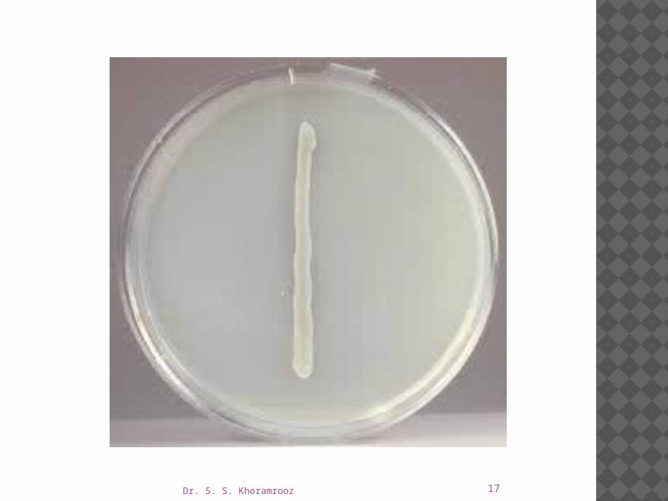
Procedure Inoculate by making a single streak line using
inoculum from an agar slant or plate.
One plate may be inoculated with up to eight isolates by spot inoculation (1/8 to 1/4 inch) or streak inoculation (a single 1- to 2-inch line).
Incubate at 35 ± 2°C for 24-48 hours.

Dr. S. S. Khoramrooz 20
Plates should be incubated in an inverted position. Incubate tubes with loosened caps.
Following incubation, flood DNase Test Agar plates with 1N HCl reagent and observe for reaction.
Reagent addition is not required with DNase Test Agar with Methyl Green or with DNase Test Agar with Toluidine Blue.
Dr. S. S. Khoramrooz 21
Expected Results
A clear area surrounding growth (band/spot inocula) on DNase Test Agar after the addition of 1N HCl indicates a positive reaction, DNase activity.
A negative reaction is indicated by no clearing and a cloudy precipitate around colonies and throughout medium due to precipitated salts in the medium.
A positive reaction on DNase Test Agar with Methyl Green is a distinct clear zone surrounding growth in an otherwise green-colored medium.
The color of the medium remains unchanged if the test is negative.
On DNase Test Agar with Toluidine Blue, DNase activity is indicated by pink to red zones surrounding growth.
The color of the medium remains unchanged if the test is negative.

Dr. S. S. Khoramrooz 22
MANNITOL SALT AGAR
Intended Use Mannitol Salt Agar is used for the selective
isolation and enumeration of staphylococci from clinical and nonclinical materials.

Dr. S. S. Khoramrooz 23
Principles of the Procedure
The 7.5% concentration of sodium chloride results in the partial or complete inhibition of bacterial organisms other than staphylococci.
Mannitol fermentation, as indicated by a change in the phenol red indicator, aids in the differentiation of staphylococcal species.
Agar is a solidifying agent.
Dr. S. S. Khoramrooz 24

Dr. S. S. Khoramrooz 25
Procedure
Incubate plates at 35 ― 2‹C in an aerobic �atmosphere for 24-48 hours, or as instructed in the standard reference.

Dr. S. S. Khoramrooz 26
Expected Results
Coagulase-positive staphylococci produce growth of yellow colonies with yellow zones.
Coagulase negative staphylococci produce small red colonies with no color change to the medium.
Micrococcus produce large, white to orange colonies, with no color change to the medium.
Most other bacteria will be inhibited.

Dr. S. S. Khoramrooz 27

Dr. S. S. Khoramrooz 28
BISMUTH SULFITE AGAR
Intended Use Bismuth Sulfite Agar is a highly selective medium
used for isolating Salmonella spp., particularly Salmonella Typhi, from food and clinical specimens.

Dr. S. S. Khoramrooz 29
Principles of the Procedure
Dextrose is an energy source.
Bismuth sulfite indicator and brilliant green are complementary in inhibiting gram-positive bacteria and members of the coliform group, while allowing Salmonella to grow luxuriantly.
Ferrous sulfate is included for detection of H2S production.
When H2S is present, the iron in the formula is precipitated, giving positive cultures the characteristic brown to black color with metallic sheen.
Dr. S. S. Khoramrooz 30

Dr. S. S. Khoramrooz 31
For isolation of Salmonella spp. from clinical specimens, inoculate fecal specimens and rectal swabs onto a small area of one quadrant of the Bismuth Sulfite Agar plate and streak for isolation.
This will permit the development of discrete colonies.
Incubate plates at 35°C. Examine at 24 hours and again at 48 hours for
colonies resembling Salmonella spp.

Dr. S. S. Khoramrooz 32
Expected results
The typical discrete S. Typhi surface colony is black and surrounded by a black or brownish-black zone which may be several times the size of the colony.
By reflected light, preferably daylight, this zone exhibits a distinctly characteristic metallic sheen.
Plates heavily seeded with S. Typhi may not show this reaction except near the margin of the mass inoculation.

Dr. S. S. Khoramrooz 33
In these heavy growth areas, this organism frequently appears as small light green colonies.
This fact emphasizes the importance of inoculating plates so that some areas are sparsely populated with discrete S. Typhi colonies.
Other strains of Salmonella produce black to green colonies with little or no darkening of the surrounding medium.
Dr. S. S. Khoramrooz 34

Dr. S. S. Khoramrooz 35
Heat with frequent agitation and boil for 1 minute to completely dissolve the powder.
DO NOT AUTOCLAVE.
Use the medium the same day it is prepared.

Dr. S. S. Khoramrooz 36
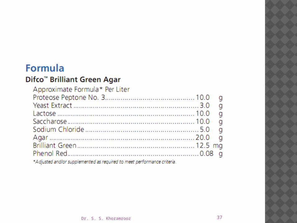
BRILLIANT GREEN AGAR
Intended Use Brilliant Green Agar is a highly selective medium for the
isolation of Salmonella other than S. Typhi from feces and other materials.
Principles of the Procedure Brilliant green dye inhibits gram-positive bacteria and a
majority of gram-negative bacilli.
Phenol red serves as a pH indicator and yields a yellow color as a result of acid production in the fermentation of the lactose and/or sucrose in the medium.
Dr. S. S. Khoramrooz 37
Dr. S. S. Khoramrooz 38
Procedure
A less selective medium and a nonselective medium should also be streaked to increase the chance of recovery when the population of gram-negative organisms is low and to provide an indication of other organisms present in the specimen.
Incubate plates, protected from light, at 35 ± 2°C for 18-24 hours.
If negative after 24 hours, reincubate an additional 24 hours.

Dr. S. S. Khoramrooz 39
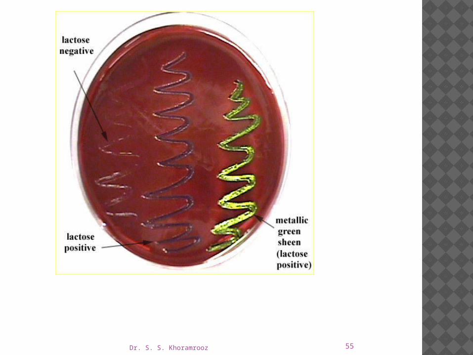
Salmonella on BPLS Agar. The colonies are red because the bacterium does not ferment lactose or sucrose.
Escherichia coli on BPLS Agar. The colonies are yellow due to the low pH which is caused by the production of acid during fermentation of lactose and/or sucrose.

Dr. S. S. Khoramrooz 40
Dr. S. S. Khoramrooz 41
DECARBOXYLASE DIFFERENTIAL MEDIA Intended Use Decarboxylase media are used in the biochemical differentiation
of gram-negative enteric bacilli based on the production:
Arginine dihydrolase Lysine decarboxylase Ornithine decarboxylase
Decarboxylase Medium Base, with added arginine, lysine or ornithine is used for the same purpose.
Lysine Decarboxylase Broth is used for differentiating microorganisms based on lysine decarboxylation.
Dr. S. S. Khoramrooz 42
Summary and Explanation
Moeller introduced the decarboxylase media for detecting the production of lysine and ornithine decarboxylase and arginine dihydrolase.
These media are a useful adjunct to other biochemical tests for the speciation and identification of the Enterobacteriaceae and other gram-negative bacilli.
The production of OD is particularly useful for differentiating Klebsiella and Enterobacter species.
Klebsiella species are non-motile and, except for K. ornithinolytica, do not produce ornithine decarboxylase, while most Enterobacter species are motile and, except for E. agglomerans, usually produce this enzyme.

Dr. S. S. Khoramrooz 43
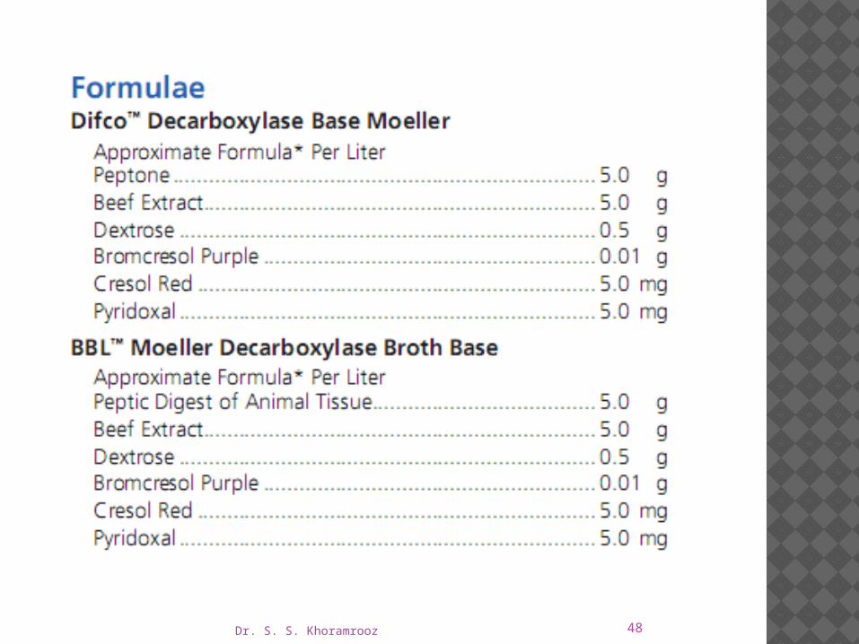
Principles of the Procedure
Pyridoxal is an enzyme co-factor for the amino acid decarboxylase.
Dextrose is a fermentable carbohydrate.
Bromcresol purple and cresol red are pH indicators.
The amino acids lysine, ornithine or arginine are added to the basal medium at a concentration of 10.0 g/L to detect the production of the enzyme specific for these substrates.
Dr. S. S. Khoramrooz 44
When the medium is inoculated with a bacterium that is able to ferment dextrose, acids are produced that lower the pH of the medium and change the color of the indicator from purple to yellow.
The acidic condition also stimulates decarboxylase activity.
If the organism produces the appropriate enzyme, the amino acid in the medium is degraded, yielding a corresponding amine.

Dr. S. S. Khoramrooz 45

Dr. S. S. Khoramrooz 46
Decarboxylation of lysine yields cadaverine.
while decarboxylation of ornithine yields putrescine.
Arginine is first hydrolyzed to form ornithine, which is then decarboxylated to form putrescine.
The production of these amines elevates the pH of the medium, changing the color of the indicator from yellow to purple or violet.
If the organism does not produce the appropriate enzyme, the medium remains acidic (yellow).

Dr. S. S. Khoramrooz 47
Each isolate to be tested must also be inoculated into a tube of the basal medium that does not contain the amino acid.
If this tube becomes alkaline, the test is invalid.
To obtain the appropriate reactions, the inoculated tubes must be protected from air with a layer of sterile mineral oil.
Exposure to air may cause alkalinization at the surface of the medium, which could cause a decarboxylase-negative organism to appear positive.
Dr. S. S. Khoramrooz 48
Dr. S. S. Khoramrooz 49
Expected Results
Compare the color of tubes of media containing the specific amino acids with the color of control tubes of basal media (without amino acid) that have been inoculated with the same isolate.
If inoculated control tubes show an alkaline reaction, the test is invalid; i.e.,
Improperly performed or the test organisms
Degrade the peptone sufficiently to produce an alkaline reaction in the absence of a specific amino acid.
The medium becomes purple to violet if the reaction is positive (alkaline).
A yellow color indicates a negative test; i.e., the organism does not produce the appropriate enzyme.

Dr. S. S. Khoramrooz 50

Dr. S. S. Khoramrooz 51
EOSIN METHYLENE BLUE AGAR
Intended Use
Eosin Methylene Blue Agar, Levine is a slightly selective and differential plating medium for the isolation of gram-negative enteric bacteria.
Principles of the Procedure
The eosin Y and methylene blue dyes in Levine EMB Agar render the medium slightly selective in that they inhibit gram- positive bacteria to a limited degree.

Dr. S. S. Khoramrooz 52
These dyes also play a role in differentiating between lactose fermenters and lactose nonfermenters due to the presence or absence of dye uptake in the bacterial colonies.
Coliforms, as lactose-fermenting organisms, are visualized as blue-black colonies, whereas colonies of Salmonella and Shigella, as lactose nonfermenters, appear colorless, transparent or amber.
Some gram-positive bacteria, such as fecal streptococci, staphylococci and yeasts, will grow on this medium and usually form pinpoint colonies.

Dr. S. S. Khoramrooz 53

Dr. S. S. Khoramrooz 54
Expected Results Typical colonial morphology on Eosin Methylene
Blue Agar, Levine is as follows:
Dr. S. S. Khoramrooz 55
Dr. S. S. Khoramrooz 56
HEKTOEN ENTERIC AGAR
Intended Use
Hektoen Enteric (HE) Agar is a moderately selective medium used in qualitative procedures for the isolation and cultivation of gram-negative enteric microorganisms, especially Shigella, from a variety of clinical and nonclinical specimens.
Dr. S. S. Khoramrooz 57
Principles of the Procedure
The selective nature of Hektoen Enteric Agar is due to the incorporation of bile salts in the formulation.
These substances inhibit gram-positive organisms but also can be toxic for some gram-negative strains.
58
This medium contains three carbohydrates, lactose, sucrose (saccharose) and salicin, for optimal differentiation of enteric pathogens
The lactose concentration is higher than in many other media used for enterics in order to aid in the visualization of enteric pathogens and minimize the problem of delayed lactose fermentation.
Ferric ammonium citrate and sodium thiosulfate in the medium enable the detection of hydrogen sulfide production.
The indicator system, consisting of acid fuchsin and bromthymol blue, has a lower toxicity than that of many other enteric media, resulting in improved recovery of enteric pathogens.
Dr. S. S. Khoramrooz

Dr. S. S. Khoramrooz 59
Procedure A nonselective medium should also be streaked to
increase the chance of recovery when the population of gram-negative organisms is low and to provide an indication of other organisms present in the specimen.
Incubate plates, protected from light, at 35 ± 2°C for 18-24 hours.

Dr. S. S. Khoramrooz 60
DO NOT AUTOCLAVE.

Dr. S. S. Khoramrooz 61

Dr. S. S. Khoramrooz 62

Dr. S. S. Khoramrooz 63
Salmonella mixed with normal fecal flora .

Dr. S. S. Khoramrooz 64

Dr. S. S. Khoramrooz 65
CLED AGAR
Intended Use CLED Agar is used for the isolation, enumeration
and presumptive identification of microorganisms from urine.

Dr. S. S. Khoramrooz 66
Principles of the Procedure
Lactose is included to provide an energy source for organisms capable of utilizing it by a fermentative mechanism.
The cystine permits the growth of “dwarf colony” coliforms.
Bromthymol blue is used as a pH indicator to differentiate lactose fermenters from lactose nonfermenters.
Dr. S. S. Khoramrooz 67
Organisms that ferment lactose will lower the pH and change the color of the medium from green to yellow.
Electrolyte sources are reduced in order to restrict the swarming of Proteus species.

Dr. S. S. Khoramrooz 68
Bacteriuria is determined by inoculating the surface of an agar medium using 0.1 mL of a 102 dilution of the urine sample or using a calibrated loop (0.001 mL) of the undiluted sample.
Current guidelines are that for a single isolate a density of >105 CFU/mL indicates infection, <104 CFU/mL indicates urethral or vaginal contamination, and between 104 and 105 CFU/mL needs to be evaluated based on clinical information.
Dr. S. S. Khoramrooz 69
Procedure Inoculate the medium as soon as possible after the
specimen is received in the laboratory.
It is recommended that quantitative methods be used for culturing urine specimens.
Incubate at 35 ― 2‹C for 24-48 hours.�

Dr. S. S. Khoramrooz 70
Expected Results Count the number of colonies on the plate or dipstick.
Multiply by an appropriate number to convert the count to CFU per mL of sample.
Contaminant bacteria usually appear in low numbers which vary in colonial morphology.
Urinary pathogens will usually yield high counts having uniform colonial morphology and should be subcultured directly to routine media for identification and susceptibility testing.

Dr. S. S. Khoramrooz 71
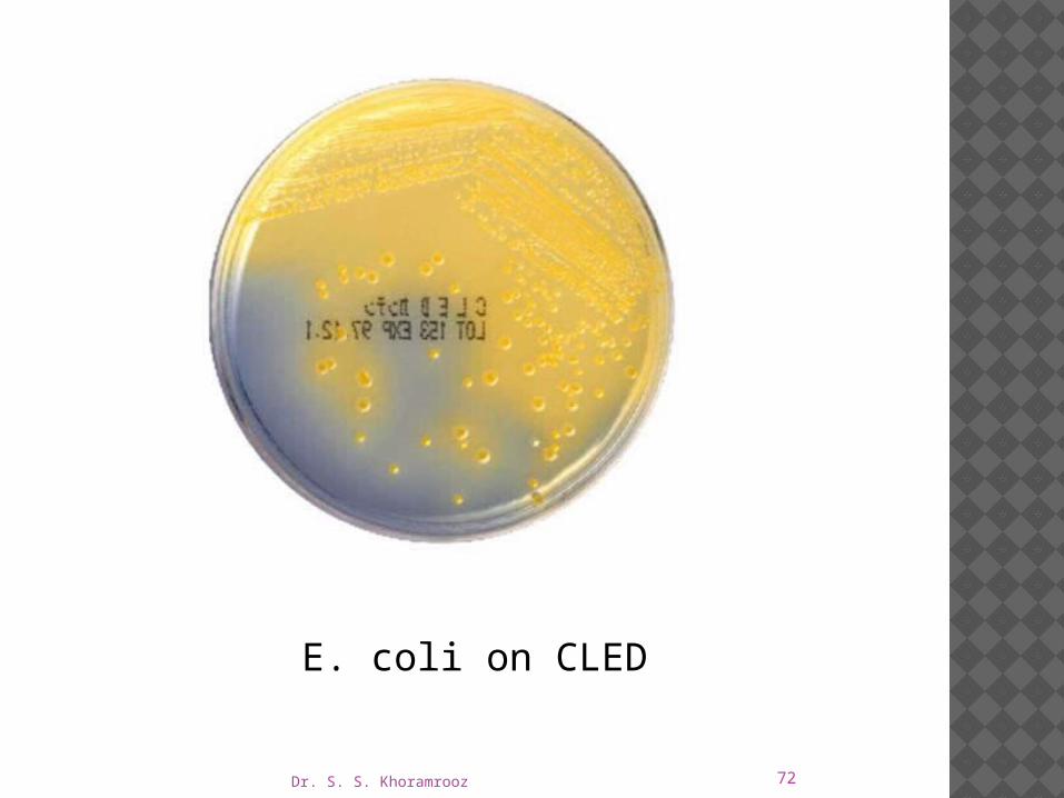
Typical colonial morphology on CLED Agar is as follows:
Dr. S. S. Khoramrooz 72
E. coli on CLED

Dr. S. S. Khoramrooz 73

Dr. S. S. Khoramrooz 74
LAURYL TRYPTOSE BROTH LAURYL SULFATE BROTH
Intended Use Lauryl Tryptose Broth and Lauryl Sulfate Broth,
which are also known as Lauryl Sulfate Tryptose (LST) Broth, are used for the detection of coliform organisms in materials of sanitary importance.
Dr. S. S. Khoramrooz 75
PRINCIPLES OF THE PROCEDURE Lactose provides a source of fermentable
carbohydrate for coliform organisms.
The fermentation of lactose with gas formation is a presumptive test for coliforms.
Sodium lauryl sulfate inhibits organisms other than coliforms.
Dr. S. S. Khoramrooz 76
This medium is used for the detection of coliforms in foods and dairy products.
It is now the medium of choice for use in the presumptive phase of the Standard Total Coliform Multiple-Tube (MPN) Test for the microbiological examination of water.

Dr. S. S. Khoramrooz 77
Dr. S. S. Khoramrooz 78
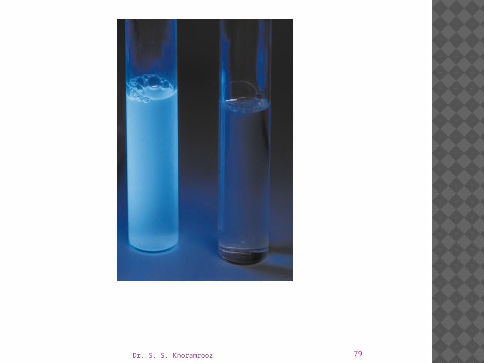
Expected Results After incubation of the tubes with loosened caps at 35 ± 0.5°C
for 24 hours, examine for turbidity and for gas production in the Durham fermentation tubes.
If no gas has formed and been trapped in the inverted tube, reincubate and reexamine after 48 hours.
Turbidity of the medium accompanied by formation of gas in any amount in the Durham tubes within 48 hours is a positive presumptive test for the presence of coliforms in the sample.
The result should be confirmed by additional standard testing.
Dr. S. S. Khoramrooz 79

Dr. S. S. Khoramrooz 80
LYSINE IRON AGAR
Intended Use Lysine Iron Agar is used for the differentiation of
enteric organisms based on their ability to decarboxylate or deaminate lysine and to form hydrogen sulfide.
Principles of the Procedure Dextrose serves as a source of fermentable carbohydrate.
The pH indicator, bromcresol purple, is changed to a yellow color at or below pH 5.2 and is purple at or above pH 6.8.

Dr. S. S. Khoramrooz 81
Ferric ammonium citrate and sodium thiosulfate are indicators of hydrogen sulfide formation.
Lysine is the substrate for use in detecting the enzymes, lysine decarboxylase and lysine deaminase.
Cultures of enteric bacilli that produce hydrogen sulfide cause blackening of the medium due to the production of ferrous sulfides.
Dr. S. S. Khoramrooz 82
Those that produce lysine decarboxylase produce an alkaline reaction (purple color) or neutral reaction in the butt of the medium.
Organisms that deaminate the lysine cause the development of a red slant over an acid butt.
Gas may be formed but its formation is often irregular or suppressed.

Dr. S. S. Khoramrooz 83

Dr. S. S. Khoramrooz 84

Dr. S. S. Khoramrooz 85

Dr. S. S. Khoramrooz 86
Procedure Using an inoculating needle, stab the butt twice
then streak the slant with growth from a pure culture.
Incubate tubes with loosened caps for 18-48 hours at 35 ± 2°C in an aerobic atmosphere.
Triple Sugar Iron Agar slants should be inoculated in parallel unless results from this medium have already been obtained to distinguish coliforms from Shigella, for example.
Dr. S. S. Khoramrooz 87
Expected Results
Lysine decarboxylation is detected in the butt by an alkaline (purple) reaction.
Lysine deamination is detected by a red slant.
Hydrogen sulfide production is detected by the formation of a black precipitate.
A negative reaction (purple slant and yellow butt) indicates fermentation of dextrose only.

Dr. S. S. Khoramrooz 88
Hydrogen sulfide may not be detected in this medium by organisms that are negative for lysine decarboxylase activity since acid production in the butt may suppress its formation.
For this reason H2S-producing Proteus species do not blacken this medium

Dr. S. S. Khoramrooz 89
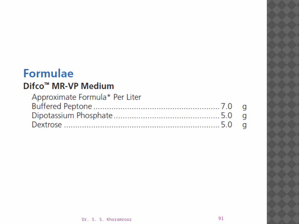
MR-VP MEDIUM • MR-VP BROTHIntended Use MR-VP Medium and MR-VP Broth (Methyl Red-
Voges Proskauer Medium/Broth, also known as Buffered Peptone- Glucose Broth) are used for the differentiation of bacteria by means of the methyl red and Voges-Proskauer reactions.

Dr. S. S. Khoramrooz 90
Principles of the Procedure
Methyl red-positive organisms produce high levels of acid during fermentation of dextrose, overcome the phosphate buffer system and produce a red color upon the addition of the methyl red pH indicator.
In the Voges-Proskauer test, the red color produced by the addition of potassium hydroxide to cultures of certain microbial species is due to the ability of the organisms to produce a neutral end product, acetoin (acetylmethylcarbinol), from the fermentation of dextrose.
The acetoin is oxidized in the presence of oxygen and alkali to produce a red color.
This is a positive Voges-Proskauer reaction.
Dr. S. S. Khoramrooz 91
Dr. S. S. Khoramrooz 92
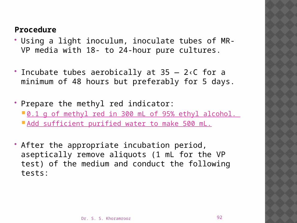
Procedure Using a light inoculum, inoculate tubes of MR-VP media with
18- to 24-hour pure cultures.
Incubate tubes aerobically at 35 ― 2‹C for a minimum of 48 �hours but preferably for 5 days.
Prepare the methyl red indicator: 0.1 g of methyl red in 300 mL of 95% ethyl alcohol. Add sufficient purified water to make 500 mL.
After the appropriate incubation period, aseptically remove aliquots (1 mL for the VP test) of the medium and conduct the following tests:
Dr. S. S. Khoramrooz 93
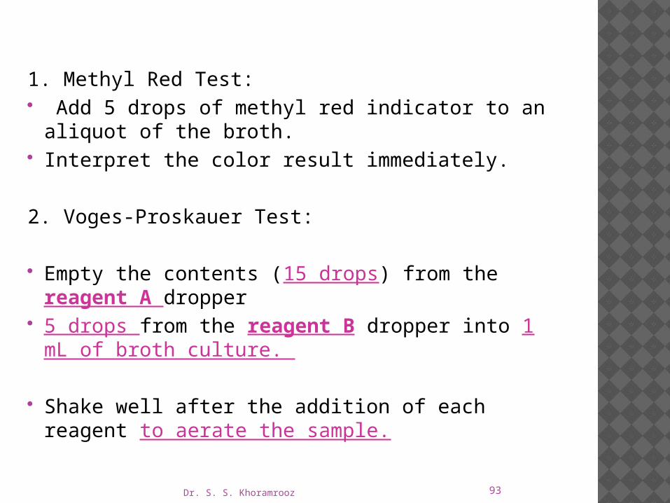
1. Methyl Red Test: Add 5 drops of methyl red indicator to an aliquot of the
broth. Interpret the color result immediately.
2. Voges-Proskauer Test:
Empty the contents (15 drops) from the reagent A dropper 5 drops from the reagent B dropper into 1 mL of broth
culture.
Shake well after the addition of each reagent to aerate the sample.

Dr. S. S. Khoramrooz 94
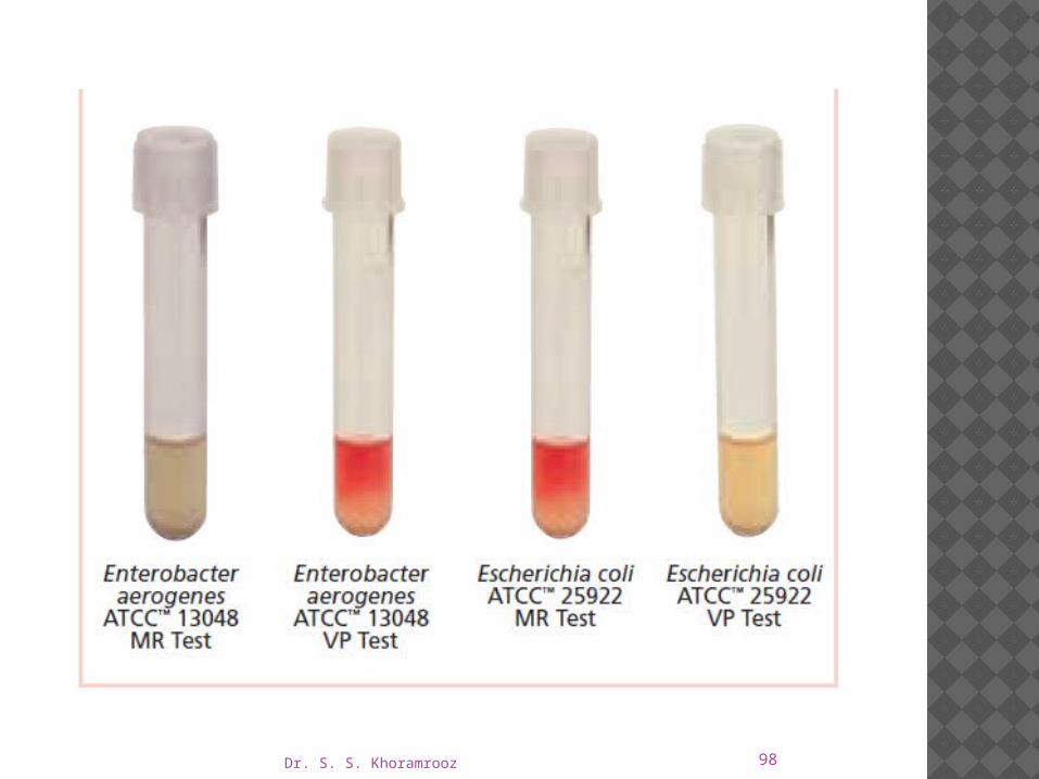
Expected Results 1. Methyl Red Test a. Positive – red color at surface of the medium. b. Negative – yellow color at surface of the medium.
2. Voges-Proskauer Test A positive reaction is indicated by the development of a
distinct red color which occurs within 5 minutes.
Certain species within Enterobacteriaceae genera may react differently or give variable results.
Consult appropriate texts for reactions of specific species.
Dr. S. S. Khoramrooz 95
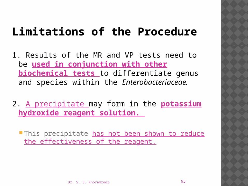
Limitations of the Procedure
1. Results of the MR and VP tests need to be used in conjunction with other biochemical tests to differentiate genus and species within the Enterobacteriaceae.
2. A precipitate may form in the potassium hydroxide reagent solution.
This precipitate has not been shown to reduce the effectiveness of the reagent.
Dr. S. S. Khoramrooz 96
3. Most members of the family Enterobacteriaceae give either a positive MR test or a positive VP test.
However, certain organisms such as Hafnia alvei and Proteus mirabilis may give a positive result for both tests.
4. Incubation time for the Methyl Red test cannot be shortened by increasing the dextrose concentration in the medium or by heavily inoculating the broth.
5. Incubate MR-negative tests for more than 48 hours and test again.

Dr. S. S. Khoramrooz 97
6. Read the VP test at 48 hours. Increased incubation may produce acid conditions in the broth that will interfere with reading the results.
7. VP reagents must be added in the order and the amounts specified or a weak-positive or false-negative reaction may occur.
A weak-positive reaction may be masked by a copper-like color which may form due to the reaction of KOH and α-naphthol.
8. Read the VP test within 1 hour of adding the reagents. The KOH and α-naphthol may react to form a copper-like color, causing a potential false-positive interpretation.
9. Due to the possible presence of acetoin, diacetyl or related substances in certain raw materials, the use of media low in these substances (such as MR-VP media) is recommended for this test.
Dr. S. S. Khoramrooz 98
Dr. S. S. Khoramrooz 99
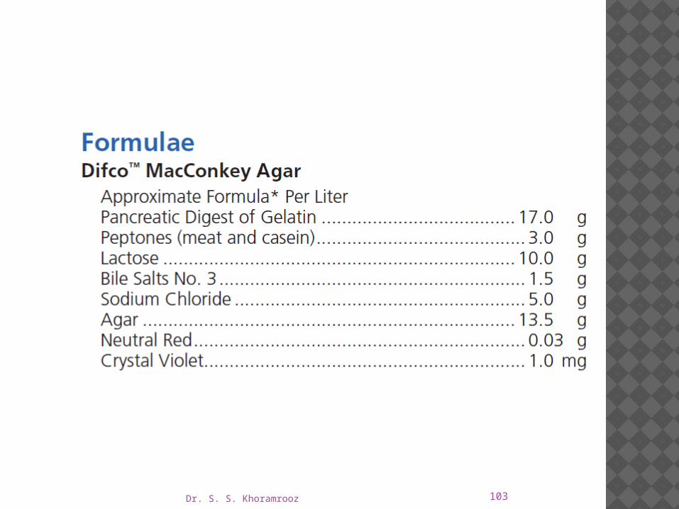
MACCONKEY AGARS
Intended Use MacConkey agars are slightly selective and differential
plating media mainly used for the detection and isolation of gram-negative organisms from clinical, dairy, food, water, pharmaceutical, cosmetic, and other industrial sources.
MacConkey Agar is used for isolating and differentiating lactose-fermenting from lactose-nonfermenting gram-negative enteric bacilli.
MacConkey Agar Base is used with added carbohydrate in differentiating coliforms based on fermentation reactions.
Dr. S. S. Khoramrooz 100
MacConkey Agar without Crystal Violet is used for isolating and differentiating enteric microorganisms while permitting growth of staphylococci and enterococci.
The medium can be used also to separate Mycobacterium fortuitum and M. chelonae from other rapidly growing mycobacteria.
MacConkey Agar without Crystal Violet or Salt and MacConkey Agar without Salt are used for isolating and differentiating gram-negative bacilli while suppressing the swarming of most Proteus species.
Dr. S. S. Khoramrooz 101
Principles of the Procedure
Lactose is a fermentable carbohydrate.
When lactose is fermented, a local pH drop around the colony cause a color change in the pH indicator (neutral red) and bile precipitation.

Dr. S. S. Khoramrooz 102
Bile salts, bile salts no. 3, oxgall and crystal violet are selective agents that inhibit growth of gram-positive organisms.
Magnesium sulfate is a source of divalent cations. Agar is the solidifying agent.
Dr. S. S. Khoramrooz 103

Dr. S. S. Khoramrooz 104
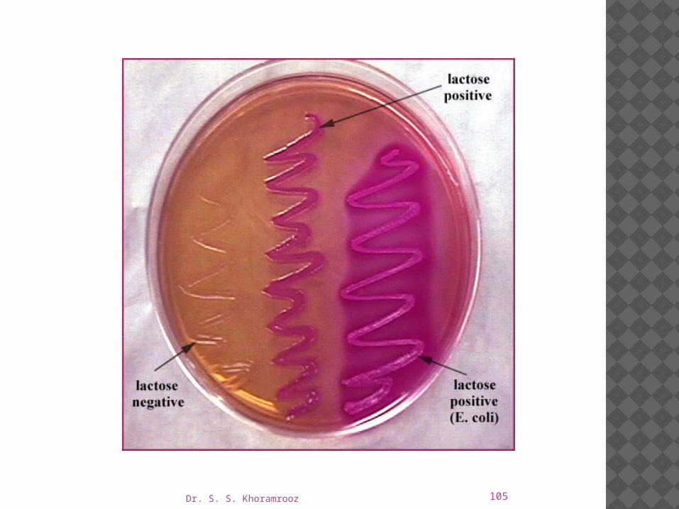
Expected Results Lactose-fermenting organisms grow as pink to
brick-red colonies with or without a zone of precipitated bile.
Lactose-nonfermenting organisms grow as colorless or clear colonies.
Swarming by Proteus spp. is reduced on MacConkey agars without salt.
Dr. S. S. Khoramrooz 105

Dr. S. S. Khoramrooz 106
Limitations of the Procedure
1. Although MacConkey media are selective primarily for gram-negative enteric bacilli, biochemical and, if indicated, serological testing using pure cultures are recommended for complete identification.
2. Incubation of MacConkey Agar plates under increased CO2 has been reported to reduce the growth and recovery of a number of strains of gram-negative bacilli.

Dr. S. S. Khoramrooz 107
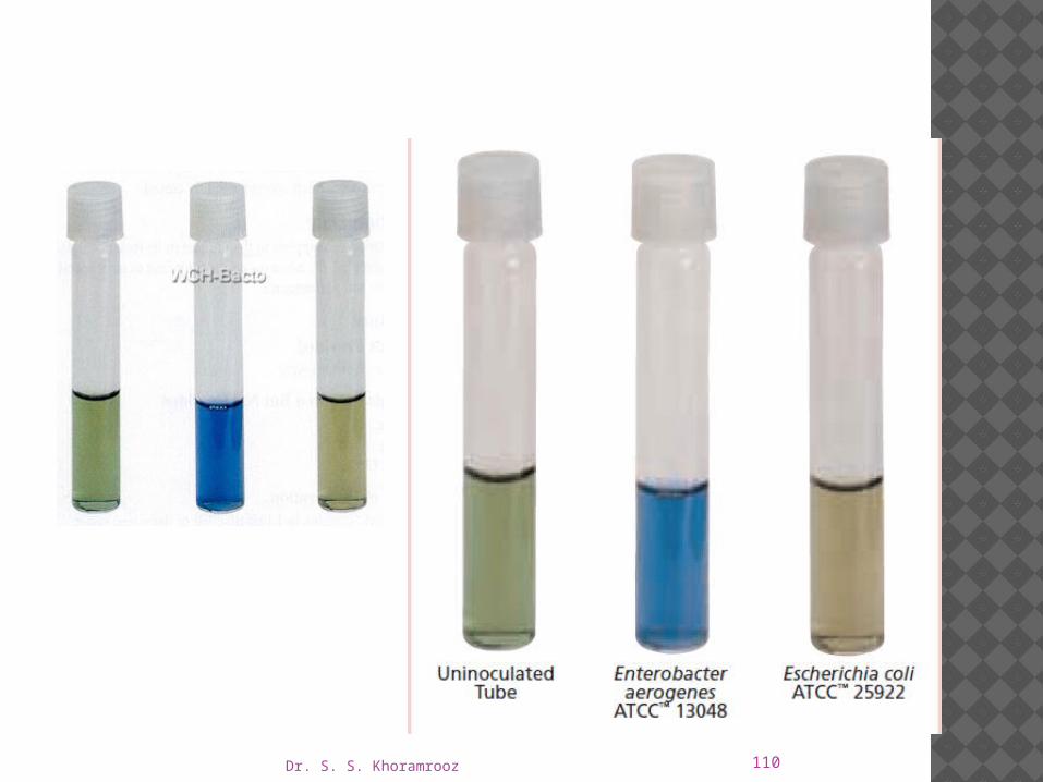
MALONATE BROTHIntended Use
Malonate Broth is used for differentiating Enterobacter from Escherichia based on malonate utilization.
Principles of the Procedure
Malonate Broth contains ammonium sulfate, which is the sole source of nitrogen in the medium;
Sodium malonate is the sole source of carbon.
Increased alkalinity resulting from malonate utilization causes the indicator, bromthymol blue, to change color from green to blue.

Dr. S. S. Khoramrooz 108

Dr. S. S. Khoramrooz 109
Procedure
1. Inoculate tubes with a loopful of test organism.
2. Incubate at 35 ― 2‹C for 18-48 hours.�3. Examine tubes for a change in the color of the
medium from green to blue.
Expected Results Malonate utilization is indicated by a change in the
color of the medium from green to blue: Positive: Blue Negative: Green
Dr. S. S. Khoramrooz 110
Dr. S. S. Khoramrooz 111
OF BASAL MEDIUM Intended Use
OF (Oxidation Fermentation) media are used for the determination of oxidative and fermentative metabolism of carbohydrates by gram-negative rods on the basis of acid reaction in either the open or closed system.
Summary and Explanation OF Medium was developed by Hugh and Leifson who
described the taxonomic significance of fermentative versus oxidative metabolism of carbohydrates by gram-negative bacteria.
Dr. S. S. Khoramrooz 112
They showed that when an organism is inoculated into two tubes of OF Basal Medium containing a carbohydrate and the medium in one of the tubes is covered with melted petrolatum prior to incubation, the patterns of metabolism are of differential significance.
Oxidative organisms only produce an acid reaction in the open tube with little or no growth and no acid formation in the covered tube.
Fermentative organisms will produce an acid reaction in both types of tubes.
Changes in the covered agar are considered to be due to true fermentation, while changes in the open tubes are due to oxidative utilization of the carbohydrate present.
If the carbohydrate is not utilized by either method, there is no acid production in either tube.

Dr. S. S. Khoramrooz 113
Principles of the Procedure Dextrose is the most important carbohydrate for
use in OF Basal Medium; however, certain organisms may metabolize other carbohydrates even if they are unable to utilize dextrose.
Prepared tubed media containing arabinose, dextrose, dulcitol, fructose, galactose, lactose, maltose, mannose, raffinose, rhamnose, salicin, sorbitol, sucrose and xylose are provided.

Dr. S. S. Khoramrooz 114

Dr. S. S. Khoramrooz 115
Procedure
Inoculate a pair of OF tubes of each carbohydrate used with each organism being tested.
The tubes should be stabbed to approximately 1/4 inch from the bottom using an inoculating needle and a light inoculum.
Overlay one tube of each pair with sterile mineral oil.
Incubate tubes at 35 ± 2°C in an aerobic atmosphere for 48 hours.
Do not discard as negative until after 4 days of incubation.
Dr. S. S. Khoramrooz 116
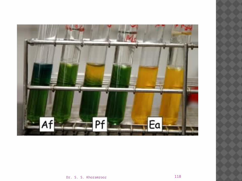
Expected Results Record results as acid (A) or alkaline/no change (–).
Also record whether or not the organism is motile as evidenced by the appearance of growth away from the line of inoculation.
Typical reaction patterns are as follows.
Dr. S. S. Khoramrooz 117
Limitations of the Procedure
1. The acid reaction produced by oxidative organisms is apparent at the surface and gradually spreads throughout the medium.
If the oxidation is weak or slow, however, an initial alkaline reaction at the surface of the open tube may persist for several days and eventually convert to an acid reaction.
2. If an organism is unable to grow on OF Basal Medium, Cowan recommends adding either 2% serum or 0.1% yeast extract to each carbohydrate tube.
Dr. S. S. Khoramrooz 118

Dr. S. S. Khoramrooz 119
SIM MEDIUM
Intended Use
SIM Medium is used to differentiate enteric bacilli on the basis of sulfide production,indole formation and motility.
Dr. S. S. Khoramrooz 120
Summary and Explanation
Hydrogen sulfide production, indole formation and motility are distinguishing characteristics which aid in the identification of the Enterobacteriaceae, especially Salmonella and Shigella.
SIM Medium, therefore ,is useful in the process of identification of enteric pathogens.

Dr. S. S. Khoramrooz 121
Principles of the Procedure
Sodium thiosulfate and ferrous ammonium sulfate are indicators of hydrogen sulfide production.
The ferrous ammonium sulfate reacts with H2S gas to produce ferrous sulfide,a black precipitate.
The casein peptone is rich in tryptophan,which is attacked by certain microorganisms resulting in the production of indole.
The indole is detected by the addition of chemical reagents following the incubation period.
Dr. S. S. Khoramrooz 122
Motility detection is possible due to the semisolid nature of the medium.
Growth radiating out from the central stab line indicates that the test organism is motile.

Dr. S. S. Khoramrooz 123
Dr. S. S. Khoramrooz 124
Procedure Loosen caps, boil and cool before use.
Using growth from a pure culture, stab an inoculating needle two-thirds of the distance to the bottom in the center of the tube.
Incubate tubes with loosened caps for18-24 hours at 35±2°C in anaerobic atmosphere.

Dr. S. S. Khoramrooz 125

Dr. S. S. Khoramrooz 126
SS AGAR Intended Use SS Agar and Salmonella Shigella Agar are
moderately selective and differential media for the isolation of pathogenic enteric bacilli, especially those belonging to the genus Salmonella.
This formulation is not recommended for the primary isolation of Shigella.
Dr. S. S. Khoramrooz 127
Principles of the Procedure
SS Agar and Salmonella Shigella Agar are designated as moderately selective media based upon the degree of inhibition of gram-positive microorganisms that they inhibit due to their content of bile salts, brilliant green and citrates.
Differentiation of enteric organisms is achieved by the incorporation of lactose in the medium.
Organisms that ferment lactose produce acid which, in the presence of the neutral red indicator, results in the formation of red colonies.
Lactose nonfermenters form colorless colonies.

Dr. S. S. Khoramrooz 128
The latter group contains the majority of the intestinal pathogens, including Salmonella and Shigella.
The sodium thiosulfate and ferric citrate enable the detection of hydrogen sulfide production as evidenced by colonies with black centers.
Dr. S. S. Khoramrooz 129
Procedure
A nonselective medium should also be streaked to increase the chance of recovery when the population of gram-negative organisms is low and to provide an indication of other organisms present in the specimen.
Incubate plates, protected from light, at 35 ± 2°C for 18-24 hours.
If negative after 24 hours, reincubate an additional 24 hours.

Dr. S. S. Khoramrooz 130

Dr. S. S. Khoramrooz 131
Expected Results Typical colonial morphology on Salmonella
Shigella Agar is as follows:
Dr. S. S. Khoramrooz 132

Dr. S. S. Khoramrooz 133
Limitation of the Procedure Due to the relatively high level of selectivity, some
Shigella strains may not grow on SS Agar and Salmonella Shigella Agar and, therefore, these media are not recommended for the primary isolation of Shigella.
Media recommended for the isolation of Shigella are Hektoen Enteric and XLD agars.

Dr. S. S. Khoramrooz 134
SELENITE BROTH • SELENITE-F BROTH
Intended Use Selenite Broth (Selenite-F Broth) is used as an
enrichment medium for the isolation of Salmonella from feces, urine, water, foods and other materials of sanitary importance.
Principles of the Procedure The peptone provides essential nitrogenous and carbon
compounds.
The lactose in the medium serves to maintain a uniform pH.

Dr. S. S. Khoramrooz 135
When selenite is reduced by the growth of bacteria, alkali is produced, and such increase in pH would lessen the toxicity of the selenite and result in overgrowth of extraneous bacteria.
The acid produced by lactose fermentation serves to maintain a neutral or slightly decreased pH.
The function of the phosphate is two-fold; it serves to maintain a stable pH and lessens the toxicity of the selenite, thus increasing the capacity of the medium.
Sodium selenite inhibits many species of grampositive and gram-negative bacteria including enterococci and coliforms.

Dr. S. S. Khoramrooz 136

Dr. S. S. Khoramrooz 137
Dr. S. S. Khoramrooz 138
Procedure
For feces and other solid materials, suspend 1-2 g of the specimen in the broth (approximately 10-15% by volume) and emulsify with an inoculating needle, if necessary.
Incubate tubes with loosened caps at 35 ± 2°C for up to 24 hours.
Subcultures should be made after 12-18 hours of incubation, if possible.
Coliforms will tend to overgrow the pathogens if incubated longer than 24 hours.
Dr. S. S. Khoramrooz 139
Expected Results After incubation, there should be an increase in the
number of pathogens that the medium is designed to select for and enrich.
Subculture onto appropriate selective and differential media (e.g., MacConkey Agar, Hektoen Enteric Agar, XLD Agar, XLT4 Agar, CHROMagar™ Salmonella) to isolate pathogens for identification.

Dr. S. S. Khoramrooz 140
Limitation of the Procedure Enrichment broths should not be used as the sole
isolation medium.
They are to be used in conjunction with selective and nonselective plating media to increase the probability of isolating pathogens, especially when they may be present in small numbers.
Dr. S. S. Khoramrooz 141
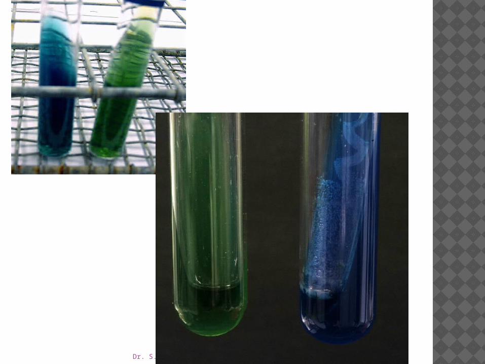
SIMMONS CITRATE AGAR
Intended Use Simmons Citrate Agar is used for the differentiation of
gram negative bacteria on the basis of citrate utilization.
Principles of the Procedure Organisms able to utilize ammonium dihydrogen
phosphate and sodium citrate as the sole sources of nitrogen and carbon, respectively, will grow on this medium and produce an alkaline reaction as evidenced by a change in the color of the bromthymol blue indicator from green (neutral) to blue (alkaline).

Dr. S. S. Khoramrooz 143
Procedure
Inoculate slants with growth from a pure culture using a light inoculum.
Incubate all tubes for 4 days at 35 ± 2°C in an aerobic atmosphere.
Expected Results
A positive reaction is indicated by growth with an intense blue color in the slant.
A negative reaction is evidenced by no growth to trace growth with no change in color (medium remains dark green).
Dr. S. S. Khoramrooz 144
Dr. S. S. Khoramrooz 145
TRIPLE SUGAR IRON AGAR TSI AGAR
Intended Use
Triple Sugar Iron Agar (TSI Agar) is used for the differentiation of gram-negative enteric bacilli based on carbohydrate fermentation and the production of hydrogen sulfide.
Principles of the Procedure
TSI Agar contains three sugars (dextrose, lactose and sucrose),
Phenol red for detecting carbohydrate fermentation
Dr. S. S. Khoramrooz 146
Ferrous ammonium sulfate for detection of hydrogen sulfide production (indicated by blackening in the butt of the tube).
Carbohydrate fermentation is indicated by the production of gas and a change in the color of the pH indicator from red to yellow.
To facilitate the detection of organisms that only ferment dextrose, the dextrose concentration is one-tenth the concentration of lactose or sucrose.
The small amount of acid produced in the slant of the tube during dextrose fermentation oxidizes rapidly, causing the medium to remain red or revert to an alkaline pH.
In contrast, the acid reaction (yellow) is maintained in the butt of the tube because it is under lower oxygen tension.

Dr. S. S. Khoramrooz 147
After depletion of the limited dextrose, organisms able to do so will begin to utilize the lactose or sucrose.
To enhance the alkaline condition of the slant, free exchange of air must be permitted by closing the tube cap loosely.
If the tube is tightly closed, an acid reaction (caused solely by dextrose fermentation) will also involve the slant.

Dr. S. S. Khoramrooz 148
Dr. S. S. Khoramrooz 149
Procedure
To inoculate, carefully touch only the center of an isolated colony on an enteric plated medium with a cool, sterile needle, stab into the medium in the butt of the tube, and then streak back and forth along the surface of the slant.
Several colonies from each primary plate should be studied separately, since mixed infections may occur.

Dr. S. S. Khoramrooz 150
Incubate with caps loosened at 35°C and examine after 18-24 hours for carbohydrate fermentation, gas production and hydrogen sulfide production.
Any combination of these reactions may be observed.
Do not incubate longer than 24 hours because the acid reaction in the slant of lactose and sucrose fermenters may revert to an alkaline reaction.

Dr. S. S. Khoramrooz 151

Dr. S. S. Khoramrooz 152
Expected Results
Carbohydrate fermentation is indicated by a yellow coloration of the medium.
If the medium in the butt of the tube becomes yellow (acidic), but the medium in the slant becomes red (alkaline), the organism being tested only ferments dextrose (glucose).
A yellow (acidic) color in the slant and butt indicates that the organism being tested ferments dextrose, lactose and/or sucrose.
A red (alkaline) color in the slant and butt indicates that the organism being tested is a nonfermenter.

Dr. S. S. Khoramrooz 153
Hydrogen sulfide production results in a black precipitate in the butt of the tube.
Gas production is indicated by splitting and cracking of the medium.
Dr. S. S. Khoramrooz 154
LIMITATIONS OF THE PROCEDURE
1. Hydrogen sulfide production may be evident on Kligler Iron Agar but negative on Triple Sugar Iron Agar.
Studies by Bulmash and Fulton showed that the utilization of sucrose could suppress the enzymatic mechanisms responsible for H2S production.
Padron and Dockstader8 found that not all H2S-positive Salmonella are positive on TSI.
2. Sucrose is added to TSI to eliminate some sucrose-fermenting lactose-nonfermenters such as Proteus and Citrobacter spp.
3. Further biochemical tests and serological typing must be performed for definite identification and confirmation of organisms.
Dr. S. S. Khoramrooz 155
4. Do not use an inoculating loop to inoculate a tube of Triple Sugar Iron Agar.
While stabbing the butt, mechanical splitting of the medium occurs, causing a false positive result for gas production.
5. A pure culture is essential when inoculating Triple Sugar Iron Agar.
If inoculated with a mixed culture, irregular observations may occur.
6. Tubes should be incubated with caps loosened.
This allows a free exchange of air, which is necessary to enhance the alkaline condition on the slant.
Dr. S. S. Khoramrooz 156
UREA MEDIAUREA AGAR • UREA BROTHIntended Use Urea Agar and Urease Test Broth are used for the
differentiation of organisms, especially the Enterobacteriaceae, on the basis of urease production.

Dr. S. S. Khoramrooz 157
Principles of the Procedure
The urea medium of Rustigian and Stuart is particularly suited for the differentiation of Proteus species from other gram negative enteric bacilli capable of utilizing urea.
Unable to do so in Urease Test Broth because of limited nutrients and the high buffering capacity of the medium.
To provide a medium with greater utility, Urea Agar was devised by Christensen with peptone and dextrose included and reduced buffer content to promote more rapid growth of many of the Enterobacteriaceae and permit a reduction in incubation time.

Dr. S. S. Khoramrooz 158
The complete Urea Agar contains 15.0 g/L of agar in addition to the ingredients in the base medium.
When organisms utilize urea, ammonia is formed during incubation which makes the reaction of these media alkaline, producing a red-pink color.
Consequently, urease production may be detected by the change in the phenol red indicator.

Urease medium

Dr. S. S. Khoramrooz 160
161
DIRECTIONS FOR PREPARATION FROM
Dehydrated Product BBL™ Urea Agar Base
1. Dissolve 29 g of the powder in 100 mL of purified water. Mix thoroughly. Sterilize by filtration.
2. Suspend 15 g of agar in 900 mL of purified water.
3. Autoclave at 121°C for 15 minutes.
4. Cool to 50°C and add 100 mL of the sterile Urea Aga Base.
5. Mix thoroughly and dispense aseptically in sterile tubes.
6. Cool tubed medium in a slanted position so that deep butts are formed.
7. Do not remelt the complete medium.
8. Test samples of the finished product for performance using stable, typical control cultures.
Dr. S. S. Khoramrooz
Dr. S. S. Khoramrooz 162
Procedure
Using a heavy inoculum (2 loopfuls) of growth from an 18- to 24-hour pure culture (TSI Agar or other suitable medium), inoculate the broth or agar (streaking back and forth over the entire slant surface).
Dr. S. S. Khoramrooz 163
Do not stab the butt since it serves as a color control.
For broth, shake tubes gently to suspend the bacteria.
Incubate tubes with loosened caps at 35 ― 2‹C in an �incubator or water bath.
Observe reactions after 2, 4, 6, 18, 24 and 48 hours.
For agar, continue to check every day for a total of 6 days; even longer incubation periods may be necessary.

Dr. S. S. Khoramrooz 164
EXPECTED RESULTS
The production of urease is indicated by an intense pink-red (red-violet) color on the slant or throughout the broth.
The color may penetrate into the agar (butt); the extent of the color indicates the rate of urea hydrolysis.

Dr. S. S. Khoramrooz 165
A negative reaction is no color change.
The agar medium remains pale yellow to buff; the broth remains yellowish orange.
Dr. S. S. Khoramrooz 166
LIMITATIONS OF THE PROCEDURE
Urea Agar Base
1. The alkaline reaction produced in this medium after prolonged incubation may not be caused by urease activity.
False positive reactions may occur due to the utilization of peptones (especially in slant agar by Pseudomonas aeruginosa, for example) or other proteins which raise the pH due to protein hydrolysis and the release of excessive amino acid residues.
To eliminate possible protein hydrolysis, perform a control test with the same test medium without urea.
2. Do not heat or reheat the medium because urea decomposes very easily.

Dr. S. S. Khoramrooz 167
3. Urea Agar detects rapid urease activity of only the urease positive Proteus species.
For results to be valid for the detection of Proteus, the results must be read within the first 2-6 hours after incubation.
Urease-positive Enterobacter, Citrobacter or Klebsiella, in contrast, hydrolyze urea much more slowly, showing only slight penetration of the alkaline reaction into the butt of the medium in 6 hours and requiring 3-5 days to change the reaction of the entire butt.

Dr. S. S. Khoramrooz 168
Urea Broth
1. To rule out false positives due to protein hydrolysis (as opposed to urea hydrolysis) that may occur in the medium after prolonged incubation, perform a control test with the same test medium without urea.
2. Do not heat or reheat the medium because urea decomposes very easily.
Dr. S. S. Khoramrooz 169
3. The high buffering system in this medium masks urease activity in organisms that are delayed positive.
This medium is therefore recommended for the detection of urease activity in all Proteus spp., Providencia rettgeri and urease-positive Providencia stuartii.
M. morganii slowly hydrolyzes urea and may require approximately a 36 hour incubation for a strong urease-positive reaction to occur.
If in doubt as to a result, compare with an uninoculated tube or incubate for an additional 24 hours.
4. Variations in the size of the inoculum can affect the time required to reach positive (alkaline, pH 8.1) results.
Dr. S. S. Khoramrooz 170
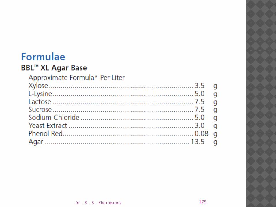
XLD AGARIntended Use XL (Xylose Lysine) Agar Base is used for the
isolation and differentiation of enteric pathogens and, when supplemented with appropriate additives, as a base for selective enteric media.
XLD Agar is the complete Xylose Lysine Desoxycholate Agar, a moderately selective medium recommended for isolation and differentiation of enteric pathogens, especially Shigella species.

Dr. S. S. Khoramrooz 171
Principles of the Procedure
Xylose is incorporated into the medium because it is fermented by practically all enterics except for the shigellae.
This property enables the differentiation of Shigella species.
Lysine is included to enable the Salmonella group to be differentiated from the nonpathogens.
Without lysine, salmonellae rapidly would ferment the xylose and be indistinguishable from nonpathogenic species.
172
After the salmonellae exhaust the supply of xylose, the lysine is attacked via the enzyme lysine decarboxylase, with reversion to an alkaline pH, which mimics the Shigella reaction.
To prevent similar reversion by lysine-positive coliforms, lactose and sucrose (saccharose) are added to produce acid in excess.
Degradation of xylose, lactose and sucrose generates acid products, which in the presence of the pH indicator phenol red, causes a color change in the medium from red to yellow.
Dr. S. S. Khoramrooz

Dr. S. S. Khoramrooz 173
To add to the differentiating ability of the formulation, an H2S indicator system, consisting of sodium thiosulfate and ferric ammonium citrate, is included for the visualization of the hydrogen sulfide produced, resulting in the formation of colonies with black centers.
The nonpathogenic H2S producers do not decarboxylate lysine; therefore, the acid reaction produced by them prevents the blackening of the colonies.
Dr. S. S. Khoramrooz 174
XLD Agar is both a selective and differential medium.
It utilizes sodium desoxycholate as the selective agent and, therefore, it is inhibitory to gram-positive microorganisms.
Dr. S. S. Khoramrooz 175

Dr. S. S. Khoramrooz 176

Dr. S. S. Khoramrooz 177
Expected Results
Degradation of xylose, lactose and sucrose generates acid products, causing a color change in the medium from red to yellow.
Hydrogen sulfide production under alkaline conditions causes colonies to develop black centers.
This reaction is inhibited by the acid conditions that accompany carbohydrate fermentation.
Lysine decarboxylation in the absence of lactose and sucrose fermentation causes reversion to an alkaline condition and the color of the medium changes back to red.
Typical colonial morphology and reactions on XLD Agar are as follows:

Dr. S. S. Khoramrooz 178

Dr. S. S. Khoramrooz 179

Dr. S. S. Khoramrooz 180

Dr. S. S. Khoramrooz 181
Limitations of the Procedure
1. Red, false-positive colonies may occur with some Proteus and Pseudomonas species.
2. Incubation in excess of 48 hours may lead to false-positive results.
3. S. Paratyphi A, S. Choleraesuis, S. pullorum and S. gallinarum may form red colonies without black centers, thus resembling Shigella species.
4. Some Proteus strains will give black-centered colonies on XLD Agar.

Dr. S. S. Khoramrooz 182
TRANSPORT MEDIUM
Intended Use
Transport Medium Amies and Transport Medium (Stuart, Toshach and Patsula) are used for collecting, transporting and preserving microbiological specimens.
Cary and Blair Transport Medium is used for collecting, transporting and preserving microbiological specimens, particularly those containing Vibrio cholerae.

Dr. S. S. Khoramrooz 183
Summary and Explanation
Transport media are chemically defined, semisolid, nonnutritive, phosphate buffered media that provide a reduced environment.
Transport media are formulated to maintain the viability of microorganisms without significant increase in growth.
In 1948, Moffett, Young and Stuart described a medium for transporting gonococcal specimens to the laboratory.

Dr. S. S. Khoramrooz 184
The ability of Stuart’s medium to maintain the viability of gonococci during transport led other researchers to explore its use with a variety of specimens.
This medium is currently recommended for throat, vaginal and wound samples.
Dr. S. S. Khoramrooz 185

Dr. S. S. Khoramrooz 186
In 1964, Cary and Blair modified Stuart’s medium by substituting inorganic phosphates for glycerophosphate and raising the pH to 8.4.
The modified medium was effective in maintaining the viability of Salmonella and Shigella in fecal samples.
Due to its high pH, Cary and Blair Transport Medium is also effective in maintaining the viability of Vibrio cultures for up to four weeks.
Cary and Blair Transport Medium is currently recommended for fecal and rectal samples.
Dr. S. S. Khoramrooz 187
Transport Medium Amies is recommended for throat, vaginal and wound samples.
Amies media are especially suited for specimens containing Neisseria gonorrhoeae.

Dr. S. S. Khoramrooz 188

Dr. S. S. Khoramrooz 189
Procedure
1. Obtain specimen with sterile swab. Insert specimen swab(s) into the upper third of the medium in the transport container.
2. Cut with sterile scissors or break-off the protruding portion of the swab stick. Tightly screw the lid on the bottle or vial.
3. Label the bottle or vial and send to the laboratory with minimum delay.
Specimens may be refrigerated until ready for shipment.
4. Submit to laboratory within 24 hours for culture and analysis.

Dr. S. S. Khoramrooz 190
Expected Results
Survival of bacteria in a transport medium depends on many factors including the type and concentration of bacteria in the specimen, the formulation of the transport medium, the temperature and duration of transport and inoculation to appropriate culture media within 24 hours.
Optimal growth and typical morphology can only be expected following direct inoculation and appropriate cultivation.

Dr. S. S. Khoramrooz 191
THE END


