IN THE NAME OF GOD. #BURNS BURNS INCIDENCE Approx. 140 Burn Centers in U. S. Approx. 1.25 million...
66
IN THE NAME OF GOD
-
Upload
david-gaines -
Category
Documents
-
view
215 -
download
0
Transcript of IN THE NAME OF GOD. #BURNS BURNS INCIDENCE Approx. 140 Burn Centers in U. S. Approx. 1.25 million...

IN THE NAME OF GOD
#BURNS
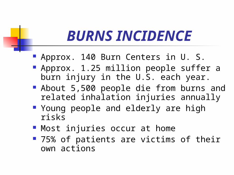
BURNS INCIDENCE Approx. 140 Burn Centers in U. S. Approx. 1.25 million people suffer a
burn injury in the U.S. each year. About 5,500 people die from burns and
related inhalation injuries annually Young people and elderly are high risks Most injuries occur at home 75% of patients are victims of their own
actions
Burns Devastating Heals slowly Disfiguring Long months of
rehabilitation Occupational
therapy
Nurse’s role: Be knowledgeable
of the pathophysiology of burns
Help meet biopsychosocial needs
Carry out measures to meet physical needs

Phases of Burn Care Emergent/Resuscitative Phase (onset of injury
to completion of fluid resuscitation) On the scene care (ABC, initial wound care,
injury) Management of fluid loss & shock
Acute/intermediate Phase (beg. of diuresis to near completion of wound closure) Maintenance of resp. & circulatory status, fluid &
electrolyte balance, GI function Infection prevention, wound care, pain
management, nutritional support Rehabilitation Phase (wound closure to optimal
level) Wound healing, restoring maximal functional
activity, alterations in self-image & lifestyle

Burns Emergency management
cold wet towels, cool water, no direct ice, cover with sterile dressing
don’t break blisters lotion only after a burn is completely cooled wrap loosely to avoid putting pressure on burned
skin ABC Prevent shock Fluid replacement Flush area thoroughly with water or saline
(chemical burns)

Burns Short term effects of burns
fl volume deficit hypovolemic shock infection
Long term effects of burns scarring (full thickness burns) contraction

Burns Minor burns
<15% TBSA with <2% full thickness
Moderate burns partial thickness burns 15-25% TBSA
with 2-10% full thickness

Burns Major Burns
>25% TBSA or >10% full thickness involvement of hands, feet, face or genitalia
Survival due to refinement of fluid resuscitation management and early transfer to burn unit.

Burns Facial burns have the possibility of
airway impairment Important to know the
circumstance of burn:greater risk of airway involvement if burn occurred in closed space
Always suspect a head injury if a patient had an electrical burn which resulted from a fall

Mechanism of Burn Injuries
flame (thermal) 55% contact with fire etc. (smoke inhalation can be as
damaging as thermal) get pt. Supine to decrease facial burn
(STOP, DROP, ROLL) prolonged exposure to cold
considered thermal burn

Mechanism of Burn Injuries
Scald 33% most common for children
chemical 10% severity depends on duration of contact,
conc. Strength of chemical, amt. Of tissue exposed
alkaline more serious than acid irrigate area immediately (most chem labs
have high pressure showers) powder-brush off; do not inhale

Mechanism of Burn Injuries
chemical con’t chemical burns destroy tissue through
protein coagulation rather than heat some acids (sulfuric and muriatic) also
destroy tissue through heat production in chemical reaction with tissue
first aid take of clothes (chemical on clothes)

Mechanisms of Burn Injuries
First aid of Chemical con’t if powder, dust off before irrigate so not attempt to neutralize agent (takes
valuable time) running water will dilute the chemical
Electrical <10% voltage running through body can do a lot
of damage (leaves an exit wound as well as entrance wound); evaluate internal damage along the path of the current

Mechanisms of Burn Injuries
Electrical con’t as electrical current goes through
tissue, it produces heat causing thermal coagulation necrosis
immediate extent of injury unknown (electrical current pathway)
deep muscle burn-burgundy color urine

Classification of Burns Burn injuries are
described according to: Depth of injury Extent of body
surface area (BSA) injured
Depth Superficial Partial-
thickness Deep partial-thickness Full-thickness
BSA Rule of nines Lund & Bowder
method Palm method
Classification of Burns First degree (superficial partial-
thickness) eg. Sunburn involvement: usually epidermis only symptoms: tingling, hypersthesia (inc.
sensitivity to pain), skin reddened,blanched with pressure, minimal or no edema
how it heals: some peeling over a week, no scar

Types of Burns Second degree burn (partial-
thickness) eg. Scalding burns involvement:epidermis and part of dermis symptoms: hyperesthesia, sensitivity to
cold, blistered mottled red base, broken epidermis , weeping surface
how it heals: new epidermis grows in 1-3 weeks
Types of Burns Third degree (full thickness)
eg. Fire burn, prolonged exposure to hot liquid involvement: epidermis, entire dermis, and
sometimes subcutaneous tissue, muscle, and bone
symptoms: painless, s/s shock, hematuria, hemolysis of blood likely,skin and fat exposed edema, skin (dry, pale white, or charred)
how it heals: needs skin grafting unless very small

Burns Rules of nine (total equals 100%)
arms 9 each (4 1/2 front, 4 1/2 back) legs 18 each (9 front, 9 back) trunk 36 (front 18 and back 18) head and neck 9 (41/2 front, 4 1/2 back) perineum 1
A superficial burn can bring on a severe systemic reaction when it covers a large body surface area

Burns Suspect inhalation injury if victim
has burns on head, neck, or anterior chest
s/s inhalation injury dyspnea, carbonaceous sputum,
wheezing. Hoarseness (caused by laryngeal edema), altered mentation
Burns Pain
continuous problem with second-degree burns but not third degree because nerves have not completely been destroyed as with third degree
when eschar removed from third degree, other pain mechanisms become operative

Burns Assessing a patient with burns
ABCs history and physical, rules of
nine,pulmonary status,check peripheral pulses on burned extremities,vital signs, urine output, labs fluid replacement, prevention of infection, psychological needs
temp usually hypothermic
Physiologic Changes con’t
Pulmonary carbon monoxide most common
cause of inhalation injury because it is a byproduct of the combustion of organic materials and therefore present in smoke (need 100% O2 due to cell hypoxia
upper airway injury, pulmonary edema, etc
Physiologic Changes in Burn Injury
Cardiovascular outpouring of catecholamines from SNS
(sympathetic nervous system) with injury which leads to peripheral blood vessel constriction and increase pulse rate
vessels become more permeable due to injury and allow fluid and colloids to leak into surrounding tissues (third spacing)

Physiologic Changes con’t
Cardiovascular peripheral vascular vasoconstriction
further decreases cardiac output with third spacing less intravascular
fluid which leads to low blood volume and low cardiac output which contributes to inadequate tissue perfusion
Physiologic Changes con’t
Renal decreased renal blood flow which
leads to glomerular damage GI
hypovolemia leads to gastric dilatation and paralytic ileus
Curling’s ulcer-stress ulcer
Physiologic Changes con’t
Fluid and Electrolytes hyponatremia (first week of acute
phase) as water shifts from interstitial to the vascular space
hyperkalemia (immediate after burn) results from massive cell destruction
hypokalemia may occur later with fluid shifts and inadequate potassium intake

Burns Pyschosocial
self-concept body image
There is a rapid fluid and electrolyte change taking place fluid loss leads to decreased blood volume
which leads to thicker blood and decreased efficiency of circulation. There is a inc cellular elements of blood which leads to inc Hct

Burns What happens in third spacing
burns produce a dilatation of capillaries and the small vessels in the area of the burn leading to increase capillary permeability. The plasma seeps out into the surrounding tissues which produce blisters and edema..
The capillary walls that are damaged permit plasma proteins to move into interstitial (extracellular) spaces

Burns Third spacing con’t
the developed capillary permeability allows plasma proteins to go through the barrier.
There is decreased osmotic pressure in blood vessels and increased osmotic pressure in interstitial space and fluid accumulates at the burn sites in blister leading to third spacing loss. Fluid shift is from vascular compartment to third space.

Burns Fluid loss with burns great. Fluid
loss by evaporation 20 times greater than normal. Loss due to evaporation called white bleeding.
Burns First 24-48 hours
large amounts of Na+ move with fluid from intravascular (within vessels) to interstitial fluid (Normally Na+ present approximately in same proportions both in intravascular and interstitial areas)
Increase in release of aldosterone and antidiuretic hormone as a result of general stress response (this inc amt of Na+ and H2O retained. Pt. Becomes oliguric.

Burns First 24-48 hours con’t
K+ elevated in blood since tissue destruction and oliguria (hyperkalemia)
Patients’ feet cold due to inc. BMR (body’s reaction to try to replace heat lost from evaporation)

Burns 48-72 hours
Na+ shifts back to intravascular space (capillaries begin to regain their integrity)
Diuresis leads to low K+ (hypokalemia) RBC loss from a microangiopathic anemia
(severeness depends on extent of burn) RBC destruction initially caused by heat of
the burn and later by hemolysis of heat damaged cells (result not only anemia but possible

Burns 48-72 hours con’t
kidney damage. Damaged RBC’s release Hgb which is filtered by kidneys.
Myoglobin is released by damaged muscle tissue. Both are filtered by kidneys and kidney tubules. They may get plugged which leads to acute tubule necrosis.
Diuresis phase lasts 3-5 days after burns. Vascular leakage recovery complete.
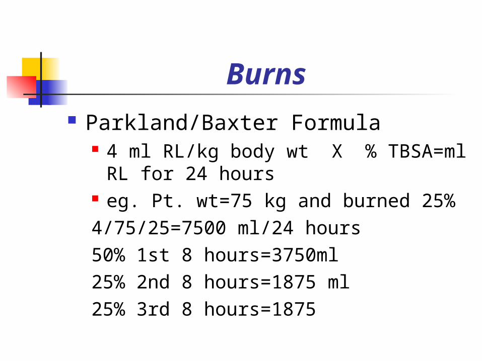
Burns Parkland/Baxter Formula
4 ml RL/kg body wt X % TBSA=ml RL for 24 hours
eg. Pt. wt=75 kg and burned 25%4/75/25=7500 ml/24 hours50% 1st 8 hours=3750ml25% 2nd 8 hours=1875 ml25% 3rd 8 hours=1875
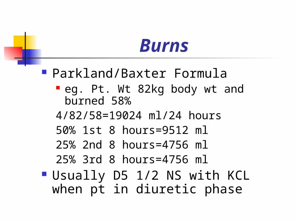
Burns Parkland/Baxter Formula
eg. Pt. Wt 82kg body wt and burned 58%
4/82/58=19024 ml/24 hours50% 1st 8 hours=9512 ml25% 2nd 8 hours=4756 ml25% 3rd 8 hours=4756 ml
Usually D5 1/2 NS with KCL when pt in diuretic phase
Burns Goal: perfuse vital organs as fully
as possible without overload Nutrition
greater protein requirement due to negative nitrogen balance
2 times calories 2 times protein

Burns Full thickness burns result in death
of skin and subcutaneous tissue Compartment syndrome
pain, pallor, decreased capillary refill, decreased peripheral pulses, decreased sensation, impaired movement (Poor peripheral tissue perfusion)

Burns IV best route for pain relief (peripheral
vasoconstriction limits absorption of drug given IM or SQ route)
Open wound-use bed cradle Circumferential burns usually involve an
extremity edema may shut off circulation and an
escharotomy or fasciotomy may be necessary to

Burns relieve the constriction and return
normal blood flow Infection remains a threat until all
second degree burns have healed and third degree burns have been closed by grafting (second degree could become third degree if infection goes deeper)
Burns (Grafts) Graft-a piece of tissue separated
completely from its normal and original position and transferred by one or more stages to correct a distant defect
Free graft: completely separated from their donor site (blood supply completely interrupted) survival of graft depends on vascular bed
from recipient site

Burns (Grafts) Free flap graft (free-tissue transfer)
cover a variety of wounds, cover exposed tendons, bones, major blood vessels.
Completely severed from the body and transferred to another site
Pedicle graft a segment of tissue that has been left
attached at one end (pedicle) while other end has been

Burns (grafts) Pedicle graft (con’t)
moved to recipient area used when thick pieces of skin that
could not survive an interruption of blood supply are transplanted
usually about 3-4 weeks for sufficient blood supply to be established

Burns (grafts) Split thickness graft
varies from thin to nearly full thickness of skin
Full thickness graft composed of a full depth of
skin(epidermis and dermis) gives best cosmetic appearance so used for
face, neck, hands

Burns (grafts) Pre-op skin grafts
hgb and clotting time noted since their levels can affect healing process
tissues need to be free from infection pt in optimum physical condition pt teaching

Burns (grafts) Necessary for a graft to take
recipient bed must be adequately vascularized
graft must be in complete contact with the bed
immobilization must be assured area must be free from infection

Burns (grafts) Types of grafts
allografts (cadaver) temporary xenografts animal (pigskin) autografts (self)
split thickness-epidermis 8-12 thousandth of an inch thick
full thickness-full depth of skin sheet grafts are put on joint areas (areas
that stress) and secured with staples or sutures

Bone (grafts) Autografts (con’t)
mesh grafts-donor skin expand to cover large area expands graft 1 1/2 -9 times its original surface
culture epithelial growth medium-grows 50-70 times initial sample (donor site heals in 1-2 weeks)
Debridement Surgical (excise) Mechanical or enzymatic (commercial
preparation) natural (body and bacterial enzymes dissolve
eschar
“Cultured Skin” ref UCSD 2001
Growing cultured skin from samples taken from patients
Copyrighted materials have been deleted from this slide

Burns (medications) Silver nitrate 0.5% (rarely used)
con’t wet dressing effectively prevents cross infection
>0.5% injures the tissue and not effective<0.5%
danger of electrolyte imbalance (especially Na and K) since the electrolytes are withdrawn from the body fluids and also from the dressing.
Turns black in sunlight and stains clothes and hands black
Burns (medications) Silvadene
wide-spectrum antimicrobial that is nonstaining and relatively painless
no systemic metabolic abnormalities however is contraindicated in pregnant women near term and premature infants
does not penetrate the eschar as well as sulfamylon

Burns (medications) Sulfamyalon acetate
interferes with bacterial cellular metabolism diffuses rapidly through burned skin and
eschar used for gram-negative organism burning sensation after applied topically may cause metabolic acidosis and is a
carbonic andryrase inhibitor may cause a rash
Burns (medications) Furacin-nitrofurazone (gauze or
cream) a synthetic broad-spectrum antibacterial inhibits enzymes required for
carbohydrate metabolism in bacteria
Xeroform-a fine mesh gauze with antimicrobial action
Case Study A 42-year old patient is brought to the ED
after being rescued from a house fire, where firefigthers found her unconscious in a bedroom closet. She has sustained burns to her right arm, right chest, and both lower extremities. On admission, she arouses to painful stimuli only. Her VS are BP 140/74, HR 112, RR 30/min and labored. She is afebrile. She has facial edema and visible soot in the oral pharynx and nares. Crackles are heard on auscultation, with decreased resp. excursion. Stridor is audible. The affected skin areas are white and inelastic, surrounded by heperemic, moist-looking tissue. She has pain on pinprick in the hyperemic tissue only.

Case Study cont. Lab results reveal a mildly
elevated glucose level, elevated Hg and Hct. Urine specific gravity of 1.030. Low PaO2 and an elevated carboxyhemoglobin level, at 37%.
What is your treatment plan? Nursing Diagnosis?
What to do about the pain during PT
U of W Harborview Burn Center is using VR (virtual reality) a non-pharmacological analgesic (distraction) used in addition to traditional levels of opioids
during wound care and physical therapy found VR worked much better than Nintendo64 pilot study showed dramatic drops in pain
during treatments

Why VR works for pain Pain perception is largely psychological pain requires conscious attention VR draws pt into another world (this
drains a lot of attentional resources leaving less attention available to process pain signals) in snow world for example the patient fly
through icy canyons etc (pts experience burning sensation during wound care so this game is designed to put out the fire)

NY Hospital-Cornell Medical Center Burn Center (1998 data)
5,000 outpatients seen/yr (>1,000 children)
1,300 inpatients seen/yr team approach(surgeons,nurses,
therapists, nutritionists, social workers)
mission develop an increasing effective
teaching and

NY Hospital-Cornell Medical Center Burn Center (1998 data)
NY’s mission con’t public awareness methods having the highest standards of clinical and
therapeutic excellence continued expansion of research in all
phases of thermal injury an optimum environment in which patients
may recover with the help and expertise of NY’s hospital-wide administrative, medical and paramedical specialists
UW Burn Center Approach to deep burn wounds
remove wound surgically within 1st post burn week
immediate grafts after wound removed to provide best functional and cosmetic results
Treatment for (TEN) Toxic Epidermal Necrolysis disease resulting from a drug reaction whereby the body’s entire epidermis sloughs

UW Burn Center treatment of TEN con’
use of pig skin and immaculate supportive care
mortality has been reduced from 70% to 15%
hospital stay has been reduced from months to average of 3 weeks
UCSD Burn Center 450 admits/year
Brave Heart Kids Program Activities Work on promoting self-esteem

Baltimore Regional Burn Center (John Hopkins)
1800/year outpatients Michael D. Hendrix Research Center
Died from burn and ARDS in 1995 Delta plane crash in Georgia
Family donated large amount of money to research Research
Infection prevention Wound healing New ways to culture skin Treatment of ARDS Immunologic response to burns

Prevention of Burn Injuries
Proper education and supervision childproof items in electrical sockets keep dangerous items (matches) out
of reach Safety measures for the elderly teach small children 911 smoke detectors in house STOP, DROP, AND ROLL