In situ spectroscopy on intact Leptospirillum ferrooxidans ... · tein called rusticyanin, and an...
10
ORIGINAL RESEARCH ARTICLE published: 12 April 2012 doi: 10.3389/fmicb.2012.00136 In situ spectroscopy on intact Leptospirillum ferrooxidans reveals that reduced cytochrome 579 is an obligatory intermediate in the aerobic iron respiratory chain Robert C. Blake II* and Megan N. Griff College of Pharmacy, Xavier University of Louisiana, New Orleans, LA, USA Edited by: David Emerson, Bigelow Laboratory for Ocean Sciences, USA Reviewed by: James Hemp, University of Illinois at Urbana-Champaign, USA David Emerson, Bigelow Laboratory for Ocean Sciences, USA *Correspondence: Robert C. Blake II, College of Pharmacy, Xavier University of Louisiana, 1 Drexel Drive, New Orleans, LA 70125, USA. e-mail: [email protected] Electron transfer reactions among colored cytochromes in intact bacterial cells were moni- tored using an integrating cavity absorption meter that permitted the acquisition of accurate absorbance data in suspensions of cells that scatter light.The aerobic iron respiratory chain of Leptospirillum ferrooxidans was dominated by the redox status of an abundant cellu- lar cytochrome that had an absorbance peak at 579 nm in the reduced state. Intracellular cytochrome 579 was reduced within the time that it took to mix a suspension of the bacte- ria with soluble ferrous iron at pH 1.7. Steady state turnover experiments were conducted where the initial concentrations of ferrous iron were less than or equal to that of the oxygen concentration. Under these conditions, the initial absorbance spectrum of the bacterium observed under air-oxidized conditions was always regenerated from that of the bacterium observed in the presence of Fe(II). The kinetics of aerobic respiration on soluble iron by intact L. ferrooxidans conformed to the Michaelis–Menten formalism, where the reduced intracellular cytochrome 579 represented the Michaelis complex whose subsequent oxi- dation appeared to be the rate-limiting step in the overall aerobic respiratory process. The velocity of formation of ferric iron at any time point was directly proportional to the concen- tration of the reduced cytochrome 579. Further, the integral over time of the concentration of the reduced cytochrome was directly proportional to the total concentration of ferrous iron in each reaction mixture.These kinetic data obtained using whole cells were consistent with the hypothesis that reduced cytochrome 579 is an obligatory steady state interme- diate in the iron respiratory chain of this bacterium. The capability of conducting visible spectroscopy in suspensions of intact cells comprises a powerful post-reductionist means to study cellular respiration in situ under physiological conditions for the organism. Keywords: Leptospirillum ferrooxidans, electron transfer, aerobic respiration, chemolithotroph, acidophile, cytochrome 579, in situ spectroscopy, integrating sphere INTRODUCTION Certain chemolithotrophic bacteria inhabit ore-bearing geolog- ical formations exposed to the atmosphere and obtain all of their energy for growth from the oxidation and dissolution of minerals within the ore. Energy is derived from oxidative phos- phorylation coupled to respiratory electron transfer. The ability to respire aerobically on soluble ferrous ions under strongly acidic conditions is currently known to be expressed by at least 34 species in 14 genera distributed throughout the Gram-negative (Markosyan, 1972; Huber and Stetter, 1989; Kelly and Wood, 2000; Hallberg et al., 2010), Gram-positive (Clark and Norris, 1996; Norris et al., 1996; Johnson et al., 2008, 2009; Guo et al., 2009; Jiang et al., 2009), and Archaea bacteria (Segerer et al., 1986; Huber et al., 1989; Huber and Stetter, 1991; Karavaiko et al., 1994; Golyshina et al., 2000, 2009). Given the genetic diversity within this collection of phenotypically related bacte- ria, it would not be surprising to learn that phylogenetically distinct groups of bacteria express different electron transfer bio- molecules and pathways to accomplish aerobic respiration on soluble iron. Classic reductionist studies that involve the structural and functional characterization of highly purified proteins in dilute solution have described a bewildering variety of different redox- active electron transport proteins in cell-free extracts derived from iron-grown Gram-negative (Cox and Boxer, 1978; Hart et al., 1991; Blake et al., 1992; Yarzábal et al., 2002, 2004), Gram-positive (Blake et al., 1993; Takai et al., 2001; Dinarieva et al., 2010), and Archaea (Hettmann et al., 1998; Dopson et al., 2005; Auernik and Kelly, 2008) bacteria. The most promising efforts to date have focused on the iron respiratory chain of Acidithiobacillus ferrooxi- dans, where an iron “respirasome” super complex has been defined that is comprised of 2 c -type cytochromes, a blue copper pro- tein called rusticyanin, and an aa 3 -type terminal oxidase (Castelle et al., 2008). The proteins in the aerobic iron respiratory pathway of At. ferrooxidans do not appear to be expressed in many of the phylogenetically distinct bacteria that also respire on iron. Simi- larly, redox-active proteins expressed in other iron-grown bacteria do not appear to be expressed in iron-grown At. ferrooxidans. Comparative analyses conducted using those relevant bacterial genomes where partial or complete DNA sequence data is available www.frontiersin.org April 2012 |Volume 3 | Article 136 | 1
Transcript of In situ spectroscopy on intact Leptospirillum ferrooxidans ... · tein called rusticyanin, and an...
ORIGINAL RESEARCH ARTICLEpublished: 12 April 2012
doi: 10.3389/fmicb.2012.00136
In situ spectroscopy on intact Leptospirillum ferrooxidansreveals that reduced cytochrome 579 is an obligatoryintermediate in the aerobic iron respiratory chainRobert C. Blake II* and Megan N. Griff
College of Pharmacy, Xavier University of Louisiana, New Orleans, LA, USA
Edited by:
David Emerson, Bigelow Laboratoryfor Ocean Sciences, USA
Reviewed by:
James Hemp, University of Illinois atUrbana-Champaign, USADavid Emerson, Bigelow Laboratoryfor Ocean Sciences, USA
*Correspondence:
Robert C. Blake II, College ofPharmacy, Xavier University ofLouisiana, 1 Drexel Drive, NewOrleans, LA 70125, USA.e-mail: [email protected]
Electron transfer reactions among colored cytochromes in intact bacterial cells were moni-tored using an integrating cavity absorption meter that permitted the acquisition of accurateabsorbance data in suspensions of cells that scatter light.The aerobic iron respiratory chainof Leptospirillum ferrooxidans was dominated by the redox status of an abundant cellu-lar cytochrome that had an absorbance peak at 579 nm in the reduced state. Intracellularcytochrome 579 was reduced within the time that it took to mix a suspension of the bacte-ria with soluble ferrous iron at pH 1.7. Steady state turnover experiments were conductedwhere the initial concentrations of ferrous iron were less than or equal to that of the oxygenconcentration. Under these conditions, the initial absorbance spectrum of the bacteriumobserved under air-oxidized conditions was always regenerated from that of the bacteriumobserved in the presence of Fe(II). The kinetics of aerobic respiration on soluble iron byintact L. ferrooxidans conformed to the Michaelis–Menten formalism, where the reducedintracellular cytochrome 579 represented the Michaelis complex whose subsequent oxi-dation appeared to be the rate-limiting step in the overall aerobic respiratory process. Thevelocity of formation of ferric iron at any time point was directly proportional to the concen-tration of the reduced cytochrome 579. Further, the integral over time of the concentrationof the reduced cytochrome was directly proportional to the total concentration of ferrousiron in each reaction mixture.These kinetic data obtained using whole cells were consistentwith the hypothesis that reduced cytochrome 579 is an obligatory steady state interme-diate in the iron respiratory chain of this bacterium. The capability of conducting visiblespectroscopy in suspensions of intact cells comprises a powerful post-reductionist meansto study cellular respiration in situ under physiological conditions for the organism.
Keywords: Leptospirillum ferrooxidans, electron transfer, aerobic respiration, chemolithotroph, acidophile,
cytochrome 579, in situ spectroscopy, integrating sphere
INTRODUCTIONCertain chemolithotrophic bacteria inhabit ore-bearing geolog-ical formations exposed to the atmosphere and obtain all oftheir energy for growth from the oxidation and dissolution ofminerals within the ore. Energy is derived from oxidative phos-phorylation coupled to respiratory electron transfer. The ability torespire aerobically on soluble ferrous ions under strongly acidicconditions is currently known to be expressed by at least 34species in 14 genera distributed throughout the Gram-negative(Markosyan, 1972; Huber and Stetter, 1989; Kelly and Wood,2000; Hallberg et al., 2010), Gram-positive (Clark and Norris,1996; Norris et al., 1996; Johnson et al., 2008, 2009; Guo et al.,2009; Jiang et al., 2009), and Archaea bacteria (Segerer et al.,1986; Huber et al., 1989; Huber and Stetter, 1991; Karavaikoet al., 1994; Golyshina et al., 2000, 2009). Given the geneticdiversity within this collection of phenotypically related bacte-ria, it would not be surprising to learn that phylogeneticallydistinct groups of bacteria express different electron transfer bio-molecules and pathways to accomplish aerobic respiration onsoluble iron.
Classic reductionist studies that involve the structural andfunctional characterization of highly purified proteins in dilutesolution have described a bewildering variety of different redox-active electron transport proteins in cell-free extracts derived fromiron-grown Gram-negative (Cox and Boxer, 1978; Hart et al.,1991; Blake et al., 1992; Yarzábal et al., 2002, 2004), Gram-positive(Blake et al., 1993; Takai et al., 2001; Dinarieva et al., 2010), andArchaea (Hettmann et al., 1998; Dopson et al., 2005; Auernik andKelly, 2008) bacteria. The most promising efforts to date havefocused on the iron respiratory chain of Acidithiobacillus ferrooxi-dans, where an iron“respirasome” super complex has been definedthat is comprised of 2 c-type cytochromes, a blue copper pro-tein called rusticyanin, and an aa3-type terminal oxidase (Castelleet al., 2008). The proteins in the aerobic iron respiratory pathwayof At. ferrooxidans do not appear to be expressed in many of thephylogenetically distinct bacteria that also respire on iron. Simi-larly, redox-active proteins expressed in other iron-grown bacteriado not appear to be expressed in iron-grown At. ferrooxidans.Comparative analyses conducted using those relevant bacterialgenomes where partial or complete DNA sequence data is available
www.frontiersin.org April 2012 | Volume 3 | Article 136 | 1
Blake II and Griff Respiratory cytochromes in Leptospirillum ferrooxidans
(Chen et al., 2005; Ram et al., 2005; Valdes et al., 2008; Clum et al.,2009; Siezen and Wilson, 2009) have not yet provided significantinsight into other iron respiratory proteins or pathways. There islittle information in the DNA databases to compare with becausethe proteins in the aerobic iron respiratory pathway of At. ferroox-idans do not appear to be universal among those bacteria thatrespire on iron. In either case, actual respiratory electron transferin the intact organism is not directly observed. Rather, the func-tional properties of the intact electron transfer chain are inferredfrom observations on isolated molecules.
This paper introduces a new means to study respiratory elec-tron transfer reactions in situ in intact bacteria under physiologicalconditions. The premise is that accurate UV-visible spectroscopyof electron transfer reactions among colored cytochromes can beconducted in highly turbid suspensions if the live bacteria areirradiated in an isotropic homogeneous field of incident measur-ing light. Under those conditions, the absorbed radiant power isindependent of scattering effects (Elterman, 1970; Fry et al., 1992;Javorfi et al., 2006; Hodgkinson et al., 2009). We conducted equi-librium and kinetic studies on the Fe(II)-dependent reductionand O2-dependent oxidation of cytochromes in intact Leptospir-illum ferrooxidans at pH 1.7. We used a commercial integratingcavity absorption meter (ICAM) where the cuvette comprised areflecting cavity completely filled with the absorbing suspension.L. ferrooxidans was selected because it is only known to respireon one substrate, reduced iron (Harrison, 1984). We observedthat a cytochrome with a reduced spectral peak at 579 nm is anobligatory intermediate in the aerobic iron respiratory chain of L.ferrooxidans.
MATERIALS AND METHODSCELL CULTURELeptospirillum ferrooxidans DSMZ 2705 was cultured autotroph-ically on soluble ferrous ions at 30˚C in the medium describedelsewhere (Tuovinen and Kelly, 1973), adjusted to pH 1.5 andamended with 44 g/l of FeSO4·7H2O. Cells grown to station-ary phase were harvested by centrifugation, washed three timeswith 0.02 M H2SO4, pH 1.7, and resuspended in sufficient 0.02 MH2SO4 to achieve a stock suspension of 1.5 × 10¹⁰ cells/ml. Thestock suspension was stored at 4˚C for up to 2 weeks while spec-troscopic experiments were conducted on aliquots of the cells.Previous stock suspensions of this organism have been stored indilute sulfuric acid at 4˚C for over 6 weeks before changes in thebacterium’s energy metabolism could be detected.
QUANTIFICATION OF BACTERIAAbsolute numbers of L. ferrooxidans cells were determined by elec-trical impedance measurements in a Multisizer 4 particle counter(Beckman Coulter, Inc., Brea, CA, USA) fitted with a 30-μm aper-ture. The instrument was programmed to siphon 50 μl of samplethat contained Isoton II as the electrolyte. The current appliedacross the aperture was 600 μA. Voltage pulses attendant withimpedance changes as particles passed through the aperture weremonitored with an instrument gain of four.
Relative numbers of L. ferrooxidans cells were determined byphoton correlation scattering spectroscopy with a DelsaNanoC particle size analyzer, also from Beckman Coulter, Inc. Cell
densities were adjusted to 8.3 × 10⁶ cells/ml in 0.02 M sulfuric acidto give an attenuator obscuration of 47%. Determination of therelative numbers of light scattering species as a function of parti-cle diameter was accomplished by the time domain method withoperating and analysis software provided by Beckman Coulter, Inc.
ABSORBANCE MEASUREMENTS WITH CELL SUSPENSIONSAbsorbance measurements on intact cells in suspension were con-ducted in an Olis CLARiTY 1000 A spectrophotometer (On LineInstrument Systems, Inc., Bogart, GA, USA) that employed a novelICAM. In a typical experiment, identical 4.2 ml solutions thatcontained ferrous sulfate in 0.02 M sulfuric acid, pH 1.7, wereadded to both the sample and reference observation cavities ofthe spectrophotometer. After recording a stable baseline from 350to 650 nm, 140 μl were withdrawn from the sample cavity andreplaced with 140 μl of the stock cell suspension of L. ferrooxi-dans. Apparent absorbance spectra (typically 6.2 s−1) were thencollected until any visible absorbance changes had ceased. Rawapparent absorbance values were converted to absorbance valuesper cm using Fry’s method (Fry et al., 2010) as described in the text.
RESULTSTHE REDOX STATE OF ELECTRON TRANSFER PROTEINS CAN BEMONITORED IN SITU IN INTACT BACTERIA UNDER PHYSIOLOGICALCONDITIONSThe principal features of the novel CLARiTY spectrophotometerused to conduct absorbance measurements in turbid solutions areincluded in the schematic diagram shown in Figure 1. The sampleand reference observation cells of this dual beam spectropho-tometer were each comprised of a 4.2-ml spherical quartz cuvettefused with a 6-mm ID quartz tube. Each quartz chamber was sur-rounded by a tightly packed proprietary white powder that servedto maximize diffuse reflectance of light on the exterior walls ofthe spherical flask. The apertures in the reflecting sphere throughwhich the measuring light entered and the transmitted/scatteredlight exited to the photomultiplier tube were positioned at a 90˚angle such that the light had to undergo many reflections andcell transversals before it was quantified using the photomultipliertube. A white Teflon plug with a 6-mm OD was inserted into thequartz tube to minimize the loss of light out of the neck. A 1.0-cm white stir bar was included in the sample chamber to facilitatesample mixing and suspension of any particulate matter.
The data shown in Figure 2 illustrate how the intact cells of L.ferrooxidans were quantified. The solid line in Figure 2 shows theabsolute counts as a function of particle size as determined witha suspension of L. ferrooxidans in the Multisizer 4. The Multi-sizer determined the number and size of intact bacteria suspendedin an electrically conductive liquid by forcing a measured vol-ume of the suspension to flow through a small aperture with animmersed electrode on either side. A current passing through theaperture between the two electrodes enabled the bacteria to bedetected by the momentary changes in the electrical impedanceas they passed through the aperture, since each bacterium dis-placed its own volume of electrolyte solution within the apertureitself. These changes in impedance were detected as a series ofvoltage pulses, for which the height and duration of each pulsewere proportional to the volume of the bacterium that produced
Frontiers in Microbiology | Microbiological Chemistry April 2012 | Volume 3 | Article 136 | 2

Blake II and Griff Respiratory cytochromes in Leptospirillum ferrooxidans
it. Each pulse was counted and allocated to 1 of 400 arbitrary sizecategories, or channels. Each channel encompassed a narrow rangeof volumes that were converted to spherical equivalents and rep-resented by the corresponding spherical diameters on the abscissaof Figure 2. The solid line in Figure 2 shows the number of countsin each size range for spherical equivalents with diameters from0.6 to 2.0 μm.
The dashed line in Figure 2 shows the relative number of L.ferrooxidans cells as a function of particle size as determined bylaser light diffraction. Light scattered by the bacterial suspensionproduced a diffraction pattern from which the relative numbers ofthe scattering species at each particle size were calculated. Like theMultisizer, the particle sizes determined by laser light diffractionare expressed as spherical equivalents and are also represented bythe corresponding spherical diameters on the abscissa of Figure 2.
FIGURE 1 | Simplified model of an integrating cavity absorption meter
designed to permit accurate absorbance measurements in media that
scatter light.
FIGURE 2 | Comparison of L. ferrooxidans cell populations by
electrical impedance (solid line) and static light diffraction methods
(dashed line). Electrical impedance measurements yielded absolutenumbers of cells (left ordinate) as a function of size; light diffractionmeasurements yielded relative numbers of cells (right ordinate) as afunction of size. The scale of each ordinate was chosen to facilitatecomparison of the curves. The area under the electrical impedancecurve was 81.2% of that under the light diffraction curve.
Close correspondence between the electrical impedance and thelight diffraction curves was observed down to 0.6 μm, indicatingthat the two instruments were monitoring the same populationof particles. Bacteria with effective diameters less than 0.6 μmwere underrepresented by the electrical impedance method, bywhich observations were limited to particles with effective diam-eters between 2 and 60% of the 30-μm aperture employed, whilethe light diffraction method was capable of resolving particles witheffective diameters smaller than 0.1 μm. On the basis of compar-isons such as that illustrated in Figure 2, it was determined that19.8% of the bacterial cell counts were below the limit of resolu-tion of the electrical impedance method with the 30-μm aperture.Accordingly, absolute counts of L. ferrooxidans cells obtained bythe electrical impedance method were multiplied by 1.23 to cor-rect for the percentage of bacterial counts that were below the limitof resolution of the instrument.
Cell pellets of L. ferrooxidans, which were a pinkish tan color,were obtained by centrifugation of batch cultures grown to sta-tionary phase on soluble ferrous iron. Curve a in Figure 3 showsthe absorbance spectrum of oxidized L. ferrooxidans that wasobtained in the CLARiTY spectrophotometer in sulfuric acid, pH1.7. Even though the cell suspension contained 5 × 10⁸ cells/mland was roughly as turbid as non-fat milk, the resulting absorbancespectrum contained no evidence of the light scattering artifactsthat one would observe by conducting the same absorbance mea-surements using a conventional linear spectrophotometer. Thespectrum in curve a showed a clearly defined Soret peak at 422 nmand a broad α,β band at around 520 nm.
The goal of the initial spectroscopic experiments was simplyto determine whether the pink protein(s) in the cells changedcolor when the cells were suspended in sulfuric acid and subse-quently exposed to excess concentrations of soluble ferrous ionsunder physiological conditions. Curve b in Figure 3 shows theabsorbance spectrum that was obtained when the cells of oxidizedL. ferrooxidans were exposed to 100 mM ferrous sulfate in sulfuricacid, pH 1.7. Exposure to excess soluble iron caused the apparent
FIGURE 3 | Apparent absorbance spectra of intact L. ferrooxidans
collected in the CLARiTY in the absence (a) and presence (b) of
100 mM ferrous sulfate in aerobic sulfuric acid, pH 1.7. The final bacterialsuspension contained 2.1 × 10⁹ cells of L. ferrooxidans in 4.2 ml. Curve cshows the apparent absorbance spectrum of 50 μM ferric ions under thesame solution conditions in the absence of bacteria.
www.frontiersin.org April 2012 | Volume 3 | Article 136 | 3

Blake II and Griff Respiratory cytochromes in Leptospirillum ferrooxidans
Soret peak to split into two peaks with maxima at 418 and 439 nm.In addition, a new absorbance peak appeared with a maximumabsorbance at 579 nm. The absorbance spectrum represented bycurve b appeared immediately after mixing the bacterial suspen-sion in the observation cell that contained the 100-mM solubleiron (≤0.5 s). This observation indicated that the iron-dependentreduction of the cytochrome(s) in intact L. ferrooxidans was essen-tially complete within the 0.5-s mixing time in the observation cellof the spectrophotometer. The resulting spectrum was very stableand did not vary for at least an hour after mixing. Although thelive cells could respire aerobically on the soluble iron, the greatmolar excess of iron over molecular oxygen in the observationcell dictated that the cytochromes in the cells would remain pre-dominantly reduced as the oxygen in the chamber was consumed.Evidence for aerobic respiration was taken from the increase inabsorbance at wavelengths below 400 nm in curve b. Curve c inFigure 3 shows the absorbance spectrum of 50 μM ferric sulfatein sulfuric acid, pH 1.7. The increase in the absorbance observedat low wavelengths in curve b was assumed to be due to ferric ionsproduced as a consequence of aerobic respiration on iron by thebacteria.
The goal of subsequent spectroscopic experiments was tolower the concentration of soluble iron and determine whethertime-dependent changes in the cellular absorbance could be
detected. Fe(II) concentrations were chosen such that the totalnumber of electrons available for aerobic respiration was lowerthan the electron-accepting capacity of the >200 μM O2 in theair-saturated suspension. The data in Figure 4A show selectedabsorbance spectra that were obtained when cells of L. ferrooxi-dans were exposed to 100 μM ferrous sulfate in sulfuric acid, pH1.7. The seven absorbance spectra shown in the figure were selectedfrom a data set where 6.2 complete scans from 350 to 650 nm werecollected every second for 400 s. Once again, the spectrum of thebacteria produced in the presence of Fe(II) was generated withinthe operational dead time of mixing, roughly 0.5 s. In this case,however, subsequent aerobic respiration under the conditions ofexcess molecular oxygen produced time-dependent changes in theobserved spectra of whole cells. The reduced peaks at 439 and579 nm disappeared over a period of 400 s, while the oxidizedSoret peak at 422 nm gradually reappeared. In addition, there was aconcomitant increase in absorbance at wavelengths below 400 nm.These spectral changes were consistent with the hypothesis that thecells respired aerobically on the soluble iron until the ferrous ironwas completely oxidized.
The primary absorbance spectra shown in Figures 3 and 4A arepresented as “apparent absorbance” because of prior reports thatspectra obtained using integrated cavity absorption meters appeardistorted when compared with corresponding spectra of the same
FIGURE 4 | Absorbance spectra obtained at different time points after
2.1 × 10⁹ cells of intact L. ferrooxidans were introduced into 4.2 ml of
100 μM ferrous sulfate in sulfuric acid, pH 1.7. (A) Apparent absorbancespectra collected in the CLARiTY at seven time points after mixing,ranging from 1.0 to 400 s. Each arrow indicates whether the apparentabsorbance at that wavelength increased or decreased with time. (B)
Absorbance spectra obtained when the apparent absorbance values in (A)
were converted to equivalent absorbance values per cm using the Fryequation (see text) to adjust for mean path length differences at differentwavelengths. The absorbances at the longer wavelengths correspond tothe more sensitive scale shown on the right ordinate. (C) Differencespectra, representing the absolute spectrum of the iron-reduced L.ferrooxidans minus those obtained at selected time points following theinitial reduction of the bacteria.
Frontiers in Microbiology | Microbiological Chemistry April 2012 | Volume 3 | Article 136 | 4

Blake II and Griff Respiratory cytochromes in Leptospirillum ferrooxidans
materials obtained using a conventional linear spectrophotometer(Elterman, 1970; Fry et al., 1992; Javorfi et al., 2006; Hodgkinsonet al., 2009). Unlike single path length spectrophotometers wherethe Beer–Lambert law governs non-linearity in the measured lightintensity as a function of analyte concentration, the measuringlight in an integrating sphere makes multiple random passes withdifferent path lengths that exacerbate the apparent non-linearitywith analyte concentration. While others have used semi-empiricalmethods to provide distortion-free spectra (Javorfi et al., 2006),we utilized the approach suggested by Fry because it is based onderivations from first principles (Fry et al., 1992, 2010). Fry arrivedat the following exact expression for fractional absorption in anICAM:
1 − T = 1 − exp [−α(ν)LG]
1 − ρ(1 − h) exp [−α(ν)LG](1)
where T equals the observed transmittance in the ICAM, α(ν)equals the corresponding absorbance that would be observed ina single path length spectrophotometer, LG represents the meangeometric path length which for a sphere equals 2/3 of the sphere’sdiameter, ρ represents the reflectance at the walls of the cavity andis a number between 0 and 1, and h is the fraction of the cavitywall that is open for ports. Equation 1 was rearranged to
α(v) = − ln
[10−A
1 − ρ (1 − h)(1 − 10−A
)]/
LG (2)
where A is the absorbance value observed in the CLARiTY andρ(1 − h) was determined in separate experiments to be equal to0.92 (data not shown).
The spectra shown in Figure 4B were obtained when theabsorbance values in Figure 4A were converted to equivalentabsorbance values per cm using Eq. 2. The converted absorbancevalues in Figure 4B illustrate the enhanced sensitivity achievedin the CLARiTY where the mean path length of the measuringlight is increased by multiple reflections around the interior ofthe cavity. The conversion also eliminated the apparent distortionin the raw absorbance values wherein the Soret peak appearedto be attenuated compared to the heme absorbance at longerwavelengths. Thus those wavelengths where greater absorptionoccurred received relatively higher values in the converted datathan did those wavelengths where lower absorption occurred.
Figure 4C shows difference spectra representing the absoluteabsorbance spectrum of the iron-reduced L. ferrooxidans minusthose obtained at various time points following the rapid reduc-tion of the bacteria. The two prominent peaks of the differencespectrum occur at 443 and 579 nm. The observation of at least fourrelatively well-defined isosbestic points is extraordinary consider-ing that the spectra were acquired in a highly turbid suspensionof bacteria. An isosbestic point is a specific wavelength at whichtwo or more absorbing species have the same molar absorptivity.The existence of those four well-defined isosbestic points suggeststhat there is only one principal iron-responsive cytochrome thatis visible in the aerobic iron respiratory chain of L. ferrooxidans.
REDUCED CYTOCHROME 579 IS AN OBLIGATORY INTERMEDIATEDURING AEROBIC RESPIRATION ON SOLUBLE IRON BY INTACTL. FERROOXIDANSThe next goal was to determine whether the time-dependentabsorbance changes observed when intact L. ferrooxidans wasexposed to soluble ferrous ions could be correlated with theappearance of product ferric ions as the bacterium respired aero-bically on reduced iron. The kinetic data shown in Figure 5A wereextracted from the data set that yielded the selected spectra shownin Figure 4A. The change in absorbance at 355 nm, which repre-sented predominantly the absorbance due to the time-dependentaccumulation of ferric ions, increased in a roughly linear fashionuntil the limiting concentration of ferrous ions was completelydepleted at around 320 s after the start of the bacterial-catalyzedreaction. The changes in absorbance at 443 nm, which representedthe peak of the difference spectrum in the Soret region shownin Figure 4C, slowly decreased over the time course of the reac-tion until about 300 s, when the absorbance then decreased rapidlyback to the initial absorbance observed in the bacterium under air-oxidized conditions. It was evident that the cellular cytochromereturned to the oxidized state at about the same time that theproduct formation ceased.
The close correspondence between the change in product for-mation and the transient changes in the redox state of cytochrome579 in intact L. ferrooxidans was consistent with the mini-mal kinetic mechanism shown in Figure 5B. Briefly, the iron-dependent reduction of cytochrome 579 in the intact bacteriumis depicted as a relatively rapid reaction, which is consistent withthe observation that it is complete within the 0.5-s dead time ofthe mixing even when the concentration of soluble ferrous ions isonly 100 μM. The bacterium with its reduced cellular cytochromeis then shown as reacting with molecular oxygen to regenerate oxi-dized cytochrome/bacterium in a slower reaction that constitutesthe rate-limiting catalytic step. The exchange of electrons betweenthe bacterium and soluble iron is depicted as a reversible reactionuntil or if experimental observations indicate otherwise.
The kinetic mechanism shown in Figure 5B is readily modeledby the Michaelis–Menten formalism where the intact bacteriumthat contains the reduced cytochrome 579 represents the Michaeliscomplex. It follows that the velocity of product formation shouldbe a hyperbolic function of the concentration of the substrateferrous ions, as shown below:
−d [Fe(II)]
dt= d [Fe(III)]
dt= Vmax [Fe(II)]
KM + [Fe(II)](3)
where V max is the maximum velocity of the bacterial-catalyzedaerobic oxidation of ferrous ions and K M is the Michaelis constantfor soluble iron. Figure 5C shows the dependence of the velocityof the absorbance changes at 355 nm on the concentration of fer-rous ions that remained in solution when intact cells were mixedwith 100 μM ferrous ions. The data points in Figure 5C representthe tangents to the 355-nm curve in Figure 5A that correspondto the same time points as those at the different concentrations offerrous ion shown on the abscissa of Figure 5C. The values for therectangular hyperbola drawn through the data points in Figure 5C
www.frontiersin.org April 2012 | Volume 3 | Article 136 | 5

Blake II and Griff Respiratory cytochromes in Leptospirillum ferrooxidans
FIGURE 5 |The absorbance of the reduced cytochrome in intact L.
ferrooxidans correlates with the absorbance of ferric ions produced
during aerobic respiration on soluble iron. (A) Time courses of theabsorbance changes at 355 and 443 nm obtained when 2.1 × 10⁹ cellsof L. ferrooxidans were mixed with 100 μM ferrous sulfate in sulfuricacid, pH 1.7. (B) Schematic representation of the kinetic mechanism foraerobic respiration on soluble iron as catalyzed by intact L. ferrooxidans.
The cross-hatched and solid rods represent bacteria that containoxidized and iron-reduced cytochromes, respectively. (C) Dependenceof the velocity of the absorbance change at 355 nm on theconcentration of ferrous ions that remain in solution. (D) Dependence ofthe velocity of the absorbance changes at 355 nm on the difference inabsorbance values at 443 nm, representing the value at 1.0 s minus thevalue at time t.
were derived from a non-linear least-squares fit of Eq. 3 to the datawith an apparent V max of 3.6 × 10⁻⁷ Ms−1 and a K M of 8.7 μM.
Another prediction that follows from the application of theMichaelis–Menten formalism to the kinetic mechanism shown inFigure 5B is that the rate of appearance of ferric ions shouldbe directly proportional to the concentration of the reducedcytochrome 579 as shown in Eq. 4:
−d [Fe(II)]
dt= d [Fe(III)]
dt= kcat
[reduced cytochrome 579
](4)
Figure 5D shows the dependence of the velocity of the absorbancechanges at 355 nm on the concentration of reduced cytochrome579. The velocities, or tangents to the 355-nm curve in Figure 5A,are the same values as those used in Figure 5C. In Figure 5D, therelative concentrations of reduced cytochrome 579 are representedon the abscissa by the differences in absorbance between thoseobserved at 443 nm at corresponding time points in Figure 5Aminus the stable absorbance observed at 443 nm at the end of thereaction. The direct proportionality predicted by Eq. 4 is evident,and the slope of the least-squares line in Figure 5D yielded a valuefor kcat of 1.4 s−1.
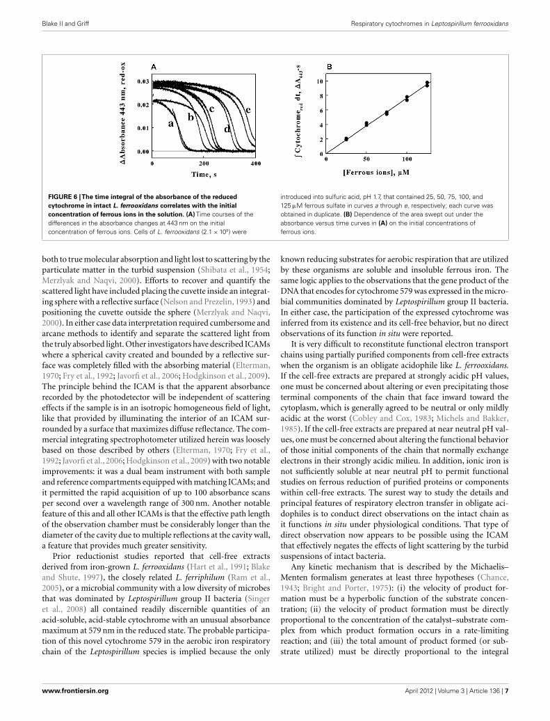
A third prediction that follows from the application of theMichaelis–Menten formalism to the kinetic mechanism shown inFigure 5B is shown in Figure 6. Equation 4 may be integrated to
yield the following:
[Fe(II)]Total = kcat
∫ [reduced cytochrome 579
]dt (5)
where the integral on the right side of the equality represents thetotal area swept out by the absorbance of the reduced cytochromeover time as illustrated by the 443-nm time course shown inFigure 5A. The kinetic curves in Figure 6A show the time coursesof the absorbance changes at 443 nm that were obtained when con-stant concentrations of L. ferrooxidans were mixed with differenttotal concentrations of ferrous ions from 25 to 125 μM. Althoughthe duplicate curves in Figure 6A appeared to differ slightly, thetotal areas under each pair of curves were remarkably similar, asshown by the five pairs of closely matched data points in Figure 6B.The direct proportionality predicted by Eq. 5 is evident, and theslope of the least-squares line in Figure 6B yielded a value for kcat
of 1.3 s−1.
DISCUSSIONThree things can happen to the measuring light in a conventionallinear spectrophotometer equipped with a standard 1-cm cuvette:it can be transmitted; it can be absorbed; or it can be scatteredwithout absorption. When the sample is not optically clear, theapparent attenuation of the measuring light reflects losses due
Frontiers in Microbiology | Microbiological Chemistry April 2012 | Volume 3 | Article 136 | 6
Blake II and Griff Respiratory cytochromes in Leptospirillum ferrooxidans
FIGURE 6 |The time integral of the absorbance of the reduced
cytochrome in intact L. ferrooxidans correlates with the initial
concentration of ferrous ions in the solution. (A) Time courses of thedifferences in the absorbance changes at 443 nm on the initialconcentration of ferrous ions. Cells of L. ferrooxidans (2.1 × 10⁹) were
introduced into sulfuric acid, pH 1.7, that contained 25, 50, 75, 100, and125 μM ferrous sulfate in curves a through e, respectively; each curve wasobtained in duplicate. (B) Dependence of the area swept out under theabsorbance versus time curves in (A) on the initial concentrations offerrous ions.
both to true molecular absorption and light lost to scattering by theparticulate matter in the turbid suspension (Shibata et al., 1954;Merzlyak and Naqvi, 2000). Efforts to recover and quantify thescattered light have included placing the cuvette inside an integrat-ing sphere with a reflective surface (Nelson and Prezelin, 1993) andpositioning the cuvette outside the sphere (Merzlyak and Naqvi,2000). In either case data interpretation required cumbersome andarcane methods to identify and separate the scattered light fromthe truly absorbed light. Other investigators have described ICAMswhere a spherical cavity created and bounded by a reflective sur-face was completely filled with the absorbing material (Elterman,1970; Fry et al., 1992; Javorfi et al., 2006; Hodgkinson et al., 2009).The principle behind the ICAM is that the apparent absorbancerecorded by the photodetector will be independent of scatteringeffects if the sample is in an isotropic homogeneous field of light,like that provided by illuminating the interior of an ICAM sur-rounded by a surface that maximizes diffuse reflectance. The com-mercial integrating spectrophotometer utilized herein was looselybased on those described by others (Elterman, 1970; Fry et al.,1992; Javorfi et al., 2006; Hodgkinson et al., 2009) with two notableimprovements: it was a dual beam instrument with both sampleand reference compartments equipped with matching ICAMs; andit permitted the rapid acquisition of up to 100 absorbance scansper second over a wavelength range of 300 nm. Another notablefeature of this and all other ICAMs is that the effective path lengthof the observation chamber must be considerably longer than thediameter of the cavity due to multiple reflections at the cavity wall,a feature that provides much greater sensitivity.
Prior reductionist studies reported that cell-free extractsderived from iron-grown L. ferrooxidans (Hart et al., 1991; Blakeand Shute, 1997), the closely related L. ferriphilum (Ram et al.,2005), or a microbial community with a low diversity of microbesthat was dominated by Leptospirillum group II bacteria (Singeret al., 2008) all contained readily discernible quantities of anacid-soluble, acid-stable cytochrome with an unusual absorbancemaximum at 579 nm in the reduced state. The probable participa-tion of this novel cytochrome 579 in the aerobic iron respiratorychain of the Leptospirillum species is implied because the only
known reducing substrates for aerobic respiration that are utilizedby these organisms are soluble and insoluble ferrous iron. Thesame logic applies to the observations that the gene product of theDNA that encodes for cytochrome 579 was expressed in the micro-bial communities dominated by Leptospirillum group II bacteria.In either case, the participation of the expressed cytochrome wasinferred from its existence and its cell-free behavior, but no directobservations of its function in situ were reported.
It is very difficult to reconstitute functional electron transportchains using partially purified components from cell-free extractswhen the organism is an obligate acidophile like L. ferrooxidans.If the cell-free extracts are prepared at strongly acidic pH values,one must be concerned about altering or even precipitating thoseterminal components of the chain that face inward toward thecytoplasm, which is generally agreed to be neutral or only mildlyacidic at the worst (Cobley and Cox, 1983; Michels and Bakker,1985). If the cell-free extracts are prepared at near neutral pH val-ues, one must be concerned about altering the functional behaviorof those initial components of the chain that normally exchangeelectrons in their strongly acidic milieu. In addition, ionic iron isnot sufficiently soluble at near neutral pH to permit functionalstudies on ferrous reduction of purified proteins or componentswithin cell-free extracts. The surest way to study the details andprincipal features of respiratory electron transfer in obligate aci-dophiles is to conduct direct observations on the intact chain asit functions in situ under physiological conditions. That type ofdirect observation now appears to be possible using the ICAMthat effectively negates the effects of light scattering by the turbidsuspensions of intact bacteria.
Any kinetic mechanism that is described by the Michaelis–Menten formalism generates at least three hypotheses (Chance,1943; Bright and Porter, 1975): (i) the velocity of product for-mation must be a hyperbolic function of the substrate concen-tration; (ii) the velocity of product formation must be directlyproportional to the concentration of the catalyst–substrate com-plex from which product formation occurs in a rate-limitingreaction; and (iii) the total amount of product formed (or sub-strate utilized) must be directly proportional to the integral
www.frontiersin.org April 2012 | Volume 3 | Article 136 | 7

Blake II and Griff Respiratory cytochromes in Leptospirillum ferrooxidans
over time of the concentration of the limiting catalyst com-plex. The behavior of the intact L. ferrooxidans and its reducedcytochrome 579 fulfilled the predictions of all three hypotheses.The V max obtained from the non-linear fit of Eq. 3 to the datain Figure 5C was 3.6 × 10⁻⁷ mol of iron oxidized/s/liter. This rateequals 2.6 × 10⁻¹⁵ mol of iron oxidized/hour/cell, because therewere 5 × 10⁸ cells/ml in the observation cavity. If one assumes thatthere are roughly 6 × 10⁻¹⁴ g of carbon per cell of L. ferrooxidans(Blake et al., 1994), then the maximum rate of iron oxidationbecomes approximately 0.5 mol of iron oxidized/hour/mole ofcellular carbon. This value is slightly lower than those reportedelsewhere for L. ferrooxidans at temperatures from 30 to 40˚C andslightly different pH values (Eccleston et al., 1985). Similarly, thevalue of K M of 8.7 μM that was derived from the data in Figure 5Cis quite a bit lower than those ranging from 250 to 500 μM asreported by others for the same strain of L. ferrooxidans (Ecclestonet al., 1985). The origins of these differences are unknown.
It must be noted that direct observations on electron trans-port reactions in intact bacteria also have their limitations if oneor more of the colored components has a small absorption coef-ficient or is present in considerably lower concentrations thanthose of the other components. The studies described above offera case in point. If cytochrome 579 participates in the aerobiciron respiratory chain of L. ferrooxidans, then what are the othercomponents and why are they not equally prominent in the vis-ible absorbance spectra collected using the intact bacteria? Theremarkably clear isosbestic points observed in Figures 4B,C indi-cate that cytochrome 579 is by far and away the principal absorbingspecies that undergoes changes in its redox state during the reac-tion. Given the extreme acid stability of the cytochrome 579 thatwas purified from cell-free extracts of L. ferriphilum (Ram et al.,2005), one can hypothesize that the cytochrome 579 functionsoutside of the plasma membrane and is perhaps the initial cellu-lar electron acceptor from ferrous ions in the acidic milieu. It isunlikely that a single acid-stable, acid-soluble cytochrome is boththe initial iron oxidase in the acid milieu and the terminal oxidasethat reduces molecular oxygen. There must be subsequent electrontransfer reactions across the plasma membrane to one or morecomponents on the cytosolic side to be consistent with the Mitchellhypothesis as it is applied to oxidative phosphorylation in obligateacidophiles (Ingledew, 1982). Perhaps these putative componentseither possess lower absorption coefficients or they are presentat much lower concentrations than the prominent cytochrome579. In addition to the exergonic transfer of electrons from Fe(II)to molecular oxygen, L. ferrooxidans must also accomplish the“uphill” transfer of electrons from Fe(II) to NAD+ or NADP+.
Presumably the formation of reduced pyridine dinucleotides foranabolic metabolism must also require electron transport pro-teins that, in principle, could be observed as they function in theintact bacterium. There were no indications of additional electrontransport proteins in the spectra shown above.
Attempts to identify other electron transfer cytochromes inthe aerobic iron respiratory chain of intact L. ferrooxidans usingclassical respiratory inhibitors were unsuccessful. Sodium cyanidecomplexes spontaneously with ferrous ions to form ferrocyanide(Blake et al., 1991). Similarly, when 100 μM sodium azide wasmixed with 1.0 mM ferrous ions at pH 1.7, a new absorbance bandwith a peak at 380 nm formed rapidly and spontaneously in thespectrophotometer (data not shown). This spectrum was identicalto that published previously for a aqueous complex of Fe(II) withazide (Chacarolli et al., 2000). Interestingly, when 100 μM sodiumazide was mixed with intact bacteria in the absence of Fe(II) at pH1.7, the cytochrome 579 was reduced to the same extent as thatshown in Figure 3 or Figure 4 (data not shown). When L. ferroox-idans was heat-inactivated by incubating a suspension at 90˚C for30 min, the cytochromes that were still visible in the intact cell didnot show any change when exposed to 100 mM Fe(II).
Despite the limitations discussed above, the direct and accurateobservation of absorbance changes in situ in intact organisms isa useful complement to traditional reductionist approaches andrecent advances in proteomic and transcriptomic studies. Thecolored prosthetic groups of most electron transport proteinscomprise intrinsic spectrophotometric probes whereby transientchanges in the oxidation–reduction state of the proteins may bemonitored with great sensitivity. There is no better means toestablish physiological relevance in a metabolic function than todirectly observe it as it occurs in the intact bacterium. The move-ment of electrons through electron transfer complexes is centralto energy production in all living cells. The ability to conductdirect spectrophotometric studies under non-invasive physiologi-cal conditions represents a new and powerful approach to examinethe extents and rates of biological events in situ without dis-rupting the complexity of the live cellular environment. Studiessuch as these should increase our fundamental understanding ofbiological energy transduction.
ACKNOWLEDGMENTSThis research was supported by grants DE-SC0007229 from theUnited States Department of Energy, 1G12RR026260 from theNational Institutes of Health, and W911NF-07-1-001 from theUnited States Department of Defense through its Army ResearchOffice.
REFERENCESAuernik, K. S., and Kelly, R. M. (2008).
Identification of components ofelectron transport chains in theextremely thermoacidophilic cre-narchaeon Metallosphaera sedulathrough iron and sulfur compoundoxidation transcriptomes. Appl.Environ. Microbiol. 74, 7723–7732.
Blake, R. C. II, Howard, G. T., andMcGinness, S. (1994). Enhancedyield of iron-oxidizing bacteria by
in situ electrochemical reductionof soluble iron in the growthmedium. Appl. Environ. Microbiol.60, 2704–2710.
Blake, R. C. II, and Shute, E. A. (1997).“Purification and characterizationof a novel cytochrome from Lep-tospirillum ferrooxidans,” in Inter-national Biohydrometallurgy Sym-posium ‘97 – BIOMINE97 (Glen-side: Australian Mineral Founda-tion), PB3.1–PB3.10.
Blake, R. C. II, Schute, E. A.,Greenwood, M. M., Spencer, G.M., and Ingeldew, W. J. (1993).Enzymes of aerobic respiration oniron. FEMS Microbiol. Rev. 11,9–18.
Blake, R. C. II, Schute, E. A.,Waskovsky, J., and Harrison, A.P. Jr. (1992). Respiratory compo-nents in acidophilic bacteria thatrespire on iron. Geomicrobiol. J. 10,173–192.
Blake, R. C. II, White, K. J., and Shute, E.A. (1991). Mixed ligand complexesof iron with cyanide and phenan-throline as new probes of metallo-protein electron transfer reactivity. J.Biol. Chem. 266, 19203–19211.
Bright, H. J., and Porter, D. J. T.(1975). “Flavoprotein oxidases,” inThe Enzymes XII: Oxidation Reduc-tion Part B, ed. P. D. Boyer(New York, NY: Academic Press),421–505.
Frontiers in Microbiology | Microbiological Chemistry April 2012 | Volume 3 | Article 136 | 8
Blake II and Griff Respiratory cytochromes in Leptospirillum ferrooxidans
Castelle, C., Guiral, M., Malarte, G.,Ledgham, F., Leroy, G., Brugna,M., and Giudici-Orticoni, M. T.(2008). A new iron-oxidizing/O2-reducing supercomplex spanningboth inner and outer membranes,isolated from the extreme acidophileAcidithiobacillus ferrooxidans. J. Biol.Chem. 283, 25803–25811.
Chacarolli, C. J., Andrade, J. F.,Guimarales, O. M., Balbo, V.R., Venezuela, C. S., and Teruel,F. S. (2000). Spectrophotomet-ric study of iron oxidation inthe iron(II)/azide/tetrahyrofuransystem and some analytical appli-cations. Anal. Chim. Acta 411,217–222.
Chance, B. (1943). The kinetics of theenzyme-substrate compound of per-oxidase. J. Biol. Chem. 151, 553–577.
Chen, L., Brugger, K., Skovgaard, M,Redder, P., She, Q., Torarinsson, E.,Greve, B., Awayez, M., Zibat, A.,Klenk, H. P., and Garrett, R. A.(2005). The genome of Sulfolobusacidocaldarius, a model organism ofthe Crenarchaeota. J. Bacteriol. 187,4992–4999.
Clark, D. A., and Norris, P. R. (1996).Acidimicrobium ferrooxidans gen.nov., sp. nov.: mixed-culture ferrousiron oxidation with Sulfobacillusspecies. Microbiology 142, 785–790.
Clum, A., Nolan, M., Lang, E., Del Rio,T. G., Tice, H., Copeland, A., Cheng,J. F., Lucas, S., Chen, F., Bruce, D.,Goodwin, L., Pitluck, S., Ivanova, N.,Mavromatis,K.,Mikhailova,N.,Pati,A., Chen, A., Palaniappan, K., Göker,M., Spring, S., Land, M., Hauser, L.,Chang, Y. J., Jeffries, C. C., Chain, P.,Bristow, J., Eisen, J. A., Markowitz,V., Hugenholtz, P., Kyrpides, N.C., Klenk, H. P., and Lapidus, A.(2009). Complete genome sequenceof Acidimicrobium ferrooxidans typestrain. Stand. Genomic Sci. 1, 38–45.
Cobley, J. G., and Cox, J. C. (1983).Energy conservation in acidophilicbacteria. Microbiol. Rev. 47, 579–595.
Cox, J. C., and Boxer, D. H. (1978). Thepurification and some properties ofrusticyanin, a blue copper proteininvolved in iron (II) oxidation fromThiobacillus ferroxidans. Biochem. J.174, 497–502.
Dinarieva, T. Y., Zhuravleva, A. E.,Pavlenko, O. A., Tsaplina, I. A.,and Netrusov, A. I. (2010). Fer-rous iron oxidation in moderatelythermophilic acidophile Sulfobacil-lus sibiricus N1T. Can. J. Microbiol.56, 803–808.
Dopson, M., Baker-Austin, C., andBond, P. L. (2005). Analysis of dif-ferential protein expression duringgrowth states of Ferroplasma strains
and insights into electron transportfor iron oxidation. Microbiology 151,4127–4137.
Eccleston, M., Kelly, D. P., and Wood, A.P. (1985). “Autotrophic growth andiron oxidation and inhibition kinet-ics of Leptospirillum ferrooxidans,” inPlanetary Ecology, eds D. E. Caldwell,J. A. Brierley, and C. L. Brierley (NewYork, NY: Van Nostrand Reinhold),263–272.
Elterman, P. (1970). Integrating cav-ity spectroscopy. Appl. Opt. 9,2140–2142.
Fry, E. S., Kattawar, G. W., and Pope,R. M. (1992). Integrating cavityabsorption meter. Appl. Opt. 31,2055–2065.
Fry, E. S., Kattawar, G. W., Strycker, B. D.,and Zhai, P. W. (2010). Equivalentpath lengths in an integrating cavity:comment. Appl. Opt. 49, 575–577.
Golyshina, O. V., Pivovarova, T. A.,Karavaiko, G. I., Kondrateva, T.F., Moore, E. R., Abraham, W.R., Lunsdorf, H., Timmis, K. N.,Yakimov, M. M., and Golyshin, P.N. (2000). Ferroplasma acidiphilumgen. nov., sp. nov., an acidophilic,autotrophic, ferrous-iron-oxidizing,cell-wall-lacking, mesophilic mem-ber of the Ferroplasmaceae fam.nov., comprising a distinct lineageof the Archaea. Int. J. Syst. Evol.Microbiol. 50, 997–1006.
Golyshina, O. V., Yakimov, M. M.,Lunsdorf, H., Ferrer, M., Nimtz,M., Timmis, K. N., Wray, V., Tin-dall, B. J., and Golyshin, P. N.(2009). Acidiplasma aeolicum gen.nov., sp. nov., a novel euryarchaeonof the family Ferroplasmaceae iso-lated from a hydrothermal pool, andtransfer of Ferroplasma cupricumu-lans to Acidiplasma cupricumulanscomb. nov. Int. J. Syst. Evol. Micro-biol. 59, 2815–2823.
Guo, X., You, X. Y., Liu, L. J., Zhang, J.Y., Liu, S. J., and Jiang, C. Y. (2009).Alicyclobacillus aeris sp. nov., a novelferrous iron- and sulfur-oxidizingbacterium isolated from a coppermine. Int. J. Syst. Evol. Microbiol. 59,2415–2420.
Hallberg, K. B., Gonzalez-Toril,E., and Johnson, D. B. (2010).Acidithiobacillus ferrivorans, sp. nov.;facultatively anaerobic, psychro-tolerant iron- and sulfur-oxidizingacidophiles isolated from metalmine-impacted environments.Extremophiles 14, 9–19.
Harrison, A. P. Jr. (1984). The acidicthiobacilli and other acidophilicbacteria that share their habitat.Annu. Rev. Microbiol. 38, 265–292.
Hart, A. J., Murrell, J. C., Poole, R. K.,and Norris, P. R. (1991). An acid-
stable cytochrome in iron-oxidizingLeptospirillum ferrooxidans.FEMS Microbiol. Lett. 81,89–94.
Hettmann, T., Schmidt, C. L.,Anemuller, S., Zahringer, U.,Moll, H., Petersen, A., and Schafer,G. (1998). Cytochrome b558/566from the archaeon Sulfolobus aci-docaldarius. J. Biol. Chem. 273,12032–12040.
Hodgkinson, J., Masiyano, D., andTatam, R. P. (2009). Using inte-grating spheres as absorption cells:path-length distribution and appli-cation of Beer’s law. Appl. Opt. 48,5748–5758.
Huber, G., Spinnler, C., Gambacorta,A., and Stetter, K. O. (1989). Met-allosphaera sedula gen. and sp. nov.represents a new genus of aero-bic, metal-mobilizing, thermoaci-dophilic archaebacteria. Syst. Appl.Microbiol. 12, 38–47.
Huber, G., and Stetter, K. O. (1991). Sul-folobus metallicus, sp. nov., a novelstrictly chemolithoautotrophic ther-mophilic archaeal species of metal-mobilizers. Syst. Appl. Microbiol. 14,372–378.
Huber, H., and Stetter, K. O. (1989).Thiobacillus prosperous sp. nov., rep-resents a new group of halotoler-ant metal-mobilizing bacteria iso-lated from a marine geothermalfield. Arch. Microbiol. 151, 479–485.
Ingledew, W. J. (1982). The bioen-ergetics of an acidophilicchemolithotroph. Biochim. Biophys.Acta 683, 89–117.
Javorfi, T., Erostyak, J., Gal, J., Buzady,A.,Menczel, L., Grab, G., and Naqvi, K.R. (2006). Quantitative spectropho-tometry using integrating cavities.J. Photochem. Photobiol. B Biol. 82,127–131.
Jiang, C. Y., Liu, Y., Liu, Y. Y., You, X.Y., Guo, X., and Liu, S. J. (2009).Alicyclobacillus ferrooxydans sp. nov.,a ferrous-oxidizing bacterium fromsolfataric soil. Int. J. Syst. Evol. Micro-biol. 58, 2898–2903.
Johnson, D. B., Bacelar-Nicolau, P.,Okibe, N., Thomas, A., and Hall-berg, K. B. (2009). Ferrimicrobiumacidiphilum gen. nov., sp. nov.and Ferrithrix thermotolerans gen.nov., sp. nov.: heterotrophic, iron-oxidizing, extremely acidophilicactinobacteria. Int. J. Syst. Evol.Microbiol. 59, 1082–1089.
Johnson, D. B., Joulian, C., d’Hugues,P., and Hallberg, K. B. (2008). Sul-fobacillus benefaciens sp. nov., an aci-dophilic facultative anaerobic Firmi-cute isolated from mineral bioleach-ing operations. Extremophiles 12,789–798.
Karavaiko, G. I., Golyshina, O. V.,Troitskii, A. V., Valieho-Roman,K. M., Golovacheva, R. S., andPivovarova, T. A. (1994). Sul-furococcus yellowstonii sp. nov., anew species of iron- and sulfur-oxidizing thermoacidophilicarchaebacteria. Mikrobiologiya 63,668–682.
Kelly, D. P., and Wood, A. P. (2000).Reclassification of some species ofThiobacillus to the newly desig-nated genera Acidithiobacillus gen.nov., Halothiobacillus gen. nov.and Thermithiobacillus gen. nov.Int. J. Syst. Evol. Microbiol. 50,511–516.
Markosyan, G. E. (1972). A new aci-dophilic iron bacterium Leptospiril-lum ferrooxidans. Biol. Zh. Arm. 25,26–33.
Merzlyak, M. N., and Naqvi, K. R.(2000). On recording the trueabsorption spectrum and the scat-tering spectrum of a turbid sample:application to cell suspensions ofthe cyanobacterium Anabaena vari-abilis. J. Photochem. Photobiol. B Biol.58, 123–129.
Michels, M., and Bakker, E. P.(1985). Generation of a large,protonophore-sensitive protonmotive force and pH difference inthe acidophilic bacteria Thermo-plasma acidophilum and Bacillusacidocaldarius. J. Bacteriol. 161,231–237.
Nelson, N. B., and Prezelin, B. B.(1993). Calibration of an integrat-ing sphere for determining theabsorption coefficient of scatter-ing suspensions. Appl. Opt. 32,6710–6717.
Norris, P. R., Clark, D. A., Owen, J. P.,and Waterhouse, S. (1996). Charac-teristics of Sulfobacillus acidophilussp. nov. and other moderatelythermophilic mineral-sulphide-oxidizing bacteria. Microbiology142, 775–783.
Ram, R. J., VerBerkmoes, N., Thelen,M. P., Tyson, G. W., Baker, B. J.,Blake, R. C. II, Shah, M., Het-tich, R., and Banfield, J. F. (2005).Community proteomics of a nat-ural microbial biofilm. Science 308,1915–1920.
Segerer, A., Neuner, A., Kristjansson,J. K., and Stetter, K. O. (1986).Acidianus infernus gen. nov.,sp. nov., and Acidianus brierleyicomb. nov.: facultatively aerobic,extremely acidophilic, thermophilicsulfur-metabolizing archaebac-teria. Int. J. Syst. Bacteriol. 36,559–564.
Shibata, K., Benson, A. A., andCalvin, M. (1954). The absorption
www.frontiersin.org April 2012 | Volume 3 | Article 136 | 9
Blake II and Griff Respiratory cytochromes in Leptospirillum ferrooxidans
spectra of suspensions of livingmicroorganisms. Biochim. Biophys.Acta 15,461–470.
Siezen, R. J., and Wilson, G. (2009).Bioleaching genomics. Microb.Biotechnol. 2, 297–303.
Singer, S. W., Chan, C. S., Zemla,A., VerBerkmoes, N. C., Hwang,M., Hettlich, R. L., Banfield, J. L.,and Thelen, M. P. (2008). Char-acterization of cytochrome 579, anunusual cytochrome isolated froman iron-oxidizing microbial com-munity. Appl. Environ. Microbiol. 74,4454–4462.
Takai, M., Kamimura, K., and Sugio,T. (2001). A new iron oxidasefrom a moderately thermophiliciron oxidizing bacterium strainTI-1. Eur. J. Biochem. 268,1653–1658.
Tuovinen, O. H., and Kelly, D. P.(1973). Studies on the growth ofThiobacillus ferrooxidans. I. Use ofmembrane filters and ferrous ironagar to determine viable num-bers, and comparison with 14CO2-fixation and iron oxidation as mea-sures of growth. Arch. Microbiol. 22,285–296.
Valdes, J., Pedroso, I., Quatrini,R., Tettelin, H., Blake, R. C.II, Malek, J., Eisen, J. A., andHolmes, D. S. (2008). Model-ing Acidithiobacillus ferrooxidansmetabolism: from genome sequenceto industrial applications. BMCGenomics 9, 597. doi:10.1186/1471-2164-9-597
Yarzábal, A., Appia-Ayme, C., Ratouch-niak, J., and Bonnefoy, V. (2004).Regulation of the expression of the
Acidithiobacillus ferrooxidans rusoperon encoding two cytochromesc, a cytochrome oxidase andrusticyanin. Microbiology 150,2113–2123.
Yarzábal, A., Brasseur, G., Appia-Ayme,C., Ratchouchniak, J., Lund, K.,Lemesle-Meunier, D., DeMoss, J.A., and Bonnefoy, V. (2002). Thehigh molecular weight cytochromec Cyc2 of Acidithiobacillus ferrooxi-dans is an outer membrane protein.J. Bacteriol. 184, 313–317.
Conflict of Interest Statement: Theauthors declare that the research wasconducted in the absence of any com-mercial or financial relationships thatcould be construed as a potential con-flict of interest.
Received: 22 November 2011; accepted: 21March 2012; published online: 12 April2012.Citation: Blake II RC and Griff MN(2012) In situ spectroscopy on intactLeptospirillum ferrooxidans reveals thatreduced cytochrome 579 is an obligatoryintermediate in the aerobic iron respira-tory chain. Front. Microbio. 3:136. doi:10.3389/fmicb.2012.00136This article was submitted to Frontiers inMicrobiological Chemistry, a specialty ofFrontiers in Microbiology.Copyright © 2012 Blake II and Griff. Thisis an open-access article distributed underthe terms of the Creative Commons Attri-bution Non Commercial License, whichpermits non-commercial use, distribu-tion, and reproduction in other forums,provided the original authors and sourceare credited.
Frontiers in Microbiology | Microbiological Chemistry April 2012 | Volume 3 | Article 136 | 10