In Situ Monitoring of Laser-Assisted Hydrothermal Growth...
9
741 © 2013 Wiley-VCH Verlag GmbH & Co. KGaA, Weinheim wileyonlinelibrary.com 1. Introduction Zinc oxide (ZnO) can form a variety of single-crystalline nanostructures including nanowires, nanorods, nanobelts, nanoplates, and nanorings. [1,2] One-dimensional (1D) ZnO nanostructures such as nanowires and nanorods are prom- ising, especially due to their mechanical flexibility and physical directionality along the longitudinal c-axis that enable integration of ZnO into featured nanodevices. Many functional characteristics of 1D ZnO nanomaterials have been demonstrated and exploited for device applications. For instance, piezoelectricity of ZnO nanowires has been In Situ Monitoring of Laser-Assisted Hydrothermal Growth of ZnO Nanowires: Thermally Deactivating Growth Kinetics Jung Bin In, Hyuk-Jun Kwon, Daeho Lee, Seung Hwan Ko, and Costas P. Grigoropoulos* extensively explored for application in nanogenerators. [2] Due to the wide direct band gap (3.37 eV) and large exciton binding (60 meV), 1D ZnO nanomaterials can be also applied to (opto-) electronic devices including ultra violet (UV) light sensors, [3] lasers, [4] and light emitting devices. [5] Recently, integration of ZnO nanowires as photoanodes in dye sensi- tized solar cells attracted significant attention, related to solar energy technology. [6,7] Various synthesis methods have been suggested to grow this versatile nanomaterial in a controlled and efficient way. Among them, physical vapor deposition and hydrothermal methods are most prevailing. In physical vapor deposition, a powder form of Zn or ZnO source is vaporized at high temperatures and subsequently deposited on the substrate placed downstream. This method is capable of epitaxial growth of large-scale ZnO nanowire arrays, and nanoscale patterning of nanowires can be achieved when combined with gold catalyst templates (vapor-liquid-sold (VLS) mech- anism [4,8–10] ). However, growth temperatures of the physical vapor deposition are too high to be applied to the polymeric substrates that are ubiquitous to modern flexible device tech- nology. Moreover, the growth system is relatively expensive and hard to maintain. In contrast, hydrothermal methods enable ZnO nanowire growth in an aqueous precursor solution and require only moderate process temperatures (generally below 100 °C under atmospheric pressure). DOI: 10.1002/smll.201301599 The laser-assisted hydrothermal growth kinetics of a cluster of ZnO nanowires are studied based on optical in situ growth monitoring. The growth yields are orders of magnitude higher than those of conventional hydrothermal methods that use bulk heating. This remarkable improvement is attributed to suppression of precursor depletion occurring by homogeneous growth reactions, as well as to enhanced mass transport. The obtained in situ data show gradually decaying growth kinetics even with negligible precursor consumption. It is revealed that the growth deceleration is caused by thermal deactivation resulting from heat dissipation through the growing nanowires. Finally, it is demonstrated that the tailored temporal modulation of the input power enables sustained growth to extended dimensions. These results provide a key to highly efficient use of growth precursors that has been pursued for industrial use of this functional metal oxide semiconductor. ZnO Nanowires Dr. J. B. In, H.-J. Kwon, Dr. D. Lee, Prof. C. P. Grigoropoulos Department of Mechanical Engineering University of California Berkeley Berkeley, CA, 94720-1740, USA Email: [email protected] Prof. S. H. Ko [+] Department of Mechanical Engineering Korea Advanced Institute of Science and Technology (KAIST) 291 Daehak-ro, Yuseong-gu, Daejeon, 305–701, Korea [+] Present address: Department of Mechanical and Aerospace Engineering, Seoul National University, Gwanak-ro 1, Gwanak-gu, Seoul, 151-744, Korea small 2014, 10, No. 4, 741–749
Transcript of In Situ Monitoring of Laser-Assisted Hydrothermal Growth...
741© 2013 Wiley-VCH Verlag GmbH & Co. KGaA, Weinheim wileyonlinelibrary.com
1 . Introduction
Zinc oxide (ZnO) can form a variety of single-crystalline
nanostructures including nanowires, nanorods, nanobelts,
nanoplates, and nanorings. [ 1,2 ] One-dimensional (1D) ZnO
nanostructures such as nanowires and nanorods are prom-
ising, especially due to their mechanical fl exibility and
physical directionality along the longitudinal c-axis that
enable integration of ZnO into featured nanodevices. Many
functional characteristics of 1D ZnO nanomaterials have
been demonstrated and exploited for device applications.
For instance, piezoelectricity of ZnO nanowires has been
In Situ Monitoring of Laser-Assisted Hydrothermal Growth of ZnO Nanowires: Thermally Deactivating Growth Kinetics
Jung Bin In , Hyuk-Jun Kwon , Daeho Lee , Seung Hwan Ko , and Costas P. Grigoropoulos *
extensively explored for application in nanogenerators. [ 2 ]
Due to the wide direct band gap (3.37 eV) and large exciton
binding (60 meV), 1D ZnO nanomaterials can be also applied
to (opto-) electronic devices including ultra violet (UV) light
sensors, [ 3 ] lasers, [ 4 ] and light emitting devices. [ 5 ] Recently,
integration of ZnO nanowires as photoanodes in dye sensi-
tized solar cells attracted signifi cant attention, related to solar
energy technology. [ 6,7 ]
Various synthesis methods have been suggested to grow
this versatile nanomaterial in a controlled and effi cient way.
Among them, physical vapor deposition and hydrothermal
methods are most prevailing. In physical vapor deposition,
a powder form of Zn or ZnO source is vaporized at high
temperatures and subsequently deposited on the substrate
placed downstream. This method is capable of epitaxial
growth of large-scale ZnO nanowire arrays, and nanoscale
patterning of nanowires can be achieved when combined
with gold catalyst templates (vapor-liquid-sold (VLS) mech-
anism [ 4,8–10 ] ). However, growth temperatures of the physical
vapor deposition are too high to be applied to the polymeric
substrates that are ubiquitous to modern fl exible device tech-
nology. Moreover, the growth system is relatively expensive
and hard to maintain. In contrast, hydrothermal methods
enable ZnO nanowire growth in an aqueous precursor
solution and require only moderate process temperatures
(generally below 100 °C under atmospheric pressure). DOI: 10.1002/smll.201301599
The laser-assisted hydrothermal growth kinetics of a cluster of ZnO nanowires are studied based on optical in situ growth monitoring. The growth yields are orders of magnitude higher than those of conventional hydrothermal methods that use bulk heating. This remarkable improvement is attributed to suppression of precursor depletion occurring by homogeneous growth reactions, as well as to enhanced mass transport. The obtained in situ data show gradually decaying growth kinetics even with negligible precursor consumption. It is revealed that the growth deceleration is caused by thermal deactivation resulting from heat dissipation through the growing nanowires. Finally, it is demonstrated that the tailored temporal modulation of the input power enables sustained growth to extended dimensions. These results provide a key to highly effi cient use of growth precursors that has been pursued for industrial use of this functional metal oxide semiconductor.
ZnO Nanowires
Dr. J. B. In, H.-J. Kwon, Dr. D. Lee, Prof. C. P. GrigoropoulosDepartment of Mechanical Engineering University of California Berkeley Berkeley , CA , 94720-1740 , USAEmail: [email protected]
Prof. S. H. Ko[+]
Department of Mechanical EngineeringKorea Advanced Institute of Science and Technology (KAIST) 291 Daehak-ro , Yuseong-gu , Daejeon , 305–701 , Korea [+]Present address: Department of Mechanical and Aerospace Engineering, Seoul National University, Gwanak-ro 1, Gwanak-gu, Seoul, 151-744, Korea
small 2014, 10, No. 4, 741–749
J. B. In et al.
742 www.small-journal.com © 2013 Wiley-VCH Verlag GmbH & Co. KGaA, Weinheim
full papers
Furthermore, this wet process is relatively cheap and easily
scalable. Despite these promising features, the conven-
tional hydrothermal method exhibits very low growth yields
as well as slow growth rates (i.e., a few micrometers long
from overnight growth). [ 11 ] (From now on, the conventional
hydrothermal growth is designated as the established solu-
tion growth method where the thermal activation is pro-
vided by bulk heating of the solution.) This low growth
effi ciency can hinder ZnO nanomaterials from being com-
mercially available and readily applicable to the aforemen-
tioned devices. Yet, the kinetic aspects of hydrothermal
growth of ZnO nanowires (or nanorods) and the origin
of such constrained growth yields have not been actively
investigated.
Recently, Yeo et al. [ 12 ] demonstrated rapid site-selective
growth of ZnO nanowires and its potential application to UV
sensors. By irradiating ZnO seeded substrates with a focused
laser beam as a heat source, they could grow a cluster of ZnO
nanowires up to ≈ 10 μ m in length for 30 min, which is signifi -
cantly faster than growth speeds of the conventional hydro-
thermal growth. This study implies that the feasible growth
rates achievable in hydrothermal growth are actually much
higher than those previously reported. In this respect, it is
important to understand the growth kinetics of such effi cient
laser growth and thereby identify limiting factors of the con-
ventional hydrothermal growth. The obtained knowledge will
be valuable not only in improving growth kinetics of laser
growth but also in developing a new effi cient large-area syn-
thesis technique.
In this paper, we demonstrate in situ optical imaging
of the ZnO nanowire clusters growing by the laser-assisted
hydrothermal method. The localized laser heating minimizes
homogeneous growth reactions and provides a unique chance
for direct optical observation of the growing nanowires; oth-
erwise, optically dispersive reaction products would render
the liquid medium translucent. [ 11 ] We fi nd that the nanowire
growth can reach length up to 60 μ m within an hour. On the
basis of the in situ data, we discuss the underlying mechanism
of the ultra-high yield of the laser-assisted hydrothermal
growth. We also use the in situ data for study of the growth
kinetics and to explore growth deceleration and cessation
mechanisms of the laser-assisted hydrothermal growth of
ZnO nanowires. Finally, our in situ growth kinetics reveals
a pronounced effect of thermal transport during the crystal
growth, which has been rarely observed in other (near-) iso-
thermal growth of 1D nanomaterials.
2 . Results and Discussion
2.1 . Laser-Assisted Hydrothermal Growth
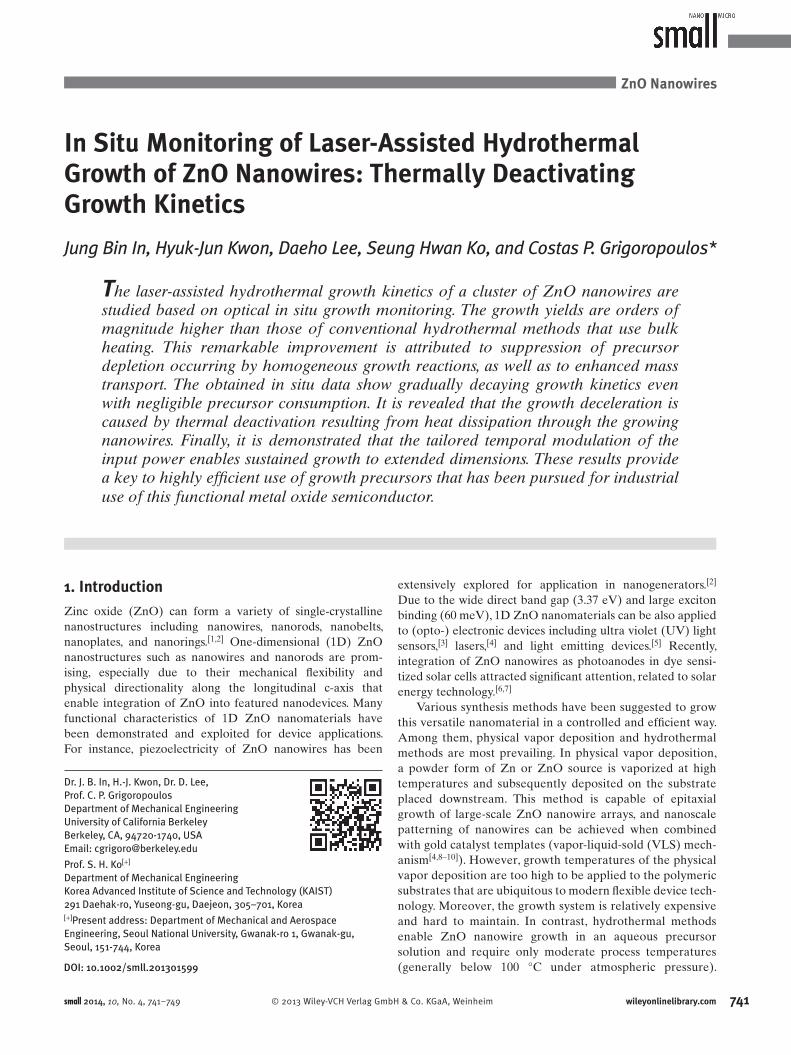
For this study, the growth substrates with a light absorp-
tion layer (Au/Cr; 90 nm/ 80 nm) on top were coated with
ZnO crystalline seeds (diameter: ∼ 6 nm in average) prior
to growth. This method hindered spontaneous nucleation
of additional growth sites and thereby we could selectively
capture growth kinetics of the nanowires emanating from
the pre-defi ned seeds. As shown in Figure 1 a, b, the growth
substrate was horizontally placed in our custom-built trans-
parent growth reactor that was equipped with optical win-
dows. Liquid inlet and outlet channels were introduced to the
reactor so that the precursor solution could be replenished,
replaced or removed without losing the original position of
the laser beam spot. However, we note that during a single
growth run we did not replenish the precursor, so the growth
was conducted basically in a closed reactor system. The fl ow
channels were used only for etching experiments that will
be discussed later. We used an almost ubiquitous combina-
tion of precursors: an aqueous mixture solution of equimolar
Figure 1. (a) Photo image of the in situ growth monitoring setup. (b) Schematic of the growth reactor. The nanowire cluster snapshot is the in situ image captured during ZnO nanowire growth that corresponds to Figure (c–f). The upper half shows the mirror image of the real nanowire cluster. (c) SEM image (side view) of a representative ZnO nanowire cluster grown with a laser power of 26.3 mW for 1 hr (scale bar: 10 μ m). (d) Magnifi ed SEM image of the marked area of (c) (scale bar: 2 μ m). (e) SEM image (top view) of the same sample (scale bar: 10 μ m). (f) Magnifi ed SEM image of the marked area of (e) (scale bar: 2 μ m).
small 2014, 10, No. 4, 741–749

In Situ Monitoring of Laser-Assisted Hydrothermal Growth of ZnO Nanowires
743www.small-journal.com© 2013 Wiley-VCH Verlag GmbH & Co. KGaA, Weinheim
(25 mmol each) zinc nitrate (Zn(NO 3 ) 2 , 98%, Sigma-Aldrich)
and hexamethyltetramine (HMTA; (CH 2 ) 6 N 4 , ≥ 99.0%,
Sigma-Aldrich). Polyethylenimine (PEI; C 2 H 5 N) has been
also frequently used to enhance the aspect ratio of hydro-
thermally grown ZnO nanowires. In this study, however,
we assume that PEI induces different growth kinetics, for
instance by suppressing radial growth and homogeneous
nucleation processes. [ 6,13 ] Hence, we did not use PEI in our
experiments to capture more fundamental growth kinetics.
A laser beam (continuous, λ = 532 nm) was focused ver-
tically onto the substrate. All growth experiments were con-
ducted under ambient conditions. It should be noted that in
the hydrothermal growth of this study, light irradiation was
directed to the backside of the light-absorbing layer (see
Figure S1 in the Supporting Information); thus, the opposite
front side (or the seed side) was exposed to the precursor
solution. This is because the direct front irradiation can opti-
cally interfere with the growing nanowires, resulting into mul-
tiple scattering and backscattering into the ambient. [ 12 ] In this
case, understanding of the growth mechanism can be further
hampered by transient light absorption (or scattering) that
evolves with elongation of nanowires. Moreover, the direct
irradiation can also involve possible photolytic interaction
with the precursor molecules or the material deposits. There-
fore, we adopted the back irradiation scheme to mitigate the
listed complications. As a result, the role of the laser irradia-
tion essentially reduced to a localized heat source for thermal
activation of the growth process. (See the Experimental Sec-
tion for more details of the laser growth experiment.)
Figure 1 c–f shows the scanning electron microscope
(SEM) images of a representative ZnO nanowire cluster
grown by the focused laser beam (power: 26.3 mW/ beam
diameter d : 32.6 μ m) for 1 h. The cluster has a pseudo-hem-
ispherical shape with decreasing lengths of the nanowires
from the spot center to the perimeter. This trend refl ects
the lateral thermal gradient induced by the Gaussian beam
shape. It is well known that the competitive growth mecha-
nism between randomly oriented nanowires results in growth
alignment to the normal direction of the substrate (Figure 1 f).
In our spot-wise growth, however, edge effects are so pro-
nounced that the nanowires near the perimeter of the cluster
grow inclined to the horizontal direction (Figure 1 d). These
morphological features confi rm random crystalline orienta-
tions of our initial seeds. [ 14,15 ]
2.2 . In Situ Monitoring of Growth Kinetics and Data Post-Processing
In order to explore the in situ growth kinetics, a simple
imaging system (consisting of a 20× objective lens, a zoom
lens, and a charge-coupled device (CCD) camera) was
directed perpendicularly to the reactor window (Figure 1 a).
Here we adjusted the magnifi cation of the zoom lens so that
1 CCD pixel corresponded to about 1 μ m in real length. This
optical confi guration enabled direct side-view observation
of a growing nanowire cluster. The oblique illumination of a
light-emitting device (LED) provided high-contrast images
that benefi ted from the light scattering incurred by the ZnO
nanowire cluster. The image data were saved into a movie fi le
for the next post-processing.
Figure 2 a pictorially describes the post-processing with
real image data and the corresponding kinetic curve. Image
processing was conducted to detect the boundary edges that
defi ne the shape of the nanowire cluster. In this study, we
focus on axial growth kinetics of ZnO nanowires at the center
of the cluster, so the transient edge position of the center
was traced and converted to height information. Finally, the
kinetic data were presented as a “nanowire height vs. growth
time” plot. However, it should be noted that we do not show
the kinetic data obtained during the fi rst 60 s after the laser
shutter was opened. This is because the initially small volume
of the nanowire cluster does not scatter suffi cient light,
resulting in a poor image contrast. The following image pro-
cessing on this blurry image mostly failed to provide reliable
Figure 2. (a) Illustration of postprocessing with real data. The set of snapshots shows the in situ optical images and post-processed outline images that were captured at the corresponding time. All the images have been rotated by 180°. (The real growth occurred downward with the base of the nanowires pinned to the fl ipped seed surface.) The kinetic data were collected from the highest pixel position of each image. (b) Comparison of the apparent growth rate to other hydrothermal growth data (fi nal length vs. growth time) reported from different groups. Morin et al . took advantage of a continuous fl ow reactor (CFR) to mitigate precursor depletion and their growth yield is relatively high.
small 2014, 10, No. 4, 741–749

J. B. In et al.
744 www.small-journal.com © 2013 Wiley-VCH Verlag GmbH & Co. KGaA, Weinheim
full papers
information on the edge positions. The accuracy of the meas-
urement was evaluated based on comparison to SEM of the
samples. The deviation was estimated less than 1.5 μ m in all
samples and the edge information showed a great match with
real morphologies (see Figure S2 in the Supporting Informa-
tion). Additional information on the image processing is pro-
vided in the Experimental Section.
2.3 . Rapid Ultra-High Yield Growth Kinetics
The laser-assisted hydrothermal growth is distinguished by
starkly rapid growth speed and the resulting high growth
yield. (In this paper, we consider growth yield based on the
laser irradiation area.) For instance, focused laser irradia-
tion of 45.7 mW ( d : 36.3 μ m) produced almost 60 μ m-long
nanowires for an hour (Figure 2 a). The growth rates can
increase further with higher laser power, but the laser power
is limited below the level of bubble generation. As shown
in Figure 2 b, the apparent growth rate (or growth yield) of
our laser-assisted hydrothermal growth is remarkably fast:
tens of micrometers for an hour, which is approximately two orders of magnitude higher than other conventional hydro-
thermal growth rates obtained under similar conditions. [ 14–20 ]
The growth parameters of the compared studies are listed in
Table S1 of Supporting Information.
The extraordinary growth improvement might have
been attributed to possible unconventional morphologies of
nanowires that can affect apparent axial growth kinetics. [ 21 ]
However, the structures of the nanowires grown by the laser
method do not seem to be signifi cantly altered. As shown in
Figure 1 f, the produced nanowires maintain the typical prism
structure with the basal plane (0001) normal to the growth
direction. In Figure 3 , we also observe morphological evo-
lution with growth time that has been commonly shown in
the conventional hydrothermal growth. Since axial growth is
accompanied by relatively slow, albeit evident radial growth,
longer nanowires have larger diameters. [ 14,17 ] As nanowire
lengths exceed inter-nanowire distances, some of the inclined
nanowires stop growing because of spatial interference
with other neighboring nanowires. As a result, the number
density decreases with longer growth,
which can be accounted for by geometrical
selection. [ 15 ]
In addition, the growth might be
affected by the underlying gold surface.
For instance, Xu et al. reported seedless
growth of vertically aligned ZnO nanow-
ires on an annealed thin gold fi lm. [ 22 ] In
our laser growth, however, most of the
nanowires were randomly oriented as
aforementioned. Moreover, a control
experiment (isothermal growth with the
same substrate at 95 °C) produced very
short nanowires ( ≈ 520 nm/h) in accord-
ance with observations of isothermal
hydrothermal growth. Thus, here we
confi rm that the gold fi lm does not infl u-
ence hydrothermal growth kinetics under
the examined conditions. From these fi ndings, we postulate
that the ultra-high yield of the laser-assisted growth is due
to amplifi ed driving force for crystallization (that is, higher
supersaturation) at the growth interface.
The most fundamental difference of our laser-assisted
growth from other conventional hydrothermal processes
lies in the thermal environment of the growth reaction zone.
Thus, we focus on thermal aspects of the laser-assisted growth
to elucidate the mechanism of the ultra-high yield growth.
In the case of conventional hydrothermal growth, the entire
pool of the precursor solution is subject basically to the same
growth temperature. Mostly, the boundary layer thickness of
precursor concentration is much smaller than reactor dimen-
sions. Therefore, the thermally activated precursor molecules
can spontaneously nucleate and homogeneously grow into
ZnO crystals dispersed in liquid before they diffuse into the
heterogeneous growth sites of the substrate. Indeed, many
studies have reported signifi cant precursor consumption by
homogeneous growth reactions. [ 19,23 ]
In principle, the aforementioned challenge can be over-
come either by using a continuous fl ow reactor (CFR) system
that continuously provides fresh nutrients to keep a con-
stant growth condition, [ 19,24 ] or by minimizing the volume
of the heat-affected zone while maintaining a suffi ciently
high temperature at the growth interface. The laser-assisted
hydrothermal growth offers a unique opportunity that cor-
responds to the latter. As the laser beam is focused onto the
substrate, a temperature gradient develops from the thin light
absorption layer down to the liquid medium. Consequently,
the reaction area is confi ned to the proximity of the sub-
strate surface and the growing ZnO nanowires. This growth
localization suppresses precursor consumption from homoge-
neous reactions in liquid far from the substrate and thereby
improves effi ciency of precursor utilization.
In addition, mass transport of precursor molecules can be
signifi cantly facilitated in the spot-wise laser-assisted growth.
In a dilute precursor solution, mass transport can be an
important rate-limiting step of growth kinetics. For instance,
under a similar level of precursor concentration (16 mmol),
Boecker et al. [ 17 ] reported a higher growth yield near the edge
of seeded substrate region, which is a typical phenomenon of
Figure 3. A series of SEM images (top view at the center of the laser spot) of nanowires grown with varying growth time (from 5 to 3600 s). The calculated power density of laser irradiation on the image area is approximately 5.9 kW cm −2 . All the images are at the same magnifi cation. The scale bar of the bottom right image indicates 400 nm.
small 2014, 10, No. 4, 741–749

In Situ Monitoring of Laser-Assisted Hydrothermal Growth of ZnO Nanowires
745www.small-journal.com© 2013 Wiley-VCH Verlag GmbH & Co. KGaA, Weinheim
diffusion-limited growth. In the case of our spot-wise growth
by focused laser, such diffusion enhancement can be ampli-
fi ed. [ 25,26 ] Finally, laser heating enables a rapid temperature
ramp to a growth point due to the small volume of the heat-
affected zone. In contrast, conventional reaction baths can
experience long transient heat-up, depending on the heat
capacity of the precursor pool. This advantageous effect of
rapid heating was explored in previous studies of microwave-
assisted hydrothermal growth. [ 27,28 ] Possible superheating
at the growth spot can result in rapid growth. [ 12 ] However,
our enhanced growth kinetics was obtained in a wide range
of laser power (as will be shown later in Figure 5 ). In this
regard, here we do not consider superheating as a differenti-
ating factor.
2.4 . Growth Deceleration
Notwithstanding the benefi cial outcomes of the laser growth,
the laser-assisted growth is still limited by gradually deac-
tivating kinetics as already shown in Figure 2 a. Here we
explore the underlying deactivation mechanism. First, we
revisit the aforementioned precursor consumption and quan-
titatively evaluate its effect on growth kinetics based on our
in situ kinetic data. In Figure 4 a, run #1 and #2 show repre-
sentative kinetic curves obtained with the same laser power
(53.8 mW) but different laser beam diameters (36.3 μ m and
39.6 μ m, respectively). The nanowires of run #1 grew faster
and longer than run #2, obviously due to the higher laser
energy density at the spot center. Meanwhile, the growth rate
of run #1 decelerated faster than that of run #2. This expo-
nentially decaying growth kinetics resembles the growth
model that Zhou et al. [ 23 ] proposed to account for signifi cant
precursor consumption of their homogeneous ZnO nanowire
growth. However, calculation of the precursor utilization
promptly proves that this is not the case in our laser growth.
Even by a conservative calculation, consumed precursor for
a single laser growth experiment is estimated less than 0.1%
of the entire precursor (solution volume: 3 cc). More obvi-
ously, when we repeated the same laser growth (run #3)
immediately following run #2, we reproduced almost iden-
tical growth kinetics even without replenishing the precursor
solution. From these fi ndings, we safely rule out growth decay
by precursor depletion.
Another possibility could be growth inhibition by accu-
mulating poisonous materials such as secondary zinc-based
material deposits [ 15 ] and ionic metal impurities. [ 29 ] These
materials can cover active growth sites hindering access of
precursor molecules even with existing supersaturation. In
order to examine the validity of the poisoning theory, we
conducted a sequence of in situ growth−etching−regrowth
experiments. The etching was to remove possible poisons
so that the following regrowth could reactivate the previous
decaying growth kinetics. First, we grew a nanowire cluster
long enough to show clear growth deactivation. Then, the
laser was turned off and the precursor solution was removed.
A continuous fl ow of deionized water was applied to rinse
the growth substrate and the reactor. For etching, a dilute
HCl (0.5 mmol) was introduced to the reactor in order to
slightly etch the growth front. According to the recorded in
situ images, the nanowires were etched away from the tips by
approximately 1 μ m for 1 h. After the second rinse, the iden-
tical but fresh precursor solution was introduced again and
the regrowth was fi nally conducted under the same condition.
The growth kinetics obtained from the etching experiment is
shown in Figure 4 b. As shown by the SEM images, the etching
process roughened the nanowire surface. By regrowth, how-
ever, the original hexagonal prism structure was completely
recovered. Importantly, the regrowth kinetics does not show
distinct reactivation by this etching-regrowth scheme. Rather,
it appears that the original trend is extended by the regrowth.
Therefore, we suggest that the poisoning mechanism cannot
account for the growth deactivation of the laser-assisted
growth at least for the examined growth time (1 h).
Up to this point, it has been presumed that the tempera-
ture at the growth front changes negligibly during growth.
Considering thermal confi nement of focused laser irradia-
tion, however, we expect a signifi cant temperature gradient
in the reaction zone that can impact on the resulting growth
kinetics. [ 30 ] Indeed, our fi nite element method (FEM) simula-
tion study (COMSOL Multiphysics) suggests that the length
Figure 4. (a) Representative in situ kinetic curves that were obtained with different beam diameters (run #1: 36.3 μ m, run #2, #3: 39.6 μ m) under the same laser irradiation power (53.8 mW). The inset shows the SEM image of Run #1 (scale bar: 20 μ m). The maximum height is about 58.8 μ m (SEM measurement). (b) Kinetic data of the etching experiment. The insets show SEM of ZnO nanowires before and after the regrowth. Both scale bars indicate 1 μ m.
small 2014, 10, No. 4, 741–749

J. B. In et al.
746 www.small-journal.com © 2013 Wiley-VCH Verlag GmbH & Co. KGaA, Weinheim
full papersscale of the heat-affected zone induced by the focused laser
beam is comparable to the size of a nanowire cluster (see
Figure S3 in Supporting Information). Then we can raise a
question about how the temperature of the growth interface
(especially the tip of the growing nanowire) will respond to
the evolving thermal environment.
One of the relatively few discussions about the infl u-
ence of a temperature gradient on growth kinetics of 1D
nanomaterials is the study of Louchev et al . [ 31 ] They pro-
posed that the kinetic deceleration in growth of carbon
nanotubes (CNTs) by chemical vapor deposition (CVD)
could be attributed to thermal cool-down at the cata-
lyst tips of the growing CNT array. Analogously, the
growing ZnO nanowires can act as effective thermal fi ns
that enhance the heat transfer rate from the beam spot to
the solution medium. Even though emphasized phonon-
boundary scattering reduces the thermal conductivity of
ZnO nanowires by more than one order, ZnO nanowires
still have a higher thermal conductivity ( ∼ 10 W m −1 K −1 ,
when the diameter is ∼ 200 nm) [ 32 ] than water. Furthermore,
an amplifi ed cooling effect is reasonably expected in solution
growth, due to signifi cantly higher heat dissipation of water
(quite generally, 200−1500 W m −2 K −1 in free convection and
0.6−0.7 W m −1 K −1 in conduction) than that of general gases
(2−25 W m −2 K −1 in free convection and 0.02−0.2 W m −1 K −1
in conduction). [ 33 ] (See the Supporting Information for fur-
ther discussion on thermal transport processes.) In contrast
to CVD processes, the growth temperature of the laser
method is sustained from the thin light-absorbing fi lm that
has a small heat capacity. Thus, the temperature at the base as
well as at the tip of the nanowire can immediately respond to
heat loss through the nanowire structure, resulting in growth
deceleration.
In order to fi nd experimental evidence on the thermally
deactivated growth, we conducted systematic growth experi-
ments (1 hour of growth time each) by changing the laser
power. The beam diameter was set at 39.6 μ m. Figure 5 shows
each kinetic curve and the resulting trend of the growth
heights. As shown in Figure 5 a, the growth rate is obviously
proportional to laser power due to the increased growth tem-
perature. At a higher temperature, the growth reactions shift
forward with the increased concentration of OH − ion most
likely caused by faster decomposition of HMTA. [ 11,23 ] Figure 5 b
also indicates that the nanowire height (obtained by 1 h
growth) increases linearly with higher laser power. Nanow-
ires could not be grown below a specifi c level of laser power,
which should be related to the equilibrium temperature of
the growth reactions. [ 20 ]
Interestingly, the growth with the lowest laser power
(19.8 mW) generated secondary deposits near the rim area
of the growth spot. The SEM images in Figure 5 b show that
the secondary material covers the nanowires that already
grew, implying that this material nucleated and grew at a
later stage of the laser growth. However, growth with higher
laser power did not generate such secondary deposits for the
same growth time. Furthermore, when we reduced the laser
power less than 18.1 mW we found that the secondary mate-
rial completely covered the beam spot area without nanow-
ires observable by SEM. The material identity is under our
investigation, but we could fi nd morphological resemblance
from several studies on hydrothermal generation of zinc
hydroxide precipitates. [ 34,35 ] Especially in accordance with
our observation, Olson et al. reported possible zinc hydroxide
nucleation on the ZnO nanowires at a reduced level of super-
saturation. [ 15 ] Therefore, we postulate that this secondary
material is indicative of low growth temperatures.
If this is the case, the secondary material should be also
generated under any higher laser power by extended growth;
eventually, the enhanced heat transfer of longer nanowires
will reduce the tip temperature to the level responsible for
generation of the secondary material. Indeed, when we con-
ducted a laser growth experiment at suffi ciently high laser
power (57.7 mW, d : 39.6 μ m) for 7 h, we found a similar mate-
rial deposit growing from the center vertex of the nanowire
cluster. Figure 6 a–c shows the in situ snapshots, ex situ optical
microscope (bright fi eld), and SEM images of the nanowire
cluster, respectively. Figure 6 a especially reveals that the
secondary material started to grow from about 3 h after the
initiation of the laser irradiation. Here, we note that this sec-
ondary material can be regarded as a growth poison from the
Figure 5. (a) In situ kinetic curves obtained with different laser powers ( d : 39.6 μ m). The SEM images show the side views of each sample. The scale bars commonly indicate 20 μ m. (The size of every image box corresponds to 60 μ m × 60 μ m.) The maximum height of each cluster measured by SEM is: (i) 49. 41 μ m, (ii) 40.35 μ m, (iii) 32.03 μ m, (iv) 20.24 μ m. (b) Plot of fi nal height (1 h growth) vs laser power. This plot shows the height data obtained by SEM measurement.
small 2014, 10, No. 4, 741–749

In Situ Monitoring of Laser-Assisted Hydrothermal Growth of ZnO Nanowires
747www.small-journal.com© 2013 Wiley-VCH Verlag GmbH & Co. KGaA, Weinheim
perspective of poisoning theory. [ 15 ] As previously mentioned
in our etching and regrowth experiment, we believe that this
poisoning mechanism manifests itself rather at the fi nal stage
of the ZnO growth and thereby it is more related to a perma-
nent growth termination.
To further support the proposed thermal deactivation,
we conducted a laser growth experiment by increasing the
laser power stepwise from 26.3 mW to 53.8 mW every 10 min
during a single growth run. Figure 7 shows the corresponding
in situ kinetic data. The lower curve shows the regular growth
with constant laser power (26.3 mW), and the upper curve
indicates the power-manipulated growth run. Indeed, the
increasing laser power delayed deceleration of growth speed.
Here the laser beam was focused tightly, so the beam spot size
was 32.6 μ m. Interestingly, the fi rst increase of laser power
at 600 sec (from 26.3 mW to 33.4 mW) did not generate a
bubble due to effi cient heat dissipation through the already
grown nanowires. Otherwise, the 33.4 mW laser power would
instantly generate a bubble by excessive heat. This result vali-
dates the role of the growing nanowires as thermal fi ns. Con-
sequently, we confi rm the presence of thermal cool-down at
the growth front that is responsible for the observed growth
deceleration and the subsequent growth termination.
2.5 . Effi cient Hydrothermal Growth
Our results provide profound implications on further devel-
opment for effi cient hydrothermal synthesis of ZnO nanow-
ires. (1) The localized heat-affected zone develops over time
to a suffi cient spatial extent for sustaining the related growth
reactions up to tens of micrometers of the nanowire height.
In this regard, the relatively large volume of conventional
chemical bath reactor systems and thereby the amount of
wasted precursor can be reasonably reduced. Rather, in-
line precursor replenishment will be a more effi cient way
to support further extended growth time or continuous
batch runs. (2) While the growth rate increases with higher
light power, boiling phenomena limit the possible enhance-
ment by further increase of light power. However, pressur-
izing the growth reactor will suppress boiling and allow a
wider window of laser power for faster growth. (3) We also
expect that the laser heating will enable reproducible growth
on substrates not only because it decouples substrate growth
kinetics from complicated homogeneous reaction kinetics [ 11 ]
but also because it is free of effects of reactor walls. (4) As
this is a purely thermal approach, one can still apply previ-
ously invented chemical growth modifi cation (i.e. improving
aspect ratio by using chemical capping agents [ 1,6 ] ). (5) The
proposed method does not necessarily require laser irra-
diation; in practice, the technique is readily compatible with
other rapid thermal processes such as Joule-heating of thin
conductive elements, [ 36 ] which can be a promising solution
for large-area growth. In this case, we also expect thermally
deactivating growth kinetics (see 1D thermal fi n model in
Supporting Information). (6) A relatively large beam spot
and the accompanying heat diffusion limit the resolution of
selective growth that is crucial for single-nanowire device fab-
rication. A high magnifi cation process can improve the reso-
lution to a degree, but the required tight control hinders its
practical use in industry. Moreover, the demonstrated facile
coating of crystalline seeds results in uncontrolled orientation
of nanowires. To resolve this dimensional restriction, density-
controlled accurate positioning of seed material should be
pursued. [ 37 ] (7) When it comes to the total energy consump-
tion necessary for mass production of nanowires (J/kg),
the energy effi ciency of laser growth can hardly match that of
a well-insulated conventional growth system. This is because
Figure 7. The lower curve indicates growth kinetics with constant laser power (26.3 mW). The upper curve indicates growth with stepwise increasing laser power (every 10 min). Each inset shows the SEM of the corresponding nanowire cluster. We note that the lower right inset is identical to Figure 1 c. Both of the images have been resized at the same scale (scale bar: 20 μ m).
Figure 6. (a) In situ optical images of the ZnO nanowire cluster grown for the extended growth time (7 hours). A secondary material starts to grow at the cluster top center (*) from about 3 hr. (b) Ex situ optical microscope image (bright fi eld) of the fi nal structure. (c) SEM image of the corresponding cluster (scale bar: 40 μ m). All the images have been rotated by 180° from the real growth confi guration.
small 2014, 10, No. 4, 741–749

J. B. In et al.
748 www.small-journal.com © 2013 Wiley-VCH Verlag GmbH & Co. KGaA, Weinheim
full papersof the limited energy effi ciency of the laser system itself,
wasted light by refl ection, and heat dissipation to the solu-
tion medium. In this respect, we envision another possibility
of using even sunlight as a light source for this photo-thermal
process. [ 38 ] For instance, sunlight irradiation on 1 cm 2 can
provide about 135 mW light power, which can be reasonably
utilized as a local heat source of our technique when focused.
3 . Conclusion
In summary, we have demonstrated ultra-high yields of
laser-assisted hydrothermal growth of ZnO nanowires and
the related growth kinetics. In situ monitoring of the growth
kinetics reveals that the laser growth enables effi cient use of
the growth precursor by suppressing homogeneous growth
reactions. In contrast to the conventional hydrothermal
growth where the precursor solution and the growing
nanowires are subject to an isothermal environment, local-
ized heating of laser induces growth deceleration stemming
from the enhanced heat transfer (that is, cooling) through the
growing nanowire thermal fi ns. As the growth temperature
decreases to the level of the secondary material deposition,
possible growth poisoning can affect the growth kinetics and
result in growth termination. This growth deactivation can be
mitigated by providing the growth sites with sustained heat
(i.e. increasing light power). Our results therefore provide
a fundamental basis for development of a high-effi ciency
hydrothermal growth system.
4 . Experimental Section
Seed Substrate Preparation : We deposited a thin Au/Cr light-absorption layer (90 nm/ 80 nm) on a quartz glass wafer (560 μ m thick) by electron beam evaporation. A seed solution consisting of crystalline ZnO nanoparticle seeds (size: ∼ 6 nm, 0.019% (wt.), prepared by the modifi ed procedure [ 39 ] of Pacholski method [ 40 ] ) was spin-coated on the substrate at 3000 rpm for 1 min. Then the coated surface was dried by blowing a nitrogen gas, and heated on a hot plate (120 °C) for 3 min to enhance adsorption of the particles. Each coating step was repeated 3 times to increase the seed density. After completion of the coating procedure, the coated wafer was split into pieces. The pieces possibly under an edge effect of spin coating were selectively removed. The opposite (or irradiation) side of the quartz wafer was cleaned with an ethanol-soaked cotton swab to make an optically clean surface.
Laser-Assisted Hydrothermal Growth : A growth substrate was mounted on a polydimethylsiloxane (PDMS) block, and this assembly was placed in the growth reactor built with trans-parent glass slides to allow optical imaging. We used two sepa-rate syringe pumps to inject and remove solutions. The growth precursor ( ∼ 3 mL) was introduced through a 0.22 μ m fi lter unit (Millipore). As illustrated in Figure 1 b, this precursor volume lev-eled the solution free surface at the substrate position without submerging the substrate. This leveling was necessary to get a consistent focus without interference due to the different
refractive index of the solution. A glass slide capped the reactor to minimize evaporation of the solution. The measured evapora-tion during laser irradiation was negligible ( ∼ 15 μ m/h decrease in level). A continuous diode-pumped solid-state laser ( λ = 532 nm, LambdaPro) was used for laser irradiation. We applied different combinations of absorptive neutral density (ND) fi lters to control the laser power. Each laser power was measured with a power meter (Coherent) and it showed a stable output (Figure S1b) for the general growth time (1 hour). A plano-convex lens (LA1951-A, Thorlabs) was placed above the reactor to focus the laser beam on the Au/Cr absorption layer. The size (1/e 2 ) of the focused beam spot was approximately 32.6 μ m in diameter and could be enlarged by adjusting the working distance (lens-to-object dis-tance). Whenever a new sample was loaded, the working distance was carefully set with multiple alignment stages and an optical micrometer (LS7030M, Keyence). When we applied laser on the substrate, the growth spot was positioned closer to the camera side for better image quality, but kept about 150 μ m away from the substrate edge to avoid possible enhanced heat transfer very near the edge. As very limited area of the substrate was used for growth, multiple growth experiments could be conducted in a single batch. We especially note that the series of growth experi-ments in this study was conducted on a single substrate sample for consistency.
In Situ Growth Monitoring System and Image Processing : In conjunction with the laser-assisted growth of ZnO nanowire clusters, we adopted a rather straightforward optical imaging method to explore real-time growth kinetics of ZnO nanowires. The system consists of basic components: a 20× objective lens, a zoom lens, a CCD camera (resolution: 640 × 480, frame rate: 29.98 fps) for image capture, and a LED ( λ = 375 nm, LED370E, Thorlabs) for oblique illumination. A higher magnifi cation might help get a better resolution, but the accompanying reduced depth of focus undermined the image quality due to the 3-dimen-sional structure of the nanowire cluster. A fl uorescence imaging fi lter ( λ = 460 nm/ fwhm = 60 nm, MF460–60, Thorlabs) was installed in the monitoring optics to selectively cut off the laser light ( λ = 532 nm) from the light-scattered image. A recorded video that corresponds to run #3 of Figure 4 a is provided in Sup-porting Information.
Post-processing of the Image Data : The original movie data (recorded for 1 hour) has a large amount of data, so the movie was reduced to 1/64 size by using Microsoft Windows Movie Maker. For image processing we used a built-in function “edge()” of MATLAB to detect edges from the raw image data. We did not con-duct additional data processing (such as data smoothing) on the fi nal kinetic curves. Thus, the seemingly stepwise increase in the kinetic curves presented in this paper does not refl ect real abrupt growth behavior but originates from the limited spatial resolution of the growth monitoring system.
Supporting Information
Supporting Information is available from the Wiley Online Library or from the author.
small 2014, 10, No. 4, 741–749

In Situ Monitoring of Laser-Assisted Hydrothermal Growth of ZnO Nanowires
749www.small-journal.com© 2013 Wiley-VCH Verlag GmbH & Co. KGaA, Weinheim
Acknowledgements
This work was supported by the NSF SINAM NSEC. J.B.I. thanks Dr. Sang-Gil Ryu for advice on laser experimentation.
[1] S. Xu , Z. L. Wang , Nano. Res. 2011 , 4 , 1013 . [2] Z. L. Wang , MRS Bull. 2012 , 37 , 814 . [3] C. S. Lao , M. C. Park , Q. Kuang , Y. L. Deng , A. K. Sood , D. L. Polla ,
Z. L. Wang , J. Am. Chem. Soc. 2007 , 129 , 12096 . [4] M. H. Huang , S. Mao , H. Feick , H. Q. Yan , Y. Y. Wu , H. Kind ,
E. Weber , R. Russo , P. D. Yang , Science 2001 , 292 , 1897 . [5] Y. F. Hsu , Y. Y. Xi , K. H. Tam , A. B. Djurisic , J. M. Luo , C. C. Ling ,
C. K. Cheung , A. M. C. Ng , W. K. Chan , X. Deng , C. D. Beling , S. Fung , K. W. Cheah , P. W. K. Fong , C. C. Surya , Adv. Funct. Mater. 2008 , 18 , 1020 .
[6] M. Law , L. E. Greene , J. C. Johnson , R. Saykally , P. D. Yang , Nat. Mater. 2005 , 4 , 455 .
[7] S. H. Ko , D. Lee , H. W. Kang , K. H. Nam , J. Y. Yeo , S. J. Hong , C. P. Grigoropoulos , H. J. Sung , Nano Lett. 2011 , 11 , 666 .
[8] R. S. Wagner , W. C. Ellis , Appl. Phys. Lett. 1964 , 4 , 89 . [9] A. M. Morales , C. M. Lieber , Science 1998 , 279 , 208 . [10] M. H. Huang , Y. Y. Wu , H. Feick , N. Tran , E. Weber , P. D. Yang , Adv.
Mater. 2001 , 13 , 113 . [11] W. S. Jang , T. Il Lee , J. Y. Oh , S. H. Hwang , S. W. Shon , D. H. Kim ,
Y. N. Xia , J. M. Myoung , H. K. Baik , J. Mater. Chem. 2012 , 22 , 20719 .
[12] J. Yeo , S. Hong , M. Wanit , H. W. Kang , D. Lee , C. P. Grigoropoulos , H. J. Sung , S. H. Ko , Adv. Funct. Mater. 2013 , DOI: 10.1002/adfm.201203863.
[13] C. K. Xu , P. Shin , L. L. Cao , D. Gao , J. Phys. Chem. C 2010 , 114 , 125 .
[14] L. E. Greene , M. Law , J. Goldberger , F. Kim , J. C. Johnson , Y. F. Zhang , R. J. Saykally , P. D. Yang , Angew. Chem. Int. Ed. 2003 , 42 , 3031 .
[15] T. Y. Olson , A. A. Chernov , B. A. Drabek , J. H. Satcher , T. Y. J. Han , Chem. Mater. 2013 , 25 , 1363 .
[16] S. H. Ko , I. Park , H. Pan , N. Misra , M. S. Rogers , C. P. Grigoro-poulos , A. P. Pisano , Appl. Phys. Lett. 2008 , 92 , 154102 .
[17] J. E. Boercker , J. B. Schmidt , E. S. Aydil , Cryst. Growth Des. 2009 , 9 , 2783 .
[18] S. Guillemin , V. Consonni , E. Appert , E. Puyoo , L. Rapenne , H. Roussel , J. Phys. Chem. C 2012 , 116 , 25106 .
[19] S. A. Morin , M. J. Bierman , J. Tong , S. Jin , Science 2010 , 328 , 476 .
[20] A. R. Rachamim , S. H. Dalai , S. M. L. Pfaendler , M. E. Swanwick , A. J. Flewitt , W. I. Milne , Mater. Res. Soc. Symp. Proc. 2009 , 1174 , 1174 - V11–08 .
[21] B. Liu , H. C. Zeng , Nano Res. 2009 , 2 , 201 . [22] S. Xu , N. Adiga , S. Ba , T. Dasgupta , C. F. J. Wu , Z. L. Wang , ACS
Nano 2009 , 3 , 1803 . [23] Z. Z. Zhou , Y. L. Deng , J. Phys. Chem. C 2009 , 113 , 19853 . [24] J. J. Richardson , F. F. Lange , Cryst. Growth Des. 2009 , 9 , 2576 . [25] D. J. Hwang , S. G. Ryu , C. P. Grigoropoulos , Nanotechnology
2011 , 22 , 385303 . [26] D. E. Kotecki , I. P. Herman , J. Appl. Phys. 1988 , 64 , 4920 . [27] S. M. Mahpeykar , J. Koohsorkhi , H. Ghafoori-fard , Nanotech-
nology 2012 , 23 , 165602 . [28] H. E. Unalan , P. Hiralal , N. Rupesinghe , S. Dalal , W. I. Milne ,
G. A. J. Amaratunga , Nanotechnology 2008 , 19 , 255608 . [29] J. Joo , B. Y. Chow , M. Prakash , E. S. Boyden , J. M. Jacobson , Nat.
Mater. 2011 , 10 , 596 . [30] D. Bäuerle , Laser Processing and Chemistry, 4th ed. , Springer ,
Berlin-Heidelberg, Germany 2011 , pp. 39 – 83 . [31] O. A. Louchev , T. Laude , Y. Sato , H. Kanda , J. Chem. Phys. 2003 ,
118 , 7622 . [32] C. T. Bui , R. G. Xie , M. R. Zheng , Q. X. Zhang , C. H. Sow , B. W. Li ,
J. T. L. Thong , Small 2012 , 8 , 738 . [33] F. P. Incropera , D. P. DeWitt , Introduction to heat transfer, 4th
ed. , Wiley , New York, USA 2002 , pp. 2 – 41 . [34] G. M. Hua , L. D. Zhang , J. Dai , L. H. Hu , S. Y. Dai , Appl. Phys. A
2011 , 102 , 275 . [35] M. K. Lee , T. H. Shih , P. C. Chen , J. Electrochem. Soc. 2009 , 156 ,
H268 . [36] C. Y. Jin , Z. Y. Li , R. S. Williams , K. C. Lee , I. Park , Nano Lett.
2011 , 11 , 4818 . [37] S. G. Ryu , E. Kim , J. H. Yoo , D. J. Hwang , B. Xiang , O. D. Dubon ,
A. M. Minor , C. P. Grigoropoulos , ACS Nano 2013 , 7 , 2090 . [38] M. T. Lee , M. Werhahn , D. J. Hwang , N. Hotz , R. Greif ,
D. Poulikakos , C. P. Grigoropoulos , Int. J. Hydrogen Energy 2010 , 35 , 118 .
[39] B. Sun , H. Sirringhaus , Nano Lett. 2005 , 5 , 2408 . [40] C. Pacholski , A. Kornowski , H. Weller , Angew. Chem. Int. Ed.
2002 , 41 , 1188 .
Received: May 24, 2013Revised: July 14, 2013Published online: September 20, 2013
small 2014, 10, No. 4, 741–749