IN-SILICO STRUCTURAL ANALYSIS AFFECTING...
43
IN-SILICO STRUCTURAL ANALYSIS AFFECTING THERMOSTABILITY IN RECOMBINANT PSYCHROPHILIC CHITINASE (CHI II) FROM Glaciozyma antarctica PI12 AMALINA BINTI AMER SHAH A thesis submitted in fulfilment of the requirement for the award of the degree of Master of Engineering (Bioprocess) Faculty of Chemical and Energy Engineering Universiti Teknologi Malaysia JANUARY 2017
Transcript of IN-SILICO STRUCTURAL ANALYSIS AFFECTING...

IN-SILICO STRUCTURAL ANALYSIS AFFECTING THERMOSTABILITY IN
RECOMBINANT PSYCHROPHILIC CHITINASE
(CHI II) FROM Glaciozyma antarctica PI12
AMALINA BINTI AMER SHAH
A thesis submitted in fulfilment of the
requirement for the award of the degree of
Master of Engineering (Bioprocess)
Faculty of Chemical and Energy Engineering
Universiti Teknologi Malaysia
JANUARY 2017

iii
The day has finally come. When I can say it out loud
‘I made it’
To mama & bapak,
this is for you
iv
ACKNOWLEDGEMENT
Alhamdulillah, all praises to Allah, Whom I am grateful for His guidance.
Without His grant, this thesis could be possible. His divine and complex creations
never failed to amaze me. I am thankful to my supervisor, Prof Dr. Rosli Md Illias
for his encouragement and guidance through all these years. Not to forget, Encik
Yaakop for the technical support provided. I would like to express my gratitude to
Ministry of Science, Technology and Innovation Malaysia for the scholarship and
research funding.
My utmost appreciation to mama and bapak, my sibling, Liyana, Luqman and
Amirah for their endless love, encouragement and prayer. To my best friends Anita,
Hazwani and Suhailis for their endless motivation and support. My deep
appreciation also goes to my labmates, for your help, knowledge and unwavering
support through ups and downs. Thanks for this friendship. I wish you guys luck. Not
to forget, Dr Aizi Nor Mazila. Thank you so much for your knowledge and guidance
in my early journey. I also wish to show my appreciation to those who directly or
indirectly involved in finishing this thesis.
To all of you, thank you so much. I am blessed

v
ABSTRACT
Cold-adapted enzymes are significant with structure flexibility and high
catalytic activity at low temperature. High structural flexibility could be due to
combination of several features such as weak intramolecular bonds, decreased
compactness of hydrophobic core and reduced number of proline and arginine
residues. However, to compensate the structural flexibility, cold-adapted enzymes
are also thermolabile which causes them to be easily inactivated at elevated
temperature. Therefore, it would be more interesting and beneficial if more stable
cold-adapted enzymes are produced to fulfill the industrial needs. In this study, a
novel cold-adapted chitinase (CHI II) from Glaciozyma antarctica PI12 was
rationally designed to improve their thermostability thus make them more resistant to
increased temperature. Four CHI II mutants were designed through rational design
named as A157Q, I134P, mutant Loop and Y257R by manipulating the structural
hydrophobicity, introduction of proline in the loop regions, introduction of arginine
salt bridges and loop shortening. Mutant Loop was designed by removing 9 residues
in loop regions thus makes loop involved became shorter. Stability of all mutants
was first predicted through a computational approach where all structures were
subjected to 10 ns molecular dynamics simulation at three temperatures; 273 K, 288
K and at 300 K. Based on the simulation, it was found that mutants I134P, mutant
Loop and Y257R exhibited structural stability at 300 K. This conclusion was made
based on low and stable root-mean square deviation (RMSD) value at 300 K in
comparison to RMSD values at 288 K and 273 K. Low RMSD values indicated
mutant structure experienced low structural deviation throughout the simulation.
Besides, this observation is correlated with reduction of structure compactness
(radius of gyration), reduced solvent accessible surface area and increased numbers
of hydrogen and salt bridges. However, mutant A157Q experienced structure
destabilization at 300 K. Substitution of helix-preferred residue, alanine with a
thermolabile residue, glutamine had caused A157Q structure become loosely packed
at 300 K indicating a thermal denaturation. To support the theoretical model, CHI II
and all mutants were then cloned into Pichia pastoris expression vector pPICZαC
and expressed in P. pastoris (GS115).

vi
ABSTRAK
Enzim tahan sejuk adalah dikenali dengan struktur yang fleksibel serta
aktiviti bermangkin yang tinggi pada suhu rendah. Fleksibiliti struktur adalah
disebabkan oleh kombinasi beberapa ciri seperti ikatan intramolekul yang lemah,
penurunan kepadatan teras hidrofobik dan pengurangan sisa prolina dan arginina.
Walau bagaimanapun, untuk menebus kembali fleksibiliti strukturnya, enzim tahan
sejuk bersifat termolabil yang menyebabkannya mudah untuk ternyahaktif pada suhu
lampau tinggi. Oleh itu, ianya sangat menarik dan bermanfaat jika enzim tahan sejuk
yang lebih stabil dapat dihasilkan bagi memenuhi keperluan industri. Dalam kajian
ini, enzim kitinase tahan sejuk (CHI II) dari organisma Glaciozyma antarctica PI12
telah direkabentuk secara rasional untuk meningkatkan tahap termostabilnya dan
menjadikan CHI II lebih tahan kepada peningkatan suhu. Empat mutan CHI II telah
di rekabentuk melalui rekabentuk rasional yang dinamakan sebagai A157Q, I134P,
Gelung mutan dan Y257R dengan mengubah suai kehidrofobikan struktur,
memperkenalkan prolina di kawasan gelung, memperkenalkan titian garam arginina
serta pemendekan gelung. Gelung mutan telah direkabentuk dengan pemotongan 9
sisa di kawasan gelung menyebabkan gelung yang terlibat menjadi semakin pendek.
Kestabilan kesemua mutan diramalkan terlebih dahulu melalui pendekatan
pengkomputeran di mana kesemua struktur mutan tertakluk kepada simulasi 10 ns
dinamik molekul yang dijalankan pada tiga suhu iaitu pada 273 K, 288 K dan 300 K.
Berdasarkan simulasi yang dijalankan, mutan I134P, gelung mutan dan Y257R
menunjukkan nilai sisihan punca min kuasa dua (RMSD) yang rendah dan stabil
pada suhu 300 K, jika dibandingkan dengan nilai RMSD pada 288 K dan 273 K.
Nilai RMSD yang rendah menggambarkan struktur mutan telah mengalami sisihan
berstruktur rendah keseluruhan simulasi dijalankan. Sebaliknya, pemerhatian ini
berkorelasi dengan penurunan kepadatan struktur (jejari legaran), penurunan luas
permukaan boleh capai pelarut dan peningkatan ikatan hidrogen serta titian garam.
Walau bagaimanapun, mutan A157Q mengalami penurunan kestabilan struktur pada
300 K. Penggantian sisa pilihan heliks, iaitu alanina dengan sisa termolabil,
glutamina telah menyebabkan struktur A157Q menjadi longgar pada suhu 300 K
menandakan penyahaslian terma. Untuk menyokong model teori ini, CHI II dan
kesemua mutan telah diklon ke dalam vektor ekspresi Pichia pastoris pPICZαC dan
dinyatakan dalam P. pastoris (GS115).
vii
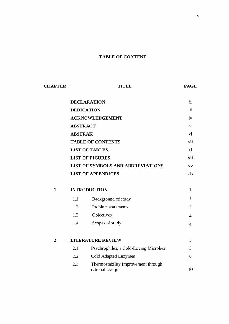
TABLE OF CONTENT
CHAPTER TITLE PAGE
DECLARATION ii
DEDICATION iii
ACKNOWLEDGEMENT iv
ABSTRACT v
ABSTRAK vi
TABLE OF CONTENTS vii
LIST OF TABLES xi
LIST OF FIGURES xii
LIST OF SYMBOLS AND ABBREVIATIONS xv
LIST OF APPENDICES xix
1 INTRODUCTION 1
1.1 Background of study
1.2 Problem statements
1.3 Objectives
1.4 Scopes of study
1
3
4
4
2 LITERATURE REVIEW 5
2.1 Psychrophiles, a Cold-Loving Microbes
2.2 Cold Adapted Enzymes
2.3 Thermostability Improvement through
rational Design
5
6
10

viii
2.3.1 Proline Substitution to Improve Protein
Thermostability 12
2.3.2 Improving Thermostability by Loop
Shortening 13
2.3.3 Introduction of Salt Bridges to Improve
Protein Thermostability 13
2.3.4 Substitution of Surface Alanine Residue
to a More Hydrophilic Residues 14
2.4 Thermostability Studies Through Molecular
Dynamic (MD) Simulation 14
2.5 Chitin 16
2.6 Chitinase, a Chitinolytic Enzyme 18
2.6.1 Chitinase Subfamiliy and
Classifications 19
2.6.2 Functional Domain of Fungal
Chitinases 23
2.6.3 Roles of Chitinases in Various
Organisms 25
2.7 A novel Chitinase (CHI II) From Glaciozyma
antarctica PI12 26
2.8 Chitinases and Their Applications 28
2.8.1 Bioconversion of Chitin in Production
of Single Cell Proteins 28
2.8.2 Enzymatic Hydrolysis in Production of
Chitooligosaccharides 29
2.8.3 Biocontrol Agents 30
2.9 Heterologous Expression in Pichia pastoris 30
3 MATERIALS AND METHODS 33
3.1 Introduction 33
3.2 Source of Sequences 35
3.3 Tools and Software 35
3.4 Mutation Design Strategy 38
3.5 Construction of A157Q, I134P and Y257R 3D
Models 38
3.6 Homology Modeling of Mutant Loop 38
3.7 Molecular Dynamics (MD) Simulation 39

ix
3.7.1 Preparation Stage 40
3.7.2 Production Stage 40
3.7.3 Trajectories Analysis for CHI II and
Mutants
41
3.7.3.1 Root Mean Square Deviation
(RMSD)
41
3.7.3.2 Root Mean Square Fluctuation
(RMSF) 41
3.7.3.3 Radius of Gyration 41
3.7.3.4 Analysis of Secondary
Structure for CHI II and it
Mutants
42
3.7.3.5 Solvent Accessible Surface
Area (SASA) 42
3.7.3.6 Analysis of Hydrogen Bonds
and Salt Bridges
42
3.8 Cloning of CHI II and It Mutants into Pichia
pastoris Expression System
42
3.9 Expression of Recombinant CHI II in Pichia
Pastoris strains
45
3.10 Screening of Mutant Expression 45
3.10.1 SDS PAGE Analysis 45
3.10.2 Western Blotting 46
3.10.3 Chitinase Assay 47
4 RESULTS AND DISCUSSION 49
4.1 Mutants Design Strategy 49
4.1.1 Introduction of Salt Bridge in Mutant
Y257R
49
4.1.2 Loop Shortening in Mutant Loop 52
4.1.3 Introduction of Proline in Loop Regions
for Mutant I134P
55
4.1.4 Substitution of Alanine to Glutamine in
Mutant A157Q
56
4.2 Construction of Mutants 3D Models 57
4.2.1 Construction of A157Q, I134P and
Y257R 3D Models Using PyMol
58

x
4.2.2 Homology Modeling of Mutant Loop 59
4.3 Molecular Dynamics (MD) Simulation of CHI II
and Mutants
64
4.4 Comparative Analysis of MD Simulation
Trajectories
65
4.4.1 Root Mean Square Deviation (RMSD) 65
4.4.2 Root Mean Square Fluctuation (RMSF) 71
4.4.3 Radius of Gyration 75
4.4.4 Analysis of Secondary Structure for
CHI II and Its Mutants
79
4.4.5 Solvent Accessible Surface Area
(SASA)
84
4.4.6 Analysis of Hydrogen Bonds 85
4.4.7 Analysis of Salt Bridges 87
4.5 Cloning of CHI II and It Mutants into Pichia
pastoris
92
4.6 Expression of CHI II Mutants into P. Pastoris
GS115
96
5 CONCLUSION 100
5.1 Conclusions 100
5.2 Recommendations 101
REFERENCES 102
APPENDIX A-F 119
xi
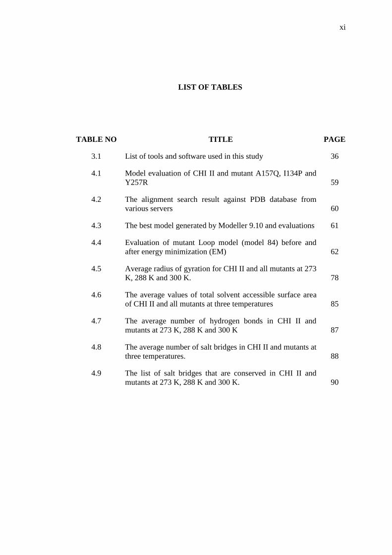
LIST OF TABLES
TABLE NO TITLE PAGE
3.1 List of tools and software used in this study 36
4.1 Model evaluation of CHI II and mutant A157Q, I134P and
Y257R 59
4.2 The alignment search result against PDB database from
various servers 60
4.3 The best model generated by Modeller 9.10 and evaluations 61
4.4 Evaluation of mutant Loop model (model 84) before and
after energy minimization (EM) 62
4.5 Average radius of gyration for CHI II and all mutants at 273
K, 288 K and 300 K. 78
4.6 The average values of total solvent accessible surface area
of CHI II and all mutants at three temperatures 85
4.7 The average number of hydrogen bonds in CHI II and
mutants at 273 K, 288 K and 300 K 87
4.8 The average number of salt bridges in CHI II and mutants at
three temperatures. 88
4.9 The list of salt bridges that are conserved in CHI II and
mutants at 273 K, 288 K and 300 K. 90
xii
LIST OF FIGURES
FIGURE NO TITLE PAGE
2.1 Factors contributed to the structural flexibility of cold-
adapted enzymes 8
2.2 The structures of amino acids arginine, proline and
glycine
9
2.3 Dry chitin flakes and the arrangement of chitin polymer 16
2.4 The arrangement of chitin microfibrils in three
polymorph forms
17
2.5 Illustration of cleavage pattern of chitinolytic enzymes 19
2.6 The classification of chitinase GH18 and GH19
subfamilies and subgroup 21
2.7 The domain organization of fungal chitinases in GH18
family
25
2.8 The predicted structure of CHI II by using threading
method.
28
3.1 Illustration of operational framework of this study 34
3.2 Three stages of MD simulation consists of preparation,
production and analysis stages
39
3.3 The overview of OE-PCR strategy to obtain a mutant’s
gene
43
4.1 The location of three salt bridges found in CHI I
structures
51
4.2 Multiple sequence alignment showed the mutation site of
Y257R
52
4.3 The structure alignment between CHI II and its template
1ITX
53
4.4 Multiple sequence alignment showed the mutation site of
mutant Loop
55
4.5 Multiple sequence alignment showed the mutation site or
I134P
56

xiii
4.6 Multiple sequence alignment showed the mutation site of
A157Q
57
4.7 Ramachandran plot of mutant Loops model (a) before
energy minimization and (b) after energy minimization
63
4.8 RMSD overlay of CHI II and four mutant at 273 K 67
4.9 RMSD overlay of CHI II and four mutants at 288 K 68
4.10 RMSD overlay of CHI II and four mutants at 300 K 69
4.11 An overview of CHI II and mutants structure snapshots
in 5 ns interval at 300 K simulation
70
4.12 The overlay RMSF graph of CHI II and all mutants at
273 K
73
4.13 The overlay RMSF graph of CHI II and all mutants at
288 K
74
4.14 The overlay RMSF graph of CHI II and all mutants at
300 K
75
4.15 The overlay Rg graph of CHI II and all mutants at 273 K 76
4.16 The overlay Rg graph of CHI II and all mutants at (a)
288 K and (b) 300 K
77
4.17 Secondary structure assignment for CHI II and mutants
at 273 K
81
4.18 Secondary structure assignment for CHI II and mutants
at 288 K
82
4.19 Secondary structure assignment for CHI II and mutants
at 300 K
83
4.20 The interaction maps between Arg57 and Asp205 which
involved carboxylate group of Asp205 and guanidium
group of Arg257.
91
4.21 The formation and breaking of salt bridge Asp205 and
Arg257 which was introduced in Y257R at three
temperatures.
91
4.22 PCR product of first PCR and second PCR reaction for
all mutants.
93
4.23 Linearization of pPICZαC and A157Q constructed
vector
94
4.24 Formation of A157Q-GS115 colonies on YPD and
YPDS plate (a) YPDS plate with 100 µg/ml of zeocin,
(b) YPD agar with 500 µg/ml of zeocin, (c) YPD agar
with 1000 µg/ml of zeocin and (d) YPD agar with 2000
µg/ml of zeocin
95

xiv
4.25 PCR colony for all mutant gene integration into P.
pastoris GS115.
96
4.26 SDS-PAGE analysis of CHI II and mutant A157Q 97
4.27 SDS-PAGE analysis of mutant Loop, I134P and Y257R. 98

xv
LIST OF SYMBOLS AND ABBREVIATIONS
% - percent
* - asterisk
.top - Topology file
~ - About
> - Greater
≥ - Greater or equal to
° - Degree
°C - degree celcius
½ - Half
3D - Three dimensional
A - Alanine
Å - Angstrom
Arg or R - Arginine
Asn - Asparagine
Asp or D - Aspartic acid/aspartate
BD - Binding domain
C. congregates - Coprinellus congregates
C. immitis - Coccidioodes immitis
CAZy - Carbohydrate Active Enzyme database
CBD - Chitin binding domain
CID - Chitinase insertion domain
Cl- - Chloride ion
Cys - Cysteine
Cα - Carbon alpha

xvi
DNA - Deoxyribonucleic acid
DOPE - Discrete optimized protein energy
DSSP - Dictionary of secondary structure prediction
EC - enzyme commission
F - Phenylalanine
G. antarctica - Glaciozyma Antarctica
GH - Glycosyl hydrolase
GlcN - Glucosamine
GlcNAc - N-acetylglucosamine
Glu or E - Glutamic acid/glutamate
Glu or Q - Glutamine
Gly or G - Glycine
GPI - Glycosylphosphatidylinositol
H - Hydrogen
H. atriviridis - Hypocrea atroviridis
His or H - Hisitidne
ID - Identifier
Ile or I - Isoleucine
K - Kelvin
kDa - Kilodalton
kJ/mol - kilojoule per mol
L - Leucine
Lys or K - Lysine
LysM - Lysin motif
MD - Molecular dynamics
MSA - Multiple sequence alignment
N - Nitrogen
N Asparagine
Na+ - Sodium ion
Nm - Nanometer
Ns - Nanoseconds
O - Oxygen
PDB - Protein data bank

xvii
pKa - Acid dissociation constant
PME - Particle mesh ewald
Pro or P - Proline
Rg - Radius of gyration
RMSD - Root mean square deviation
RMSF - Root mean square fluctuation
S. cerivisae - Saccharomyces cerivisae
SASA - Solvent accessible surface area
SCP - Single cell protein
Ser or S - Serine
sp. - Species
SPC - Simple point charge
T - Threonine
T. atroviride - Trichoderma atroviride
T. aurantiacus - Thermoascus aurantiacus
T. lanuginosus - Thermomyces lanuginosus
TIM - Triosephosphate isomerase
Tm - Midpoint temperature
Tyr orY - Tyrosine
V - Valine
VMD - Visual molecular dynamic
α - Alpha
β - Beta
γ - Gamma
φ - Phi
ψ - Psi
µg/µl - Microgram per microliter
µl - Microliter
µmol - Micromole
BMGY - Buffered glycerol complex medium
BMMY - Buffered methanol complex medium
Bp - Base pair
EDTA - Ethylenediaminetetraacetic acid
xviii
LB - Luria Bertani
M - Molarity
ml - Milliliter
mM - Millimole
OE - Overlapping extension
P. pastoris - Pichia pastoris
PCR - Polymerase chain reaction
U - Unit activity
V - Voltage
v/v - Volume over volume
YPD - Yeast extract peptone dextrose
YPDS - Yeast extract peptone dextrose sorbitol
zeoR - Zeocin resistance

xix
LIST OF APPENDICES
APPENDIX TITLE PAGE
A Tutorial for MD Simulation 119
B Routine and Media 123
C Primer Sequences 128
D pPICZαC Vector Map 129
E Nucleotide and Protein Sequences of CHI II and All
mutants
130
F Sequencing Result 132
CHAPTER 1
INTRODUCTION
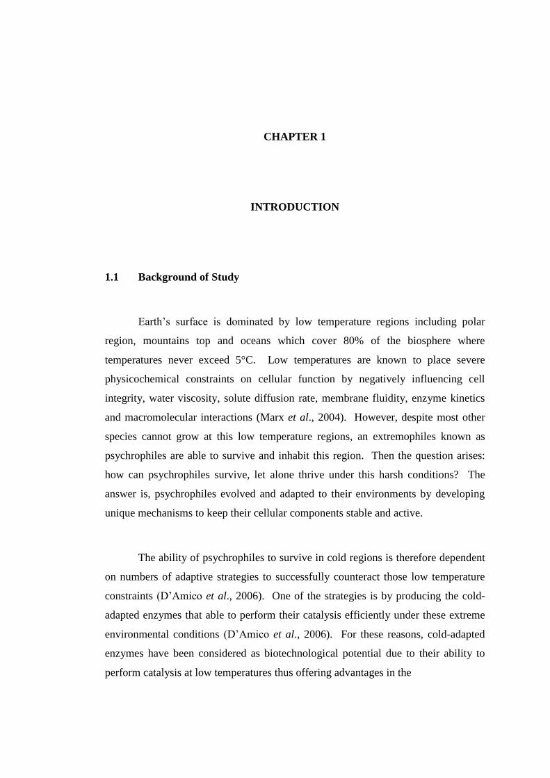
1.1 Background of Study
Earth’s surface is dominated by low temperature regions including polar
region, mountains top and oceans which cover 80% of the biosphere where
temperatures never exceed 5°C. Low temperatures are known to place severe
physicochemical constraints on cellular function by negatively influencing cell
integrity, water viscosity, solute diffusion rate, membrane fluidity, enzyme kinetics
and macromolecular interactions (Marx et al., 2004). However, despite most other
species cannot grow at this low temperature regions, an extremophiles known as
psychrophiles are able to survive and inhabit this region. Then the question arises:
how can psychrophiles survive, let alone thrive under this harsh conditions? The
answer is, psychrophiles evolved and adapted to their environments by developing
unique mechanisms to keep their cellular components stable and active.
The ability of psychrophiles to survive in cold regions is therefore dependent
on numbers of adaptive strategies to successfully counteract those low temperature
constraints (D’Amico et al., 2006). One of the strategies is by producing the cold-
adapted enzymes that able to perform their catalysis efficiently under these extreme
environmental conditions (D’Amico et al., 2006). For these reasons, cold-adapted
enzymes have been considered as biotechnological potential due to their ability to
perform catalysis at low temperatures thus offering advantages in the

2
environmental application and energy savings in industrials processes (Gianese et al.,
2001).
While other enzymes are subject to cold denaturation and suffered the loss of
activity at low temperatures, cold-adapted enzymes are resistant to cold denaturation
with efficient catalytic activity. Their survival is correlated with their structural
flexibility that was believed as a compensation for the freezing effect in cold habitats
(Johns & Somero 2004). Structures flexibility of cold-adapted enzymes is the result
of combination of several features such as increased numbers of hydrophobic side
chains that are exposed to the solvent, a decrease in the compactness of hydrophobic
core, a higher number of glycine and lysine residues, a reduced number of proline and
arginine residues and weakening of intramolecular bonds (Rodrigues & Tiedje 2008).
However, because of their structural flexibility, cold-adapted enzymes become less
stable and also thermolabile which cause them to denature at elevated temperature
(Siddiqui & Cavicchioli 2006).
Therefore, cold-adapted enzymes are often engineered either through rational
design or directed evolution to improve its thermostability. Thermostability is defined
as improved long-term survival under mild conditions and increased ability to remain
active under harsh industrial condition but still retains its catalytic efficiency (Wijma
et al., 2013). In this study, a cold-adapted chitinase named as CHI II was used as the
subject understudied. Chitinase (EC 3.2.2.14) are categorized under glycosyl
hydrolases (GH) family and can be found in wide range of organisms such as bacteria,
fungi, yeasts, plants and mammals. Capabilities of chitinase to hydrolyse chitin to a
low molecular weight chitooligomers cause them to have broad potential in industrial,
agricultural and medicinal functions (Dahiya et al., 2006; Liu et al., 2013; Patil et al.,
2000; Park & Kim 2010; Khan et al., 2015).
CHI II was previously isolated from Glaciozyma antarctica PI12 and it's three-
dimensional (3D) structures had been modeled by Ramli et al. (2011, 2012). Based
on the structure analysis and primary sequence analysis, several characteristics related
to cold adaptations were found in CHI II. CHI II was identified to have less number
of salt bridges and arginine residues, increase in surface hydrophobicity and reduced
3
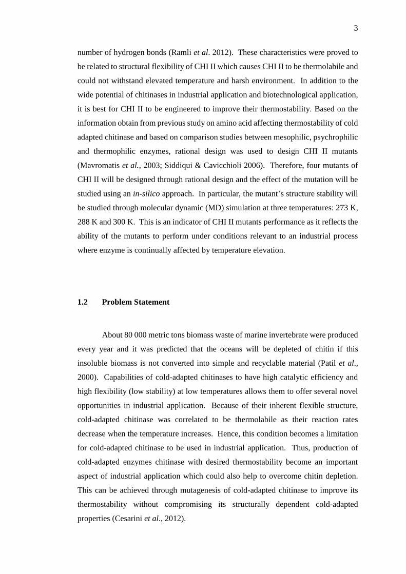
number of hydrogen bonds (Ramli et al. 2012). These characteristics were proved to
be related to structural flexibility of CHI II which causes CHI II to be thermolabile and
could not withstand elevated temperature and harsh environment. In addition to the
wide potential of chitinases in industrial application and biotechnological application,
it is best for CHI II to be engineered to improve their thermostability. Based on the
information obtain from previous study on amino acid affecting thermostability of cold
adapted chitinase and based on comparison studies between mesophilic, psychrophilic
and thermophilic enzymes, rational design was used to design CHI II mutants
(Mavromatis et al., 2003; Siddiqui & Cavicchioli 2006). Therefore, four mutants of
CHI II will be designed through rational design and the effect of the mutation will be
studied using an in-silico approach. In particular, the mutant’s structure stability will
be studied through molecular dynamic (MD) simulation at three temperatures: 273 K,
288 K and 300 K. This is an indicator of CHI II mutants performance as it reflects the
ability of the mutants to perform under conditions relevant to an industrial process
where enzyme is continually affected by temperature elevation.
1.2 Problem Statement
About 80 000 metric tons biomass waste of marine invertebrate were produced
every year and it was predicted that the oceans will be depleted of chitin if this
insoluble biomass is not converted into simple and recyclable material (Patil et al.,
2000). Capabilities of cold-adapted chitinases to have high catalytic efficiency and
high flexibility (low stability) at low temperatures allows them to offer several novel
opportunities in industrial application. Because of their inherent flexible structure,
cold-adapted chitinase was correlated to be thermolabile as their reaction rates
decrease when the temperature increases. Hence, this condition becomes a limitation
for cold-adapted chitinase to be used in industrial application. Thus, production of
cold-adapted enzymes chitinase with desired thermostability become an important
aspect of industrial application which could also help to overcome chitin depletion.
This can be achieved through mutagenesis of cold-adapted chitinase to improve its
thermostability without compromising its structurally dependent cold-adapted
properties (Cesarini et al., 2012).

4
1.3 Objectives
The main objective of this study is to analyse the effect of amino acids
substitution, loop shortening and introduction of the salt bridge in the non-catalytic
region on CHI II thermostability through in-silico approach.
1.4 Scopes of Study
The scope of this study are:
a) Design four CHI II mutants through rational design.
b) Construction of four mutants three-dimensional (3D) structures using
mutagenesis plugin in PyMOL and homology modeling by using Modeller.
c) Performing the Molecular Dynamics (MD) simulation of CHI II and its four
mutants at three different temperatures; 273 K, 288 K and 300 K.
d) Performing comparative trajectories analysis on CHI II and its mutants to study
the effect of mutation on CHI II thermostability.
102
REFERENCES
Abedi Karjiban, R., Abdul Rahman, M. B., Basri, M., Salleh, A. B., Jacobs, D., &
Abdul Wahab, H. (2009). Molecular dynamics study of the structure, flexibility
and dynamics of thermostable L1 lipase at high temperatures. The Protein
Journal, 28(1), 14–23.
Abraham, M. J., Murtola, T., Schulz, R., Páall, S., Smith, J. C., Hess, B., & Lindah, E.
(2015). Gromacs: High performance molecular simulations through multi-level
parallelism from laptops to supercomputers. SoftwareX, 1-2, 19–25.
Adrangi, S., & Faramarzi, M. A. (2013). From bacteria to human: A journey into the
world of chitinases. Biotechnology Advances.
Aghajari, N., Feller, G., Gerday, C., & Haser, R. (1998). Crystal structures of the
psychrophilic alpha-amylase from Alteromonas haloplanctis in its native form
and complexed with an inhibitor. Protein Science : A Publication of the Protein
Society, 7, 564–572.
Aghajari, N., Feller, G., Gerday, C., & Haser, R. (1998). Structures of the
psychrophilic Alteromonas haloplanctis α-amylase give insights into cold
adaptation at a molecular level. Structure, 6(12), 1503–1516.
Aghajari, N., Van Petegem, F., Villeret, V., Chessa, J.-P., Gerday, C., Haser, R., &
Van Beeumen, J. (2003). Crystal structures of a psychrophilic metalloprotease
reveal new insights into catalysis by cold-adapted proteases. Proteins: Structure,
Function, and Bioinformatics, 50(4), 636–647.
Alias, N., Ahmad Mazian, M., Salleh, A. B., Basri, M., & Rahman, R. N. Z. R. A.
(2014). Molecular cloning and optimization for high level expression of cold-
adapted serine protease from antarctic yeast Glaciozyma antarctica PI12. Enzyme
Research, 2014.
Anbar, M., Gul, O., Lamed, R., Sezerman, U. O., & Bayer, E. A. (2012). Improved
thermostability of Clostridium thermocellum endoglucanase Cel8A by using

103
consensus-guided mutagenesis. Applied and Environmental Microbiology, 78(9),
3458–3464.
Andersen, C. A., Bohr, H., & Brunak, S. (2001). Protein secondary structure: Category
assignment and predictability. FEBS Letters, 507(1), 6–10.
Arnórsdóttir, J., Helgadóttir, S., Thorbjarnardóttir, S. H., Eggertsson, G., &
Kristjánsson, M. M. (2007). Effect of selected Ser/Ala and Xaa/Pro mutations on
the stability and catalytic properties of a cold adapted subtilisin-like serine
proteinase. Biochimica et Biophysica Acta - Proteins and Proteomics, 1774(6),
749–755.
Bajaj, K., Madhusudhan, M. S., Adkar, B. V., Chakrabarti, P., Ramakrishnan, C., Sali,
A., & Varadarajan, R. (2007). Stereochemical criteria for prediction of the effects
of proline mutations on protein stability. PLoS Computational Biology, 3(12),
2465–2475.
Bauvois, C., Jacquamet, L., Huston, A. L., Borel, F., Feller, G., & Ferrer, J.-L. (2008).
Crystal structure of the cold-active aminopeptidase from Colwellia
psychrerythraea, a close structural homologue of the human bifunctional
leukotriene A4 hydrolase. Journal of Biological Chemistry, 283 (34), 23315–
23325.
Benrezkallah, D., Dauchez, M., & Krallafa, A. M. (2015). Molecular dynamics of the
salt dependence of a cold-adapted enzyme: endonuclease I. Journal of
Biomolecular Structure & Dynamics, (2), 1–11.
Berglund, L., Brunstedt, J., Nielsen, K. K., Chen, Z., Mikkelsen, J. D., & Marcker, K.
A. (1995). A proline-rich chitinase from Beta vulgaris. Plant Molecular Biology,
27(1), 211–216.
Bhuyan, M. S. I., & Gao, X. (2011). A protein-dependent side-chain rotamer library.
BMC Bioinformatics, 12(14), 1–12.
Borders, C. L., Broadwater, J. A., Bekeny, P. A., Salmon, J. E., Lee, A. S., Eldridge,
A. M., & Pett, V. B. (1994). A structural role for arginine in proteins: multiple
hydrogen bonds to backbone carbonyl oxygens. Protein Science : A Publication
of the Protein Society, 3(4), 541–8.
Borgi, M. A., Srih-Belguith, K., Ben Ali, M., Mezghani, M., Tranier, S., Haser, R., &
Bejar, S. (2004). Glucose isomerase of the Streptomyces sp. SK strain:
Purification, sequence analysis and implication of alanine 103 residue in the
enzyme thermostability and acidotolerance. Biochimie, 86(8), 561–568.
104
Bosshard, H. R., Marti, D. N., & Jelesarov, I. (2004). Protein stabilization by salt
bridges: Concepts, experimental approaches and clarification of some
misunderstandings. Journal of Molecular Recognition.
Bryksin, A. V., & Matsumura, I. (2010). Overlap extension PCR cloning: A simple
and reliable way to create recombinant plasmids. BioTechniques, 48(6), 463–465.
Bussink, A. P., Speijer, D., Aerts, J. M. F. G., & Boot, R. G. (2007). Evolution of
mammalian chitinase(-like) members of family 18 glycosyl hydrolases. Genetics,
177(2), 959–970.
Cavicchioli, R., Siddiqui, K. S., Andrews, D., & Sowers, K. R. (2002). Low-
temperature extremophiles and their applications. Current Opinion in
Biotechnology, 13(3), 253–261.
Cesarini, S., Bofill, C., Pastor, F. I. J., Reetz, M. T., & Diaz, P. (2012). A thermostable
variant of P. Aeruginosa cold-adapted LipC obtained by rational design and
saturation mutagenesis. Process Biochemistry, 47(12), 2064–2071.
Chakravarty, S., & Varadarajan, R. (2000). Elucidation of determinants of protein
stability through genome sequence analysis. FEBS Letters, 470(1), 65–69.
Chan, C. H., Yu, T. H., & Wong, K. B. (2011). Stabilizing salt-bridge enhances protein
thermostability by reducing the heat capacity change of unfolding. PLoS ONE,
6(6).
Chan, M. K., Mukund, S., Kletzin, a, Adams, M. W., & Rees, D. C. (1995). Structure
of a hyperthermophilic tungstopterin enzyme, aldehyde ferredoxin
oxidoreductase. Science (New York, N.Y.), 267(5203), 1463–1469.
Chaudhari, S. S., Arakane, Y., Specht, C. A., Moussian, B., Boyle, D. L., Park, Y.
Muthukrishnan, S. (2011). Knickkopf protein protects and organizes chitin in the
newly synthesized insect exoskeleton. Proceedings of the National Academy of
Sciences, 108 (41), 17028–17033.
Chen, J.-K. K., Shen, C.-R. R., & Liu, C.-L. L. (2010). N-acetylglucosamine:
production and applications. Marine Drugs, 8(9), 2493–2516.
Chen, W.-M., Chen, G.-H., Chen, C.-S., & Jiang, S.-T. (2009). Cloning, expression
and purification of Bacillus cereus endochitinase in the Escherichia coli AD494
(DE3) pLysS Expression System. Bioscience, Biotechnology, and Biochemistry,
73(5), 1172–1174.

105
Choi, J. H., & Lee, S. Y. (2004). Secretory and extracellular production of recombinant
proteins using Escherichia coli. Applied Microbial and Biotechnology, 64(5),
625–635.
Collinge, D. B., Kragh, K. M., Mikkelsen, J. D., Nielsen, K. K., Rasmussen, U., &
Vad, K. (1993). Plant chitinases. The Plant Journal : For Cell and Molecular
Biology, 3(1), 31–40.
Colovos, C., & Yeates, T. O. (1993). Verification of protein structures: patterns of
nonbonded atomic interactions. Protein Science : A Publication of the Protein
Society, 2(9), 1511–1519.
Criswell, A. R., Bae, E., Stec, B., Konisky, J., & Phillips, G. N. (2003). Structures of
thermophilic and mesophilic adenylate kinases from the genus Methanococcus.
Journal of Molecular Biology, 330(5), 1087–1099.
D’Amico, S., Claverie, P., Collins, T., Georlette, D., Gratia, E., Hoyoux, A. Gerday,
C. (2002). Molecular basis of cold adaptation. Philosophical Transactions of the
Royal Society of London. Series B, Biological Sciences, 357(1423), 917–925.
D’Amico, S., Collins, T., Marx, J.-C., Feller, G., & Gerday, C. (2006). Psychrophilic
microorganisms: challenges for life. EMBO Reports, 7(4), 385–389.
Daggett, V., & Levitt, M. (1993). Protein unfolding pathways explored through
molecular dynamics simulations. Journal of Molecular Biology, 232, 600–619.
Dahiya, N., Tewari, R., & Hoondal, G. S. (2006). Biotechnological aspects of
chitinolytic enzymes: A review. Applied Microbiology and Biotechnology, 71(6),
773–782.
Daly, R., & Hearn, M. T. W. (2005). Expression of heterologous proteins in Pichia
pastoris: A useful experimental tool in protein engineering and production.
Journal of Molecular Recognition, 18(2), 119-138.
Davies, G., & Henrissat, B. (1995). Structures and mechanisms of glycosyl hydrolases.
Structure, 3(9), 853–859.
De Maayer, P., Anderson, D., Cary, C., & Cowan, D. A. (2014). Some like it cold:
understanding the survival strategies of psychrophiles. EMBO Reports, 15(5),
508–17.
DeLano, W. L. (2002). The PyMOL molecular graphics system, Version 1.1.
Schrödinger LLC,

106
Dick, M., Weiergräber, O. H., Classen, T., Bisterfeld, C., Bramski, J., Gohlke, H., &
Pietruszka, J. (2016). Trading off stability against activity in extremophilic
aldolases. Scientific Reports, 6, 17908. http://doi.org/10.1038/srep17908
Donald, J. E., Kulp, D. W., & DeGrado, W. F. (2011). Salt bridges: Geometrically
specific, designable interactions. Proteins: Structure, Function and
Bioinformatics, 79(3), 898–915.
Du, X., Sang, P., Xia, Y.-L., Li, Y., Liang, J., Ai, S.-M., Liu, S.-Q. (2016).
Comparative thermal unfolding study of psychrophilic and mesophilic subtilisin-
like serine proteases by molecular dynamics simulations. Journal of
Biomolecular Structure and Dynamics, 2(1), 1–18.
Dunbrack, R., & Cohen, F. (1997). Bayesian statistical analysis of protein side-chain
rotamer preferences. Protein Science, 6(8). 1661-1681.
Duo-Chuan, L. (2006). Review of fungal chitinases. Mycopathologia, 161(6), 345–
360.
Durham, E., Dorr, B., Woetzel, N., Staritzbichler, R., & Meiler, J. (2009). Solvent
accessible surface area approximations for rapid and accurate protein structure
prediction. Journal of Molecular Modeling, 15(9), 1093–1108.
Eisenberg, D., Luthy, R., & Bowie, J., (1997). VERIFY3D: Assessment of protein
models with three-dimensional profiles. Methods in Enzymology, 277, 396–406.
Elcock, A. H. (1998). The stability of salt bridges at high temperatures: implications
for hyperthermophilic proteins. Journal of Molecular Biology, 284(2), 489–502.
Eswar, N., Webb, B., Marti-Renom, M. a, Madhusudhan, M. S., Eramian, D., Shen,
M.-Y. Sali, A. (2007). Comparative protein structure modeling using
MODELLER. Current Protocols in Protein Science, 5(6), 1-5.
Fan, H., & Mark, A. E. (2004). Refinement of homology-based protein structures by
molecular dynamics simulation techniques. Protein Science : A Publication of the
Protein Society, 13(1), 211–20.
Feller, G., Narinx, E., Arpigny, J. L., Aittaleb, M., Baise, E., Genicot, S., & Gerday,
C. (1996). Enzymes from psychrophilic organisms. FEMS Microbiology
Reviews, 18(2-3), 189–202.
Feller, G., Zekhnini, Z., Lamotte-Brasseur, J., & Gerday, C. (1997). Enzymes from
cold-adapted microorganisms. The class C beta-lactamase from the antarctic
psychrophile Psychrobacter immobilis A5. European Journal of Biochemistry /
FEBS, 244(1), 186–91.

107
Fields, P. A., Dong, Y., Meng, X., & Somero, G. N. (2015). Adaptations of protein
structure and function to temperature: there is more than one way to “skin a cat.”
Journal of Experimental Biology, 218(12), 1801–1811.
Gan, Z., Yang, J., Tao, N., Liang, L., Mi, Q., Li, J., & Zhang, K. Q. (2007). Cloning
of the gene Lecanicillium psalliotae chitinase Lpchi1 and identification of its
potential role in the biocontrol of root-knot nematode Meloidogyne incognita.
Applied Microbiology and Biotechnology, 76(6), 1309–1317.
Georlette, D., Jónsson, Z. O., Van Petegem, F., Chessa, J.-P., Van Beeumen, J.,
Hübscher, U., & Gerday, C. (2000). A DNA ligase from the psychrophile
Pseudoalteromonas haloplanktis gives insights into the adaptation of proteins to
low temperatures. European Journal of Biochemistry, 267(12), 3502–3512.
Gianese, G., Argos, P., & Pascarella, S. (2001). Structural adaptation of enzymes to
low temperatures. Protein Engineering Design and Selection, 14(3), 141–148.
Ginalski, K. (2006). Comparative modeling for protein structure prediction. Current
Opinion in Structural Biology, 16(2), 7-172
Goldstein, R. A. (2007). Amino-acid interactions in psychrophiles, mesophiles,
thermophiles, and hyperthermophiles: Insights from the quasi-chemical
approximation. Protein Science, 16(9), 1887–1895.
González, M. A. (2011). Force fields and molecular dynamics simulations. Society for
Neuroscience, 12, 169–200.
Goodrick, J. C., Xu, M., Finnegan, R., Schilling, B. M., Schiavi, S., Hoppe, H., &
Wan, N. C. (2001). High-level expression and stabilization of recombinant
human chitinase produced in a continuous constitutive Pichia pastoris expression
system. Biotechnology and Bioengineering, 74(6), 492–497.
Goomber, S., Kumar, A., Singh, R., & Kaur, J. (2016). Point mutation Ile137-Met near
surface conferred psychrophilic behaviour and improved catalytic efficiency to
Bacillus Lipase of 1.4 subfamily. Biotechnology, 178(4), 753–765.
Gregoret, L. M., & Sauer, R. T. (1998). Tolerance of a protein helix to multiple alanine
and valine substitutions. Folding & Design, 3(2), 119–126.
Grover, A. (2012). Plant chitinases: Genetic diversity and physiological roles. Critical
Reviews in Plant Sciences, 31(1), 57–73.
Gruber, S. G., & Seidl-Seiboth, V. (2012). Self versus non-self: Fungal cell wall
degradation in Trichoderma. Microbiology, 158(1), 26-34.

108
Guo, J., Ren, H., Ning, L., Liu, H., & Yao, X. (2012). Exploring structural and
thermodynamic stabilities of human prion protein pathogenic mutants D202N,
E211Q and Q217R. Journal of Structural Biology, 178(3), 225–232.
Guvench, O., & MacKerell, A. D. (2008). Comparison of protein force fields for
molecular dynamics simulations. Methods in Molecular Biology, 443, 63–88.
Hamid, R., Khan, M. A., Ahmad, M. M. M., Ahmad, M. M. M., Abdin, M. Z.,
Musarrat, J., & Javed, S. (2013). Chitinases: An update. Journal of Pharmacy &
Bioallied Sciences, 5(1), 21–29.
Hartl, L., Zach, S., & Seidl-Seiboth, V. (2012). Fungal chitinases: diversity,
mechanistic properties and biotechnological potential. Applied Microbiology and
Biotechnology, 93(2), 533–543.
Heckman, K. L., & Pease, L. R. (2007). Gene splicing and mutagenesis by PCR-driven
overlap extension. Nature Protocols, 2(4), 924–932.
Henrissat, B., & Bairoch, A. (1993). New families in the classification of glycosyl
hydrolases based on amino acid sequence similarities. Biochemical Journal,
293(3), 781–788.
Herrera-Estrella, A., & Chet, I. (1999). Chitinases in biological control. Experientia
Supplementum, 87, 171–184.
Hong, L., & Lei, J. (2009). Scaling law for the radius of gyration of proteins and its
dependence on hydrophobicity. Journal of Polymer Science, Part B: Polymer
Physics, 47(2), 207–214.
Humphrey, W., Dalke, A., & Schulten, K. (1996). VMD: Visual molecular dynamics.
Journal of Molecular Graphics, 14(1), 33–38.
Ji, L., & Jinyuan, L. (2003). A novel cotton gene encoding a new class of chitinase.
Acta Botanica Sinica, 45(12), 1489-1496.
Joshi, S., & Satyanarayana, T. (2015). In vitro engineering of microbial enzymes with
multifarious applications: Prospects and perspectives. Bioresource Technology,
176, 273–283.
Kabsch, W., & Sander, C. (1983). Dictionary of protein secondary structure: Pattern
recognition of hydrogen-bonded and geometrical features. Biopolymers, 22(12),
2577–2637.
Karshikoff, A., & Jelesarov, I. (2008). Salt bridges and conformational flexibility:
Effect on protein stability. Biotechnology and Biotechnological Equipment,
22(1), 606–611.

109
Kasprzewska, A. (2003). Plant chitinases - regulation and function. Cellular and
Molecular Biology Letters, 8(3), 809–824.
Khan, F. I., Bisetty, K., Singh, S., Permaul, K., & Hassan, M. I. (2015). Chitinase from
Thermomyces lanuginosus SSBP and its biotechnological applications.
Extremophiles, 19(6), 1055–1066.
Khoushab, F., & Yamabhai, M. (2010). Chitin research revisited. 8(7), 1988-
2012.Marine Drugs.
Knape, K., Linder, T., Wolschann, P., Beyer, A., & Stary-Weinzinger, A. (2011). In-
silico analysis of conformational changes induced by mutation of aromatic
binding residues: Consequences for drug binding in the hERG K+ channel. PLoS
ONE, 6(12),
Kobayashi, K., Kuwae, S., Ohya, T., Ohda, T., Ohyama, M., Ohi, H, Ohmura, T.
(2000). High-level expression of recombinant human serum albumin from the
methylotrophic yeast Pichia pastoris with minimal protease production and
activation. Journal of Bioscience and Bioengineering, 89(1), 55–61.
Kokkinidis, M., Glykos, N. M., & Fadouloglou, V. E. (2012). Protein flexibility and
enzymatic catalysis. Structural and Mechanistic Enzymology Bringing Together
Experiments and Computing 87, 181–218.
Korkegian, A., Black, M. E., Baker, D., & Stoddard, B. L. (2005). Computational
thermostabilization of an enzyme. Science, 308(5723), 857–60.
Kraut, D. A., Carroll, K. S., & Herschlag, D. (2003). Challenges in enzyme mechanism
and energetics. Annual Review of Biochemistry, 72, 517–71.
Krebitz, M., Wagner, B., Ferreira, F., Peterbauer, C., Campillo, N., Witty, M,
Breiteneder, H. (2003). Plant-based heterologous expression of Mal d 2, a
thaumatin-like protein and allergen of apple (Malus domestica), and its
characterization as an antifungal protein. Journal of Molecular Biology, 329(4),
721–730.
Kulakova, L., Galkin, A., Nakayama, T., Nishino, T., & Esaki, N. (2003).
Improvement of thermostability of cold-active serine alkaline protease from the
psychrotrophic bacterium Shewanella sp. strain Ac10 by rational mutagenesis.
Journal of Molecular Catalysis B: Enzymatic, 22(1–2), 113–117.
Kumar, S., & Nussinov, R. (2001). How do thermophilic proteins deal with heat?
Cellular and Molecular Life Sciences, 58(9), 1216–1233.
110
Kumar, S., & Nussinov, R. (2004). Experiment-guided thermodynamic simulations on
reversible two-state proteins: Implications for protein thermostability.
Biophysical Chemistry, 111(3), 235–246.
Kumar, S., Tsai, C. J., & Nussinov, R. (2002). Maximal stabilities of reversible two-
state proteins. Biochemistry, 41(17), 5359–5374.
Kumar, S., Tsai, C.-J., & Nussinov, R. (2000). Factors enhancing protein
thermostability. Protein Engineering, 13(3), 179–191.
Kumar, V., Sharma, N., & Bhalla, T. C. (2014). In silico analysis of β-galactosidases
primary and secondary structure in relation to temperature adaptation. Journal of
Amino Acids, 2014, 475839.
Kumwenda, B., Litthauer, D., Bishop, Tastan, O., & Reva, O. (2013). Analysis of
protein thermostability enhancing factors in industrially important Thermus
bacteria species. Evolutionary Bioinformatics, 2013(9), 327–342.
Laemmli, U. K. (1970). Cleavage of structural proteins during the assembly of the
head of bacteriophage T4. Nature, 227(5259), 680–685.
Laskowski, R. a., MacArthur, M. W., Moss, D. S., & Thornton, J. M. (1993).
PROCHECK: a program to check the stereochemical quality of protein structures.
Journal of Applied Crystallography, 26(11), 283–291.
Li, H., & Greene, L. H. (2010). Sequence and structural analysis of the chitinase
insertion domain reveals two conserved motifs involved in chitin-binding. PLoS
ONE, 5(1).
Li, X., Zhang, Z., & Song, J. (2012). Computational enzyme design approaches with
significant biological outcomes: progress and challenges. Computational and
Structural Biotechnology Journal, 2(3), e201209007.
Lindorff-Larsen, K., Maragakis, P., Piana, S., Eastwood, M. P., Dror, R. O., & Shaw,
D. E. (2012). Systematic validation of protein force fields against experimental
data. PLoS ONE, 7(2).
Liu, L., Liu, Y., Shin, H. D., Chen, R., Li, J., Du, G., & Chen, J. (2013). Microbial
production of glucosamine and N-acetylglucosamine: Advances and
perspectives. Applied Microbiology and Biotechnology, 97(14), 6149-6158.
Liu, Z. H., Yang, Q., Hu, S., Zhang, J. D., & Ma, J. (2008). Cloning and
characterization of a novel chitinase gene (chi46) from Chaetomium globosum
and identification of its biological activity. Applied Microbiology and
Biotechnology, 80(2), 241–252.
111
Lobanov, M. Y., Bogatyreva, N. S., & Galzitskaya, O. V. (2008). Radius of gyration
as an indicator of protein structure compactness. Molecular Biology, 42(4), 623-
628.
Macauley-Patrick, S., Fazenda, M. L., McNeil, B., & Harvey, L. M. (2005).
Heterologous protein production using the Pichia pastoris expression system.
Yeast, 22(4), 70-249.
Macouzet, M., Simpson, B. K., & Lee, B. H. (2005). Expression of a cold-adapted fish
trypsin in Pichia pastoris. FEMS Yeast Research, 5(9), 851–857.
Maiorov, V. N., & Crippen, G. M. (1994). Significance of root-mean-square deviation
in comparing three-dimensional structures of globular proteins. Journal of
Molecular Biology, 235(2), 625–634.
Margesin, R., Neuner, G., & Storey, K. B. (2007). Cold-loving microbes, plants, and
animals - Fundamental and applied aspects. Naturwissenschaften, 94(2), 77–99.
Martínez, R., & Schwaneberg, U. (2013). A roadmap to directed enzyme evolution
and screening systems for biotechnological applications. Biological Research,
46(4), 395–405.
Martinez, R., Schwaneberg, U., & Roccatano, D. (2011). Temperature effects on
structure and dynamics of the psychrophilic protease subtilisin S41 and its
thermostable mutants in solution. Protein Engineering, Design and Selection,
24(7), 533–544.
Mavromatis, K., Feller, G., Kokkinidis, M., & Bouriotis, V. (2003). Cold adaptation
of a psychrophilic chitinase: a mutagenesis study. Protein Engineering, 16(7),
497–503.
Mavromatis, K., Tsigos, I., Tzanodaskalaki, M., Kokkinidis, M., & Bouriotis, V.
(2002). Exploring the role of a glycine cluster in cold adaptation of an alkaline
phosphatase. European Journal of Biochemistry, 269(9), 2330–2335.
Merzendorfer, H., & Zimoch, L. (2003). Chitin metabolism in insects: structure,
function and regulation of chitin synthases and chitinases. The Journal of
Experimental Biology, 206(24), 4393–4412.
Metpally, R. P. R., & Reddy, B. V. B. (2009). Comparative proteome analysis of
psychrophilic versus mesophilic bacterial species: Insights into the molecular
basis of cold adaptation of proteins. BMC Genomics, 10(1), 11.
Miller, G. L. (1959). Use of dinitrosalicylic acid reagent for determination of reducing
sugar. Analytical Chemistry, 31(3), 426–428.
112
Miyazaki, K., Wintrode, P. L., Grayling, R. a, Rubingh, D. N., & Arnold, F. H. (2000).
Directed evolution study of temperature adaptation in a psychrophilic enzyme.
Journal of Molecular Biology, 297(4), 1015–1026.
Mohammadi, S., Parvizpour, S., Razmara, J., Abu Bakar, F. D., Illias, R. M., Mahadi,
N. M., & Murad, A. M. (2016). Structure prediction of a novel exo-β-1,3-
glucanase: Insights into the cold adaptation of psychrophilic yeast Glaciozyma
antarctica PI12. Interdisciplinary Sciences: Computational Life Sciences, 1–12.
Moret, M. A., & Zebende, G. F. (2007). Amino acid hydrophobicity and accessible
surface area. Physical Review E - Statistical, Nonlinear, and Soft Matter Physics,
75(1). 20-119.
Mosimann, S., Meleshko, R., & James, M. N. G. (1995). A critical assessment of
comparative molecular modeling of tertiary structures of proteins. Proteins:
Structure, Function, and Bioinformatics, 23(3), 301–317.
Müller, S., Sandal, T., Kamp-Hansen, P., & Dalbøge, H. (1998). Comparison of
expression systems in the yeasts Saccharomyces cerevisiae, Hansenula
polymorpha, Klyveromyces lactis, Schizosaccharomyces pombe and Yarrowia
lipolytica. Cloning of two novel promoters from Yarrowia lipolytica. Yeast,
14(14), 1267–1283.
Nakamura, H. (2011). Application of glucosamine on human disease - Osteoarthritis.
in Carbohydrate Polymers, 84, 835–839.
Nasseri, a. T., Rasoul-Amini, S., Morowvat, M. H., & Ghasemi, Y. (2011). Single Cell
Protein: Production and Process. American Journal of Food Technology, 6(2),
103-116.
Noorbatcha, I. A., Sultan, A. M., Salleh, H. M., & Amid, A. (2013). Understanding
thermostability factors of Aspergillus niger PhyA phytase: A molecular dynamics
study. Protein Journal, 32(4), 309–316.
Ohnuma, T., Numata, T., Osawa, T., Mizuhara, M., Lampela, O., Juffer, A. H.
Fukamizo, T. (2011). A class V chitinase from Arabidopsis thaliana: gene
responses, enzymatic properties, and crystallographic analysis. Planta, 234(1),
123–137.
Onaga, S., & Taira, T. (2008). A new type of plant chitinase containing LysM domains
from a fern (Pteris ryukyuensis): Roles of LysM domains in chitin binding and
antifungal activity. Glycobiology, 18 (5), 414–423.

113
Pack, S. P., & Yoo, Y. J. (2004). Protein thermostability: Structure-based difference
of amino acid between thermophilic and mesophilic proteins. Journal of
Biotechnology, 111(3), 269–277.
Papaleo, E., Tiberti, M., & Invernizzi, G. (2016). Molecular Dynamics Simulations to
Study Structure-Function Relationship in Psychrophilic Enzymes, Biotechnology
of Extremophiles: Advances and Challenges, 675–698.
Park, B. K., & Kim, M.-M. (2010). Applications of chitin and its derivatives in
biological medicine. International Journal of Molecular Sciences, 11(12), 5152–
5164.
Parvizpour, S., Razmara, J., Ramli, A. N. M., Md Illias, R., & Shamsir, M. S. (2014).
Structural and functional analysis of a novel psychrophilic β-mannanase from
Glaciozyma antarctica PI12. Journal of Computer-Aided Molecular Design,
28(6), 685–698.
Patil, R. S., Ghormade, V., & Deshpande, M. V. (2000). Chitinolytic enzymes: An
exploration. Enzyme and Microbial Technology. 26(7), 473-483.
Paul, M., Hazra, M., Barman, A., & Hazra, S. (2014). Comparative molecular
dynamics simulation studies for determining factors contributing to the
thermostability of chemotaxis protein “CheY”. Journal of Biomolecular
Structure & Dynamics, 32(6), 928–49.
Pikkemaat, M. G., Linssen, A. B. M., Berendsen, H. J. C., & Janssen, D. B. (2002).
Molecular dynamics simulations as a tool for improving protein stability. Protein
Engineering, 15(3), 185–192.
Ramli, A. N. M., Mahadi, N. M., Shamsir, M. S., Rabu, A., Joyce-Tan, K. H., Murad,
A. M. A., & Md. Illias, R. (2012). Structural prediction of a novel chitinase from
the psychrophilic Glaciozyma antarctica PI12 and an analysis of its structural
properties and function. Journal of Computer-Aided Molecular Design, 26(8),
947–961.
Ramli, A., Mahadi, N., Rabu, A., Murad, A., Bakar, F., & Illias, R. (2011). Molecular
cloning, expression and biochemical characterisation of a cold-adapted novel
recombinant chitinase from Glaciozyma antarctica PI12. Microbial Cell
Factories, 10(1), 94.
Rathore, A. S., & Gupta, R. D. (2015). Chitinases from bacteria to human: Properties,
applications, and future perspectives. Enzyme Research, 19, 709-791.

114
Raval, A., Piana, S., Eastwood, M. P., Dror, R. O., & Shaw, D. E. (2012). Refinement
of protein structure homology models via long, all-atom molecular dynamics
simulations. Proteins: Structure, Function and Bioinformatics, 80(8), 2071–
2079.
Revah-Moiseev, S., & Carroad, P. A. (1981). Conversion of the enzymatic hydrolysate
of shellfish waste chitin to single-cell protein. Biotechnology and Bioengineering,
23(5), 1067–1078.
Rhodes, R. G., Atoyan, J. A., & Nelson, D. R. (2010). The chitobiose transporter,
chbC, is required for chitin utilization in Borrelia burgdorferi. BMC
Microbiology, 10, 21.
Rinaudo, M. (2006). Chitin and chitosan: Properties and applications. Progress in
Polymer Science, 31(7), 603–632.
Rodrigues, D. F., & Tiedje, J. M. (2008). Coping with our cold planet. Applied and
Environmental Microbiology, 74(6), 1677–1686.
Romanos, M. (1995). Advances in the use of Pichia pastoris for high-level gene
expression. Current Opinion in Biotechnology, 6(5), 527–533.
Russell, R. J. M., Ferguson, J. M. C., Hough, D. W., Danson, M. J., & Taylor, G. L.
(1997). The crystal structure of citrate synthase from the hyperthermophilic
archaeon Pyrococcus furiosus at 1.9 Å resolution. Biochemistry, 36(33), 9983–
9994.
Russell, R. J., Gerike, U., Danson, M. J., Hough, D. W., & Taylor, G. L. (1998).
Structural adaptations of the cold-active citrate synthase from an Antarctic
bacterium. Structure, 6(3), 351–361.
Sakaguchi, M., Matsuzaki, M., Niimiya, K., Seino, J., Sugahara, Y., & Kawakita, M.
(2007). Role of proline residues in conferring thermostability on aqualysin I.
Journal of Biochemistry, 141(2), 213–220.
Sali, A., & Blundell, T. L. (1993). Comparative protein modelling by satisfaction of
spatial restraints. Journal of Molecular Biology, 234(3), 779–815.
Seidl, V. (2008). Chitinases of filamentous fungi: a large group of diverse proteins
with multiple physiological functions. Fungal Biology Reviews, 22(1), 36–42.
Seidl, V., Huemer, B., Seiboth, B., & Kubicek, C. P. (2005). A complete survey of
Trichoderma chitinases reveals three distinct subgroups of family 18 chitinases.
FEBS Journal, 272(22), 5923–5939.

115
Senthilkumar, B., Meshachpaul, D., Sethumadhavan, R., & Rajasekaran, R. (2015).
Selection of effective and highly thermostable Bacillus subtilis lipase A template
as an industrial biocatalyst -A modern computational approach. Frontiers in
Biology, 10(6), 508–519.
Shao, J., Tanner, S. W., Thompson, N., & Cheatham, T. E. (2007). Clustering
molecular dynamics trajectories: Characterizing the performance of different
clustering algorithms. Journal of Chemical Theory and Computation, 3(6), 2312–
2334.
Shokrollahzade, S., Sharifi, F., Vaseghi, A., Faridounnia, M., & Jahandideh, S. (2015).
Protein cold adaptation: Role of physico-chemical parameters in adaptation of
proteins to low temperatures. Journal of Theoretical Biology, 383, 130–137.
Siddiqui, K. S., & Cavicchioli, R. (2006). Cold-adapted enzymes. Annual Review of
Biochemistry, 75, 403–433.
Sinha, J., Plantz, B. A., Inan, M., & Meagher, M. M. (2005). Causes of proteolytic
degradation of secreted recombinant proteins produced in methylotrophic yeast
Pichia pastoris: Case study with recombinant ovine interferon-τ Biotechnology
and Bioengineering, 89(1), 102–112.
Smalas, A. O., Heimstad, E. S., Hordvik, A., Willassen, N. P., & Male, R. (1994). Cold
adaption of enzymes: Structural comparison between salmon and bovine trypsins.
Proteins: Structure, Function and Genetics, 20(2), 149–166.
Sokalingam, S., Madan, B., Raghunathan, G., & Lee, S.-G. (2013). In silico study on
the effect of surface lysines and arginines on the electrostatic interactions and
protein stability. Biotechnology and Bioprocess Engineering, 18(1), 18–26.
Somero, G. N. (2004). Adaptation of enzymes to temperature: Searching for basic
“strategies.” Comparative Biochemistry and Physiology, 139(3), 321–333.
Sowka, S., Wagner, S., Krebitz, M., Arija-Mad-Arif, S., Yusof, F., Kinaciyan, T,
Breiteneder, H. (1998). cDNA cloning of the 43-kDa latex allergen Hev b 7 with
sequence similarity to patatins and its expression in the yeast Pichia pastoris.
European Journal of Biochemistry, 255, 213–219.
Suzuki, K., Sugawara, N., Suzuki, M., Uchiyama, T., Katouno, F., Nikaidou, N., &
Watanabe, T. (2002). Chitinases A, B, and C1 of Serratia marcescens 2170
produced by recombinant Escherichia coli: enzymatic properties and synergism
on chitin degradation. Bioscience, Biotechnology, and Biochemistry, 66(5),
1075–1083.

116
Swiontek Brzezinska, M., Jankiewicz, U., Burkowska, A., & Walczak, M. (2014).
Chitinolytic microorganisms and their possible application in environmental
protection. Current Microbiology, 68(1), 71–81.
Taira, T., Hayashi, H., Tajiri, Y., Onaga, S., Uechi, G., Iwasaki, H. Fukamizo, T.
(2009). A plant class V chitinase from a cycad (Cycas revoluta): Biochemical
characterization, cDNA isolation, and posttranslational modification.
Glycobiology, 19 (12), 1452–1461.
Takaya, N., Yamazaki, D, Horiuchi, H., Ohta, A., & Takagi, m. (1998). Cloning and
characterization of a chitinase-encoding gene (chiA) from Aspergillus nidulans,
disruption of which decreases germination frequency and hyphal growth.
Bioscience, Biotechnology, and Biochemistry, 62(1), 60–65.
Tambunan, U. S. F., Randy, A., & Parikesit, A. A. (2014). Design of Candida
antarctica Lipase B thermostability improvement by introducing extra disulfide
bond into the enzyme. Journal of Biological Sciences, 14(2), 108.
Tang, C. M., Chye, M. L., Ramalingam, S., Ouyang, S. W., Zhao, K. J., Ubhayasekera,
W., & Mowbray, S. L. (2004). Functional analyses of the chitin-binding domains
and the catalytic domain of Brassica juncea chitinase BjCHI1. Plant Molecular
Biology, 56(2), 285–298.
Tarek, M., & Tobias, D. J. (2002). Role of protein-water hydrogen bond dynamics in
the protein dynamical transition. Physical Review Letters, 88(13), 138101.
Thompson, M. J., & Eisenberg, D. (1999). Transproteomic evidence of a loop-deletion
mechanism for enhancing protein thermostability. Journal of Molecular Biology,
290(2), 595–604.
Tronelli, D., Maugini, E., Bossa, F., & Pascarella, S. (2007). Structural adaptation to
low temperatures - Analysis of the subunit interface of oligomeric psychrophilic
enzymes. FEBS Journal, 274(17), 4595–4608.
Tyler, L., Bragg, J. N., Wu, J., Yang, X., Tuskan, G. a, & Vogel, J. P. (2010).
Annotation and comparative analysis of the glycoside hydrolase genes in
Brachypodium distachyon. BMC Genomics, 11(1), 600.
Van Loon, L. C., Rep, M., & Pieterse, C. M. J. (2006). Significance of inducible
defense-related proteins in infected plants. Annual Review of Phytopathology, 44,
135–62.

117
Vieille, C., & Zeikus, J. G. (1996). Thermozymes: Identifying molecular determinants
of protein structural and functional stability. Trends in Biotechnology, 14(6), 183-
190.
Vieira, D. S., & Degrève, L. (2009). An insight into the thermostability of a pair of
xylanases: the role of hydrogen bonds. Molecular Physics, 107(1), 59–69.
Vyas, P., & Deshpande, M. (1991). Enzymatic hydrolysis of chitin by Myrothecium
Verbucaria chitinase complex and its utilization to produce SCP. The Journal of
General and Applied Microbiology, 37(3), 267–275.
Vyas, V. K., Ukawala, R. D., Ghate, M., & Chintha, C. (2012). Homology modeling
a fast tool for drug discovery: Current perspectives. Indian Journal of
Pharmaceutical Sciences, 74(1), 1–17.
Wang, S. L., Yen, Y. H., Tsiao, W. J., Chang, W. T., & Wang, C. L. (2002). Production
of antimicrobial compounds by Monascus purpureus CCRC31499 using shrimp
and crab shell powder as a carbon source. Enzyme and Microbial Technology,
31(3), 337–344.
Wang, S. Y., Wu, S. J., Thottappilly, G., Locy, R. D., & Singh, N. K. (2001). Molecular
cloning and structural analysis of the gene encoding Bacillus cereus exochitinase
Chi36. Journal of Bioscience and Bioengineering, 92(1), 59–66.
Watanabe, K., Hata, Y., Kizaki, H., Katsube, Y., & Suzuki, Y. (1997). The refined
crystal structure of Bacillus cereus oligo-1,6-glucosidase at 2.0 A resolution:
structural characterization of proline-substitution sites for protein
thermostabilization. Journal of Molecular Biology, 269(1), 142–153.
Wiederstein, M., & Sippl, M. J. (2007). ProSA-web: Interactive web service for the
recognition of errors in three-dimensional structures of proteins. Nucleic Acids
Research, 35(2).
Wijma, H. J., Floor, R. J., & Janssen, D. B. (2013). Structure- and sequence-analysis
inspired engineering of proteins for enhanced thermostability. Current Opinion
in Structural Biology. 23(4), 588-594.
Wintrode, P. L., Zhang, D., Vaidehi, N., Arnold, F. H., & Goddard, W. A. (2003).
Protein dynamics in a family of laboratory evolved thermophilic enzymes.
Journal of Molecular Biology, 327(3), 745–757.
Yan, B. X., & Sun Qing, Y. (1997). Glycine residues provide flexibility for enzyme
active sites. Journal of Biological Chemistry, 272(6), 3190–3194.

118
Yang, H., Liu, L., Li, J., Chen, J., & Du, G. (2015). Rational Design to Improve Protein
Thermostability: Recent Advances and Prospects. ChemBioEng Reviews, 2(2),
87–94.
Yi, Z. L., Pei, X. Q., & Wu, Z. L. (2011). Introduction of glycine and proline residues
onto protein surface increases the thermostability of endoglucanase CelA from
Clostridium thermocellum. Bioresource Technology, 102(3), 3636–3638.
Zhang, H., Li, J., Wang, J., Yang, Y., & Wu, M. (2014). Determinants for the improved
thermostability of a mesophilic family 11 xylanase predicted by computational
methods. Biotechnology for Biofuels, 7(1), 3.
Zhang, X.-C., Wu, X., Findley, S., Wan, J., Libault, M., Nguyen, H. T. Stacey, G.
(2007). Molecular evolution of lysin motif-type receptor-like kinases in plants.
Plant Physiology, 144, 623–36.
Zhang, Z., Yuen, G. Y., Sarath, G., & Penheiter, a R. (2001). Chitinases from the Plant
Disease Biocontrol Agent, Stenotrophomonas maltophilia C3. Phytopathology,
91(2), 204–11.
Zhao, J., Wang, J., An, L., Doerge, R. W., Chen, Z. J., Grau, C. R., Osborn, T. C.
(2007). Analysis of gene expression profiles in response to Sclerotinia
sclerotiorum in Brassica napus. Planta, 227(1), 13–24.
Zhou, C., Xue, Y., & Ma, Y. (2010). Enhancing the thermostability of α-glucosidase
from Thermoanaerobacter tengcongensis MB4 by single proline substitution.
Journal of Bioscience and Bioengineering, 110(1), 12–17.
Zhou, X. S., & Zhang, Y. X. (2002). Decrease of proteolytic degradation of
recombinant hirudin produced by Pichia pastoris by controlling the specific
growth rate. Biotechnology Letters, 24(17), 1449–1453.
Zhou, X. X., Wang, Y. B., Pan, Y. J., & Li, W. F. (2008). Differences in amino acids
composition and coupling patterns between mesophilic and thermophilic
proteins. Amino Acids, 34(1), 25-33.
Zhu, Q., Arakane, Y., Beeman, R. W., Kramer, K. J., & Muthukrishnan, S. (2008).
Functional specialization among insect chitinase family genes revealed by RNA
interference. Proceedings of the National Academy of Sciences, 105 (18), 6650–
6655.

119
APPENDIX A
APPENDIX A: TUTORIAL FOR MD SIMULATION
1) Process the pdb file withpdb2gmx
pdb2gmx –ignh –f inputabf.pdb –o abf.pdb –p abf.top –water spce
Pdb2gmx command converts the pdb file to a gromacs file and write the topology.
Points to ponder:
What is the total mass of your protein?
What is the total charge of your protein?
Open the topology file (abf.top) using Gedit, see how the force define protein
2) Set-up box for simulation
editconf –bt cubic –f abf.pdb –o abf_bsolv.pdb –d 2.0
Editconf specify the simulation box. ‘-d’ sets the dimension of the box 2.0 nm (20A).
It should at no less than 0.9 nm for most system.
3) Solvate the box
genbox –cp abf_bsolv.pdb –cs spc216.gro –o abf_bion.pdb –p abf.top
Points to ponder:
Does the size of your box change after the solvation?
How many SOL molecules were added into your simulation box?
Any change to your topology file?
4) Neutralize the protein system
grompp –f ion.mdp –c abf_bion.pdb –p abf.top –o ion.tpr –maxwarn 5
genion –s ion.tpr –o abf_b4em.pdb –nname CL (–pname NA) –nn 2 (–np 2) –p abf.top
–g ion.log
Type “13” for SOL
Points to ponder:
120
How many NA and CL had been added into the system?
Is there any charge in the topology file?
What do the flags used along the genion?
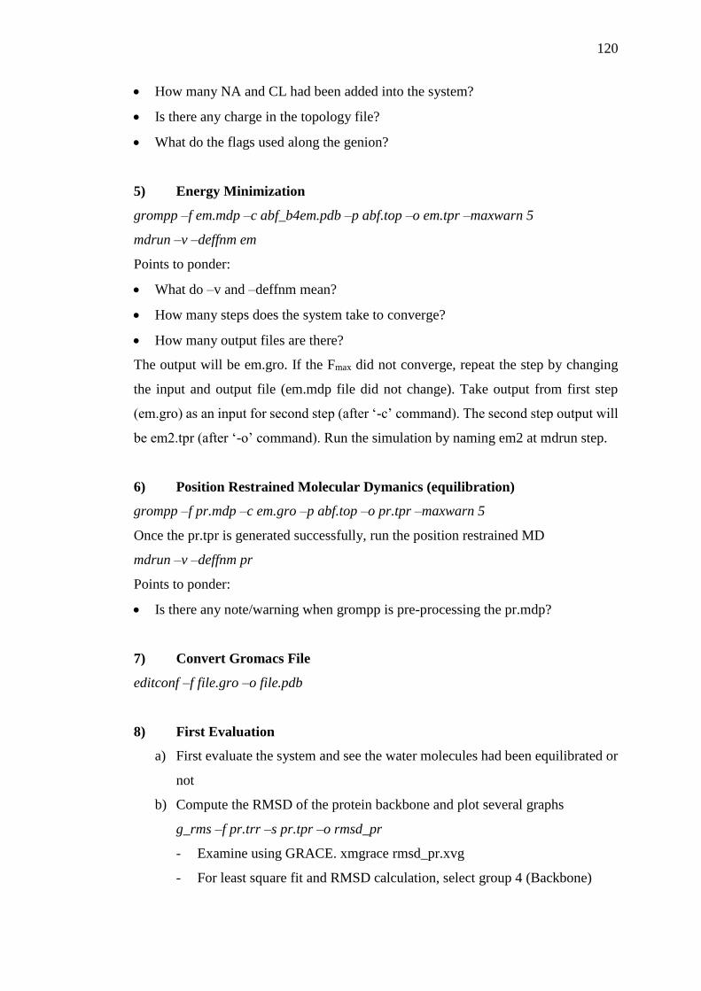
5) Energy Minimization
grompp –f em.mdp –c abf_b4em.pdb –p abf.top –o em.tpr –maxwarn 5
mdrun –v –deffnm em
Points to ponder:
What do –v and –deffnm mean?
How many steps does the system take to converge?
How many output files are there?
The output will be em.gro. If the Fmax did not converge, repeat the step by changing
the input and output file (em.mdp file did not change). Take output from first step
(em.gro) as an input for second step (after ‘-c’ command). The second step output will
be em2.tpr (after ‘-o’ command). Run the simulation by naming em2 at mdrun step.
6) Position Restrained Molecular Dymanics (equilibration)
grompp –f pr.mdp –c em.gro –p abf.top –o pr.tpr –maxwarn 5
Once the pr.tpr is generated successfully, run the position restrained MD
mdrun –v –deffnm pr
Points to ponder:
Is there any note/warning when grompp is pre-processing the pr.mdp?
7) Convert Gromacs File
editconf –f file.gro –o file.pdb
8) First Evaluation
a) First evaluate the system and see the water molecules had been equilibrated or
not
b) Compute the RMSD of the protein backbone and plot several graphs
g_rms –f pr.trr –s pr.tpr –o rmsd_pr
- Examine using GRACE. xmgrace rmsd_pr.xvg
- For least square fit and RMSD calculation, select group 4 (Backbone)
121
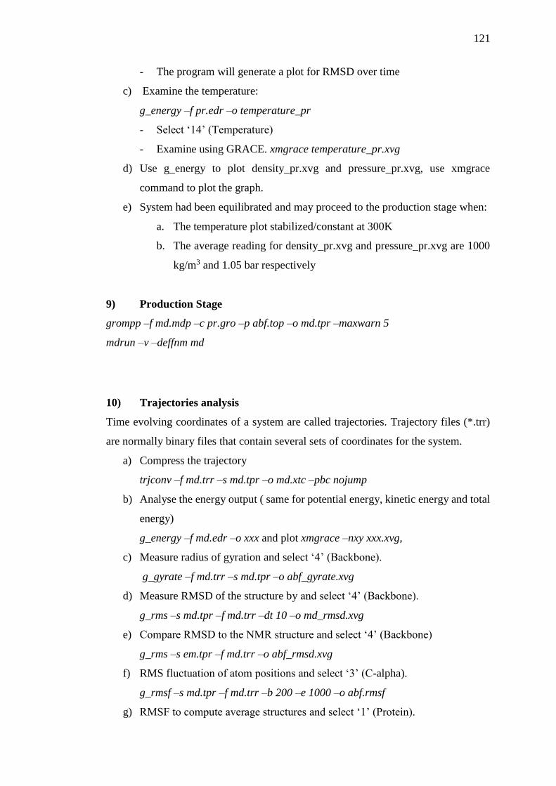
- The program will generate a plot for RMSD over time
c) Examine the temperature:
g_energy –f pr.edr –o temperature_pr
- Select ‘14’ (Temperature)
- Examine using GRACE. xmgrace temperature_pr.xvg
d) Use g_energy to plot density_pr.xvg and pressure_pr.xvg, use xmgrace
command to plot the graph.
e) System had been equilibrated and may proceed to the production stage when:
a. The temperature plot stabilized/constant at 300K
b. The average reading for density_pr.xvg and pressure_pr.xvg are 1000
kg/m3 and 1.05 bar respectively
9) Production Stage
grompp –f md.mdp –c pr.gro –p abf.top –o md.tpr –maxwarn 5
mdrun –v –deffnm md
10) Trajectories analysis
Time evolving coordinates of a system are called trajectories. Trajectory files (*.trr)
are normally binary files that contain several sets of coordinates for the system.
a) Compress the trajectory
trjconv –f md.trr –s md.tpr –o md.xtc –pbc nojump
b) Analyse the energy output ( same for potential energy, kinetic energy and total
energy)
g_energy –f md.edr –o xxx and plot xmgrace –nxy xxx.xvg,
c) Measure radius of gyration and select ‘4’ (Backbone).
g_gyrate –f md.trr –s md.tpr –o abf_gyrate.xvg
d) Measure RMSD of the structure by and select ‘4’ (Backbone).
g_rms –s md.tpr –f md.trr –dt 10 –o md_rmsd.xvg
e) Compare RMSD to the NMR structure and select ‘4’ (Backbone)
g_rms –s em.tpr –f md.trr –o abf_rmsd.xvg
f) RMS fluctuation of atom positions and select ‘3’ (C-alpha).
g_rmsf –s md.tpr –f md.trr –b 200 –e 1000 –o abf.rmsf
g) RMSF to compute average structures and select ‘1’ (Protein).

122
g_rmsf –s md.tpr –f md.trr –b 800 –e 1000 –o abf_xvg.pdb
h) Analyse the secondary structure of model by and select ‘1’ (Protein).
do_dssp –s md.tpr –f md.trr –o abf_ss.xpm –dt 10