In silico design of anti-atherogenic...
10
In silico design of anti-atherogenic biomaterials Daniel R. Lewis a, 1 , Vladyslav Kholodovych d, e, 1 , Michael D. Tomasini b, f , Dalia Abdelhamid c , Latrisha K. Petersen b , William J. Welsh d , Kathryn E. Uhrich c , Prabhas V. Moghe a, b, * a Department of Chemical and Biochemical Engineering, Rutgers University, NJ, USA b Department of Biomedical Engineering, Rutgers University, NJ, USA c Department of Chemistry and Chemical Biology, Rutgers University, NJ, USA d Department of Pharmacology, Robert Wood Johnson Medical School, University of Medicine and Dentistry of New Jersey, Rutgers University, Piscataway, NJ, USA e High Performance and Research Computing, University of Medicine and Dentistry of New Jersey, Rutgers University, Newark, NJ, USA f Laboratory for Cellular Biophysics, The Rockefeller University, NY, USA article info Article history: Received 17 May 2013 Accepted 1 July 2013 Available online 25 July 2013 Keywords: Amphiphilic macromolecules Macrophages Atherosclerosis Molecular modeling Structure-activity relations abstract Atherogenesis, the uncontrolled deposition of modified lipoproteins in inflamed arteries, serves as a focal trigger of cardiovascular disease (CVD). Polymeric biomaterials have been envisioned to counteract atherogenesis based on their ability to repress scavenger mediated uptake of oxidized lipoprotein (oxLDL) in macrophages. Following the conceptualization in our laboratories of a new library of amphiphilic macromolecules (AMs), assembled from sugar backbones, aliphatic chains and poly(- ethylene glycol) tails, a more rational approach is necessary to parse the diverse features such as charge, hydrophobicity, sugar composition and stereochemistry. In this study, we advance a computational biomaterials design approach to screen and elucidate anti-atherogenic biomaterials with high efficacy. AMs were quantified in terms of not only 1D (molecular formula) and 2D (molecular connectivity) de- scriptors, but also new 3D (molecular geometry) descriptors of AMs modeled by coarse-grained mo- lecular dynamics (MD) followed by all-atom MD simulations. Quantitative structure-activity relationship (QSAR) models for anti-atherogenic activity were then constructed by screening a total of 1164 de- scriptors against the corresponding, experimentally measured potency of AM inhibition of oxLDL uptake in human monocyte-derived macrophages. Five key descriptors were identified to provide a strong linear correlation between the predicted and observed anti-atherogenic activity values, and were then used to correctly forecast the efficacy of three newly designed AMs. Thus, a new ligand-based drug design framework was successfully adapted to computationally screen and design biomaterials with cardio- vascular therapeutic properties. Ó 2013 Elsevier Ltd. All rights reserved. 1. Introduction Atherosclerosis is characterized as an inflammatory disease involving macrophage scavenger receptor (SR) interactions with oxidized low-density lipoproteins (oxLDL) in the vascular intima, leading to plaque initiation and growth [1]. The early stages of atherosclerosis include low-density lipoproteins (LDL) sequestra- tion and oxidation in arterial walls, followed by monocyte recruitment and differentiation into macrophages, which inter- nalize oxLDL via SR-mediated mechanisms [2,3]. This progression results in enhanced inflammatory signaling and lipid-laden foam cell formation [4]. The accumulation of foam cells can lead to a lipid filled necrotic core covered by a fibrous cap, which upon rupture can lead to thrombus formation and the clinical endpoints of myocardial infarction or stroke [5]. Conventional therapies are often limited in terms of their ability to address localized inflammation from pre-existing lipid deposits [6]. As macrophages are crucial to progressing the inflammatory cycle and responsible for the majority of lipid accumulation, they present an ideal target for therapeutics to arrest disease progression [7]. By designing biomaterial nanoassemblies that can simulate the size, amphiphilicity and anionic charge of oxLDL and thus modulate scavenger receptor-mediated oxLDL trafficking, the macrophage pathways that are key to formation of atherosclerotic plaque can be potentially targeted. Studies from our laboratories have revealed a class of amphiphilic macromolecules (AM), which can competitively block oxLDL interaction with scavenger receptors, thereby mitigating * Corresponding author. Department of Biomedical Engineering, Rutgers Uni- versity, 599 Taylor Road, Piscataway, NJ 08854, USA. E-mail address: [email protected] (P.V. Moghe). 1 Equal lead co-authors. Contents lists available at ScienceDirect Biomaterials journal homepage: www.elsevier.com/locate/biomaterials 0142-9612/$ e see front matter Ó 2013 Elsevier Ltd. All rights reserved. http://dx.doi.org/10.1016/j.biomaterials.2013.07.011 Biomaterials 34 (2013) 7950e7959
Transcript of In silico design of anti-atherogenic...

lable at ScienceDirect
Biomaterials 34 (2013) 7950e7959
Contents lists avai
Biomaterials
journal homepage: www.elsevier .com/locate/biomateria ls
In silico design of anti-atherogenic biomaterials
Daniel R. Lewis a,1, Vladyslav Kholodovych d,e,1, Michael D. Tomasini b,f, Dalia Abdelhamid c,Latrisha K. Petersen b, William J. Welsh d, Kathryn E. Uhrich c, Prabhas V. Moghe a,b,*
aDepartment of Chemical and Biochemical Engineering, Rutgers University, NJ, USAbDepartment of Biomedical Engineering, Rutgers University, NJ, USAcDepartment of Chemistry and Chemical Biology, Rutgers University, NJ, USAdDepartment of Pharmacology, Robert Wood Johnson Medical School, University of Medicine and Dentistry of New Jersey, Rutgers University, Piscataway,NJ, USAeHigh Performance and Research Computing, University of Medicine and Dentistry of New Jersey, Rutgers University, Newark, NJ, USAf Laboratory for Cellular Biophysics, The Rockefeller University, NY, USA
a r t i c l e i n f o
Article history:Received 17 May 2013Accepted 1 July 2013Available online 25 July 2013
Keywords:Amphiphilic macromoleculesMacrophagesAtherosclerosisMolecular modelingStructure-activity relations
* Corresponding author. Department of Biomedicversity, 599 Taylor Road, Piscataway, NJ 08854, USA.
E-mail address: [email protected] (P.V. Moghe).1 Equal lead co-authors.
0142-9612/$ e see front matter � 2013 Elsevier Ltd.http://dx.doi.org/10.1016/j.biomaterials.2013.07.011
a b s t r a c t
Atherogenesis, the uncontrolled deposition of modified lipoproteins in inflamed arteries, serves as a focaltrigger of cardiovascular disease (CVD). Polymeric biomaterials have been envisioned to counteractatherogenesis based on their ability to repress scavenger mediated uptake of oxidized lipoprotein(oxLDL) in macrophages. Following the conceptualization in our laboratories of a new library ofamphiphilic macromolecules (AMs), assembled from sugar backbones, aliphatic chains and poly(-ethylene glycol) tails, a more rational approach is necessary to parse the diverse features such as charge,hydrophobicity, sugar composition and stereochemistry. In this study, we advance a computationalbiomaterials design approach to screen and elucidate anti-atherogenic biomaterials with high efficacy.AMs were quantified in terms of not only 1D (molecular formula) and 2D (molecular connectivity) de-scriptors, but also new 3D (molecular geometry) descriptors of AMs modeled by coarse-grained mo-lecular dynamics (MD) followed by all-atom MD simulations. Quantitative structure-activity relationship(QSAR) models for anti-atherogenic activity were then constructed by screening a total of 1164 de-scriptors against the corresponding, experimentally measured potency of AM inhibition of oxLDL uptakein human monocyte-derived macrophages. Five key descriptors were identified to provide a strong linearcorrelation between the predicted and observed anti-atherogenic activity values, and were then used tocorrectly forecast the efficacy of three newly designed AMs. Thus, a new ligand-based drug designframework was successfully adapted to computationally screen and design biomaterials with cardio-vascular therapeutic properties.
� 2013 Elsevier Ltd. All rights reserved.
1. Introduction
Atherosclerosis is characterized as an inflammatory diseaseinvolving macrophage scavenger receptor (SR) interactions withoxidized low-density lipoproteins (oxLDL) in the vascular intima,leading to plaque initiation and growth [1]. The early stages ofatherosclerosis include low-density lipoproteins (LDL) sequestra-tion and oxidation in arterial walls, followed by monocyterecruitment and differentiation into macrophages, which inter-nalize oxLDL via SR-mediated mechanisms [2,3]. This progressionresults in enhanced inflammatory signaling and lipid-laden foam
al Engineering, Rutgers Uni-
All rights reserved.
cell formation [4]. The accumulation of foam cells can lead to a lipidfilled necrotic core covered by a fibrous cap, which upon rupturecan lead to thrombus formation and the clinical endpoints ofmyocardial infarction or stroke [5].
Conventional therapies are often limited in terms of their abilityto address localized inflammation from pre-existing lipid deposits[6]. As macrophages are crucial to progressing the inflammatorycycle and responsible for the majority of lipid accumulation, theypresent an ideal target for therapeutics to arrest disease progression[7]. By designing biomaterial nanoassemblies that can simulate thesize, amphiphilicity and anionic charge of oxLDL and thus modulatescavenger receptor-mediated oxLDL trafficking, the macrophagepathways that are key to formation of atherosclerotic plaque can bepotentially targeted. Studies from our laboratories have revealed aclass of amphiphilic macromolecules (AM), which can competitivelyblock oxLDL interactionwith scavenger receptors, therebymitigating

D.R. Lewis et al. / Biomaterials 34 (2013) 7950e7959 7951
downstream consequences [8e15]. Previous work with first gener-ation AM structures qualitatively studied the impact of chargeplacement andnet charge on oxLDL uptake inhibition, but lacked thesophistication to accurately discern the key structural features thatgovern anti-atherogenic potency [10,11]. More recent studies indi-cate that AM chemistries with similar chemical composition canelicit markedly different, and somewhat unexpected, interactionswith oxLDLwhen imbuedwith different stereochemistries [8]. Thus,more rational and computationally guided approaches would aid inelucidating the AM structure-biological activity relations anddesigning a wider library of AMs with high potency.
Given the large sizeof theAMs,manycurrentdrugdesignmethodsof developing “small molecule” therapeutics are not directly appli-cable to AM structures (due to limits in feature space). Highthroughput screening could in theory be used to synthesize a largearray of AMsbut this approach has an inherently high cost barrier andis often is plagued by large false negative and false positive rates [16].Furthermore, structural optimization of early lead compounds bychemical synthesis of analogs is both time and cost intensive andtypically based on heuristic, rather than rational drug design ap-proaches. Computational drug discovery and optimization ap-proaches,basedonquantitative structure-activity relationship(QSAR)principles, offer an efficient and economical alternative for drugdevelopment when employed in conjunction with synthetic medici-nal chemistry and experimental testing of lead compounds [17e20].
Computational (rational) drug design has made significantcontributions to the discovery of new and more efficacious thera-peutics. The general strategies employed today are broadly dividedinto ligand-based and structure-based drug design (LBDD andSBDD, respectively) [21e24]. Assuming that drug action operatesthrough the simple mechanism of drugetarget interaction, theoption to use one or both strategies in a drug discovery campaigndepends on the existing knowledge of biologically active ligands forLBDD and the 3D structure of the target protein for SBDD. Moderntechniques in LBDD rely heavily on in silico (virtual) screening ofoften vast chemical libraries, QSAR modeling, and pharmacophoremodeling [12,25e29]. Likewise, SBDD studies frequently employvirtual screening procedures using a process commonly known asligand-receptor docking [8,30e32]. LBDD and SBDD methodsremain the subject of intensive research to improve their speed,accuracy, and sophistication [33,34].
We have previously employed several different approaches toconstruct QSAR models of biological activities of drugs and bio-materials [35e38].QSARmodels associatevariations in the chemicalstructure of the subject materials, as encoded by molecular de-scriptors, with variations in their corresponding biological activity(e.g., inhibition of oxLDL uptake). QSAR modeling entails two keysteps: 1) computing values of an ensemble ofmolecular descriptors,and2) creating andvalidating the regressionor classificationmodelsby machine learning methods and statistical analysis tools. Molec-ular descriptors can be categorized as 1D (e.g., molecular weight,number of rings), 2D (e.g., electro-topological/connectivity indices)whose values are conformation invariant, or 3D (e.g., dipolemoment, surface area, radius of gyration) whose values are confor-mation dependent.
The goal of the present study was to propose a LBDD-basedapproach, using both 2D and 3D descriptors, to identify structure-activity relationships between the AMs and inhibition of oxLDLuptake and foam cell formation in human monocyte-derivedmacrophages (MDM). One of the specific objectives is to generateQSAR models as means to predict the biological activities of newAMs, thus, guiding the rational design and optimization of AMs. Acritical component of the molecular modeling strategy for thecurrent AMs, is the inclusion of 3D descriptors, which uniquelyencodes vital information such as stereochemistry of the AMs. We
hypothesize that this studywill provide a new “in silico” frameworkto correlate compositional changes in the AM biomaterials with therespective variations in anti-atherogenic potency and thus facilitatethe design of new AM structures by predicting biological activity.
2. Materials and methods
2.1. Materials
All chemicals/materials were purchased from SigmaeAldrich (Milwaukee, WI)or Fisher Scientific (Pittsburgh, PA) and used as received unless otherwise noted.Deionized (DI) water with a resistivity of 18 MU cm is obtained using PicoPure 2 UVPlus (Hydro Service and SupplieseDurham, NC). The following items were pur-chased from the indicated vendors: RPMI 1640 from ATCC (Manassas, VA), macro-phage colony stimulating factor (M-CSF) from PeproTech (Rocky Hill, NJ), 1.077 g/cm3 FicollePaque Premium from GE healthcare (Pittsburgh, PA), FBS and Hoechst33342 from Life Technologies (Grand Island, NY), 3,30-dioctadecyloxacarbocyanine(DiO) labeled oxLDL from Kalen Biomedical (Montgomery Village, MD), unlabeledoxidized LDL from Biomedical Technologies Inc. (Stoughton, MA), and human buffycoats from the Blood Center of New Jersey (East Orange, NJ).
2.2. AM synthesis and physicochemical property determination
AMs were synthesized as previously described [10e14,39e41]. The pKa valueswere estimated based on ionizable functional groups. For example, aliphatic car-boxylic acids have a pKa ofw3e5, and primary aminesw35. Attachment length wascalculated by counting the number of atoms between the functional groupresponsible for the charge and the attachment of PEG as shown in Fig. 1. In thecurrent study, 17 different AM structures were examined, varying the overall charge,hydrophobicity, sugar structure (linear vs. cyclic) and stereochemistry. Table 1summarizes the diverse range of their physicochemical properties.
2.3. Isolation and culture of hMDMs
Peripheral blood mononuclear cells (PBMCs) were isolated from human buffycoats by FicollePaque (1.077 g/cm3) density gradient. Red blood cells were lysed withACKbufferandplateletswere removedbycentrifugationat300g for10m.PBMCsweretransferred to flasks containing RPMI 1640 supplemented with 10% FBS, 1% penicillin/streptomycin. Monocyteswere selected fromPBMCs by adherence after 24 h and thencultured for 7 days in RPMI 1640 supplemented with 10% FBS, 1% penicillin/strepto-mycin and 50 ng/mL M-CSF for differentiation into macrophages. After the 7 day cul-ture, themacrophages were trypsinized and scraped from flasks, transferred intowellplates at 50,000 cells/cm2, and treatments administered after 24 h.
2.4. OxLDL uptake by hMDMs
To measure AM efficacy at inhibiting oxLDL uptake, hMDMs were incubatedwith 1 mg/mL of DiO labeled oxLDL and 10�6
M AM in RPMI 1640 for 24 h. Cells wereremoved from plates by vigorous pipetting in cold PBS with 2 mM EDTA, washedwith PBS, centrifuged and fixed in 1% paraformaldehyde. DiO fluorescence (oxLDLuptake) was measured by flow cytometry on a FACScalibur (Beckton Dickenson) inthe FL1 channel. A minimum of 15,000 events per sample were collected, andquantified using the geometric mean fluorescence intensity (MFI) of intact hMDMswith FloJo (Treestar). Results are the average of three independent experiments withtwo technical replicates per experiment. Data is presented as % oxLDL uptake in-hibition, which was calculated using the following formula:
100� 100*MFI of AM containing condition
MFI of oxLDL control¼ % oxLDL uptake inhibition:
2.5. Foam cell formation
To measure the effectiveness at preventing foam cell formation (lipid accumu-lation), AMs (10�5
M) and oxLDL (50 mg/mL)were co-incubatedwith hMDMs for 24 h.Cells were thenwashed, fixed, dehydrated with 60% isopropanol, stained with 2mg/mL Oil Red O in 60% isopropanol for 5 min, washed and the nucleus counterstainedwith 1 mg/mL Hoechst 33342. Brightfield and epifluorescent images were taken on aNikon Eclipse TE2000-S and merged using ImageJ. Images shown are representativeof two independent experiments with three technical replicates per experiment.
2.6. Statistical analysis
OxLDL uptake results are presented as mean � standard error of the mean(S.E.M.) and data evaluated by one-way ANOVA and Tukey’s test for post-hoc pair-wise comparisons between multiple conditions. A p-value of 0.05 or less wasconsidered statistically significant.
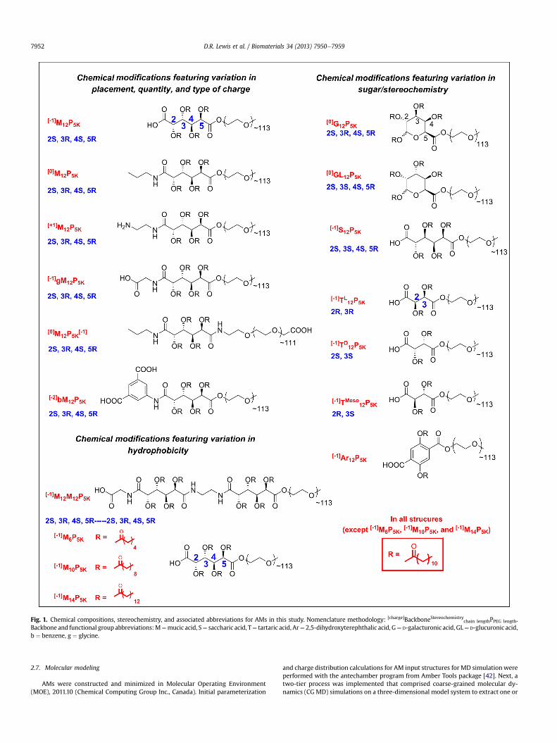
Fig. 1. Chemical compositions, stereochemistry, and associated abbreviations for AMs in this study. Nomenclature methodology: [charge]BackboneStereochemistrychain lengthPPEG length.
Backbone and functional group abbreviations:M¼mucic acid, S¼ saccharic acid, T¼ tartaric acid, Ar¼ 2,5-dihydroxyterephthalic acid, G¼ D-galacturonic acid, GL¼ D-glucuronic acid,b ¼ benzene, g ¼ glycine.
D.R. Lewis et al. / Biomaterials 34 (2013) 7950e79597952
2.7. Molecular modeling
AMs were constructed and minimized in Molecular Operating Environment(MOE), 2011.10 (Chemical Computing Group Inc., Canada). Initial parameterization
and charge distribution calculations for AM input structures for MD simulationwereperformed with the antechamber program from Amber Tools package [42]. Next, atwo-tier process was implemented that comprised coarse-grained molecular dy-namics (CG MD) simulations on a three-dimensional model system to extract one or
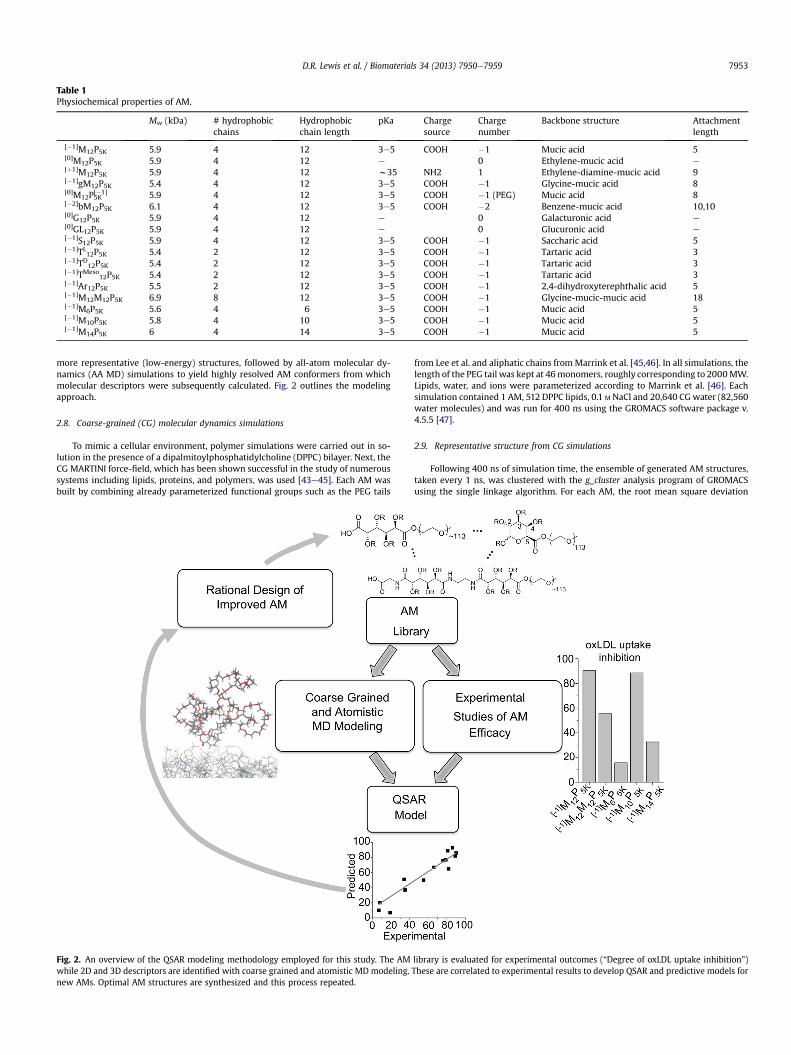
Table 1Physiochemical properties of AM.
Mw (kDa) # hydrophobicchains
Hydrophobicchain length
pKa Chargesource
Chargenumber
Backbone structure Attachmentlength
[�1]M12P5K 5.9 4 12 3e5 COOH �1 Mucic acid 5[0]M12P5K 5.9 4 12 e 0 Ethylene-mucic acid e[þ1]M12P5K 5.9 4 12 w35 NH2 1 Ethylene-diamine-mucic acid 9[�1]gM12P5K 5.4 4 12 3e5 COOH �1 Glycine-mucic acid 8[0]M12P5K[�1] 5.9 4 12 3e5 COOH �1 (PEG) Mucic acid 8[�2]bM12P5K 6.1 4 12 3e5 COOH �2 Benzene-mucic acid 10,10[0]G12P5K 5.9 4 12 e 0 Galacturonic acid e[0]GL12P5K 5.9 4 12 e 0 Glucuronic acid e[�1]S12P5K 5.9 4 12 3e5 COOH �1 Saccharic acid 5[�1]TL12P5K 5.4 2 12 3e5 COOH �1 Tartaric acid 3[�1]TD12P5K 5.4 2 12 3e5 COOH �1 Tartaric acid 3[�1]TMeso
12P5K 5.4 2 12 3e5 COOH �1 Tartaric acid 3[�1]Ar12P5K 5.5 2 12 3e5 COOH �1 2,4-dihydroxyterephthalic acid 5[�1]M12M12P5K 6.9 8 12 3e5 COOH �1 Glycine-mucic-mucic acid 18[�1]M6P5K 5.6 4 6 3e5 COOH �1 Mucic acid 5[�1]M10P5K 5.8 4 10 3e5 COOH �1 Mucic acid 5[�1]M14P5K 6 4 14 3e5 COOH �1 Mucic acid 5
D.R. Lewis et al. / Biomaterials 34 (2013) 7950e7959 7953
more representative (low-energy) structures, followed by all-atom molecular dy-namics (AA MD) simulations to yield highly resolved AM conformers from whichmolecular descriptors were subsequently calculated. Fig. 2 outlines the modelingapproach.
2.8. Coarse-grained (CG) molecular dynamics simulations
To mimic a cellular environment, polymer simulations were carried out in so-lution in the presence of a dipalmitoylphosphatidylcholine (DPPC) bilayer. Next, theCG MARTINI force-field, which has been shown successful in the study of numeroussystems including lipids, proteins, and polymers, was used [43e45]. Each AM wasbuilt by combining already parameterized functional groups such as the PEG tails
Fig. 2. An overview of the QSAR modeling methodology employed for this study. The AMwhile 2D and 3D descriptors are identified with coarse grained and atomistic MD modeling.new AMs. Optimal AM structures are synthesized and this process repeated.
from Lee et al. and aliphatic chains fromMarrink et al. [45,46]. In all simulations, thelength of the PEG tail was kept at 46monomers, roughly corresponding to 2000MW.Lipids, water, and ions were parameterized according to Marrink et al. [46]. Eachsimulation contained 1 AM, 512 DPPC lipids, 0.1 M NaCl and 20,640 CGwater (82,560water molecules) and was run for 400 ns using the GROMACS software package v.4.5.5 [47].
2.9. Representative structure from CG simulations
Following 400 ns of simulation time, the ensemble of generated AM structures,taken every 1 ns, was clustered with the g_cluster analysis program of GROMACSusing the single linkage algorithm. For each AM, the root mean square deviation
library is evaluated for experimental outcomes (“Degree of oxLDL uptake inhibition”)These are correlated to experimental results to develop QSAR and predictive models for

D.R. Lewis et al. / Biomaterials 34 (2013) 7950e79597954
(RMSD) threshold was chosen as the minimum value such that greater than 50% ofstructures were members of a single cluster. The representative structure waschosen to be the median structure (in terms of RMSD) of the largest cluster.
2.10. Reverse mapping of CG structures to atomistic AM structures
The reverse transformation technique of Rzepiela et al. was applied to convertthe MARTINI CG structures back to all-atom structures [48]. The Antechambermodule of the Amber Tools software package was used to generate atomistic to-pologies for each AM and the program acpypewas used to convert Amber topologiesinto GROMACS format [42,49]. The GROMACS utility g_fg2cg was used to generatean initial approximation of the all-atom structure by placing at random the un-derlying atoms within the volume of their corresponding CG interaction site.Simulated annealing (SA) was then used to bring the system from 1300 K to 310 Kover 100 ps to allow for rearrangement of the atoms and crossing of energeticbarriers. During SA, a restraining force is used such that the center of mass (COM) ofatoms corresponding to a given CG interaction site alignwith the COMof the CG site.Following SA, the restraining force is slowly removed over a period of 10 ps. Theresultant structure was then subject to energy minimization.
Fig. 3. The AM library shows graded efficacy of anti-atherogenesis in hMDMs AeC) The percDeG) Representative micrographs showing modulation of lipid uptake and foam cell phendrophobic modifications (C/F). Treatments with the same letter are not statistically significathe control (no AM, oxLDL only).
2.11. Atomistic MD simulations
Followed the long-range CG MD simulation for 400 ns, the average structures ofthe AMs were subjected to refinement with AA MD in aqueous solution over thesurface of the membrane bilayer, constructed to mimic the macrophage cellularsurface. Each reverse mapped AM was placed over the surface of the constructedmembrane comprising phosphatidylcholine (PC), phosphatidylethanolamine (PE)and phosphatidylserine (PS) (courtesy of Dr. William Moyle, Department of OBGYN,RWJMS, Rutgers University), neutralized with sodium ions and solvated with tleapsubroutine from Amber 12 software package [42]. High level AA MD simulationtotaling 10 ns for each of the 17 AMs contained 1 AM, 812 lipids and 82,560 watermolecules in a periodic box was performed on the specifically built and dedicatedhigh performance GPU Linux cluster.
2.12. 3D molecular descriptors and QSAR analysis
The representative low energy 3D structures emerging from the AA MD simula-tionswere subjected to QSAR analysis tofind correlations betweenpolymer structuralfeatures and their effect on oxLDL uptake. Each polymer was encoded with 1664
entage of inhibition of oxLDL uptake was plotted vs. nature of AM treatment in hMDMs.otype. AM are grouped to show effects of charge (A/D), stereochemical (B/E) and hy-nt from one another and the asterisk (*) indicates statistical significance (p < .05) from

D.R. Lewis et al. / Biomaterials 34 (2013) 7950e7959 7955
distinctmoleculardescriptorsusingDragonv.5.4 softwareprogram. Sets ofdescriptorswere limited to 3D descriptor blocks as defined in Dragon, namely Randic molecularprofiles, geometrical descriptors, RDF descriptors, 3d-MoRSE, Weighted HolisticInvariant Molecular (WHIM), GEometry, Topology, and Atom-Weights AssemblY(GETAWAY) and charge descriptors (total of 735). After removing highly correlatedpairs and descriptors with standard deviation below the program default threshold,the final set of descriptors was reduced to 115. Partial least squares (PLS) regressionmethod implemented in MOE was used to model the experimental data.
3. Results
3.1. oxLDL uptake in hMDMs
Thedifferent AMstructures havedemonstrated varying abilities toinhibit oxLDL uptake in MDMs. As shown in Fig. 3AeC, [�1]M12P5K,[0]M12P5K, [0]G12P5K, [�1]TMeso
12P5K, [�1]gM12P5K, and [�1]M10P5Kdisplay the most significant reduction in oxLDL accumulation. Incontrast, AM structures, [�1]S12P5K, [�1]Ar12P5K, and [�1]M6P5K, did notshow a statistically significant decrease in oxLDL uptake as comparedto the oxLDL control.
3.2. Foam cell formation in hMDMs
Treatment of hMDMs with oxLDL generated the foam cellphenotype as evidenced by intracellular lipid droplet accumulation(Fig. 3G). The foam cell formation results qualitatively parallel theoxLDL uptake results, and show that the AM library has differentialability to reduce lipid accumulation (Fig. 3DeF). These resultsdemonstrate the ability of AMs to mitigate actual atherosclerotic-relevant endpoints.
3.3. CGMD in conjunction with atomistic MD
The two-tiered approach used in this study provided detailedconformers for all AM structures. A schematic of an AM along withits CG representation and atomistic transformation is shown inFig. 4. Snapshots of selected AM are presented in Fig. 5.
3.4. Simulations of AM conformations
The MD results suggest that the most effective AM at inhibitingoxLDL ([�1]M12P5K, [�1]gM12P5K, [þ1]M12P5K) tend to stay in the
Fig. 4. A) The molecular and B) coarse grained structure of [�1]M12P5K shown in a ball-and-sin the MARTINI force field where CG beads are shown as transparent spheres.
extendedconformationduring theentireMDsimulation,while theirless active counterparts ([�1]S12P5K, [0]GL12P5K, [�1]M12M12P5K)generally form more compact globular structures with lauryl armspointing in the direction opposite of the cell membrane (Fig. 5).
3.5. Establishment of a QSAR model
Molecular descriptors were generated to encode various phys-icochemical properties of the AM including spatial organization,chemical composition and stereochemistry. Following filtering andprioritization of descriptors based on their information content,QSAR models were constructed using PLS regression to predict theAM efficacy (i.e., inhibition of oxLDL uptake). Several QSAR modelswere built and key descriptors that explain oxLDL uptake-relatedbehavior of AM stereo pairs were identified. A statistically strongcorrelation between predicted and observed results was achievedwith only five descriptors. Employing these descriptors, a QSARmodel was established which exhibited a strong linear correlation(r2 ¼ 0.91, rcv2 ¼ 0.77) between predicted and observed values ofoxLDL uptake (Table 2).
3.6. QSAR predictive capability
The final model was applied to predict oxLDL uptake inhibitionof new AMs, namely, [�1]M12P5K analogs with variable aliphaticarms ([�1]M6P5K, [�1]M10P5K, and [�1]M14P5K) (Fig. 6). The predictedand experimental values for the test set, comprising three newlydesigned AM with varying hydrophobic moieties were correctlyranked from low to high values. This points to the prediction abilityof the final QSAR model and its utility for optimizing AM structuresthat mitigate adverse athero-relevant endpoints.
4. Discussion
Inhibition of the athero-inflammatory phenotype of macro-phages is considered to be as a major strategic target for themanagement of atherosclerosis underlying cardiovascular disease[50e53]. While a range of recently advanced AMs show continuedpromise in inhibiting atherogenesis in inflamed macrophages, arational framework to designing improved AMs is currently lacking
tick representation. C) The conformation of [�1]M12P5K along with its CG representation

Fig. 5. AMs exhibiting high to moderate efficiency in reduction of oxLDL uptake (left column) have their aliphatic arms in an extended conformation while less effective polymers(right column) form more compact globular structures with aliphatic arms pointing in the direction opposite to the cell membrane. These snapshots of AMs were obtained after400 ns of CG MD simulation and additional 2 ns of AA MD simulation over the surface of membrane bilayer. For simplicity, hydrogen atoms are omitted. The hydrophobic heads ofthe AMs are highlighted as white sticks and the PEG tail is shown as a trace attached to the AM “head”. For visual comparison, the top three rows show stereo pairs of polymers thathave distinctive behavior in reduction of oxLDL uptake: [�1]M12P5K/[�1]S12P5K, [�1]TMeso
12P5K/[�1]TD12P5K, and [0]G12P5K/[0]GL12P5K.
D.R. Lewis et al. / Biomaterials 34 (2013) 7950e79597956

Table 2QSAR equation related oxLDL uptake inhibition and descriptors of polymers, sta-tistical analysis of the model fit and relative influence of descriptors on the QSARmodel. oxLDL uptake inhibition ¼ �617:97111þ 6528:05803*G3pþ 187:62572*HOMAþ 608:22915*Ds� 391:41561*R5uþ 1179:44106*G1u:
Relative influence of descriptors
G3p 1.000000 3rd component symmetry directional WHIM index/weighted by atomic polarizabilities
HOMA 0.329363 Harmonic oscillator model of aromaticity indexDs 0.382432 D total accessibility index/weighted by atomic
electrotopological statesR5u 0.314173 R autocorrelation of lag 5/unweightedG1u 0.245292 1st component symmetry directional WHIM index/
unweighted.
QSAR fit
Root mean square error (RMSE) 8.65491Correlation coefficient (R2) 0.91Cross-validated RMSE 14.65Cross-validated R2 0.77
WHIM descriptors (G3p, Ds, G1u): (Weighted Holistic Invariant Molecular de-scriptors), which are geometrical descriptors based on statistical indices calculatedon the projections of the atoms along principal axes.Geometrical descriptors (HOMA): different kinds of conformationally dependentdescriptors based on the molecular geometry.GETAWAY descriptors: descriptors calculated from the leverage matrix obtained bythe centered atomic coordinates (molecular influence matrix, MIM).
D.R. Lewis et al. / Biomaterials 34 (2013) 7950e7959 7957
[8,10e12,14,54]. In this work, a multiscale modeling approach wasadvanced by employing molecular descriptors of the AM chemicalstructures and geometric parameters based on conformationalchanges of the AMs on model lipid membranes. By correlatingbiological efficacy data from hMDMs with a wide range of AMdescriptors, a QSAR model was derived, which affords new insightsto optimize and predict the efficacy of new AM structures.
Developing QSAR and principal component analysis (PCA)models to isolate critical molecular features is a versatile approachapplied to a wide array of molecular feature spaces [55,56]. Even asmall library size of molecules such as the AMs show a high vari-ability in biological efficacy, as slight changes in AM structureproduce markedly different levels of efficacy at inhibiting oxLDLuptake and foam cell formation (Fig. 3). While this result is quitecommon for small molecule therapeutics, this effect was somewhatintriguing for the current AM library given the large MW and the
Fig. 6. The final QSAR model was successful at predicting the bioactivity (oxLDL uptake inhibfrom simulation is often within the S.E.M. from the experimental data. B) The linear fit of e
conformational flexibility of the AMs, thus prompting furthermechanistic modeling efforts.
Although previous studies found AM efficacy differencesdependent on charge type and charge placement [10,11], this workhas identified that the largest differences in AM efficacy arise fromvariations in stereochemical and hydrophobic modifications(Fig. 3AeC). For instance, [0]M12P5K and [0]G12P5K show relativelyhigh levels of bioactivity despite having an overall neutral chargethat should make them less effective at binding to the positivecharged SR binding domains, which mediate oxLDL binding anduptake, and similarly AM uptake. This result may indicate that thefolded conformation of the AM and the presentation of hydro-phobic moieties can contribute to improved binding. Additionally,the mucic acid sugar backbone seems to provide increased efficacyas [�1]M12P5K, [0]M12P5K, M12P5K[�1], [�2]bM12P5K, [�1]gM12P5K and[þ1]M12P5K all show higher levels of bioactivity (inhibition of oxLDLuptake) than tartaric and saccharic acid backbone AMs. However,changes in stereochemistry resulted in the most drastic differencesin inhibition efficiency, for example, with the substitution of sac-charic acid in [�1]S12P5K for mucic acid in [�1]M12P5K where the onlystructural difference is one stereocenter. Thus, [�1]M12P5K was thesecond most effective AM at inhibiting oxLDL uptake, whereas itsstereo-counterpart, [�1]S12P5K consistently showed little inhibition.
While measurements of oxLDL uptake provide early insightsinto variations in lipid influx, they do not capture the longer-termeffects of lipid accumulation, metabolism and secretion [57].Transition to the foam cell phenotype defined by large lipid drop-lets developing within the cell, is an alternative, physiologicallyrelevant marker of atherogenesis as it marks the first stage in lesiondevelopment in vivo and is often accompanied by increased in-flammatory cytokine secretion. Our results indicate that the AMlibrary shows similar effects on inhibition of the foam cell pheno-type as that on oxLDL uptake in vitro. As these results mimic thoseof oxLDL uptake, they show that AM could be effective at managingatherosclerotic disease even in areas of high oxLDL concentration.
The molecular dynamics simulations shed light into potentialmechanisms underlying how the AMs with different alkyl armsexhibit differential bioactivity. For example, the MD snapshotsindicate that the extended conformation of alkyl arms is requiredfor effective bioactivity and conversely increasingly globular AMswere found to be less effective. This divergence may arise fromstronger interactions occurring between the extended lauryl arms
ition) of the new, three member AM test set. A) The QSAR fit showing that the residualxperimental vs. predicted outcomes with R2 ¼ 0.91.

D.R. Lewis et al. / Biomaterials 34 (2013) 7950e79597958
with the membrane bilayer surface during simulation. As a result,these interactions would generate deeper and more rapid pene-tration of the AM into the cell membrane, suggesting that tighterAM binding leads to more effective inhibition of oxLDL uptake andfoam cell formation.
The advantage of using combined coarse grained and atomisticMDmodeling is that the resultant descriptors could also be used toestablishQSARmodels for predictingAMefficacy. Oneof thenotableresults from this study is that a statistically significant model wasestablished between predicted and experimental outcomes usingmerely five AM descriptors. The descriptor set, although not easilytransferable into simple chemical terms, encompasses awide rangeof AM descriptors such as aromatic rings and unsaturated bonds(HOMA descriptor), molecular geometry, size, shape and stereo-chemistry (Table 2). Among the prominent 3D descriptors are theWHIM descriptors, which are geometrical descriptors based onstatistical indices calculated on the projections of the atoms alongprincipal axes and the GETAWAY descriptors [58e61]. WHIM de-scriptors encode 3D information on molecular size, shape, symme-try and atomdistributionwith respect to invariant reference frames,while the GETAWAY descriptors encode atomic properties such asatomicmass, atomic polarizability, atomic electronegativity, van derWaals atomic volume, and the unitweight. Overall, this QSARmodelwas successful at predicting the efficacy of [�1]M12P5K analogs withvariable hydrophobicity ([�1]M6P5K, [�1]M10P5K, and [�1]M14P5K).
Despite the success of the proposed QSAR formalism, furthermodeling improvements will be forthcoming as the AM librarycontinues to evolve, to include, for example, newer and more com-plexcarbohydrate-derived core structures, aliphatic chains, andPEGchains. Moreover, the QSAR models will gain in predictive perfor-mance and statistical robustness as more experimental results areobtained formodel building. AnalogousQSAR relations could also besought in the future between the AM chemistry and downstreamactivity such as membrane binding/partitioning, or modulation ofinflammatory activity. For example, we hypothesize that certainAMs could attenuate the downstream pro-inflammatory signalingcascade either directly through the scavenger receptor-mediateduptake processes or indirectly through other unrelated mecha-nisms. The modeling approach could potentially help to tease apartthe roleof differentmembersof theAMlibrary that could triggeroneor more such pathways in silico. Improved molecular level bindingdata of how the AMs bind to scavenger receptors is necessary tofurther validate the relevance of the AM conformers on the bioac-tivity. In the absence of such information, the current modelingQSAR framework that ties together chemical and molecular levelinputs from biomaterial structures with downstream biologicaloutputs is particularly valuable. In summary, the implementation ofthe validated QSAR model, together with insights into structure-activity relationships gained from analyzing the leading moleculardescriptors, can guide the functional design of AM biomaterials astherapeutics for cardiovascular disease.
5. Conclusion
A modeling framework was developed for the design andpredictive enhancements to biomaterials that can inhibit the up-take of oxidized lipoproteins in inflammatory macrophages, aprocess called atherogenesis. An expanded biomaterial library ofamphiphilic macromolecules (AM) modulating key molecularfeatures was created that displayed high variability in the reduc-tion of oxLDL uptake and foam cell formation. As 2D molecularparameters cannot adequately describe a large 3D structure, MDsimulations were used to generate 3D structures in solution. These2D and 3D molecular models yielded several descriptors, whichwere correlated to biological activity using QSAR. This model
based upon five key descriptors was then implemented to suc-cessfully predict the efficacy of new AM structures. This workdemonstrates the unique interplay of 2D and 3D computationalapproaches combined with powerful bioactivity prediction modelsthat can be used for the rational design and optimization ofmacromolecular therapeutics.
Acknowledgments
The authors thank the National Heart, Lung and Blood Institute(R01HL107913, R21HL93753ePVM, KEU), the Coulter Foundationfor Biomedical Engineering Translational Research Award (PVM),National Institute of Biomedical Imaging and Bioengineering (T32EB005583eDRL), for financial support.
Appendix A. Supplementary data
Supplementary data related to this article can be found at http://dx.doi.org/10.1016/j.biomaterials.2013.07.011.
References
[1] Libby P, Ridker PM, Maseri A. Inflammation and atherosclerosis. Circulation2002;105(9):1135e43.
[2] Sahoo D, Drover V. The role of scavenger receptors in signaling, inflammationand atherosclerosis. In: Cheema S, editor. Biochemistry of atherosclerosis. US:Springer; 2006. p. 70e91.
[3] Fuhrman B, Partoush A, Volkova N, Aviram M. Ox-LDL induces monocyte-to-macrophage differentiation in vivo: possible role for the macrophage colonystimulating factor receptor (M-CSF-R). Atherosclerosis 2008;196(2):598e607.
[4] Berliner JA, Heinecke JW. The role of oxidized lipoproteins in atherogenesis.Free Radic Biol Med 1996;20(5):707e27.
[5] Boyle JJ. Macrophage activation in atherosclerosis: pathogenesis and phar-macology of plaque rupture. Curr Vasc Pharmacol 2005;3:63e8.
[6] Gotto AM. Antioxidants, statins, and atherosclerosis. J Am Coll Cardiol2003;41(7):1205e10.
[7] Saha P, Modarai B, Humphries J, Mattock K, Waltham M, Burnand KG, et al.The monocyte/macrophage as a therapeutic target in atherosclerosis. CurrOpin Pharmacol 2009;9(2):109e18.
[8] Hehir S, Plourde NM, Gu L, Poree DE, Welsh WJ, Moghe PV, et al. Carbohydratecomposition of amphiphilic macromolecules influences physicochemicalproperties and binding to atherogenic scavenger receptor A. Acta Biomater2012;8(11):3956e62.
[9] Iverson NM, Plourde NM, Sparks SM, Wang J, Patel EN, Shah PS, et al. Dual useof amphiphilic macromolecules as cholesterol efflux triggers and inhibitors ofmacrophage athero-inflammation. Biomaterials 2011;32(32):8319e27.
[10] Iverson NM, Sparks SM, Demirdirek B, Uhrich KE, Moghe PV. Controllableinhibition of cellular uptake of oxidized low-density lipoprotein: structure-function relationships for nanoscale amphiphilic polymers. Acta Biomater2010;6(8):3081e91.
[11] Wang J, Plourde NM, Iverson N, Moghe P, Uhrich KE. Nanoscale amphiphilicmacromolecules as lipoprotein inhibitors: the role of charge and architecture.Int J Nanomedicine 2007;2(4):697e705.
[12] Plourde NM, Kortagere S, Welsh W, Moghe PV. Structure-activity relations ofnanolipoblockers with the atherogenic domain of human macrophage scav-enger receptor A. Biomacromolecules 2009;10(6):1381e91.
[13] Chnari E, Nikitczuk JS, Wang J, Uhrich KE, Moghe PV. Engineered polymericnanoparticles for receptor-targeted blockage of oxidized low density lipo-protein uptake and atherogenesis in macrophages. Biomacromolecules2006;7(6):1796e805.
[14] Chnari E, Nikitczuk JS, Uhrich KE, Moghe PV. Nanoscale anionic macromole-cules can inhibit cellular uptake of differentially oxidized LDL. Bio-macromolecules 2006;7(2):597e603.
[15] Chnari E, Lari HB, Tian L, Uhrich KE, Moghe PV. Nanoscale anionic macro-molecules for selective retention of low-density lipoproteins. Biomaterials2005;26(17):3749e58.
[16] Mayr LM, Bojanic D. Novel trends in high-throughput screening. Curr OpinPharmacol 2009;9(5):580e8.
[17] Wang CY, Ai N, Arora S, Nagarajan K, Zauhar R, Young D, et al. Identification ofpreviously unrecognized antiestrogenic chemicals using a novel virtualscreening approach. Chem Res Toxicol 2006;19(12):1595e601.
[18] Chekmarev D, Kholodovych V, Kortagere S, Welsh W, Ekins S. Predicting in-hibitors of acetylcholinesterase by regression and classification machinelearning approaches with combinations of molecular descriptors. Pharm Res2009;26(9):2216e24.
[19] Paranjpe PV, Chen Y, Kholodovych V, Welsh W, Stein S, Sinko PJ. Tumor-targeted bioconjugate based delivery of camptothecin: design, synthesis andin vitro evaluation. J Control Release 2004;100(2):275e92.

D.R. Lewis et al. / Biomaterials 34 (2013) 7950e7959 7959
[20] Passic SR, Ferguson ML, Catalone BJ, Kish-Catalone T, Kholodovych V, Zhu W,et al. Structure-activity relationships of polybiguanides with activity againsthuman immunodeficiency virus type 1. Biomed Pharmacother 2010;64(10):723e32.
[21] Kortagere S, Welsh W. Development and application of hybrid structure basedmethod for efficient screening of ligands binding to g-protein coupled re-ceptors. J Comput Aided Mol Des 2006;20(12):789e802.
[22] Aparoy P, Kumar Reddy K, Reddanna P. Structure and ligand based drugdesign strategies in the development of novel 5-lox inhibitors. Curr MedChem 2012;19(22):3763e78.
[23] Hsieh JH, Yin S, Wang XS, Liu S, Dokholyan NV, Tropsha A. Cheminformaticsmeets molecular mechanics: a combined application of knowledge-basedpose scoring and physical force field-based hit scoring functions improvesthe accuracy of structure-based virtual screening. J Chem Inf Model2011;52(1):16e28.
[24] Mize CD, Abbott AM, Gacasan SB, Parrill AL, Baker DL. Ligand-based autotaxinpharmacophore models reflect structure-based docking results. J Mol GraphModel 2011;31(0):76e86.
[25] Bhatt HG, Patel PK. Pharmacophore modeling, virtual screening and 3D-QSARstudies of 5-tetrahydroquinolinylidine aminoguanidine derivatives as sodiumhydrogen exchanger inhibitors. Bioorg Med Chem Lett 2012;22(11):3758e65.
[26] Sushko I, Novotarskyi S, Körner R, Pandey A, Rupp M, Teetz W, et al. Onlinechemical modeling environment (ochem): web platform for data storage,model development and publishing of chemical information. J Comput AidedMol Des 2011;25(6):533e54.
[27] Dube D, Periwal V, Kumar M, Sharma S, Singh T, Kaur P. 3D-QSAR basedpharmacophore modeling and virtual screening for identification of novelpteridine reductase inhibitors. J Mol Model 2012;18(5):1701e11.
[28] Dong XO, Ebalunode J, Yang S-Y, Zheng W. Receptor-based pharmacophoreand pharmacophore key descriptors for virtual screening and QSAR modeling.Curr Comput Aided Drug Des 2011;7(3):181e9.
[29] Ebalunode J, Zheng W, Tropsha A. Application of QSAR and shape pharma-cophore modeling approaches for targeted chemical library design. In:Zhou JZ, editor. Chemical library design. Methods in molecular biology, vol.685. Humana Press; 2011. p. 111e33.
[30] Kooistra AJ, Roumen L, Leurs R, de Esch IJ, de Graaf C. From heptahelicalbundle to hits from the haystack: structure-based virtual screening for GPCRligands. Meth Enzymol 2013;522:279e336.
[31] Kamal A, Kashi Reddy M, Viswanath A. The design and development of imi-dazothiazoleechalcone derivatives as potential anticancer drugs. Expert OpinDrug Discov 2013;8(3):289e304.
[32] Krasowski MD, Hopfinger AJ. The discovery of new anesthetics by targetingGABAA receptors. Expert Opin Drug Discov 2011;6(11):1187e201.
[33] Martin TM, Harten P, Young DM, Muratov EN, Golbraikh A, Zhu H, et al. Doesrational selection of training and test sets improve the outcome of QSARmodeling? J Chem Inf Model 2012;52(10):2570e8.
[34] Ertl P, Lewis R. Iade: a system for intelligent automatic design of bioisostericanalogs. J Comput Aided Mol Des 2012;26(11):1207e15.
[35] Gubskaya AV, Kholodovych V, Knight D, Kohn J, Welsh WJ. Prediction offibrinogen adsorption for biodegradable polymers: integration of moleculardynamics and surrogate modeling. Polymer 2007;48(19):5788e801.
[36] Smith JR, Knight D, Kohn J, Rasheed K, Weber N, Kholodovych V, et al. Usingsurrogate modeling in the prediction of fibrinogen adsorption onto polymersurfaces. J Chem Inf Comput Sci 2004;44(3):1088e97.
[37] Duan P, Li S, Ai N, Hu L, Welsh WJ, You G. Potent inhibitors of human organicanion transporters 1 and 3 from clinical drug libraries: discovery and mo-lecular characterization. Mol Pharmacol 2012;9(11):3340e6.
[38] Fomovska A, Wood RD, Mui E, Dubey JP, Ferreira LR, Hickman MR, et al.Salicylanilide inhibitors of toxoplasma gondii. J Med Chem 2012;55(19):8375e91.
[39] Tian L, Yam L, Zhou N, Tat H, Uhrich KE. Amphiphilic scorpion-like macro-molecules: design, synthesis, and characterization. Macromolecules 2004;37(2):538e43.
[40] Djordjevic J, del Rosario LS, Wang J, Uhrich KE. Amphiphilic scorpion-likemacromolecules as micellar nanocarriers. J Bioact Compat Polym2008;2008(23):532e51.
[41] Wang J, del Rosario LS, Demirdirek B, Bae A, Uhrich KE. Comparison of pegchain length and density on amphiphilic macromolecular nanocarriers: self-assembled and unimolecular micelles. Acta Biomater 2009;5:883e92.
[42] Case DA, Darden TA, Cheatham TE, Simmerling CL, Wang J, Duke RE, et al.Amber 12. San Francisco: University of California; 2012.
[43] Marrink SJ, Risselada HJ, Yefimov S, Tieleman DP, de Vries AH. The MARTINIforce field: coarse grained model for biomolecular simulations. J Phys Chem B2007;111(27):7812e24.
[44] Monticelli L, Kandasamy SK, Periole X, Larson RG, Tieleman DP, Marrink S-J.The MARTINI coarse-grained force field: extension to proteins. J Chem TheoryComput 2008;4(5):819e34.
[45] Lee H, de Vries AH, Marrink S-J, Pastor RW. A coarse-grained model forpolyethylene oxide and polyethylene glycol: conformation and hydrody-namics. J Phys Chem B 2009;113(40):13186e94.
[46] Marrink SJ, de Vries AH, Mark AE. Coarse grained model for semiquantitativelipid simulations. J Phys Chem B 2003;108(2):750e60.
[47] Hess B, Kutzner C, van der Spoel D, Lindahl E. GROMACS 4: algorithms forhighly efficient, load-balanced, and scalable molecular simulation. J ChemTheory Comput 2008;4(3):435e47.
[48] Rzepiela AJ, Schäfer LV, Goga N, Risselada HJ, De Vries AH, Marrink SJ.Reconstruction of atomistic details from coarse-grained structures. J ComputChem 2010;31(6):1333e43.
[49] Sousa da Silva A, Vranken W. ACPYPEeAnteChamber PYthon Parser interfacE.BMC Res Notes 2012;5(1):367.
[50] Lewis DR, Kamisoglu K, York AW, Moghe PV. Polymer-based therapeutics:nanoassemblies and nanoparticles for management of atherosclerosis. WileyInterdiscip Rev Nanomed Nanobiotechnol 2011;3(4):400e20.
[51] Iverson N, Plourde N, Chnari E, Nackman GB, Moghe PV. Convergence ofnanotechnology and cardiovascular medicine. Biodrugs 2008;22(1):1e10.
[52] Mantovani A, Garlanda C, Locati M. Macrophage diversity and polarization inatherosclerosis: a question of balance. Arterioscler Thromb Vasc Biol2009;29(10):1419e23.
[53] Mantovani A, Sica A, Locati M. New vistas on macrophage differentiation andactivation. Eur J Immunol 2007;37(1):14e6.
[54] York AW, Zablocki KR, Lewis DR, Gu L, Uhrich KE, Prud’homme RK, et al.Kinetically assembled nanoparticles of bioactive macromolecules exhibitenhanced stability and cell-targeted biological efficacy. Adv Mater2012;24(6):733e9.
[55] Kou PM, Pallassana N, Bowden R, Cunningham B, Joy A, Kohn J, et al. Pre-dicting biomaterial property-dendritic cell phenotype relationships from themultivariate analysis of responses to polymethacrylates. Biomaterials2012;33(6):1699e713.
[56] Petersen LK, Ramer-Tait AE, Broderick SR, Kong C-S, Ulery BD, Rajan K, et al.Activation of innate immune responses in a pathogen-mimicking manner byamphiphilic polyanhydride nanoparticle adjuvants. Biomaterials 2011;32(28):6815e22.
[57] Ricci R, Sumara G, Sumara I, Rozenberg I, Kurrer M, Akhmedov A, et al.Requirement of JNK2 for scavenger receptor A-mediated foam cell formationin atherogenesis. Science 2004;306(5701):1558e61.
[58] Todeschini R, Lasagni M, Marengo E. Newmolecular descriptors for 2D and 3Dstructures. Theory J Chemom 1994;8(4):263e72.
[59] Consonni V, Todeschini R, Pavan M. Structure/response correlations andsimilarity/diversity analysis by getaway descriptors. 1. Theory of the novel 3Dmolecular descriptors. J Chem Inf Comput Sci 2002;42(3):682e92.
[60] Consonni V, Todeschini R, Pavan M, Gramatica P. Structure/response corre-lations and similarity/diversity analysis by getaway descriptors. 2. Applicationof the novel 3D molecular descriptors to QSAR/QSPR studies. J Chem InfComput Sci 2002;42(3):693e705.
[61] Kubinyi H, Folkers G, Martin YC, editors. 3D QSAR in drug design. Dordrecht(The Netherlands): Kluwer/ESCOM; 1998.