IN SILICO DESIGN OF 1,3,5-TRIAZINE DERIVATIVES AS ENOYL ...
8
Menon European Journal of Pharmaceutical and Medical Research www.ejpmr.com 465 IN SILICO DESIGN OF 1,3,5-TRIAZINE DERIVATIVES AS ENOYL ACYL CARRIER PROTEIN REDUCTASE INHIBITORS Divya Menon* and Supriya Mahajan Department of Pharmaceutical Chemistry, C. U. Shah College of Pharmacy, S. N. D. T. Women’s University, Sir Vithaldas Vidyavihar, Juhu Tara Road, Santacruz (West), Mumbai-400049, India. Article Received on 11/02/2019 Article Revised on 04/03/2019 Article Accepted on 25/03/2019 Mycobacterium tuberculosis are aerobic bacteria, with a waxy cell wall, composed of mycolic acid. [1-2] It is the main cause of tuberculosis, an infectious disease, which is the ninth leading cause of death worldwide. [3] Tuberculosis mainly affects the lungs but can affect other sites as well. It spreads when people who are sick with pulmonary TB, expel bacteria into the air by coughing. A person infected with Mycobacterium tuberculosis may not develop clinically significant disease. The viable organism may remain dormant and only reactivate when the host’s immune defenses are lowered, to take on an infectious and life-threatening form. [4] Worldwide around 10 million people fall ill with TB and it is one of the top ten causes of death. There were 5,58,000 new cases with resistance to rifampicin (RRTB), of which 82% had multi-drug resistant TB (MDR-TB). [3] The statistics does not paint an encouraging picture of the current status of the disease. The factors that are responsible for tuberculosis to have such a strong hold as a feared infectious disease are the appearance of multi-drug resistant strains, non- compliance to the drug regimen and AIDS epidemic. [5] The emergence of MDR-TB and extensively drug- resistant TB (XDR-TB) has prompted researchers to explore different pathways existing in mycobacterium and trying to find potential lead molecules. The cell wall of Mycobacterium tuberculosis is associated with its pathogenicity 5 . The main constituent of its cell wall is composed of mycolic acid. Enoyl acyl carrier protein reductase is a key fatty acid synthase type II enzyme. It catalyzes the final enzymatic step converting trans-2-enoyl-ACP to acyl-ACP in a NADH dependent reaction. Isoniazid (INH) is a front-line antitubercular drug. Activity of INH is dependent on its conversion to an acyl radical by catalase-peroxidase KatG enzyme. The acyl radical binds covalently to NADH, the co-substrate for InhA. This adduct functions as a potent inhibitor of InhA. [6-10] Clinical resistance to INH has been linked to mutations in KatG. [11-12] The novelty of direct InhA inhibitors lies in the fact that they do not have to be activated by an enzyme and hence, is less prone to develop resistance by the organism. [13] The aim of the study is to find potential leads amongst the substituted 1,3,5-triazine derivatives as direct inhibitors of the InhA enzyme by performing molecular docking studies. MATERIALS AND METHODS Computational Tools Docking studies were performed using Maestro 11, a Molecular Modeling Software from Schrödinger. Maestro is the graphical user interface for nearly all of SJIF Impact Factor 4.897 Research Article ISSN 2394-3211 EJPMR EUROPEAN JOURNAL OF PHARMACEUTICAL AND MEDICAL RESEARCH www.ejpmr.com ejpmr, 2019,6(4), 465-472 ABSTRACT Tuberculosis is an infectious disease caused by Mycobacterium tuberculosis. It affects millions of people worldwide. Isoniazid (INH) is a frontline antitubercular agent. It requires activation by the enzyme catalase- peroxidase (KatG) to convert to an active form, which forms an adduct with NADH, to inhibit the enzyme enoyl acyl carrier protein reductase (InhA). Mutations in the KatG enzyme has resulted in the development of isoniazid resistant strains of the organism. A series of 1,3,5-triazine derivatives were designed in silico and docked in the active site of the enzyme enoyl ACP reductase. These compounds were designed as direct inhibitors of the enzyme. Direct InhA inhibitors require no prior activation by any enzyme. The interaction of 24 compounds in the active site of the enzyme was studied. It was observed that some of the compounds exhibited hydrogen bond formation with the residue Tyr158, which is a conserved feature among the inhibitors of this enzyme. The potential to inhibit the enzyme was analyzed on the basis of the Glide Score (G-Score), hydrogen bonding and van der Waals interaction. Out of the 24 compounds, the ones with good G-score would be synthesized and their mechanism by inhibition of the enoyl ACP reductase would be confirmed subsequently. KEYWORDS: Tuberculosis, InhA, direct inhibitors, triazine derivatives, Glide score. *Corresponding Author: Divya Menon Department of Pharmaceutical Chemistry, C. U. Shah College of Pharmacy, S. N. D. T. Women's University, Sir Vithaldas Vidyavihar, Juhu Tara Road, Santacruz (West), Mumbai-400049, India.
Transcript of IN SILICO DESIGN OF 1,3,5-TRIAZINE DERIVATIVES AS ENOYL ...
Menon et al. European Journal of Pharmaceutical and Medical Research
www.ejpmr.com
465
IN SILICO DESIGN OF 1,3,5-TRIAZINE DERIVATIVES AS ENOYL ACYL CARRIER
PROTEIN REDUCTASE INHIBITORS
Divya Menon* and Supriya Mahajan
Department of Pharmaceutical Chemistry, C. U. Shah College of Pharmacy, S. N. D. T. Women’s University, Sir
Vithaldas Vidyavihar, Juhu Tara Road, Santacruz (West), Mumbai-400049, India.
Article Received on 11/02/2019 Article Revised on 04/03/2019 Article Accepted on 25/03/2019
Mycobacterium tuberculosis are aerobic bacteria, with a waxy cell wall, composed of mycolic acid.[1-2] It is the
main cause of tuberculosis, an infectious disease, which
is the ninth leading cause of death worldwide.[3]
Tuberculosis mainly affects the lungs but can affect other
sites as well. It spreads when people who are sick with
pulmonary TB, expel bacteria into the air by coughing. A
person infected with Mycobacterium tuberculosis may
not develop clinically significant disease. The viable
organism may remain dormant and only reactivate when
the host’s immune defenses are lowered, to take on an
infectious and life-threatening form.[4]
Worldwide around 10 million people fall ill with TB and
it is one of the top ten causes of death. There were
5,58,000 new cases with resistance to rifampicin
(RRTB), of which 82% had multi-drug resistant TB
(MDR-TB).[3] The statistics does not paint an
encouraging picture of the current status of the disease.
The factors that are responsible for tuberculosis to have
such a strong hold as a feared infectious disease are the
appearance of multi-drug resistant strains, non-
compliance to the drug regimen and AIDS epidemic.[5]
The emergence of MDR-TB and extensively drug-resistant TB (XDR-TB) has prompted researchers to
explore different pathways existing in mycobacterium
and trying to find potential lead molecules.
The cell wall of Mycobacterium tuberculosis is associated with its pathogenicity5. The main constituent
of its cell wall is composed of mycolic acid. Enoyl acyl
carrier protein reductase is a key fatty acid synthase type
II enzyme. It catalyzes the final enzymatic step
converting trans-2-enoyl-ACP to acyl-ACP in a NADH
dependent reaction. Isoniazid (INH) is a front-line
antitubercular drug. Activity of INH is dependent on its
conversion to an acyl radical by catalase-peroxidase
KatG enzyme. The acyl radical binds covalently to
NADH, the co-substrate for InhA. This adduct functions
as a potent inhibitor of InhA.[6-10] Clinical resistance to INH has been linked to mutations in KatG.[11-12] The
novelty of direct InhA inhibitors lies in the fact that they
do not have to be activated by an enzyme and hence, is
less prone to develop resistance by the organism.[13]
The aim of the study is to find potential leads amongst
the substituted 1,3,5-triazine derivatives as direct
inhibitors of the InhA enzyme by performing molecular
docking studies.
MATERIALS AND METHODS
Computational Tools Docking studies were performed using Maestro 11, a
Molecular Modeling Software from Schrödinger.
Maestro is the graphical user interface for nearly all of
SJIF Impact Factor 4.897
Research Article
ISSN 2394-3211
EJPMR
EUROPEAN JOURNAL OF PHARMACEUTICAL
AND MEDICAL RESEARCH
www.ejpmr.com
ejpmr, 2019,6(4), 465-472
ABSTRACT
Tuberculosis is an infectious disease caused by Mycobacterium tuberculosis. It affects millions of people worldwide. Isoniazid (INH) is a frontline antitubercular agent. It requires activation by the enzyme catalase-
peroxidase (KatG) to convert to an active form, which forms an adduct with NADH, to inhibit the enzyme enoyl
acyl carrier protein reductase (InhA). Mutations in the KatG enzyme has resulted in the development of isoniazid
resistant strains of the organism. A series of 1,3,5-triazine derivatives were designed in silico and docked in the
active site of the enzyme enoyl ACP reductase. These compounds were designed as direct inhibitors of the enzyme.
Direct InhA inhibitors require no prior activation by any enzyme. The interaction of 24 compounds in the active
site of the enzyme was studied. It was observed that some of the compounds exhibited hydrogen bond formation
with the residue Tyr158, which is a conserved feature among the inhibitors of this enzyme. The potential to inhibit
the enzyme was analyzed on the basis of the Glide Score (G-Score), hydrogen bonding and van der Waals
interaction. Out of the 24 compounds, the ones with good G-score would be synthesized and their mechanism by
inhibition of the enoyl ACP reductase would be confirmed subsequently.
KEYWORDS: Tuberculosis, InhA, direct inhibitors, triazine derivatives, Glide score.
*Corresponding Author: Divya Menon
Department of Pharmaceutical Chemistry, C. U. Shah College of Pharmacy, S. N. D. T. Women's University, Sir Vithaldas Vidyavihar, Juhu
Tara Road, Santacruz (West), Mumbai-400049, India.
Menon et al. European Journal of Pharmaceutical and Medical Research
www.ejpmr.com
466
Schrödinger computational programs. It was installed on
a computer system having Windows XP as an operating
system and having a configuration of 3.4 GHz Pentium-4
processor with 1 GB RAM and 160 GB Hard Disk.
The steps involved in the docking studies were as follows:
Ligand preparation
Figure 1: Ligand structure by LigPrep.
The ligands were sketched using the build panel on
Maestro and optimized by using program LigPrep, using
OPLS-2003 force field. LigPrep, which is a ligand
preparation tool, provides high quality, all atom 3D
structures.
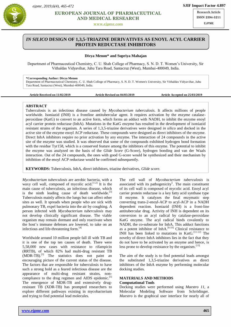
Protein preparation
A B
C
Figure 2: Protein preparation of InhA (PDB ID: 2NSD) (A) Raw protein (B) Pre-processed protein (C)
Optimized and minimized protein.
The X-ray crystallographic structure of InhA, the enoyl
acyl carrier protein reductase from Mycobacterium
tuberculosis, in complex with N-(4-methylbenzoyl)-4-
benzylpiperidine (4PI), which is a direct inhibitor of the
enzyme, was downloaded from Research Collaboratory
for Structural Bioinformatics (RCSB) having PDB ID
2NSD and a resolution of 1.9 A°. The enzyme was
processed using Protein Preparation Wizard.

Menon et al. European Journal of Pharmaceutical and Medical Research
www.ejpmr.com
467
Grid generation
Figure 3: Receptor grid generation.
The receptor grid can be generated by using the Glide
Receptor Grid Generation Wizard. The protein structure
usually has a co-crystallized ligand. During receptor grid
generation, the co-crystallized ligand around which a
grid box is to be generated is picked up. The size of the
grid can be adjusted. The co-crystallized ligand is
excluded from the receptor grid generation calculations.
Validation
Figure 4: Super-positioning of de-docked and re-
docked co-crystallized inhibitor 4PI.
The inhibitor, N-(4-methylbenzoyl)-4-benzylpiperidine
(4PI), was split from the receptor (de-docked). It was re-
docked onto the active site. The de-docked and re-
docked inhibitor 4PI were superimposed. The root mean
square deviation (RMSD) should be less than one.
Docking
A B
Figure 5: (A) 4PI docked in the active site of enzyme 2NSD (B) Isoniazid docked in the active site of enzyme
2NSD.
Docking studies were performed using the program
Glide. During docking studies, ligand was flexible and
the receptor was held rigid. The compounds were docked
in the active site of the enzyme 2NSD. The docking
results were expressed in terms of their G-scores.
RESULTS AND DISSCUSSION
Validation of the protocol
The RMSD value obtained when the de-docked and re-
docked inhibitor 4PI were superimposed, was 0.4524,
which is less than 1, showing that the docking protocol was validated.
Figure 4 shows the superimposed de-docked and re-
docked co-crystallized inhibitor 4PI.

Menon et al. European Journal of Pharmaceutical and Medical Research
www.ejpmr.com
468
Docking studies
A B
C D
Figure 6: Ligand interaction of (A) Inhibitor 4PI (B) Isoniazid (C) & (D) Compounds C18 and C15 with enzyme
2NSD.
Glide Score is an empirical scoring function designed to
maximize separation of compounds with strong binding affinity from those with little to no binding ability.
Particularly beneficial to the binding is the formation of
one or more protein-ligand hydrogen bonds, which helps
the ligand to stay bound in the active site of the receptor
thereby providing stability to the complex.
Figure 6A shows the interaction of the co-crystallized
ligand 4PI in the active pocket of enzyme 2NSD. The
unsubstituted aromatic ring shows pi-pi stacking
interaction, shown by a green line, with the residue
Phe149, and the oxygen of the carboxyl group shows
hydrogen bond formation with the side chain of the residue Tyr158, shown by dashed pink line. In Fig. 6B,
the oxygen of the carboxyl group of isoniazid is seen to
form hydrogen bond with the side chain of the residue
Tyr158.
The InhA inhibitors observed in the literature show a
signature hydrogen bond with the residue Tyr158, which is one of the catalytic residues of the enzyme. This seems
to be a conserved feature among these inhibitors.
In the compound C18 (Table 1), the nitrogen of the
1,3,5-triazine moiety forms hydrogen bond with the side
chain of the residue Tyr158. It also shows pi-pi stacking
interaction with the naphthalene ring of compound C18.
These interactions may be the reason for the high G-
score of -12.25 exhibited by this compound, higher than
the co-crystallized ligand 4PI.
Compound C15 has a G-score similar to the co-crystallized ligand 4PI. The high G-score of -11.65
exhibited by this compound can be attributed to the
presence of bulky substituent in the form of the diphenyl
group, which helps in increasing the hydrophobic contact
between the compound and the hydrophobic residues in
the active site of the enzyme 2NSD.

Menon et al. European Journal of Pharmaceutical and Medical Research
www.ejpmr.com
469
A B
C D
E
Figure 7: (A) Ugly bond (B) Bad bond (C) Hydrogen bond (D) pi-pi stacking (E) Halogen bond formed by the
compounds with various residues.
Figure 7A show the ugly bond which can be seen as a
red dashed line, the bad bond as an orange dashed line
(Fig. 7B), hydrogen bond as a yellow dashed line (Fig.
7C), pi-pi stacking interaction as a cyan dashed line (Fig.
7D) and halogen bond as a purple dashed line (Fig. 7E).
The designed compounds showed 0-2 hydrogen bonds
with not only the residue Tyr158, but also with residues
such as Arg43 and Met98. They showed halogen bonds
with residues Met199 and Arg43. Salt bridge formation
was also seen with the residue Arg43. The number of

Menon et al. European Journal of Pharmaceutical and Medical Research
www.ejpmr.com
470
ugly vdw contacts ranged from 0-1 and the bad vdw
contacts ranged from 0-8.
Compounds C1, C4, C5, C6, C9, C11, C13, C14, C16,
C17, C18, C20 and C21 have G-scores ranging from -
9.095 to -9.934. Compounds C2, C3, C7, C8, C10, C12, C19, C22, C23 and C24 have G-scores in the range of -
10.076 to -10.844. All the compounds have G-scores
higher than those of isoniazid and ethionamide. Table 1
shows various substituents and the G-scores of the
compounds.
N
N
N
Cl
O N
CH3
CH3
R Figure 8: General structure of the 1,3,5-triazine
derivatives.
Table 1: Various substituents and G-scores of the 1,3,5-triazine derivatives
Compound
No. R G-score
C1 -Cl -9.934
C2 NH-
-10.844
C3 NH-
Cl
-10.118
C4 NH-Cl
-9.771
C5 NH-Cl
Cl
-9.574
C6 NH-Br
-9.23
C7 NH-F
-10.262
C8
NH-
N+
O–
O
-10.241
C9 NH-N+
O–
O
-9.095
C10
NH-
O
CH3
-10.148

Menon et al. European Journal of Pharmaceutical and Medical Research
www.ejpmr.com
471
C11 NH-O
CH3
-9.488
C12 N-
CH3
-10.33
C13
NH-
-9.905
C14
NN
N-
-9.342
C15
N-
-11.65
C16 O N-
-9.105
C17 N-NCH3
-9.366
C18
O-
-12.25
C19
O
OH
NH-
-10.076
C20 NH-NH2
-9.401
C21 NH-OH
-9.894
C22 NH
NH-
-10.712
C23 N
CH3
CH3
CH3
CH3
NH2
-10.196

Menon et al. European Journal of Pharmaceutical and Medical Research
www.ejpmr.com
472
C24 O-
-10.456
4PI - -11.62
Isoniazid - -6.915
Ethionamide - -7.541
CONCLUSION
The discovery of novel InhA inhibitors is essential since
direct InhA inhibitors circumvent the need to be
activated by enzymes to show activity. In this study,
docking studies were carried out on a series of 1,3,5-
triazine derivatives using the software Glide. The
compounds showed good G-scores and also interaction
with the residue Tyr158, which has been observed with
other active InhA inhibitors as well. Aside from hydrogen bond with the residues Tyr158, Met98 and
Arg43, the compounds also showed interactions like pi-
pi stacking, halogen bonds and formation of salt bridges.
Hence, it can be concluded that these compounds may be
potent antimycobacterial agents. In future research work,
synthesis of these compounds will be taken up and the
synthesized compounds will be screened for in vitro
antimycobacterial activity. The mechanism of action of
these compounds by InhA inhibition will be confirmed
by InhA enzyme inhibition studies.
REFERENCES 1. Pelczar MJ, Chan ECS, Krieg NR, Microbiology, 5th
edition, Tata McGraw-Hill publication, New Delhi,
2006; 296.
2. Lone MY, Athar M, Gupta VK, Jha PC,
Identification of Mycobacterium tuberculosis enoyl-
acyl carrier protein reductase inhibitors: A combined
in silico and in vitro analysis, Journal of Molecular
Graphics and Modelling, 2017; 76: 172-180.
3. World Health Organization, Global Tuberculosis
Report, 2018.
4. Kumar V, Abbas AK, Fausto N, Robbins and Cotran, Pathologic basis of disease, 7th edition,
Elsevier, New Delhi, 2007; 381-386.
5. Takayama K, Wang C, Besra GS, Pathway to
synthesis and processing of mycolic acids in
Mycobacterium tuberculosis, Clinical Microbiology
Reviews, 2005; 18(1): 81-101.
6. More UA, Joshi SD, Aminabhavi TM, Gadad AK,
Nadagouda MN, Kulkarni VH, Design, synthesis,
molecular docking and 3D-QSAR studies of potent
inhibitors of enoyl acyl carrier protein reductase as
potential antimycobacterial agents, European
Journal of Medicinal Chemistry, 2014; 71: 199-218. 7. Saharan VD, Mahajan SS, Design of furfuraldehyde
formazans as direct inhibitors of enoyl acyl carrier
protein reductase for the treatment of tuberculosis,
American Journal of Pharmtech Research, 2015;
5(4): 367-39.
8. Saharan VD, Mahajan SS, Development of gallic
acid formazans as novel enoyl acyl carrier protein
reductase inhibitor for the treatment of tuberculosis,
Bioorganic & Medicinal Chemistry Letters, 2017;
27: 808-815.
9. Prati F, Zuccotto F, Fletcher D, Convery MA,
Fernandez Menendez R, Bates R, Encinas L, Zeng J,
Chung C, Anton PD, Mendoza-Losana A,
Mackenzie C, Green SR, Huggett M, Barros D,
Wyatt PG, Ray PC, Screening of a novel fragment
library with functional complexity against
Mycobacterium tuberculosis InhA, Chem Med Chem., 2018; 13: 672–677.
10. Heath RJ, Rock CO, Fatty acid biosynthesis as a
target for novel antibacterials, Current Opinion in
Investigational Drugs, 2004; 5(2): 146–153.
11. He X, Alian A, Stroud R, Ortiz de Montellano PR,
Pyrrolidine carboxamides as a novel class of
inhibitors of enoyl acyl carrier protein reductase
(InhA) from Mycobacterium tuberculosis, Journal of
Medicinal Chemistry, 2006; 49(21): 6308–6323.
12. Kuo MR, Morbidoni HR, Alland D, Sneddon SF,
Gourlie BB, Staveski MM, Leonard M, Gregory JS,
Janjigian AD, Yee C, Musser JM, Kreiswirth B, Iwamoto H, Perozzo R, Jacobs, Jr. WR, James C.
Sacchettini JC, Fidock DA, Targeting tuberculosis
and malaria through inhibition of enoyl reductase,
The Journal of Biological Chemistry, 2003; 278(23):
20851–20859.
13. He X, Alian A, Ortiz de Montellano PR, Inhibition
of Mycobacterium tuberculosis enoyl acyl carrier
protein reductase InhA by arylamides, Bioorganic
Medicinal Chemistry, 2007; 15(21): 6649-6658.


![John Daly Lecture: Structure-guided Drug Design for Adenosine …csbj.org/articles/e2015P002.pdf · 2019. 6. 23. · a close overlay of a 1,2,4-triazolo[1,5-a][1,3,5]triazine with](https://static.fdocuments.in/doc/165x107/60b65b22174e1f20e04c667c/john-daly-lecture-structure-guided-drug-design-for-adenosine-csbjorgarticles.jpg)