in Pulmonary - Columbia Universitycolumbiamedicine.org/education/r/Pulmonary/PE/PIOPED.pdf ·...
7
Value of the Ventilation/Perfusion Scan in Acute Pulmonary Embolism Results of the Prospective Investigation of Pulmonary Embolism Diagnosis (PIOPED) The PIOPED Investigators To determine the sensitivities and specificities of ventilation/perfusion lung scans for acute pulmonary embolism, a random sample of 933 of 1493 patients was studied prospectively. Nine hundred thirty-one underwent scintigraphy and 755 underwent pulmonary angiography; 251 (33%) of 755 demonstrated pulmo- nary embolism. Almost all patients with pulmonary embolism had abnormal scans of high, intermediate, or low probability, but so did most without pulmonary embolism (sensitivity, 98%; specificity, 10%). Of 116 patients with high-probabili- ty scans and definitive angiograms, 102 (88%) had pulmonary embolism, but only a minority with pulmonary embolism had high-probability scans (sensitivity, 41%; specificity, 97%). Of 322 with intermediate-probability scans and definitive angiograms, 105 (33%) had pulmonary embolism. Follow-up and angiography together suggest pulmonary embolism occurred among 12% of patients with low-probability scans. Clinical assessment combined with the ventilation/perfu- sion scan established the diagnosis or exclusion of pulmonary embolism only for a minority of patients\p=m-\those with clear and concordant clinical and ventilation/ perfusion scan findings. (JAMA. 1990;263:2753-2759) PERFUSION lung scans have been re¬ ported to be sensitive in detecting pul¬ monary emboli, but many other condi- For editorial comment see p 2794. tions such as pneumonia or local bronchospasm cause perfusion defects.1 Ventilation scans were added to perfu¬ sion scans with the idea that ventilation would be abnormal in areas of pneumo¬ nia or local hypoventilation, but that in pulmonary embolism ventilation would be normal.2 A number of investigators have attempted to make ventilation/ perfusion (V/Q) scans more useful for diagnosing pulmonary embolism by classifying them not just as normal or abnormal, but if abnormal, as indicating high probability, intermediate probabil¬ ity (indeterminate), or low probability of pulmonary embolism.3 Under the aus¬ pices of the National Heart, Lung, and Blood Institute, the Prospective Inves¬ tigation of Pulmonary Embolism Diag- nosis (PIOPED) investigators have as¬ sessed the diagnostic usefulness of V/Q lung scans in acute pulmonary embo¬ lism. The project protocol and consent forms were approved by the institution¬ al review boards of all participating cen¬ ters. (Participating centers and investi¬ gators are listed at the end of the article.) METHODS Patient Enrollment From January 1985 through Septem¬ ber 1986 in each of six clinical centers, all patients for whom a request for a V/Q scan or a pulmonary angiogram was made were considered for study entry. The eligible study population consisted of patients, 18 years or older, inpatients and outpatients, in whom symptoms that suggested pulmonary embolism were present within 24 hours of study entry and without contraindications to angiography such as pregnancy, serum creatinine level greater than 260 jjtmol/L, or hypersensitivity to contrast material. Once approached for the study, patients with recurrences were not approached for recruitment a sec¬ ond time. Recruitment A total of 5587 requests for V/Q scans were recorded in the six PIOPED clini¬ cal centers from January 1985 through September 1986 (Figure). Although Reprint requests to Division of Lung Diseases, Na- tional Heart, Lung, and Blood Institute, Westwood Bldg, Room 6A16, 5333 Westbard Ave, Bethesda, MD 20892 (Carol E. Vreim, PhD). at New York State Psychiatric Institute on August 3, 2009 www.jama.com Downloaded from
Transcript of in Pulmonary - Columbia Universitycolumbiamedicine.org/education/r/Pulmonary/PE/PIOPED.pdf ·...
Value of the Ventilation/Perfusion Scanin Acute Pulmonary EmbolismResults of the Prospective Investigation ofPulmonary Embolism Diagnosis (PIOPED)The PIOPED Investigators
To determine the sensitivities and specificities of ventilation/perfusion lungscans for acute pulmonary embolism, a random sample of 933 of 1493 patientswas studied prospectively. Nine hundred thirty-one underwent scintigraphy and755 underwent pulmonary angiography; 251 (33%) of 755 demonstrated pulmo-nary embolism. Almost all patients with pulmonary embolism had abnormalscans of high, intermediate, or low probability, but so did most without pulmonaryembolism (sensitivity, 98%; specificity, 10%). Of 116 patients with high-probabili-ty scans and definitive angiograms, 102 (88%) had pulmonary embolism, butonly a minority with pulmonary embolism had high-probability scans (sensitivity,41%; specificity, 97%). Of 322 with intermediate-probability scans and definitiveangiograms, 105 (33%) had pulmonary embolism. Follow-up and angiographytogether suggest pulmonary embolism occurred among 12% of patients withlow-probability scans. Clinical assessment combined with the ventilation/perfu-sion scan established the diagnosis or exclusion of pulmonary embolism only fora minority of patients\p=m-\thosewith clear and concordant clinical and ventilation/perfusion scan findings.
(JAMA. 1990;263:2753-2759)
PERFUSION lung scans have been re¬ported to be sensitive in detecting pul¬monary emboli, but many other condi-
For editorial comment see p 2794.
tions such as pneumonia or localbronchospasm cause perfusion defects.1Ventilation scans were added to perfu¬sion scans with the idea that ventilation
would be abnormal in areas of pneumo¬nia or local hypoventilation, but that inpulmonary embolism ventilation wouldbe normal.2 A number of investigatorshave attempted to make ventilation/perfusion (V/Q) scans more useful fordiagnosing pulmonary embolism byclassifying them not just as normal or
abnormal, but ifabnormal, as indicatinghigh probability, intermediate probabil¬ity (indeterminate), or low probabilityofpulmonary embolism.3 Under the aus¬pices of the National Heart, Lung, andBlood Institute, the Prospective Inves¬tigation of Pulmonary Embolism Diag-
nosis (PIOPED) investigators have as¬sessed the diagnostic usefulness ofV/Qlung scans in acute pulmonary embo¬lism. The project protocol and consentforms were approved by the institution¬al review boards ofall participating cen¬ters. (Participating centers and investi¬gators are listed at the end of thearticle.)METHODSPatient Enrollment
From January 1985 through Septem¬ber 1986 in each of six clinical centers,all patients forwhom a request fora V/Qscan or a pulmonary angiogram wasmade were considered for study entry.The eligible study population consistedof patients, 18 years or older, inpatientsand outpatients, in whom symptomsthat suggested pulmonary embolismwere present within 24 hours of studyentry and without contraindications toangiography such as pregnancy, serumcreatinine level greater than 260jjtmol/L, or hypersensitivity to contrastmaterial. Once approached for thestudy, patients with recurrences werenot approached for recruitment a sec¬ond time.
RecruitmentA total of 5587 requests for V/Q scans
were recorded in the six PIOPED clini¬cal centers from January 1985 throughSeptember 1986 (Figure). Although
Reprint requests to Division of Lung Diseases, Na-tional Heart, Lung, and Blood Institute, Westwood Bldg,Room 6A16, 5333 Westbard Ave, Bethesda, MD 20892(Carol E. Vreim, PhD).
at New York State Psychiatric Institute on August 3, 2009 www.jama.comDownloaded from

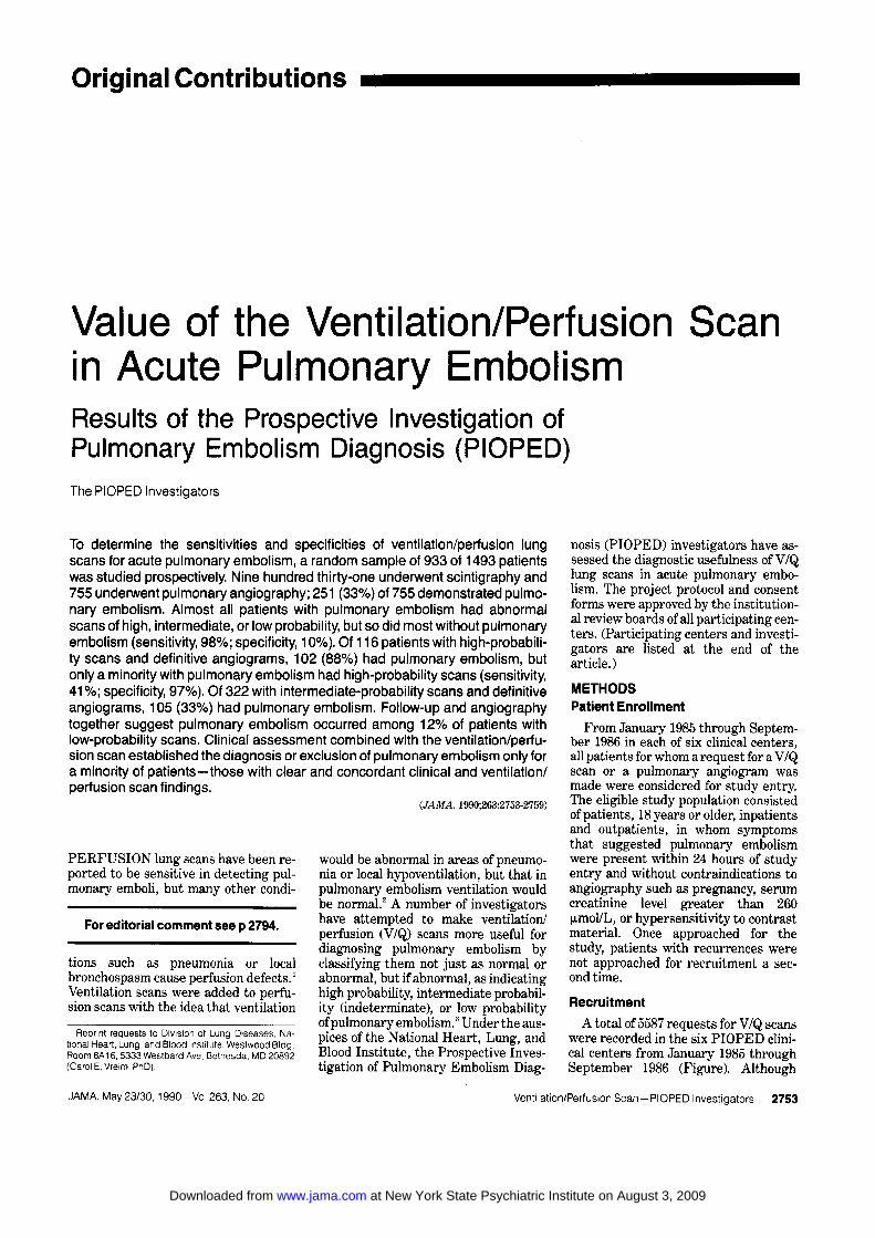
Requests for Lung Scans5587
Scan Requests Cancelled,Scans Requested for Research Purposes,Diagnoses Other Than Acute
Pulmonary Embolism,Patients in Whom AngiographyContraindicated, and Other Reasonsfor Ineligibility According toStudy Design
2571
Eligible Patients3016
_EConsent
Given1493
3_ConsentRefused
1523
Random Sample
Not Selected forSensitivity and
Specificity Analyses560
Selected forSensitivity and
Specificity Analyses933
Interpretable ScanNot Completed
2
ScanCompleted
931
AngiogramCompleted
755
XPulmonary Embolism
Present251
AngiogramNot Completed
176
Pulmonary EmbolismAbsent
480
1Pulmonary Embolism
Uncertain24
Flow chart illustrating the numbers of requests for lung scans, recruitment of patients, completion of lungscans, and results of angiography in the Prospective Investigation of Pulmonary Embolism Diagnosis.
some patients could not be thoroughlyevaluated prior to completion of the V/Qscan, clinical investigators made everyeffort to record their individual clinicalimpressions as to the likelihood of pul¬monary embolism prior to learning theresults of V/Q scans and angiography.Impressions were based on an agreed onset of information—history, results ofphysical examination, arterial blood gasanalyses, chest roentgenograms, andelectrocardiograms—but without stan¬dardized diagnostic algorithms. Themedical records of a random sample ofpatients who refused or were ineligiblefor study entry (refuser/ineligible pa¬tients) were evaluated retrospectivelyfor comparison with study patients.
Lung Scan
The protocol directed ventilation andperfusion studies with the subject in theupright position, but other positionswere acceptable. Ventilation studieswere performed with 5.6xl08 to11.1 x 108 Bq of xenon 133 using a 20%symmetric window set over the 80-keVenergy peak. They started with a100 000-count, posterior-view, first-breath image and then posterior equi¬librium (wash-in) images for two con¬secutive 120-second periods. Washoutconsisted of three serial 45-second pos¬terior views, 45-second left and rightposterior oblique views, and a final 45-second posterior view. Then, perfusion
scans were obtained with 1.5 x 10⁸ Bq oftechnetium Tc 99m macroaggregatedalbumin that contained 100 000 to700 000 particles using a 20% symmetricwindow set over the 140-keV energypeak. Particles were injected into anantecubital vein over 5 to 10 respiratorycycles, with the patient supine or atmost semierect. The perfusion imagesconsisted of anterior, posterior, bothposterior oblique, and both anterioroblique views, with 750 000 counts perimage for each. For the lateral viewwith the best perfusion, 500 000 countsper image were collected; the other lat¬eral view was obtained for the same
length of time. Scintillation cameraswith a wide field of view (38.1 cm indiameter) were used with parallel-hole,low-energy, all-purpose collimators.Perfusion scans were satisfactory orbetter in 96% of cases, ventilation scans
adequate or better in 95%.
AngiographyThe femoral-vein Seldinger tech¬
nique with a multiple side-holed, 6F to8F pigtail catheter was used. Smallamounts ofcontrast material (5 to 8 mL)were injected by hand, to check the pa¬tency of the inferior vena cava by fluo-roscopy. The catheter was directed intothe main pulmonary artery of the lungwith the greatest V/Q scan abnormali¬ty. Initial filming was in the antero-posterior projection. Seventy-six per¬cent iodinated contrast material was
injected at a rate of 20 to 35 mL/s for a
total of 40 to 50 mL (2-second injection).Film rates were three per second for 3seconds, followed by one per second for4 to 6 seconds. Depending on the size ofthe lungs, filming was not magnified or
given a low magnification of 1.4. A 12:1grid was used and roentgenographicfactors were in the range of 70 to 80kilovolts (peak) and 0.025 to 0.040 sec¬onds at 1000 mA (large focal spot of 1.2to 1.5 mm in diameter). If emboli werenot identified, injections were repeatedand magnification (1.8 to 2.0 times)oblique views were obtained of the ar¬eas suspicious for pulmonary embolism.Films were obtained with an air-gaptechnique (ie, no grid used). Roent¬genographic factors were in the range of78 to 88 kV(p) and 0.040 to 0.080 secondsat 160 mA (small focal spot of 0.3 to 0.6mm in diameter). If no emboli were
found in the first lung, or if bilateralangiography in the clinical center was
routine, identical techniques were usedfor the second lung. Angiography was
completed within 24 hours, and usuallywithin 12 hours of V/Q scans. Pulmo¬nary angiograms were adequate or bet¬ter in 95% of cases.
at New York State Psychiatric Institute on August 3, 2009 www.jama.comDownloaded from

Central Scan andAngiogram Interpretations
Two nuclear medicine readers, notfrom the center that performed thescan, independently interpreted thelung scans with chest roentgenogramsaccording to preestablished study crite¬ria (Table 1). Angiograms were likewiserandomly assigned to pairs of angio-graphers from clinical centers otherthan the originating hospital. The an¬
giogram readers interpreted the angio¬grams with lung scans as having acutepulmonary embolism present—whichrequired the identification of an embo-lus obstructing a vessel or the outline ofan embolus (filling defect) within a ves¬sel—absent, or uncertain. If two read¬ers disagreed, the interpretations were
adjudicated by readers who were se¬
lected randomly from the remainingclinical centers. If adjudicating readersdid not agree with either of the first tworeaders, scans or angiograms went topanels of nuclear medicine or angiogra¬phy readers. The final adjudicated V/Qscan readings consisted of four catego¬ries—high probability, intermediateprobability (indeterminate), low proba¬bility, and low/very low probabilitythrough normal (near normal/normal).The near-normal/normal category in¬cludes readings of very low probabilityby one reader and low probability by theother, very low probability by both,very low probability by one and normalby the other, and normal by both. Re¬fuser/ineligible patients' scans wereread in each clinical center by the clini¬cal center's PIOPED nuclear medicinereader(s) and not reread.Follow-up andOutcome Classification
Patients were contacted by telephoneat 1, 3, 6, and 12 months after studyentry. Deaths, new studies for pulmo¬nary embolism, and major bleedingcomplications were reviewed by an out¬come classification committee using allavailable information. Only 23 (2.5%) ofthe 931 patients had incomplete (16) orno (7) follow-up. Angiograms, follow-updata, and outcome classifications wereused to determine pulmonary embolismstatus as positive for patients with an¬
giograms that showed pulmonary em¬boli and for patients for whom outcomereview established the presence of pul¬monary emboli at the time of PIOPEDrecruitment. Pulmonary embolism sta¬tus was determined as negative for pa¬tients with angiograms that did notshow pulmonary emboli and no contraryoutcome review and for patients wholacked a definitive angiogram readingwho were discharged from the hospitalwithout a prescription for anticoagu-
Table 1. —PIOPED Central Scan Interpretation Categories and Criteria*
High probability22 Large (>75% of a segment) segmentai perfusion defects without corresponding ventilation or roentgeno-
graphic abnormalities or substantially larger than either matching ventilation or chest roentgenogramabnormalities
2:2 Moderate segmental (225% and s75% of a segment) perfusion defects without matching ventilation or chestroentgenogram abnormalities and 1 large mismatched segmental defect
24 Moderate segmentai perfusion defects without ventilation or chest roentgenogram abnormalitiesIntermediate probability (indeterminate)
Not falling into normal, very-low-, low-, or high-probability categoriesBorderline high or borderline lowDifficult to categorize as low or high
Low probabilityNonsegmental perfusion defects (eg, very small effusion causing blunting of the costophrenic angle, cardiomegaly,
enlarged aorta, hila, and mediastinum, and elevated diaphragm)Single moderate mismatched segmentai perfusion defect with normal chest roentgenogramAny perfusion defect with a substantially /arger chest roentgenogram abnormalityLarge or moderate segmentai perfusion defects involving no more than 4 segments in 1 lung and no more than
3 segments in 1 lung region with matching ventilation defects either equal to or larger in size and chestroentgenogram either normal or with abnormalities substantially smaller than perfusion defects
>3 Small segmentai perfusion defects (<25% of a segment) with a normal chest roentgenogramVery low probability
s3 Small segmentai perfusion defects with a normal chest roentgenogramNormal
No perfusion defects presentPerfusion outlines exactly the shape of the lungs as seen on the chest roentgenogram (hilar and aortic impressions
may be seen, chest roentgenogram and/or ventilation study may be abnormal)
•PIOPED indicates Prospective Investigation of Pulmonary Embolism Diagnosis.
Table 2.—Recruitment of Patients and Completion of Angiography*
Clinical Center
%ofEligiblePatients
Recruited
No. of PIOPED Patients WithLung Scans Who Were Selected for
Angiographie PursuitAngiograms
Obtained, No. (%)Duke University 46 137 115 (84)Henry Ford Hospital 62 228 177 (78)Massachusetts General Hospital 33 140 120 (86)University of Michigan 52 102 65 (64)University of Pennsylvania 70 168 134 (80)Yale UniversityTotal
4350
156931
144 (92)755 (81)
PIOPED indicates Prospective Investigation of Pulmonary Embolism Diagnosis.
lants and in whom no outcome eventsuggested pulmonary embolism. Pul¬monary embolism status could be deter¬mined as positive or negative for 902patients. À clinical assessment of thelikelihood of pulmonary embolism wasavailable for 887 (98%) of these patients.Statistical Methods
Probability values for the comparisonofpercentages and proportions and 95%confidence intervals (CTs) were calcu¬lated using standard z tests." A x2 testfor homogeneity of proportions wasused to compare distributions.5 Sensi¬tivity is defined as the proportion ofcases of pulmonary embolism correctlydiagnosed and specificity as the pro¬portion of diagnoses that pulmonaryembolism is absent for patients withoutpulmonary embolism. Sensitivity, spe¬cificity, and percent agreement havebeen calculated according to standardmethods for proportions.6 Analyseswere performed with the StatisticalPackage for the Social Sciences statisti¬cal software package.7 Recruitment of
900 to 1000 patients in the random sam¬
ple for PIOPED angiography was
planned to obtain estimates of sensitiv¬ity and specificity with 95% CIs no wid¬er than ± 8%. Tb determine the sensi¬tivity and specificity of V/Q lung scanswithout the biases associated with hap¬hazard patient selection (ie, conve¬nience sampling),89 a 933-patient sam¬
ple of the 1493 patients who consentedto PIOPED participation was selectedaccording to random sampling sched¬ules created separately by the data andcoordinating center for each clinical cen¬ter. The PIOPED protocol requiredthese 933 patients to undergo angiogra¬phy if their scans were abnormal. Of the933 patients selected for angiography, 1patient died before the V/Q scan couldbe completed and 1 other patient's V/Qscan was determined to be uninterpre-table. These 2 patients are not furtherreported herein.
RESULTSOf the 3016 patients eligible for
PIOPED, 1493 (50%) gave consent to
at New York State Psychiatric Institute on August 3, 2009 www.jama.comDownloaded from

Table 3.—Patient Characteristics*
Refuser/PIOPED Ineligible
_(N = 931) (N = 326)Age (mean), y 56.1 56,4Maie, % 45 44Service, %
Medical/CCU 40 36Surgical 18 21Emergency department/clinic 30 32ICU 10 10Other 1 1
Hospital mortality, % 9 10
•PIOPED indicates Prospective Investigation of Pul¬monary Embolism Diagnosis; CCU, coronary care unit;and ICU, intensive care unit.
participate in PIOPED (Figure). Theclinical centers varied in the percentageof eligible patients for whom consentcould be obtained, from 33% to 70%, andin the percentage of patients for whomangiograms were obtained among thoseselected to determine the sensitivityand specificity of V/Q lung scans
(PIOPED angiographie pursuit), from64% to 92% (Table 2). The PIOPED pa¬tients resembled refuser/ineligible pa¬tients in a variety of clinical characteris¬tics (Table 3). The PIOPED patientsand refuser/ineligible patients were dif¬ferent, however, in their lung scan ab¬normalities (P<.01). Although they hadsimilar frequencies of high-probabilityscans (13% among PIOPED patientsand 11% among refuser/ineligible pa¬tients), the PIOPED patients had inter¬mediate-probability scans almost twiceas often as refuser/ineligible patients(39% vs 22%). The PIOPED study had asmaller proportion of patients withlow-probability and near-normal/nor¬mal lung scans. Of the 931 patients whowere selected for mandatory angiogra¬phy in PIOPED, 755 (81.1%) completedangiography; 69 (7.4%) did not completeangiography because their V/Q scanswere interpreted locally as normal; and107 (11.5%) did not complete angiogra¬phy in spite of the requirements of theprotocol.Reader Agreement
Agreement among scan readers wasexcellent for high-probability (95%),very-low-probability (92%), and nor¬mal (94%) scan categories. For interme¬diate-probability (indeterminate) andlow-probability scan categories, thereaders agreed less frequently (75% and70%, respectively). In only 24 (2.6%)of 931 scans was panel adjudication nec¬
essary. Agreement among angiogramreaders was excellent for the presenceof pulmonary embolism (92%). For theabsence of pulmonary embolism andpulmonary embolism uncertain, inde¬pendent readers agreed on 83% and 89%
Table 4.—Comparison of Scan Category With Angiogram Findings
Scan CategoryPulmonaryEmbolismPresent
PulmonaryEmbolism
Absent
PulmonaryEmbolismUncertain
NoAngiogram
TotalNo.
High probability 102 14 1 124Intermediate probability 105 217 33 364Low probability 39 199 12 62 312Near normal/normal 50 131Total 251 480 931
Table 5.—Comparison of Scan Category With Angiogram Findings, Sensitivity and SpecificityScan Category Sensitivity, % Specificity, %
High probability 41 97High or intermediate probability 82 52High, intermediate, or low probability 98 10
of angiograms, respectively. In only 13(1.7%) of 755 angiograms was panel ad¬judication necessary.Scan Findings
Most (676) of the 931 patients hadintermediate- or low-probability V/Qscan readings (39% and 34%, respec¬tively) (Table 4). Only 131 (14%) hadnear-normal/normal V/Q scans and 124(13%) had high-probability scans. The176 patients who did not undergo angi¬ography, in spite of their selection formandatory angiography, had less se¬vere scan abnormalities than those whocompleted angiography (P<.01).Angiogram and Outcome Findings
Among the 755 patients who com¬pleted angiography, 251 (33%) hadthromboemboli seen on the angiogram,480 (64%) had no thromboemboli seen,and 24 (3%) had angiograms in which thepresence of thromboemboli was uncer¬tain (Table 4). For the vast majority ofpatients, 1 year of follow-up revealedclinical courses entirely consistent withangiographically established diagnoses.The outcome classification committeedisagreed with central angiography in¬terpretations for 4 patients with pulmo¬nary angiograms free of signs of acuteembolism who had pulmonary embolismat autopsies performed 2 to 6 days afterangiography. The scan interpretationswere of low probability for 3 and of in¬termediate probability (indeterminate)for 1 of these 4 patients.Scans Compared With Angiograms
One hundred two of 251 patients withangiograms that showed thromboem¬boli had high-probability V/Q scans.The sensitivity, therefore, was 41%(95% CI, 34% to 47%) (Tables 4 and 5). Ifthe patient had either a high- or inter¬mediate-probability V/Q scan, the sen-
sitivity for thromboemboli on angiogra¬phy increased to 207 (82%) of 251 (95%CI, 78% to 87%). If the patient had ei¬ther a high-, intermediate-, or low-probability V/Q scan, then 246 of 251had thromboemboli on angiography, a
sensitivity of 98% (95% CI, 96% to100%).
Only 14 (3%) of 480 patients who didnot have thromboemboli on angiogra¬phy had high-probability V/Q scans.The specificity of a high-probabilityscan—ie, the percentage of patientswith angiograms free of signs of acuteembolism who had a scan that showedother than high probability—was 97%(466/480) (95% CI, 96% to 98%). Forhigh- and intermediate-probabilityscans together, specificity was 52%(249/480 patients with angiograms freeof signs of acute embolism) (95% CI,47% to 56%). For high-, intermediate-,and low-probability scans together,specificity was 10% (50/480 patientswith angiograms free of signs of acuteembolism) (95% CI, 8% to 13%). The 36sensitivities and specificities calculatedby reproducing Table 5 for each clinicalcenter varied about the studywide sen¬sitivities and specificities—25 (69%) of36 were within ± 5% of the studywideestimates and 34 (94%) of 36 within± 10%.
Most patients with high-probabilityV/Q scans had angiographie evidence ofpulmonary embolism (102/116 definitivestudies, or a positive predictive value of88%). Of the 60 patients with previoushistories of pulmonary embolism, 20were found to have pulmonary embolion angiography. Of the 19 patients withhistories of pulmonary embolism and a
high-probability V/Q scan, only 14 werefound to have acute pulmonary embolion angiography. The positive predictivevalue of a high-probability V/Q scan inpatients with histories of pulmonary
at New York State Psychiatric Institute on August 3, 2009 www.jama.comDownloaded from

Table 6.—Pulmonary Embolism (PE) Status*
Clinical Science Probability, %
80-100 20-79 0-19 All Probabilities
PE + /NO. PE + /NO. PE-WNo. PE + /NO.Scan Category_of Patients_%_ofPatients_%_ofPatients_%_of Patients_%_
High probability 28/29 96 70/80 88 5/9 56 103/118 87
Intermediate probability_27/41_66_66/236_28_11/68_16_104/345_30Low probability_6/15_40_30/191_16_4/90_4_40/296_14Nearnormal/normal_0/5_0_4162_6_1/61_2_5/128_4Total 61/90 68 170/569 30 21/228 9 252/887 28
*PE + indicates angiogram reading that shows pulmonary embolism or determination of pulmonary embolism by the outcome classification committee on review. Pulmonaryembolism status is based on angiogram interpretation for 713 patients, on angiogram interpretation and outcome classification committee reassignment for 4 patients, and on
clinical information alone (without definitive angiography) for 170 patients.
embolism was only 74% (14/19), com¬
pared with 91% (88/97) for those withouta history of pulmonary embolism(P<.05). This difference in positive pre¬dictive values reflects a loss of specific¬ity in the high-probability V/Q scan di¬agnosis for patients with histories ofpulmonary embolism (88%) vs thosewith no prior pulmonary embolism(98%)(P<.01).
The percentage of patients whose an¬
giograms showed thromboemboli wasless in the intermediate-probability(indeterminate), low-probability, andnear-normal/normal scan categories—33%, 16%, and 9%, respectively (Table4). The frequency of angiographicallydemonstrable emboli among patientswith low-probability scans (39 [16%] of238) and near-normal/normal scans (5[9%] of55) is influenced by the relativelylarge numbers of patients (74 patientsand 76 patients, respectively) for whomangiography was not completed or in¬terpretations were uncertain in thesescan categories (Table 4). Since none ofthese patients received anticoagulantsand none developed clinically evidentpulmonary embolism during follow-up,important pulmonary emboli did not oc¬cur in this group. If all 150 patients were
regarded as not having had pulmonaryemboli, then the frequency of clinicallyimportant pulmonary emboli in patientswith low-probability scans could be noless than 39 (12%) of 312, and in patientswith near-normal/normal scans, 5 (4%)of 131.
There were 21 patients whose V/Qscans were read centrally as normal onfirst reading by both readers. Three un¬derwent angiography and none showedthromboemboli. None of the remaining18 patients received anticoagulants andnone had clinically evident pulmonaryembolism on follow-up.Clinical Assessment of theLikelihood of Pulmonary Embolism
The clinician's assessment of the like¬lihood of pulmonary embolism recorded
before the scan was performed ("priorprobability") was compared with pul¬monary embolism status as determinedby angiography and follow-up informa¬tion (Table 6) for 887 patients with priorprobability assessments and definitepulmonary embolism status. A clinicalassessment of 80% to 100% likelihood ofpulmonary embolism was made in 90patients (10%) and was correct in 61(68%) of 90. A clinical assessment of 0%to 19% likelihood of pulmonary embo¬lism was made in 228 (26%) and wascorrect in 207 (91%) of 228. Clinical as¬
sessment, therefore, was more oftencorrect in excluding pulmonary embo¬lism than in identifying pulmonary em¬bolism. In the majority of patients (569[64%]), clinical assessments were non¬committal (20% to 79% likelihood of pul¬monary embolism).
Combining clinical assessments withthe V/Q scan interpretations improvedthe overall chance of reaching a correctdiagnosis of acute pulmonary embolism(Table 6). Among patients in whom theclinical impression and the scan inter¬pretation were both of high probabilityfor pulmonary embolism, 28 (96%) of 29had pulmonary embolism. If the high-probability scan interpretation was
paired with an intermediate-likelihoodclinical assessment or a low-likelihoodclinical assessment, then the probabili¬ty that the patient had pulmonary em¬bolism fell to 70 (88%) of 80 and 5 (56%)of 9, respectively. The addition of theclinical evaluation also helped in thelow-probability and in the near-normal/normal scan categories. A low-probabil¬ity clinical assessment (0% to 19% likeli¬hood of pulmonary embolism based onclinical judgment), when paired with a
low-probability V/Q scan, correctly ex¬cluded the diagnosis of pulmonary em¬bolism in 86 (96%) of 90 patients. Thenear-normal/normal V/Q scan category,when paired with a low-likelihood clini¬cal assessment, correctly excluded pul¬monary embolism in 60 (98%) of 61patients.
COMMENT
The PIOPED study was conducted asa multicenter, prospective effort to esti¬mate the sensitivity and specificity ofthe V/Q lung scan for the diagnosis ofpulmonary embolism. Other retrospec¬tive and prospective studies have fo¬cused on positive predictive values,which are influenced by prevalence ofpulmonary embolism and patient selec¬tion. Sensitivity and specificity, howev¬er, are fundamental characteristics of a
diagnostic test and are not affected bythe prevalence of disease.10
In PIOPED, almost all patients (98%)with clinically important pulmonaryembolism had lung scans that fell intoone of the three abnormal categories—high, intermediate (indeterminate), orlow probability. If all three abnormalcategories are combined into one, thelung scan is sensitive enough to serve asa screening test for the diagnosis ofpulmonary embolism, but the specific¬ity is limited. The high-probability scanlacked sensitivity in diagnosing pulmo¬nary embolism, since it failed to identify59% of patients with this disorder.
Only 14 (3%) of 480 patients who didnot have evidence of acute pulmonaryembolism on angiography had high-probability scans (Table 4). Therefore,the specificity of a high-probability scanwas 97%. For patients with histories ofpulmonary embolism, the specificity ofthe high-probability scan was reduced.This finding is consistent with other re¬
ports of previous pulmonary embolismas a cause of V/Q scan abnormality thatmay be confused with acute pulmonaryembolism.1112 The specificity of scans ofintermediate or low probability wasmuch less than the specificity of thehigh-probability scan.
The PIOPED's study design includedpatient enumeration and recruitmentprior to scan completion to avoid bias inpatient selection. Nonetheless, patientswho ultimately had high- and interme¬diate-probability scans were more often
at New York State Psychiatric Institute on August 3, 2009 www.jama.comDownloaded from

successfully recruited for PIOPED. Ifanything, this selection bias would sug¬gest that PIOPED tends to overesti¬mate V/Q scans' sensitivities and under¬estimate specificities.
Clinical decisions are often made onthe basis of the predictive values, whichdepend not only on the test's sensitivityand specificity, but also on the preva¬lence of disease in the population stud¬ied. Based on angiogram results, theprevalence of pulmonary embolism inPIOPED was 33% (251/755) (Table 4);based on pulmonary embolism status—derived from angiogram evaluation and/or clinical evaluation—the prevalencewas 28% (Table 6), similar to the preva¬lences described in previous reports.13"21In PIOPED, the positive predictive val¬ue of the high-probability scan was 88%,whereas the negative predictive valueof a low-probability scan was 84%. Thenegative predictive value of the near-normal/normal scan category was bet¬ter at 91%. Estimates of negative pre¬dictive values increased when analysestook into account patients who did notundergo angiography, did not receiveanticoagulants, and had no evidence ofpulmonary embolism occurring during 1year of follow-up. Including these pa¬tients among those not having pulmo¬nary embolism in the analysis improvedthe negative predictive value of the low-probability scan from 84% to 88% and ofthe near-normal/normal scan from 91%to 96%. Because some instances ofacutepulmonary embolism may not have beendetected among these patients, the truenegative predictive values may be lessthan 88% for low-probability scans and96% for near-normal/normal scans, butstill ought to be closer to these lattervalues than to the 84% and 91%, whichdid not account for patients without an¬
giography results.Although pulmonary emboli did occur
in patients with scans classified in thecategories between low probability andnormal, pulmonary embolism was docu¬mented in only 5 (4%) of 131 of suchpatients. The true proportion of pa¬tients with pulmonary embolism mustbe inferred with caution, because largenumbers of patients with near-normal/normal scans were not successfully re¬cruited for the study. Only 42% of the131 PIOPED patients in this categorycompleted angiography. Only 3 ofthe 21patients with lung scans read as normalby both readers on the final readingcompleted angiography; all 3 had nor¬mal pulmonary angiograms. None of theremaining 18 had clinically evident pul¬monary emboli on follow-up. This find¬ing is consistent with the findings ofKipper et al.22
The value of combining clinical judg-
ment with the interpretation of the scanis supported by the PIOPED study. Thepredictive value of the high- and low-probability lung scans improved whensupported by similar clinical assess¬ments. For 90 patients, the negativepredictive value of the low-probabilityscan rose to 96% when accompanied by aclinical assessment of low likelihood. In29 patients, the positive predictive val¬ue of a high-probability scan increasedto 96% if supported by a high-likelihoodclinical assessment. In the PIOPED ex¬
perience, combining a lung scan inter¬pretation with a strong clinical suspi¬cion as to whether acute pulmonaryembolism is present is a sound diagnos¬tic strategy, as previously suggested byMcNeil and colleagues,20,21 but is suf¬ficient for only a minority of patients(Table 6). For a substantial number ofpatients in the PIOPED study, angiog¬raphy was required for a definitive diag¬nosis ofpulmonary embolism.
The PIOPED study employed pulmo¬nary angiography, which proved to be asafe and accurate method of diagnosingpulmonary embolism, although it is in¬vasive. The four patients (0.5%) forwhom the outcome classification com¬mittee disagreed with blinded angio¬gram interpretations that showed acutepulmonary embolism to be absent mustbe considered carefully in light of theangiographie criteria's design for acutepulmonary embolism, the variable timebetween angiographie evaluation andthe patients' deaths, and the variabilityin pathophysiology and pathological in¬terpretation of thromboemboli in evolu¬tion. In the PIOPED study, a normalangiogram almost excluded the possibil¬ity of pulmonary embolism, confirmingthe results of two previous studies.1415
The PIOPED findings extend ob¬servations made by other investiga¬tors,131220 from whom the PIOPED in¬vestigators derived study criteria forangiogram and V/Q scan interpretation.Although predictive values for patientswith high-probability scans and pa¬tients with low-probability scans in pre¬vious series are generally consistentwith the PIOPED findings, the under-representation of patients with low-probability scans in previous studieshas in the past led to an exaggeratedimpression of the sensitivity of the high-probability lung scan.
The findings of Hull and colleagues"'18in the Hamilton District Thromboembo-lism Programme are particularly inter¬esting in comparison with the PIOPEDresults. Of the 305 patients with sus¬
pected pulmonary embolism and abnor¬mal perfusion lung scans in their study,173 (57%) had adequate ventilationscans and adequate pulmonary angio-
grams. Ninety-five patients (31%) hadpulmonary emboli demonstrated on an¬
giography. The predictive values fromtheir study are similar to PIOPED re¬sults in the high-probability and inter¬mediate-probability (indeterminate)scan categories. The PIOPED study,likewise, found pulmonary emboliamong patients with scans in the low-probability category, but fewer thanthe 25% for subsegmental matched le¬sions and 40% for subsegmental mis¬matched lesions found by Hull et al. Pa¬tient referral patterns or lung scan
interpretation criteria may account forthe differences between PIOPED re¬sults and the Hamilton study results.Since angiographie studies are notavailable and clinical follow-up has notbeen applied to determine pulmonaryembolism status for the 110 patientswithout adequate angiography, for the22 patients without adequate ventila¬tion scans, and for the patients withnormal scans in the Hamilton DistrictThromboembolism Programme, com¬
parisons of estimates of sensitivity andspecificity between the two studies arenot possible.
The PIOPED results lead to a num¬ber of conclusions that settle controver¬sies about the diagnostic value of thelung scan in pulmonary embolism.23,24 Ahigh-probability scan usually indicatespulmonary embolism, but only a minor¬ity of patients with pulmonary embo¬lism have a high-probability scan. A his¬tory of pulmonary embolism decreasesthe accuracy ofdiagnoses based on high-probability scans. A low-probabilityscan with a strong clinical impressionthat pulmonary embolism is not likelymakes the possibility of pulmonary em¬bolism remote. Near-normal/normallung scans make the diagnosis of acutepulmonary embolism very unlikely. Anintermediate-probability (indetermi¬nate) scan is not of help in establishing a
diagnosis. In PIOPED, the scan com¬bined with clinical assessment permit¬ted a noninvasive diagnosis or exclusionof acute pulmonary embolism for a mi¬nority ofpatients.
This study was supported by contracts NOl-HR-34007, NO1-HR-34008, NO1-HR-34009, NOl-HR-34010, NO1-HR-34011, NO1-HR-34012, and NOl-HR-34013 from the National Heart, Lung, andBlood Institute, Bethesda, Md.
The secretarial assistance of JoAnne Decker hasbeen greatly appreciated.
Steering CommitteeThe PIOPED investigators are as follows:
Herbert A. Saltzman, MD, chairman; AbassAlavi, MD, Richard H. Greenspan, MD, Charles A.Hales, MD, Paul D. Stein, MD, Michael Terrin,MD, MPH, Carol Vreim, PhD, John G. Weg, MD;alternates: Christos Athanasoulis, MD, AlexanderGottschalk, MD.
at New York State Psychiatric Institute on August 3, 2009 www.jama.comDownloaded from

Clinical CentersDuke University
Herbert A. Saltzman, MD, principal investiga¬tor; Russell Blinder, MD, R. Edward Coleman,MD, N. Reed Dunnick, MD, William J. Fulker-son, Jr, MD, Lee Mallatratt, RN, Carl E. Ravin,MD.
Henry Ford HospitalPaul D. Stein, MD, principal investigator; Debo¬rah Adams, RN, Matthew Burke, MD, Jerry W.Froelich, MD, Kenneth V. Leeper, MD, BarryA. Lesser, MD, John Popovich, Jr, MD, P. C.Shetty, MD, James Thrall, MD.
Massachusetts General HospitalCharles A. Hales, MD, principal investigator;Christos Athanasoulis, MD, Stuart Geller, MD,Kenneth McKusick, MD, Deborah Quinn, RN,MS, B. Taylor Thompson, MD, Arthur C. Walt-man, MD.
University of MichiganJohn G. Weg, MD, principal investigator; GraceBall, RN, Kyung J. Cho, MD, Charles A. Easton,MD, Andrew Flint, MD, Thomas A. Griggs, MD,Jack E. Juni, MD, Jerold Wallis, MD, DavidWilliams, MD.
University of PennsylvaniaAbass Alavi, MD, principal investigator; Marga¬ret Ahearn-Spera, RNC, MSN, Dana R. Burke,MD, Jeffrey Carson, MD, Mark A. Kelley, MD,Gordon K. McLean, MD, Steven G. Meranze,MD, Harold I. Palevsky, MD, Sanford Schwartz,MD.
Yale UniversityRichard H. Greenspan, MD, principal investiga¬tor; Donald F. Denny, Jr, MD, Alexander Gott-schalk, MD, Lee H. Greenwood, MD, Jacob S. 0.Loke, MD, Richard A. Matthay, MD, Steven S.Morse, MD, H. Dirk Sostman, MD, FeliciaTencza, MPH.
Data and Coordinating CenterMaryland Medical Research Institute: Michael
L. Terrin, MD, MPH, principal investigator; Wil-mot Ball, MD, Mary Burke, Martha Canner, MS,Paul Canner, PhD, Margie Carroll, Martin Gold¬man, MD, Carol Handy, Elizabeth Heinz, ThomasE. Hobbins, MD, Frank Hooper, ScD, StevenKaufman, MD, Christian R. Klimt, MD, DrPH(principal investigator, September 1983 throughSeptember 1984), William F. Krol, PhD, Norman
LaFrance, MD, Gerard J. Prud'homme, MA, Shar¬on Pruitt, Pauline Raiz, Bruce Thompson, PhD,Heidi Weissman, MD.
Project OfficeNational Heart, Lung, and Blood Institute: Carol
E. Vreim, PhD, Margaret Wu, PhD.
Policy and Data SafetyMonitoring Board
Myron Stein, MD, chairman; Daniel M. Biello,MD (deceased), Sarah Greene Burger, MPH, Rob¬ert Henkin, MD, Thomas Hyers, MD, Paul S.Levy, ScD, Franklin Miller, Jr, MD, Robert E.O'Mara, MD, Morris Simon, MD, Gerard Turino,MD, George W. Williams, PhD.
Outcome Classification CommitteeMark A. Kelley, MD, chairman; Jeffrey Carson,
MD, William J. Fulkerson, MD, Thomas E. Hob-bins, MD, Richard A. Matthay, MD, Harold Pa-levsky, MD, John Popovich, Jr, MD, B. TaylorThompson, MD, John G. Weg, MD.
References
1. Wagner HN, Sabiston DC, Iio M, McAfee JG,Meyer JK, Langan JK. Regional pulmonary bloodflow in man by radioisotope scanning. JAMA.1964;187:601-603.2. Wagner HN Jr, Lopez-Majano V, Langan JK,Joshi RC. Radioactive xenon in the differential di-agnosis of pulmonary embolism. Radiology.1968;91:1168-1184.3. Biello DR, Mattar AG, McKnight RC, SiegelBA. Ventilation perfusion studies in suspected pul-monary embolism. Am J Radiol. 1979;133:1033\x=req-\1037.4. Snedecor JW, Cochran WG. Statistical Meth-ods. 6th ed. Ames: Iowa State University Press;1967.5. Cochran WG. Some methods of strengtheningthe common \g=x\2tests. Biometrics. 1954;10:417-451.6. Fleiss JL. Statistical Methods for Rates andProportions. 2nd ed. New York, NY: John Wiley &Sons Inc; 1981.7. Nie NH, ed. SPSSX User's Guide. New York,NY: McGraw-Hill International Book Co; 1983.8. Hill AB. Principles of Medical Statistics. 9thed. New York, NY: Oxford University Press Inc;1971.9. Murphy EA. Probability in Medicine. Balti-more, Md: The Johns Hopkins University Press;1979.10. Vecchio JJ. Predictive value of a single diag-
nostic test in unselected populations. N Engl JMed. 1966;274:1171-1175.11. Li DK, Seltzer SG, McNeil BJ. V/Q mis-matches unassociated with pulmonary embolism:case report and review of the literature. J NuclMed. 1978;19:1331-1333.12. Biello DR, Kumar B. Symmetrical perfusiondefects without pulmonary embolism. Eur J NuclMed. 1982;7:197-199.13. Alderson PO, Martin EC. Pulmonary embo-lism: diagnosis with multiple imaging modalities.Radiology. 1987;164:297-312.14. Cheely R, McCartney WH, Perry JR, et al.The role of noninvasive tests versus pulmonaryangiography in the diagnosis of pulmonary embo-lism. Am J Med. 1981;70:17-22.15. Novelline RA, Baitarowich OH, AthanasoulisCA, Waltman AC, Greenfield AJ, Mckusick KA.The clinical course of patients with suspected pul-monary embolism and a negative pulmonary arte-riogram. Radiology. 1978;126:561-567.16. Sasahara AA, Stein M, Simon M, Littmann D.Pulmonary angiography in the diagnosis of throm-boembolic disease. N Engl J Med. 1964;270:1075\x=req-\1081.17. Hull RD, Hirsh J, Carter CJ, et al. Pulmonaryangiography, ventilation lung scanning and venog-raphy for clinically suspected pulmonary embolismwith abnormal perfusion lung scan. Ann Intern
Med. 1983;98:891-899.18. Hull RD, Hirsh J, Carter CJ, et al. Diagnosticvalue of ventilation-perfusion lung scanning in pa-tients with suspected pulmonary embolism. Chest.1985;88:819-828.19. Poulose KP, Reba RC, Gilday DL, Deland FH,Wagner HN. Diagnosis of pulmonary embolism: acorrelative study of the clinical, scan, and angio-graphic findings. BMJ. 1970;3:67-71.20. McNeil BJ. Ventilation-perfusion studies andthe diagnoses ofpulmonary embolism: concise com-munication. J Nucl Med. 1980;21:319-323.21. McNeil BJ, Hessel SJ, Branch WT, Bjork L,Adelstein SJ. Measures of clinical efficacy, III: thevalue of the lung scan in the evaluation of youngpatients with pleuritic chest pain. J Nucl Med.1976;17:163-169.22. Kipper MS, Moser KM, Kortman KE, AshburnWL. Long-term follow-up of patients with sus-
pected pulmonary embolism and a normal lungscan. Chest. 1982;82:411-415.23. Robin ED. Over diagnosis and over treatmentof pulmonary embolism: the emperor may have noclothes. Ann Intern Med. 1977;87:775-781.24. Biello DR. Radiological (scintigraphic) evalua-tion of patients with suspected pulmonary throm-boembolism. JAMA. 1987;257:3257-3259.
at New York State Psychiatric Institute on August 3, 2009 www.jama.comDownloaded from