IN A FEMALE RUNNER WITH PELVIC INVOLVEMENT: A … Prereadings... · IN A FEMALE RUNNER WITH PELVIC...
10
The International Journal of Sports Physical Therapy | Volume 8, Number 4 | August 2013 | Page 462 ABSTRACT Background: Gluteal injuries, proximal hamstring injuries, and pelvic floor disorders have been reported in the literature among runners. Some suggest that hip, pelvis, and/or groin injuries occur in 3.3% to 11.5% of long distance runners. The purpose of this case report is to describe the differential diagnosis and treat- ment approach for a patient presenting with combined hip and pelvic pain. Case description: A 45-year-old female distance runner was referred to physical therapy for proximal hamstring pain that had been present for several months. This pain limited her ability to tolerate sitting and caused her to cease running. Examination of the patient’s lumbar spine, pelvis, and lower extremity led to the initial differential diagnosis of hamstring syndrome and ischiogluteal bursitis. The patient’s pri- mary symptoms improved during the initial four visits, which focused on education, pain management, trunk stabilization and gluteus maximus strengthening, however pelvic pain persisted. Further examina- tion led to a secondary diagnosis of pelvic floor hypertonic disorder. Interventions to address the pelvic floor led to resolution of symptoms and return to running. Outcomes: Pain level on the Visual Analog Scale decreased from 7/10 to 1/10 over the course of treatment. The patient was able to return to full sport activity and improved sitting tolerance to greater then two hours without significant discomfort. Discussion: This case suggests the interdependence of lumbopelvic and lower extremity kinematics in complaints of hamstring, posterior thigh and pelvic floor disorders. This case highlights the importance of a thorough examination as well as the need to consider a regional interdependence of the pelvic floor and lower quarter when treating individuals with proximal hamstring pain. Keywords: runner, pelvic floor, hip pain Level of Evidence: Level 4 IJSPT CASE REPORT DIFFERENTIAL DIAGNOSIS OF DEEP GLUTEAL PAIN IN A FEMALE RUNNER WITH PELVIC INVOLVEMENT: A CASE REPORT Laura Podschun, DPT, OCS 1 William J. Hanney, DPT, PhD 2 Morey J. Kolber, PT, PhD, OCS 3 Ashley Garcia, PT, DPT 2 Carey E. Rothschild, PT, DPT 2 1 Florida Hospital Sports Medicine and Rehabilitation, Orlando, FL, USA 2 University of Central Florida, Orlando, FL, USA 3 Nova Southeastern University, Ft. Lauderdale, FL, USA CORRESPONDING AUTHOR William J. Hanney University of Central Florida - Program in Physical Therapy HPA 1 – Suite 258; Orlando, FL 32816 Phone (407) 823-0217 Email: [email protected]
Transcript of IN A FEMALE RUNNER WITH PELVIC INVOLVEMENT: A … Prereadings... · IN A FEMALE RUNNER WITH PELVIC...

The International Journal of Sports Physical Therapy | Volume 8, Number 4 | August 2013 | Page 462
ABSTRACT
Background: Gluteal injuries, proximal hamstring injuries, and pelvic floor disorders have been reported in the literature among runners. Some suggest that hip, pelvis, and/or groin injuries occur in 3.3% to 11.5% of long distance runners. The purpose of this case report is to describe the differential diagnosis and treat-ment approach for a patient presenting with combined hip and pelvic pain.
Case description: A 45-year-old female distance runner was referred to physical therapy for proximal hamstring pain that had been present for several months. This pain limited her ability to tolerate sitting and caused her to cease running. Examination of the patient’s lumbar spine, pelvis, and lower extremity led to the initial differential diagnosis of hamstring syndrome and ischiogluteal bursitis. The patient’s pri-mary symptoms improved during the initial four visits, which focused on education, pain management, trunk stabilization and gluteus maximus strengthening, however pelvic pain persisted. Further examina-tion led to a secondary diagnosis of pelvic floor hypertonic disorder. Interventions to address the pelvic floor led to resolution of symptoms and return to running.
Outcomes: Pain level on the Visual Analog Scale decreased from 7/10 to 1/10 over the course of treatment. The patient was able to return to full sport activity and improved sitting tolerance to greater then two hours without significant discomfort.
Discussion: This case suggests the interdependence of lumbopelvic and lower extremity kinematics in complaints of hamstring, posterior thigh and pelvic floor disorders. This case highlights the importance of a thorough examination as well as the need to consider a regional interdependence of the pelvic floor and lower quarter when treating individuals with proximal hamstring pain.
Keywords: runner, pelvic floor, hip pain
Level of Evidence: Level 4
IJSP
TCASE REPORT
DIFFERENTIAL DIAGNOSIS OF DEEP GLUTEAL PAIN
IN A FEMALE RUNNER WITH PELVIC INVOLVEMENT:
A CASE REPORT
Laura Podschun, DPT, OCS1
William J. Hanney, DPT, PhD2
Morey J. Kolber, PT, PhD, OCS3
Ashley Garcia, PT, DPT2
Carey E. Rothschild, PT, DPT2
1 Florida Hospital Sports Medicine and Rehabilitation, Orlando, FL, USA
2 University of Central Florida, Orlando, FL, USA3 Nova Southeastern University, Ft. Lauderdale, FL, USA
CORRESPONDING AUTHOR
William J. HanneyUniversity of Central Florida - Program in Physical TherapyHPA 1 – Suite 258; Orlando, FL 32816Phone (407) 823-0217 Email: [email protected]

The International Journal of Sports Physical Therapy | Volume 8, Number 4 | August 2013 | Page 463
INTRODUCTIONLower extremity injuries are common in distance runners.1 In subjects who run more than 5 kilome-ters, the incidence of upper leg (hamstring, thigh and quadriceps) injuries has been shown to rangefrom 3.4% to 38.1%, and hip/pelvis/groin injury rates range from 3.3% to 11.5%.1 Risk factors for sustain-ing a hamstring muscle injury include: increased age (>23 years), past history of a posterior thigh injury, and past history of a knee injury or osteitis pubis.2
The musculotendinous junction of the biceps fem-oris is cited as being the most commonly involved site of hamstring strains, accounting for over 50% of hamstring injuries.3 Attachment of the proximal hamstring tendons onto the pelvis via the ischial tuberosity makes discrimination between pelvic and hamstring muscle/tendon involvement in a given injury challenging.4 Using magnetic resonance imag-ing (MRI) to confirm a clinical diagnosis of hamstring strain, Verrall et al2 found 19% of subjects diagnosed with a posterior thigh injury lacked any signs of damage to the hamstring muscle/tendon complex.These investigators further revealed that a past history of a back injury correlated with an increased risk of posterior thigh pain that mimicked a hamstring strain despite no evidence of hamstring damage.
Referred pain from the lumbo-pelvic region, the sci-atic nerve, the gluteus maximus, or piriformis muscle may present with symptoms consistent with a ham-string strain, significantly complicating differential diagnosis of a posterior hip/thigh injury.5 Researchers analyzed muscular activity via surface electromyog-raphy (EMG) and determined that the biceps femoris has an increased latency (earlier onset) and increased relative amplitude during walking in patients with low back or pelvic pain.6 This premature hamstring acti-vation in the presence of decreased stability in more proximal lumbopelvic structures may result in symp-toms appearing to originate in the hamstring mus-culature.7 Thus, the etiology of posterior thigh pain does not necessarily result from isolated hamstring pathology, and may result from other musculoskeletal sources such as: neural tension, piriformis syndrome, ischiogluteal bursitis, and lumbopelvic dysfunction.3,8
The ambiguous nature of hamstring injuries and complaints of posterior thigh pain is also evident in the lack of consensus as to the optimal rehabilitation
approach for these injuries.9-14 Rehabilitation proto-cols are often multi-modal, with emphasis varying from stretching,9 progressive strengthening,10 agil-ity exercises, pelvic stabilization,11 neural gliding,12 and spinal manipulation.13 A Cochrane Review of interventions for rehabilitation of hamstring injuries concluded that there is no consensus in the litera-ture and current practice cannot be supported nor refuted.14 These authors encouraged consideration of lumbar spine, sacroiliac, and pelvic alignment as well as postural control mechanisms in the manage-ment of hamstring injuries.14 Thus, both differential diagnosis and interventions for hamstring related pain must take into account both proximal and distal potential contributory factors. The regional interde-pendence model for patient examination may offer clinical utility for patients with complaints of glu-teal and hamstring pain since it considers regions remote to the primary site of pain for potential etio-logical contributions.15
The purpose of this case report is to describe the diag-nostic process in a runner with complaints of deep gluteal and hamstring pain as well as the rationale for treatment choices based upon clinical findings. The patient’s lack of full resolution of symptoms with initial interventions led to further diagnostic inves-tigation regarding possible pelvic floor involvement. The interrelationship between pelvic and hamstring muscle pain from the aspect of differential diagnosis through response to treatment appears to be unique in a review of existing literature.
CASE DESCRIPTIONA 45-year-old Caucasian woman was referred to physical therapy by her gynecologist with an initial diagnosis of proximal left hamstring strain related to distance running. She appeared to be in good gen-eral health, with a reported height of 5�4� and weight of 118 pounds. She presented with pain in the left ischial tuberosity with diffuse aching into the left gluteal and pubic ramus regions. The patient was an experienced marathon runner, who had participated in marathons for the past 20 years, averaging one to two races per year. She did not characterize herself as competitive in races, however, she incorporated speed work and hill runs as part of her training, nor-mally with one track session or tempo run and one hill run per week. Prior to onset of symptoms, she

The International Journal of Sports Physical Therapy | Volume 8, Number 4 | August 2013 | Page 464
reported running an average of 30 to 40 total miles per week, with a frequency of three to four runs per week. Her running pace was approximately 7:50 min-utes/mile for speed work and 9:00 to 9:30 minutes/mile for long runs. During the year she was injured, she finished marathons five and three months prior to injury and completed another marathon three months post injury. Her pre-injury marathon completion times were near 4 hours, while her post- injury marathon com-pletion time was slower at 4 hours and 30 minutes.
Chief ComplaintThe patient presented with a chief complaint of pain in the left ischial tuberosity with diffuse aching into the left gluteal and pubic ramus regions. She reported her initial injury occurred four months prior to her initial physical therapy visit. She described her mechanism of injury as “pulling a muscle” while attempting to avoid falling during a trail run on uneven terrain. She was able to complete her run despite immediate left proximal hamstring pain near the ischial tuberosity. She was unable to tolerate any speed work after the injury, but was able to continue marathon training by decreasing her speed for all runs, especially on the hilly portions. She initially tried self-treatment consisting of over the counter non-steroidal anti-inflammatory drugs (NSAIDs) as needed, ice, and gentle stretching of the hamstring and piriformis muscles. Six weeks after the initial injury, she experienced a significant exacerbation of pain after sitting on a hard wooden chair for several hours. This resulted in severe focal pain on the left ischial tuberosity with occasional tingling pain along the proximal hamstring progressing distally to the level of the fibular head. These symptoms were pri-marily exacerbated by sitting more than one hour. Following this exacerbation, the patient significantly limited her running to approximately 10-15 miles/week and performed all other training by deep water running in a pool. One month prior to initiat-ing physical therapy treatment, the patient partici-pated in a marathon that was scheduled pre-injury. Although the patient successfully completed the marathon, she had to walk parts of the course due to significant pain. At the time of her examination, she had stopped running completely and discontin-ued the use of NSAIDs since she felt they were pro-viding little to no relief. She did not tolerate sitting for longer than 15 minutes and tried to shift weight
onto her right ischial tuberosity in order to avoid exacerbating symptoms. At the time of the physical therapy examination, her primary complaint was aching and burning at the left ischial tuberosity with secondary reports of diffuse aching into the left glu-teal and pubic ramus regions.
Past Medical HistoryThe patient’s past medical history was remarkable for a prior episode of low back and pelvic pain seven months prior to the injury resulting in the aching and burning in the left ischial tuberosity. The patient noted no specific injury with this prior episode but did associate this pain with running. No complaints of radiating symptoms were noted with this episode. She consulted an orthopedic physician who ordered an MRI for the pelvis and low back when plain radio-graphs of the lumbar spine and hips were normal. MRI revealed mild osteitis pubis, a minimal left lat-eral disc bulge at L3-4 without neural compromise and mild facet hypertrophy at L4-5. Treatment at the time consisted of oral corticosteroids and rest from running for two weeks, which resulted in the resolu-tion of her symptoms. The patient was in overall good health otherwise. She also had a recent yearly gyne-cological exam where she mentioned her gluteal/hamstring pain. This physician had no significant findings with the yearly pelvic examination and felt her pain was of musculoskeletal origin, thus referring her to physical therapy. On physical therapy exami-nation, the patient had no complaints of dyspareunia nor changes in bladder or bowel function.
The patient completed a numeric pain rating scale for pain and rated her worst pain as 7/10 when sit-ting more than one hour or attempting to run hills or run at a faster speed. Pain was noted in the left glu-teal region, ischial tuberosity and pubic ramus with speed walking or running, particularly at the point of initial contact of the left lower extremity. Pain reported with running was not consistently repro-ducible and was most intense during the first several miles of a run which reduced if attempts were made to shorten her stride length. She experienced periods of no pain (0/10) with rest and casual walking but began to notice some pain while resting (2/10) that would improve with slight movements or changes in position. The patient’s goals were to resume running and tolerate sitting without pain.
The International Journal of Sports Physical Therapy | Volume 8, Number 4 | August 2013 | Page 465
Clinical DiagnosisBased upon the patient’s history, the most likely primary problem was a hamstring strain/injury. Her age and prior history of osteitis pubis increased her risk for true hamstring pathology versus other sources of posterior thigh/gluteal pain.2 Her com-plaints of pain near the fibular head, resting pain, and pain with sitting would require additional test-ing/screening to rule out other potential primary problems and help identify any possible second-ary diagnosis. The diagnosis of posterior thigh or gluteal pain is generally based on clinical findings and often by exclusion of other possible pain gen-erators in this region which makes diagnosis of the primary source of pain difficult.16 Differential diag-nosis for this patient originally focused on the fol-lowing: hamstring strain/tendinopathy, hamstring syndrome, piriformis syndrome, ischiogluteal bur-sitis, stress fracture, hip intra-articular injury, and lumbar spine (LS) dysfunction and sacroiliac (SI) dysfunction. Based on the patient’s symptoms, pel-vic floor assessment was not initially a component of the examination. However, its relationship to the patient’s complaints became more obvious as other conditions were ruled out and the primary areas of soreness began responding to the initial treatment.
Initial Examination and EvaluationA general screen of the patient revealed no significant postural deficits or gait abnormalities with ambula-tion. Gross screening of trunk motion was normal except for limited forward flexion due to complaints of hamstring tension. Sitting trunk flexion with over-pressure was normal and did not produce symptoms. Straight leg raise (SLR) testing and slump sit test reproduced concordant complaints of pain. Due to the high sensitivity (97%17 and 83%18 respectively) but limited specificity (57%17 and 55%18 respectively) of these tests, this did not necessarily rule in lumbar disc lesion or radiculopathy as the primary source of pain. Pain with these tests could also be explained by increased tension on the hamstring muscles or irritation of the sciatic nerve peripherally as it exits through the sciatic notch near the piriformis8 or as it exits lateral to the tendinous attachments of the hamstrings onto the ischial tuberosity.19 Segmental joint mobility testing of the lumbosacral spine did not reproduce symptoms. Considering the relatively recent negative diagnostic imaging in conjunction
with the physical examination findings, lumbosacral dysfunction was not considered to be a significant factor in this patient’s complaints.
Screening for possible sacroiliac dysfunction consisted of the thigh thrust, sacral thrust, SI compression, SI dis-traction and Gaenslen’s test; only the thigh thrust test was positive. Although these tests may lack diagnostic utility in isolation, this cluster of tests has a sensitivity of 91% and specificity of 87%, implicating sacroiliac dysfunction if there is reproduction of symptoms with three out of five procedures.20 Based on these results, sacroiliac dysfunction was not considered to be a pri-mary factor in the patient’s symptoms.
Assessment of active and passive hip range of motion (ROM) revealed bilateral tightness of both hip flexors and quadriceps muscles as noted via the Modified Thomas test, but all other left hip ROM was grossly normal with no clicking or popping noted. Scour testing of the hip was negative. Narvani et al21 noted high sensitivity of the Scour test for labral tears if a history of clicking was present with ROM. These negative findings in conjunction with relatively recent imaging studies of the hips without signifi-cant findings helped to rule out hip joint pathology. The possibility of the patient’s complaints stem-ming from a stress fracture merited consideration, given her age, sex, history of distance running and prior findings of osteitis pubis. However, a stress fracture was not considered likely, due to her symp-toms being markedly increased with sitting and not changing significantly with running. Testing for piri-formis syndrome included the Frieberg, Pace, and FAIR (flexion, adduction, internal rotation) tests, all of which were negative. Diagnostic utility of tests for piriformis syndrome is limited,22 but the 88% sen-sitivity for the FAIR test,23 in combination with the negative cluster of test results helped to eliminate piriformis syndrome from the differential diagnosis.
Strength testing for this patient via manual muscle testing24 demonstrated 5/5 muscle strength for all hip and knee muscles, except left hamstrings and bilat-eral gluteus maximus. Gluteus maximus was tested in prone with the knee bent and rated 4/5 bilaterally, with slight pain on the left. Hamstring testing was performed in several positions to help differentiate hamstring tendopathy from hamstring syndrome. Resisted knee flexion in prone was rated 4+/5 and

The International Journal of Sports Physical Therapy | Volume 8, Number 4 | August 2013 | Page 466
slightly painful on the left. The resistive test for ham-string syndrome is described as lying supine with the left hip flexed to 90� and the knee extended to its limit.24 In this position, resisted knee flexion was rated 4/5 with a significant increase in the patient’s com-plaints of pain. Tenderness to palpation was noted over the left ischial tuberosity, inferior pubic ramus and proximal biceps femoris tendon. The patient’s tenderness to palpation, pain with sitting and pain at rest (in bed) suggest ischiogluteal bursitis as a com-ponent of the patient’s pathology.
Initial Treating Diagnoses and Plan of CareBased upon the initial examination, the primary work-ing diagnosis was hamstring syndrome. Hamstring syndrome has been described as a gluteal sciatic pain in which there is irritation of the sciatic nerve due to scarring or fibrosis of the tendinous origins of the proximal hamstring muscles at the ischial tuberosity, often due to recurrent strains or partial tears of the proximal tissues.16 This condition is often preceded by a prior hamstring or low back injury and is commonly seen in active individuals, especially endurance ath-letes or power/sprinting/jumping athletes.16 A clini-cal diagnosis was made with a cluster of findings that included the following: pain with sitting, local pain/tenderness in the ischial tuberosity, painful resisted knee flexion with the hip flexed to 90� and the knee extended to its limit, provocation of symptoms with Straight Leg Raise or Slump Test and complaints of pain in the leg forward landing position (decelera-tion impact) while running.16,24 Pain with sitting and provocation of symptoms with Straight Leg Raise or Slump Test are key factors distinguishing hamstring syndrome from hamstring tendopathy.24 Other diag-noses that present with symptoms very similar to hamstring syndrome include: piriformis syndrome, gluteus medius insertion tendonitis, symptoms origi-nating in the lumbar spine, and ischiogluteal bursi-tis.16 Ischiogluteal bursitis differs from hamstring syndrome in that the pain with bursitis may also be felt at rest and cause difficulty in finding a comfort-able position at night.19 The patient’s tenderness to palpation, pain with sitting and pain at rest (in bed) suggested possible ischiogluteal bursitis as a component of the patient’s pathology. Based upon the initial treat-ing diagnoses, suggested non-surgical interventions include: termination of hamstring stretching in favor of lower extremity neural mobilization, education on
ways to decrease pressure/compression of the sciatic nerve near the ischial tuberosity, and trial of ionto-phoresis near the ischial tuberosity.24
Initial interventionThe initial intervention was targeted toward the ham-string and ischiogluteal bursa emphasizing education and pain management.24 The patient was instructed to use a wedge or donut relief cushion in sitting to decrease pressure on the ischial tuberosity. She was also asked to perform gentle neural mobilization at the knee and ankle daily for 30 repetitions with a three second hold.12 (Figure 1). Initial treatment also included iontophoresis to the ischial tuberosity using 4 mg/ml dexamethasone with a dose of 80 mA·min at an intensity of 4 mA. Following initial treatment, pain at rest and with sitting was somewhat improved as evident by a decrease in reported pain. Exercises added on the second visit emphasized agility, trunk stabilization, and gluteus maximus strengthening (Table 1). These exercises were chosen because they were more comfortable than resistive isolated ham-string strengthening and have been reported11 to help prevent recurrence of hamstring injury on return to sports. This patient’s exercises were modified slightly from the program of Sherry and Best11 in order to avoid exacerbation of pain with sitting on a stationary bike and painful stretch of the hamstring muscles. Glu-teal and hamstring exercises were performed in the quadruped position emphasizing hip extension with the knee flexed. This was performed to increase glu-teus maximus activation and emphasized a position that inhibited biceps femoris guarding. This was an
Figure 1. Supine neural mobilization at the knee and ankle.

The International Journal of Sports Physical Therapy | Volume 8, Number 4 | August 2013 | Page 467
attempt to normalize normal muscle firing patterns of gluteal contraction prior to biceps femoris during hip extension.25 (Figure 2)
Re-examinationBy the fourth visit, the patient reported a decrease in pain to 0/10 at rest and 3/10 with sitting on the VAS. As the pain around the ischial tuberosity decreased, the patient’s primary complaint shifted to aching in the left inferior pubic ramus. The patient described this as a deep ache that could not be palpated exter-nally. The patient was tender to palpation over the left ischial tuberosity and sacrotuberous ligament, but this did not reproduce the complaints of “deep pain”. The sign of the buttock test was performed to help rule out more serious non-musculoskeletal pathology and was negative. Based upon the diffi-culty with external palpation and noted pubis ramus pain that was not resolving with treatment, further investigation of the pelvic floor muscles was indi-
Figure 2. Gluteal and hamstring retraining exercises.
cated. Pelvic floor assessment was not initially a focus of the examination; however, its relationship to the patient’s complaints became more obvious as other conditions were ruled out and the primary areas of soreness began responding to the initial treatment.
The risks, benefits and procedure for vaginal palpa-tion of pelvic floor muscles were explained to the patient and informed consent was obtained. Inter-nal pelvic floor examination of the left levator ani and obturator internus muscles was conducted by a trained female physical therapist and revealed significant tenderness to palpation, reproduction of complaints of “deep pain”, and increased muscle tone on the left levator ani muscles as compared to the right. Manual palpation of maximal volun-tary contraction of the levator ani muscles using the modified Oxford grading scheme revealed weak contraction (2/5) and minimal lifting or excursion of the pelvic floor.26 Surface EMG was applied to the external perineum to further assess the pelvic floor muscles. Resting baseline levels were higher on the left versus right. When cued to contract the pelvic floor muscles, the patient had difficulty holding a 10 second contraction and was unable to return to baseline readings between contractions on the left side. This combination of findings helped to identify pelvic floor hypertonic dysfunction as a part of the patient’s diagnosis.27 Patients with pelvic floor muscle hypertonicity often have so much muscle tension in their “relaxed” state that they are unable to produce more contractile strength and muscle excursion with volitional contraction. Attempts to relax the pelvic floor muscles completely may be limited, delayed, or demonstrate rebound hypertonicity where there
Table 1. Phase I Hamstring Program.
The International Journal of Sports Physical Therapy | Volume 8, Number 4 | August 2013 | Page 468
is spontaneous muscle fasciculation after relaxing.27
The patient and therapist agreed that treatment for the hypertonic pelvic floor dysfunction should be incorporated into the existing plan of care.
Revised interventionExercises to address the hamstring and gluteal dysfunc-tion were progressed (Table 2) and reinforced as part of a home exercise program. Iontophoresis to the ischial tuberosity region was continued for three additional sessions. Treatment added to address pelvic hyperto-nicity emphasized soft tissue mobilization of the left levator ani and obturator internus muscles. Efficacy for manual therapy techniques to decrease muscle hypertonicity is limited; however, manual therapy techniques for pelvic floor myofascial trigger points have been shown to be effective in reducing pelvic pain in patients diagnosed with interstitial cystitis and urgency-frequency syndrome.28 Treatment consisted of compression, stretching and strumming of the leva-tor ani and obturator internus muscles followed by iso-metric contraction against resistance to enhance the reflex inhibition of the involved muscles.28 Perineal surface EMG biofeedback was utilized for two sessions to assist the patient with becoming aware of resting lev-els of pelvic floor tension and to learn “reverse Kegel” exercises that emphasize relaxation after pelvic floor contraction.27 This technique was then incorporated into the patient’s home exercise program along with the use of graduated vaginal dilators for self stretching and relaxation of the pelvic floor muscles.
OUTCOMETreatment continued for seven sessions over a two month period. By the final treatment session, the
patient’s pain on VAS had decreased to 1/10, only not-ing slight aching with sitting longer than two hours. When the patient used her wedge cushion for sitting, tolerance increased to eight hours. The patient had no complaints of pain with single leg hop on the left, walking, or running up to 15 miles. She had not yet returned to her full training program, which included running up to 22 miles per week, speed work, and hill training. The patient stated that this was due to time constraints and a desire to slowly progress with run-ning to avoid re-injury. Manual muscle testing of glu-teus maximus was 5/5 and hamstring testing in supine with 90� hip flexion was 4+/5 on the left. Digital pal-pation of pelvic floor muscle strength was good (4/5), with only minimal tenderness to palpation over the left levator ani and obturator internus muscles. The patient felt confident continuing with her home exer-cise program of agility, lower extremity strengthening, pelvic stabilization, pelvic floor relaxation and soft tis-sue mobilization as needed with dilators. Six months after therapy was initiated, the patient reported via telephone call that she had fully returned to sports and was able to run a marathon without any of the complaints that had necessitated intervention.
DISCUSSIONThe initial diagnostic challenge for this patient with proximal posterior thigh and gluteal pain was to accu-rately identify the structures and mechanism of pain generation. Definitive differential diagnosis in this region has proven difficult due to the numerous possible pain generators that have similar pain referral patterns.24 Topographical-anatomical models that view the lumbar spine, pelvis and legs as separate entities in examina-tion and treatment have not accounted for the complex
Table 2. Phase 2 Hamstring Program.
The International Journal of Sports Physical Therapy | Volume 8, Number 4 | August 2013 | Page 469
coupling of these structures from a neurophysiologic, biomechanical and functional view.29 Differential diag-nosis in this region has been primarily clinical and based on exclusion, yet exclusion proves difficult when structures being examined are so interrelated.30 The challenge of diagnostic accuracy has been compounded by the fact that most of the clinical special tests of these regions have limited evidence to support their use.31
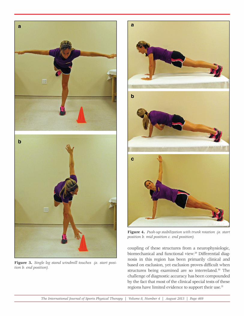
Figure 3. Single leg stand windmill touches (a. start posi-tion b. end position).
Figure 4. Push-up stabilization with trunk rotation (a. start position b. mid position c. end position).
The International Journal of Sports Physical Therapy | Volume 8, Number 4 | August 2013 | Page 470
Loss of neuromuscular stability in any part of the lumbar-pelvis-lower extremity kinetic chain has been proposed as an underlying contributor in hamstring injuries.7 Hungerford et al25 demonstrated delayed activity of the internal oblique, multifidus and gluteus maximus muscles in conjunction with early activity of the biceps femoris in patients with pelvic pain. Thus, any threat to stability of the lower limb or pel-vis could lead to increased neuromuscular demands on the hamstring muscle group and predispose this tissue to injury.7 This patient’s prior history of pelvic pain and osteitis pubis could have resulted in altered neuromuscular firing patterns and pelvic floor hyper-tonicity that contributed to the injury requiring treat-ment. The interrelationship of neuromuscular control between core stabilizer muscles and proximal ham-string muscles could also help explain why patients with hamstring strains decrease their reinjury rates when treatment emphasizes trunk stabilization and agility versus isolated hamstring strengthening.11
The interplay between the hamstring muscles and pel-vic floor pain has not been well documented. Forces generated by the biceps femoris have been shown to increase tension at the sacrotuberous ligament.29
Increased tension on the sacroiliac ligaments could, in theory, increase stress to the pelvic floor muscles, but this cause and effect relationship has not been established. The piriformis muscle has been directly associated with pelvic floor muscle pain due to its proximity to the lateral pelvic wall.30 Any effect of the hamstring muscles on pelvic floor pain would appear to be indirect due to the distance between the areas. Pain syndromes that have been associated with pelvic floor hypertonic disorders include: interstitial cystitis, vulvodynia, colorectal pain disorders, and pelvic pain/myofascial disorders.27 None of these factors appeared to affect this particular patient except for her history of pelvic pain. Although the patient had no complaints of pelvic pain on initial examination, her prior his-tory may have contributed to the subsequent presen-tation of pelvic floor pain. This leads to the question of whether prior instances of pelvic pain, such as this patient’s history of osteitis pubis, predispose the ham-string injury or if the stress on the hamstring muscles with injury created the secondary pelvic floor pain.
This case report serves to highlight the importance of considering how prior medical complaints may affect
current symptoms and the need for continual re-examination and refinement of differential diagnosis as patients respond to interventions. The concept of regional interdependence is highlighted in which pain is somehow related to a more remote dysfunction.15 In this context, osteitis pubis has been shown to be a risk factor for future hamstring injury2 and may play a role in hypertonicity of the pelvic floor muscles due to pelvic pain. Therefore, this case study helps dem-onstrate the possible interrelationship between ham-string, gluteal and pelvic pain within a single subject and highlights the need for diligent reassessment and revision of the treating diagnosis and plan of care if a patient’s complaints of pain change in response to the initial interventions provided.
Future consideration for this and similar patients must include an evaluation of the cause and effect relation-ship of their complaints. Reflecting on this patient over her presentation and course of treatment, the examiner would not have altered the sequencing of differential assessment and treatment. As the original complaints appeared more gluteal and hamstring mediated, this would still be the place to initiate treatment. Given the initial tightness in the hip flexors and quadriceps in conjunction with weakness in the gluteus maximus, another intervention that may have benefitted the patient is mobilization of the hip joint with PA glides followed by hip flexor/quadriceps stretching. The tightness in the anterior hip, whether of capsular or muscular origin, might have inhibited optimal gluteus maximus firing and helped contribute to the patient’s complaints.
CONCLUSION This patient initially presented in an out-patient ortho-pedic clinic with complaints typically assessed and treated by physical therapists who specialize in orthope-dic injuries. However, the focus of her treatment shifted to require internal pelvic assessment and treatment that requires specific clinical training. While few physical therapists may feel confident as primary therapist for both types of specialized care, all therapists who special-ize in orthopedics or women’s health should recognize the possible need for an interdisciplinary approach to patients with pelvic girdle and hamstring pain.
REFERENCES 1. Van Gent RN SD, Van Middelkoop M, Van Os AG,
Bierma-Zeinstra SM, Koes BW. Incidence and

The International Journal of Sports Physical Therapy | Volume 8, Number 4 | August 2013 | Page 471
determinants of lower extremity running injuries in long distance runners: a systematic review. Br J Sports Med. 2007;41:469-480.
2. Verrall GM SJ, Barnes PG, Fon GT, Spriggins AJ. Clinical risk factors for hamstring muscle strain injury: a prospective study with the correlation of injury by magnetic resonance imaging. Br J Sports Med. 2001;35:435-440.
3. Woods C HR, Maltby S, Hulse M, Thomas A, Hodson A. The football association medical research programme: an audit of injuries in professional football - analysis of hamstring injuries. Br J Sports Med. 2004;38:36-41.
4. Bruckner P KK. Clinical sports medicine. London: McGraw-Hill; 1993.
5. Orchard. Intrinsic and extrinsic risk factors for muscle strains in Australian football. Am J Sports Med. 2001;29(3):300-303.
6. Vogt L PK, Banzer W. Neuromuscular control of walking with chronic low-back pain. Man Ther. 2003;8:21-28.
7. Sole G MS, Sullivan SJ, Nicholson H. Running-related hamstring injuries: a neuromuscular approach. Phys Ther Reviews. 2008;13(2):102-110.
8. McCrory P BS. Nerve entrapment syndromes as a cause of pain in the hip, groin and buttock. Sports Med. 1999;27(4):261-274.
9. Malliaropoulos N PS, Papalada A, Papacostas E. The role of stretching in rehabilitation of hamstring injuries: 80 athletes follow-up. Med Sci Sport Exercise. 2004;36(5):756-759.
10. Croisier. Factors associated with recurrent hamstring injuries. Sports Med. 2004;34(10):681-695.
11. Sherry MA BT. A comparison of 2 rehabilitation programs in the treatment of acute hamstring strains. J Orthop Sports Phys Ther. 2004;34:116-125.
12. Kornberg C LP. The effect of stretching neural structures on grade one hamstring injuries. J Orthop Sports Phys Ther. 1989(June):481-487.
13. Cibulka MT RS, Delitto A, Sinacore DR. Hamstring muscle strain treated by mobilizing the sacroiliac joint. Phys Ther. 1986;66(8):1220-1223.
14. Mason DL DV, Vail A. Rehabilitation for hamstring injuries. Cochrane Database of Systematic Reviews. 2007(1).
15. Wainner RS WJ, Cleland JA, Flyn TW. Regional interdependence: a musculoskeletal examination model whose time has come. J Orthop Sports Phys Ther. 2007;37:658-660.
16. Orava. Hamstring syndrome. Op Tech Sports Med. 1997;5:143-149.
17. Vroomen PC dKM, wilmink JT, Kester AD, Knottnerus JA. Diagnostic value of history and physical examination in patients suspected of lumbosacral nerve root compression. J Neurol Neurosurg Psychiatry. 2002;72(5):630-634.
18. Stankovic R JO, Maly P, Willner S. Use of lumbar extension, slump test, physical and neurological examination in the evaluation of patients with suspected herniated nucleus pulposus: a prospective clinical study. Man Ther. 1999;4(1):25-32.
19. Puranen J OS. The hamstring syndrome: a new diagnosis of gluteal sciatic pain. Am J Sports Med. 1988;16(5):517-521.
20. Laslett M YS, Aprill CN, McDonald B. Diagnosing painful sacroiliac joints: a validity study of a McKensie evaluation and sacroiliac provocation tests. Aust J Physiotherapy. 2003;49:89-97.
21. Narvani A TE, Kendall S, Chaudhuri R, Thomas P. A preliminary report on prevalence of acetabular labrum tears in sports patients with groin pain. Knee Surg Traumatol Arthrosc. 2003;11:4003-4408.
22 Kirschner JS FP, Cole JL. Piriformis syndrome, diagnosis and treatment. Muscle Nerve. 2009;40:10-18.
23. Fishman L ZP. Electrophysiologic evidence of piriformis syndrome. Arch Phys Med Rehabil. 1992;73:359-364.
24. Sizer P. The hip: physical therapy patient management utilizing current evidence. In: M W, ed. Current concepts of orthopedic physical therapy. La Crosse: Orthopedic Section, APTA; 2006.
25. Hungerford B GW, Hodges P. Evidence of altered lumbopelvic muscle recruitment in the presence of sacroiliac joint pain. Spine. 2003;28(14):1593-1600.
26. Laycock J, Jerwood D. Pelvic fl oor muscle assessment: the PERFECT scheme. Physiotherapy. 2001;87(12):631-642.
27. Butrick CW. Pelvic fl oor hypertonic disorders: identifi cation and management. Obstet Gynecol Clin N AM. 2009;36:707-722.
28. Weiss JM. Pelvic fl oor myofascial trigger points: manual therapy for interstitial cystitis and the urgency-frequency syndrome. J Urol. 2001;166:2226-2231.
29. Van Wingerden JP VA, Snijders CJ, Stoeckart R. A functional-anatomical approach to the spine-pelvis mechanism: interaction between the biceps femoris muscle and the sacrotuberous ligament. Eur Spine J. 1993;2:140-144.
30. Papadopoulos EC KS. Piriformis syndrome and low back pain: a new classifi cation and review of the literature. Orthop Clin N Am. 2004;35:65-71.