Improving diagnostic quality – advanced analysis tools in ... › wp-content › ... · Improving...
4
Improving diagnosc quality with Spacelabs Healthcare CardioExpress SL18A 030-2501-00 Rev A An OSI SyStemS COmpAny Improving diagnosc quality – advanced analysis tools in a standard ECG cart Spacelabs Healthcare Clinical Support Team • A new standard ECG cart can offer 15- and 18- lead recording and analysis tools which lead to improved diagnosis for certain condions. • Roune 12-lead ECGs can miss significant diagnosc informaon. • Availability and ease of use are the keys to efficient workflows and effecve tests. 18-Lead ECG and vectorcardiogram (VCG) can help you to determine the origins of an arrhythmia or localise areas of myocardial damage with increased accuracy* 1,2,3,4,5 . The procedure is non-invasive and easily performed. Instant accessibility and ease of use is the key to fast, accurate results. Spacelabs Healthcare now provides advanced funcons and mul-lead capability in the standard SL18A ECG cart. This supports the clinician with valuable addional informaon and leads to opmal diagnosis and treatment for the paent. 15- and 18-lead ECG and advanced analysis – clinical diagnosc benefits • A standard 12-lead ECG can miss up to 31% of infarcons* 1 . • In pediatrics, changes in the axis during development can lead to abnormalies being missed. • VCG is superior to ECG in specific aspects of condions including Wolff-Parkinson-White, pre-excitaon, axis deviaon, Brugada syndrome, enlargement, localised electrical inacvity and associated conducon disorders. • Addional features provide benefits, but will not be used effecvely unless they are automac, quick, and simple to read. • It can be difficult to ensure that a cart with this level of capability is ready to hand when it may be needed. • 15-lead ECG can localise to RV or posterior LV • 18-lead ECG allows both RV and posterior LV to be analysed instantaneously • 18-lead ECG provides increased accuracy and ability to observe abnormalies • Vectorcardiogram is provided automacally through a simple user-interface and report format • SL18A’s automac analysis presents the informaon so as to help clinicians quickly idenfy whether the result is normal or needs further aenon • Fully featured carts can be provided within a commonly acceptable ‘standard’ cost envelope
Transcript of Improving diagnostic quality – advanced analysis tools in ... › wp-content › ... · Improving...
Improving diagnostic quality with Spacelabs Healthcare CardioExpress SL18A
030-2501-00 Rev A
An OSI SyStemS COmpAny
Improving diagnostic quality – advanced analysis tools in a standard ECG cart
Spacelabs Healthcare Clinical Support Team
• A new standard ECG cart can offer 15- and 18-lead recording and analysis tools which lead toimproved diagnosis for certain conditions.
• Routine 12-lead ECGs can miss significantdiagnostic information.
• Availability and ease of use are the keys toefficient workflows and effective tests.
18-Lead ECG and vectorcardiogram (VCG) can help you to determine the origins of an arrhythmia or localise areas ofmyocardial damage with increased accuracy*1,2,3,4,5. The procedure is non-invasive and easily performed.
Instant accessibility and ease of use is the key to fast, accurate results. Spacelabs Healthcare now provides advanced functions and multi-lead capability in the standard SL18A ECG cart. This supports the clinician with valuable additional information and leads to optimal diagnosis and treatment for the patient.
15- and 18-lead ECG and advanced analysis – clinical diagnostic benefits
• A standard 12-lead ECG can miss up to 31% ofinfarctions*1.
• In pediatrics, changes in the axis duringdevelopment can lead to abnormalities beingmissed.
• VCG is superior to ECG in specific aspects ofconditions including Wolff-Parkinson-White,pre-excitation, axis deviation, Brugada syndrome,enlargement, localised electrical inactivity andassociated conduction disorders.
• Additional features provide benefits, but will notbe used effectively unless they are automatic,quick, and simple to read.
• It can be difficult to ensure that a cart with thislevel of capability is ready to hand when it may beneeded.
• 15-lead ECG can localise to RV or posterior LV
• 18-lead ECG allows both RV and posterior LV tobe analysed instantaneously
• 18-lead ECG provides increased accuracy andability to observe abnormalities
• Vectorcardiogram is provided automaticallythrough a simple user-interface and report format
• SL18A’s automatic analysis presents theinformation so as to help clinicians quicklyidentify whether the result is normal or needsfurther attention
• Fully featured carts can be provided within acommonly acceptable ‘standard’ cost envelope
030-2501-00 SL18A White Paper (ENG) RevA 0.4.indd 1 08/03/2018 14:08:58
Improving diagnostic quality with Spacelabs Healthcare CardioExpress SL18A
030-2501-00 Rev A
Coronary Artery Disease – up to 31% of infarcts may be missed by 12-lead ECG*1,2,3,4
“Coronary artery disease is still one of the largest health threats today. The rapid and early identification of a patient with acute coronary syndrome is paramount in today’s medical world. However, a standard 12-lead ECG is not enough for comprehensive MI diagnosis.” Brian J O’Neill, 20101,2.
History – 12-lead, STEMIs and the significance of the problem• The most common ECG exam is the standard 12-lead ECG. It is simple to measure, and observing the heart from
these 12 directions provides key information for a wide range of clinical applications.• However, some areas, especially pathological change in the right ventricle and the posterior wall, cannot be
observed from the 12-lead ECG. In order to actually measure the right chest (V3R, V4R, V5R) and back (V7, V8, V9) areas, it is necessary to use additional electrode positions compared to the standard 12-lead ECG.
• *2,3,4 Posterior infarction accompanies 15-20% of STEMIs, usually occurring in the context of an inferior or lateral infarction.
• *3,4 Isolated posterior MI is less common (3-11% of infarcts). So up to 31% of infarcts could be missed on a 12-lead ECG.
Why this happens • Posterior extension of an inferior or lateral infarct implies a much larger area of myocardial damage, with an
increased risk of left ventricular dysfunction and death.• Isolated posterior infarction is an indication for emergent coronary reperfusion. However, the lack of obvious ST
elevation in this condition means that the diagnosis is often missed.• Right ventricular (RV) myocardial infarction most often occurs in the setting of inferior wall myocardial infarction.
Right ventricular infarction complicates approximately 25% (range 20% - 60%) of inferior acute myocardial infarction.
• ST-segment elevation of greatest magnitude in lead III, ST-segment elevation in lead V1, and/or ST-segment elevation in right chest leads (RV1 through RV6).
Pediatrics • Pediatric ECGs are regularly performed for a variety of conditions, including chest pain, syncope and suspected
arrhythmia. Correct interpretation can be challenging. Pediatric ECGs have to be interpreted — especially during the first year of life with due regard to the age-specific changes associated with the anatomic/physiologic development of the heart.
• 15/18-lead ECGs provide increased accuracy to help ensure that no electrical abnormalities will be missed in the test.
15-lead is better than 12-lead, but 18-lead is even better. • 15-lead isolated to localisation of either RV or posterior LV electrical mapping. • 18-lead allows both RV and posterior LV electrical signal to be analysed instantaneously. • Real-time capture guarantees accuracy — no “derived” channels. A 360-degree electrical map of the heart!• Leads are fitted in one ECG recording, so a single 10s recording captures all channels.
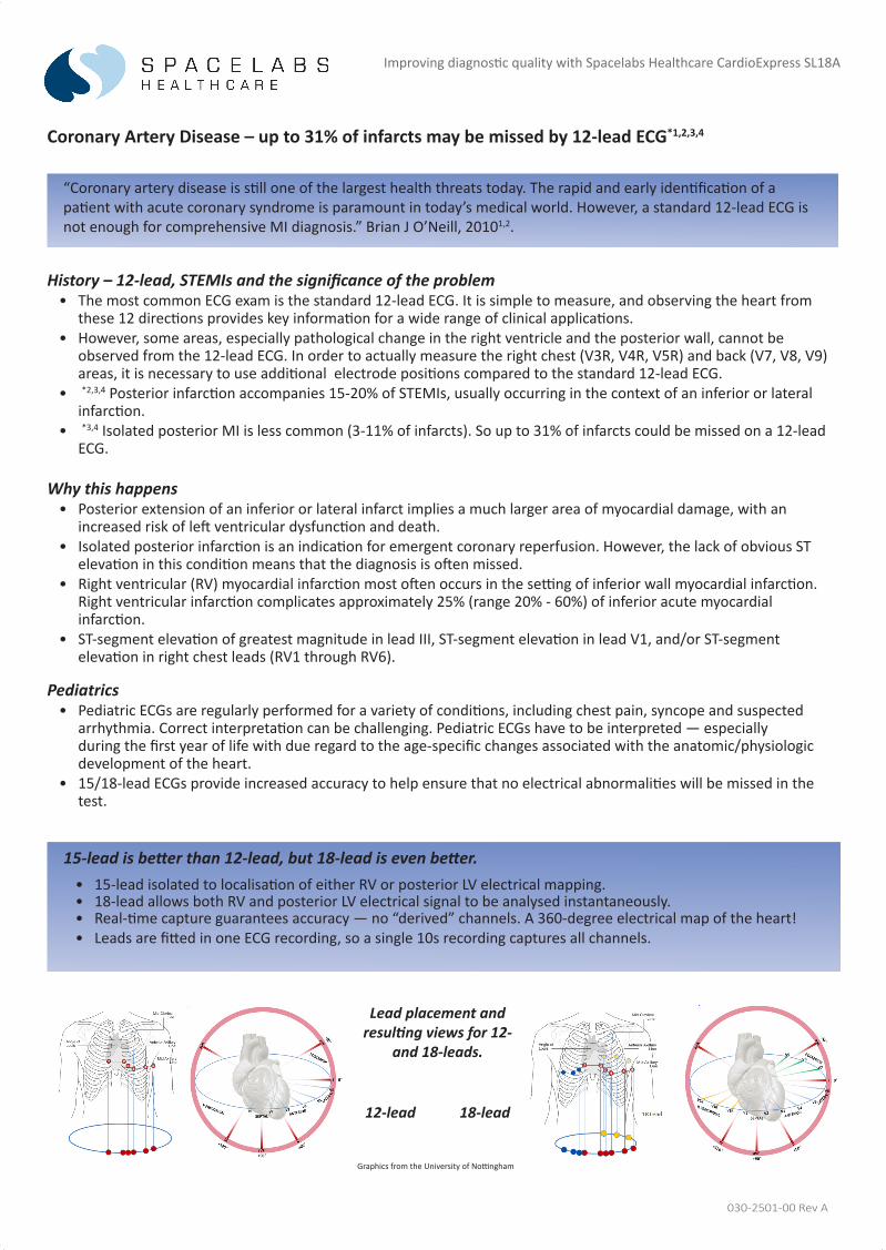
Lead placement and resulting views for 12-
and 18-leads.
12-lead 18-lead
Graphics from the University of Nottingham
030-2501-00 SL18A White Paper (ENG) RevA 0.4.indd 2 08/03/2018 14:08:58

Improving diagnostic quality with Spacelabs Healthcare CardioExpress SL18A
030-2501-00 Rev A
SEMIP Algorithm and presentation – automated and quick to review
• SEMIP is designed to assist the physician in reading and evaluatingan ECG printout with up to 18-leads. This method of measurementand interpretation was developed in cooperation with leadingcardiologists.
• Interpretation of ECGs uses Minnesota Code Classification andDiagnosis Information for Electrocardiology.
• The interpretation and graphical presentation support rapidrecognition of normal and abnormal recordings for further review.
Vectorcardiogram (VCG) is superior to ECG in specific aspects of conditions including –
• Wolff-Parkinson-White, pre-excitation, axis deviation, Brugada syndrome, enlargement, localisedelectrical inactivity and associated conduction disorders.
• Vectorcardiography (VCG) is a method ofrecording the magnitude and direction of theelectrical impulses generated by the heart. Itworks by means of a continuous series of vectorsthat form curving lines around a central pointusing the Frank Lead system to create VCG loops.
• Each cell in the heart can be represented as adipole with differing direction throughout the heartbeat. A collectionof all these small dipoles close to each other can be represented as asingle dipole. The electrical field of the heart can then be studied as afield of a single dipole — the cardiac vector. The line drawn by the tipof the cardiac vector is the VCG.
Graphic from Clinical Electrocardiographic informatics, KD Zhang
• Signal-averaged electrocardiography (SAECG) is a special electrocardiographictechnique in which multiple ECG complexes are averaged to removeinterference and reveal small variations in the QRS complex.
• These are the so-called "late potentials.” They may represent a predispositiontowards potentially dangerous ventricular tachyarrhythmia.
Signal-averaged ECG – may represent a predisposition to VT
Graphic from http://www.monte.amu.edu.pl
030-2501-00 SL18A White Paper (ENG) RevA 0.4.indd 3 08/03/2018 14:09:01

Improving diagnostic quality with Spacelabs Healthcare CardioExpress SL18A
030-2501-00 Rev A35301 Center St.,Snoqualmie, WA 98065 T+1 (425) 396 3300 ● Unit B Foxholes Centre, John Tate Road, Hertford, SG13 7DT. UK T+44 (0)1992 507700
www. Spacelabshealthcare.com ©2018 Spacelabs Healthcare Specifications subject to change without notice.
Interpretation • SEMIP automatic measurement and interpretation tested with
authoritative CSE database• Vectorcardiography, Signal-averaged ECG, HRV
15” color touchscreen and light sensor• High-resolution, clear view of the ECG traces• Automatically adjusts the brightness according to the working
environment
Silicone touchscreen keyboard • Waterproof alphanumeric keyboard to minimize the likelihood of cross-
infection
Function icons• Frequently used functions at the touch of a button
Signal quality indicator • Enables the user to check signal quality at a glance
Battery backup• For prolonged mobile use
Signal acquisition• Sample rate 16,000Hz• Digital filters for baseline drift, AC, EMG• Pacemaker detection ANSI/AAMI/ECB
Printing • Flexible formats for 9,12,15- and 18-lead reports• Internal high-resolution thermal or external USB printer• Speeds 5/6.25/10/12.5/25/50 mm/s, user selectable
Memory, storage and connectivity• Internal storage of up to 1000 10-second resting ECGs• External archiving to Spacelabs Sentinel • Export to network or USB: PDF,SCP,DICOM,FDA-XML
Networking• Port or built-in Wi-Fi for 802.11 b/g/e/l single-stream /n• 2.4GHz, 802.11n RF transceiver, high performance amplifier• WPA and WPA2 supported but not Vendor EAP Type(s)
CardioExpress SL18A – key features
12/15/18-lead Electrocardiogram with automatic and manual Interpretation, HRV, VCG and SAECG
Mattu,A., Tabas,J.A, & Barish R.A (2007) Electrocardiography in Emergency Medicine. Dallas, Tx; American College of Emergency PhysiciansSomers, M.P.,Brady,W.J.,Bateman,D.C., Mattu, A., & Perron, A.D (2003) Additional electrocardiographic leads in the ED chest patient ; Right ventricular and posterior leads. American Journal of Emergency Medicine, 21 563-567Aqel,R.A. Hage,F.G., Ellipeddi,P.,Blackmon,L.,McElderry,H.T.,Kay,G.N.,Plumb,V. & Iskandrian,A.,E (2009). Usefulness of three posterior chest leads for the detection of posterior wall acute myocardial infarction. American journal of Cardiology, 103, 159-164.Pradhan et Al Clinical significance of ST-segment elevation in posterior leads v7-v8, v9 in patients with acute inferior wall myocardial infarction 2329-9517.10001, Journal of Cardiovascular Diseases and Diagnosis, 2013 1-2, 2329-9517.10001
*1 Emergency Nurses Association, Translation into Practice - Right-sided and Posterior Electrocardiograms
*2 Brady WJ, Morris F. ABC of clinical electrocardiography: Acute myocardial infarction-Part II. BMJ. 2002; 324: 963-6.
*3 Morris F, Brady WJ. ABC of clinical electrocardiography: Acute myocardial infarction-Part I. BMJ. 2002; 324: 831-4. *4 Van Gorselen EO, Verheugt FW, Meursing BT, Oude Ophuis AJ. Posterior myocardial infarction: the dark side of the moon. Neth Heart J. 2007; 15: 16-21.[PMC PMC1847720] *5 Andres Ricardo Perez Riera MD et al, “Significance of VCG in Cardiological Diagnosis of the 21st Century” , Clin.Cardiology 30, 218-323 (2007)
References – Additional Reading –
030-2501-00 SL18A White Paper (ENG) RevA 0.4.indd 4 08/03/2018 14:09:03