Download Full Text (Final Version , 280kb) - RePub - Erasmus

Improvement of therapy for amblyopia
Sjoukje Elizabeth Loudon

The research project was initiated by the Department of Ophthalmology, Erasmus MC Uni
versity Medical Center Rotterdam, the Netherlands. The work described in this thesis was fi
nancially supported by the Health Research and Development Council of the Netherlands
(project number 2300.0020). Financial support for the printing of this thesis was received from
the Prof.dr. Henkes Stichting, Orthopad, 3M Opticlude, the Rotterdamse Vereniging Blinden
belangen and the Erasmus Universiteit Rotterdam.
ISBN 9789085592682
Copyright © 2007 S.E. Loudon, Rotterdam, the Netherlands
All rights reserved. No part of this thesis may be reproduced, stored in a retrieval system or
transmitted in any form or by any means, without the permission of the author, or when ap
propriate, of the publishers of the publications.
Layout and printing: Optima Grafische Communicatie, Rotterdam, The Netherlands
Cover design: VOF Vingerling & De Bruyne

Improvement of Therapy for Amblyopia
Verbeteren van de Behandeling voor Amblyopie
Proefschrift
ter verkrijging van de graad van doctor aan de
Erasmus Universiteit Rotterdam
op gezag van de rector magnificus
Prof.dr. S.W.J. Lamberts
en volgens besluit van het College voor Promoties.
De openbare verdediging zal plaatsvinden op
woensdag 21 februari 2007 om 15:45 uur
door
Sjoukje Elizabeth Loudon
geboren te Huddersfield, GrootBrittannië

PromoTIEcommISSIE
Promotoren Prof.dr. G. van Rij
Prof.dr. H.J. Simonsz
overige leden Prof.dr. P.J. van der Maas
Prof.dr. J. Passchier
Dr. M. Fronius
Dr. M.J. Moseley

To my parents


7
conTEnTS
Publications and manuscripts on which this thesis is based 9
chapter 1 General introduction 11
chapter 2 The history of the treatment of amblyopia 19
chapter 3 Objective survey of the prescription of occlusion therapy for
amblyopia
39
chapter 4 Visusevaluierung in einer historischen Kohorte von 137 okkludierten
Patienten, 3035 Jahre nach Ende der Okklusionsbehandlung /
Evaluation of visual acuity in a historic cohort of 137 patients treated
for amblyopia by occlusion 3035 years ago
49
chapter 5 Electronic recording of occlusion treatment for amblyopia: potential
of the new technology
65
chapter 6 Electronically measured compliance with occlusion therapy for
amblyopia is related to visual acuity increase
77
chapter 7 Predictors and a remedy for noncompliance with amblyopia
therapy in children measured with the Occlusion Dose Monitor
87
chapter 8 The influence of parental attitudes and behaviour on compliance
with amblyopia therapy and the effect of an educational programme
105
chapter 9 Physiological and mechanical properties of the eye patch: influence
on compliance and parental satisfaction
119
chapter 10 Account of the study population in The Hague: prevalence of
occluded children, causes of amblyopia and final visual acuity
131
chapter 11 General discussion and future prospects 139

8
References 147
Summary / Samenvatting 161
Dankwoord 171
About the author 175
List of Publications 177
Appendix / Colour figures 179
9
Publications and manuscripts on which this thesis is based
Chapter 2 S.E. Loudon, H.J. Simonsz The history of the treatment of amblyopia. Strabismus
2005 Jun;13(2):93106
Chapter 3 S.E. Loudon, J.R. Polling, B. Simonsz, H.J. Simonsz Objective survey of the prescrip-
tion of occlusion therapy for amblyopia. Graefes Arch Clin Exp Ophthalmol 2004
Sep;242(9):73640
Chapter 4 B. SimonszTóth, S.E. Loudon, H. van Kempendu Saar, E.S. van de Graaf, J.H. Groe
newoud, H.J. Simonsz Visusevaluierung in einer historischen Kohorte von 137 okklu-
dierten Patienten, 30-35 Jahre nach Ende der Okklusionsbehandlung / Evaluation of
visual acuity in a historic cohort of 137 patients treated for amblyopia by occlusion
30-35 years ago. Accepted for publication Klin Monatsbl Augenheilkd 2006
Chapter 5 Y. Chopovska, S.E. Loudon, L. Cirina, A. Zubcov, H.J. Simonsz, M. Luchtenberg, M.
Fronius Electronic recording of occlusion treatment for amblyopia: potential of the
new technology. Graefes Arch Clin Exp Ophthalmol 2005 Jun;243(6):53944
Chapter 6 S.E. Loudon, J.R. Polling, H.J. Simonsz Electronically measured compliance with oc-
clusion therapy for amblyopia is related to visual acuity increase. Graefes Arch Clin
Exp Ophthalmol 2003 Mar;241(3):17680
Chapter 7 S.E. Loudon, M. Fronius, C.W.N. Looman, M. Awan, B. Simonsz, P.J. van der Maas,
H.J. Simonsz Predictors and a remedy for non-compliance with amblyopia therapy in
children measured with the Occlusion Dose Monitor. Invest Ophthalmol Vis Science
2006 Oct;47(10):4393400
Chapter 8 S.E. Loudon, L. Chaker, S. de Vos, M. Fronius, J. Passchier, R.A. Harrad, C.W.N. Looman,
B. Simonsz, H.J. Simonsz Effect of an educational programme on attitudes and behav-
iour with occlusion therapy and reasons for total non-compliance. (submitted)
Chapter 9 S.E. Loudon, A.W. Wypekma, C.W.N. Looman, M. Fronius, B. Simonsz, H.J. Simonsz
Physiological properties of the eye patch and influence on compliance with occlusion
therapy. (submitted)


chap
ter 1
chap
ter 2
chap
ter 3
chap
ter 4
chap
ter 5
chap
ter 6
chap
ter 7
chap
ter 8
chap
ter 9
chap
ter 1
0ch
apte
r 11
Add
enda
Chapter 1
General introduction


13
chap
ter 1
General introduction
InTroducTIon
definition
The term ‘amblyopia’ originates from the Greek language and literally means dimness or dull
ness of vision. In time, the condition has been defined in a variety of ways, very much depend
ing on the prevailing pathophysiological concept about its etiology. In general, amblyopia
can be defined as a unilateral or bilateral decrease in visual acuity for which no organic cause
can be found on physical examination of the eye. It is caused by a refractive error (one foveal
image is more blurred than the other); strabismus (ocular misalignment causing each eye
to have a different image on the fovea) or, more rarely, deprivation of a clear retinal image
(physical obstruction, e.g. infantile cataract, ptosis) (von Noorden 1967; 1985; von Noorden
and Campos 2002). Amblyopia usually presents itself during the ophthalmological examina
tion by the ophthalmologist or the orthoptist as a reduced visual acuity in one or both eyes,
in the presence of a refractive error and/or strabismus or a deprivation. This reduced visual
acuity persists after optimum correction of any refractive error (i.e. a pair of spectacles) and it
cannot be explained by another ocular abnormality (e.g. retinopathy).
Epidemiology & screening
Amblyopia is the most common cause of monocular vision loss in children, accounting for over
90% of the visits of children to ophthalmologists and orthoptists (Attebo, et al. 1998; Moseley, et al.
1997; Sjöstrand and Abrahamsson 1997). The general estimate of the prevalence of amblyopia is
approximately 3.5%, but varies considerably in the literature (0.55.3%) due to differences in study
design, population and the examination methods used (Attebo, et al. 1998; Cole 1959; Helveston
1965; von Noorden and Campos 2002; Simons 1996; Theodore, et al. 1946; Vinding, et al. 1991). In
The Netherlands, the incidence is approximately 6500 amblyopic children each year. The national
screening programme checks for the presence of strabismus after birth, and, periodically examines
visual acuity from the age of three. The referral procedures in the Netherlands are currently studied
by the Rotterdam AMblyopia Screening Effectiveness Study (RAMSES) (Juttmann, et al. 2001). This
is a 7 year followup study, which evaluates the effectiveness and the efficiency of screening. Ap
parently, one third of all children with a positive screening test result (i.e. reduced visual acuity) are
not conclusively evaluated at an ophthalmological centre and consequently fail to profit from an
early detection and treatment. Whether this could be attributed to the many intermediate steps
between the referral and the orthoptist (parents have to make an appointment with their general
practitioner, are then referred to the ophthalmologist and finally to the orthoptist), or the lack of
understanding of the necessity of the referral by the parents is still unclear. When a positive referral
leads to a visit to the orthoptist and the reduced visual acuity is confirmed they will be prescribed
treatment, which may continue for several months up to several years.

14
Treatment
Treatment of amblyopia involves complete or incomplete exclusion of the better eye from
visual activity; hence, the use of the amblyopic eye is stimulated. The purpose of amblyopia
treatment is equal acuity in both eyes and, consequently, preventing any future disability (e.g.
choice of profession, quality of life). Early treatment, i.e. during the sensitive period of visual
development lasting up to the age of 7 years, can reduce or completely reverse the effects
of abnormal visual experiences, whereas treatment later in the critical period becomes less
effective (Birch, et al. 1990; Crawford, et al. 1983; Epelbaum, et al. 1993; MintzHittner, et al.
2000; Mitchell 1991).
The mainstay treatment has been occlusion of the better eye using an opaque patch. Howev
er, as there is little consensus amongst orthoptists concerning the necessary number of occlu
sion hours, occlusion regimens may vary from occluding the better eye a few minutes per day
to all waking hours (Tan, et al. 2003). More recently optical penalisation (selectively fogging
the image of the nonamblyopic eye by glasses) or pharmaceutical penalisation (cycloplegia
by the daily instillation of drops into the fornix of the nonamblyopic eye) was described.
Several studies have demonstrated that atropine was as effective as occlusion therapy, but
occlusion therapy caused a more rapid response, while atropine had a somewhat higher ac
ceptability by the families (Cole 2001; PEDIG 2002; 2003; 2004; 2005).
Despite screening and treatment, approximately a third of the affected children who have
been prescribed occlusion therapy do not reach visual acuity of 6/12 in the amblyopic eye and
are unable to read with the amblyopic eye. This excludes them from any future tasks that re
quire equal good vision (Jensen and Goldschmidt 1986; Vinding, et al. 1991). Matters worsen
when an amblyopic child in one study the proportion was estimated at 0.175% (Tommila
and Tarkkanen 1981) will loose the function of the better eye later in life, because of trauma
or, for example, macular degeneration. This will result in bilateral visual impairment causing
job losses, an increased morbidity and social isolation (Chua and Mitchell 2004; Fronius, et al.
2005; Rahi, et al. 2002). A decrease in quality of life in adulthood has also been described (van
de Graaf, et al. 2004). In people aged 2070, amblyopia is the most common cause of monocu
lar loss of vision (Buch, et al. 2001).
The effectiveness of occlusion therapy was questioned in a report published by Snowdon &
StewartBrown in 1997, who conducted a systematic review of the literature (Snowdon and
StewartBrown 1997). They concluded that occlusion therapy has not yet been subjected to
formal controlled trials and that much of the improvement in visual acuity could be sponta
neous and unrelated to the therapy. However, they may have overlooked the possibility that
the lack of evidence for the efficacy of occlusion could be due to low compliance rather than
to ineffectiveness of the treatment. Their report contributed to the setup of five randomised

15
chap
ter 1
General introduction
controlled trials (RCTs) that produced evidence for the effectiveness of occlusion therapy
(Awan, et al. 2005; Clarke, et al. 2003; PEDIG 2002; 2004; 2005; Stewart, et al. 2004; 2005). The
percentage of successfully treated amblyopes spans a broad range: 1993% success rates. Fac
tors that influence the outcome of treatment include age (Massie 1965; Stewart, et al. 2004;
2005), visual acuity at start of treatment (Cobb, et al. 2002; Hiscox, et al. 1992; Lithander and
Sjöstrand 1991; Smith, et al. 1995, Stewart, et al. 2004; 2005) and type of amblyopia (Cobb, et
al. 2002). The factor most frequently quoted, however, was the degree of compliance: the bet
ter eye is not patched according to the orthoptists’ prescription (Awan, et al. 2005; Dorey, et al.
2001; Lithander and Sjöstrand 1991; Simmers, et al. 1999; Simons and Preslan 1999; Smith, et
al. 1995; Stewart, et al. 2004; 2005).
compliance
In recent years, ‘compliance studies’ are receiving increased attention now that it can be mea
sured electronically (Kass, et al. 1986a; 1986b; 1987; Urquhart, 1992; 1999), with the first con
ference addressing the issue of patient compliance organised in 1974. Compliance is referred
to as the degree of correspondence between the recommendations from the health care pro
vider and the patients’ actual dosage. The first devices that measured compliance electroni
cally were developed to monitor the administration of pilocarpine eye drops in the treatment
of glaucoma (Kass, et al. 1986a; 1986b; Norell, et al. 1980). The results showed that only 76% of
the pilocarpine drops were taken as prescribed, while 6% took less than one quarter and 15%
took less than half of the prescribed dosage. However, the patients’ diaries reported to have
taken 97% of their medication. Poor compliance decreases the effectiveness of treatment and
increases costs to the health care system (Cleemput and Kesteloot 2002). In children, the issue
of noncompliance is especially challenging as the relationship is compound: the orthoptist
deals with noncompliant parents, the parents deal with a noncompliant child. In addition,
noncompliance in a parent is regarded as a more serious fault by society than noncompli
ance in an adult patient and may therefore cause feelings of insufficiency and shame. Com
pliance with any treatment for children is largely dependent on the ability of the parents or
guardian to understand and follow through with recommended treatment. Only few groups
have studied compliance electronically in children. Milgrom, et al. (1996) found 58% use of
prescribed inhaled corticosteroids in asthma in children electronically, whereas the diaries
kept by the patients or their parents reported 95% use. More than 90% of the patients exag
gerated their use of inhaled steroids, and even the least compliant reported high levels of
adherence to prescribed therapy. The authors concluded that most of the hospital admissions
for asthma were caused by noncompliance.
Since the development of the Occlusion Dose Monitor (ODM) by Fielder and Moseley (Fielder,
et al. 1994) compliance with occlusion therapy for amblyopia can be measured electronically
and therefore objectively. They developed an ODM that measured skin conductance at the

16
border of the patch. In 1997, the department of Medical Technical Development at the Aca
demical Medical Center, Amsterdam modified the FielderODM design and made it smaller.
It now measures 24x12x3.6 mm and weighs 1.8 g. It is taped to the outside of a standard eye
patch and measures the temperature difference between the front and the back of the ODM
every 2 minutes, instead of skin conductance (Figure 1). In previous pilot studies with the
ODM in patients from the Sophia Children’s Hospital Rotterdam, it was found that compliance
with occlusion therapy was low and the patterns of noncompliance remained the same for
one child and were apparently case specific (Simonsz and Polling 2001). In a second pilot
study the ODM was distributed by the orthoptist in the clinic to patients whose compliance
was thought to be low. Parents, however, interpreted the ODM as a ‘liedetector’, resulting in a
breakdown in the relationship between them and the orthoptist (Simonsz, et al. 1999).
To date, several studies have demonstrated by means of the ODM that, generally, compliance
with amblyopia treatment is low and treatment success is related to the level of compliance
(Awan, et al. 2005; Simonsz, et al. 1999; Stewart, et al. 2004; 2005).
Figure 1. a the first model of the ODM developed by Prof. Alistair Fielder and Dr. Merrick Moseley in London, United Kingdom b: in 1997 the department of Medical Technical Development at the Academic Medical Center Amsterdam modified the design and made it smaller: it now measures temperature difference between the front and the back c & d the ODM as used in the study in The Hague: it weighs 1.8 g and measures 24 x 12 x 3.6 mm.
Figuren bij Chapter 5
1a 1b 1c 1d
Figure 1. The development of the Occlusion Dose Monitor. a Year 1991: first model developed by
Fielder and Moseley in Birmingham, UK (with permission of M. Moseley). The ODM was a miniature
datalogger, which logs the patch-skin contact resistance every 64 s. b Year 1997: The Academic
Medical Center Amsterdam modified the design into an ODM containing two thermistors connected to
either the front or the back of the ODM (35x23x4 mm, 6 g). c The 2001 type used in the Netherlands
(24x12x3.6 mm, 1.8 g). d The 2002 type used in Germany (31x15x3.5 mm, 2.3 g)
2a
2b
Figure 2. Examples of recordings with the ODM, with measured temperature difference on the
ordinates. a A week’s recording. Abscissa: date of the recording. The temperature difference was about
"0" (0; 0.0625) while the child was not patched (baseline value) and higher during the periods of
1a 1b 1c 1d

17
chap
ter 1
General introduction
oBjEcTIVE And ouTLInE oF ThIS ThESIS
The main objective of the research presented in this thesis is, on the one side, to determine
whether compliance with occlusion therapy can be improved with an educational programme
explaining, without text and no animal figures, to a 4yearold child the reasons why the bet
ter eye must be patched, together with a calendar, reward stickers and a sheet containing
general information about amblyopia and its treatment; and, on the other side, to identify cer
tain predictors leading to noncompliance. Studied were the clinical parameters of the child,
the socioeconomic and ethnic parameters and the psychometric parameters. Compliance was
measured electronically by means of the Occlusion Dose Monitor.
Following this first chapter, the history of the treatment for amblyopia is given in Chapter 2.
Chapter 3 presents an inventory made in order to try and identify the variation in prescrip
tions of occlusion hours amongst orthoptists in the Netherlands and in Germany, their con
sistency in prescriptions and the main determinants when prescribing a certain number of
occlusion hours. In Chapter 4, the current visual acuity of 137 amblyopic patients treated for
amblyopia 30 years ago, is evaluated and factors associated with a poor outcome are deter
mined. Chapter 5 investigates whether the Occlusion Dose Monitor (ODM) that is used to
objectively measure compliance, is able to differentiate between measurements on the eye
and on other parts of the body. Chapter 6 presents a pilot study in which the ODM was distrib
uted via home visits by the researcher and that determined whether children whose acuity
had not improved sufficiently after six months of patching were indeed the children with low
compliance. The following three chapters (number 7, 8 and 9) present studies that are carried
out using data from the prospective randomised clinical trial in The Hague, the Netherlands.
For 30 months all newly diagnosed amblyopic children were recruited from the four clinics
in The Hague. Chapter 7 illustrates the effect of the educational cartoon story on compliance
and determines the influence of clinical and socioeconomic factors on compliance. Chapter 8
presents the effect of the educational programme on attitudes and behaviour factors and rea
sons for total noncompliance with occlusion therapy for amblyopia. Chapter 9 investigates
the physiological properties of the eye patch and its influence on compliance. Chapter 10
gives an account of the study population, causes of amblyopia and the final visual acuity.
The clinical relevance of the findings and future prospects are discussed in the eleventh and
final chapter.


chap
ter 1
chap
ter 2
chap
ter 3
chap
ter 4
chap
ter 5
chap
ter 6
chap
ter 7
chap
ter 8
chap
ter 9
chap
ter 1
0ch
apte
r 11
Add
enda
Chapter 2
The history of the treatment of amblyopia


21
chap
ter 2
The history of the treatment of amblyopia
InTroducTIon
The Greek word amblyopia means dimness or dullness of vision (ambly αμβλυσ = dull and
ops ωψ = vision) and the condition has been defined in a variety of ways in the literature.
Amblyopia is a decrease in visual acuity, usually in one eye. It persists after the correction of
the refractive error (i.e., acuity is not improved by glasses) or removal of any pathological ob
stacle to vision (i.e. cataract) and no organic cause can normally be found (Ansons 2001; von
Noorden 2002).
The general estimate of the prevalence of amblyopia hovers around 3.5%. The reported preva
lence in the literature varies considerably (0.55.3%) due to differences in study design, popu
lation and the examination methods used (Attebo, et al. 1998; Ciuffreda, et al. 1999; Cole 1959;
Helveston 1965; von Noorden 2002; Simons 1996; Theodore, et al. 1946; Vinding, et al. 1991).
In addition, the criteria used to diagnose amblyopia differ at the start of treatment, the end of
the treatment and later in life.
This developmental anomaly is mainly monocular and caused by misalignment of the eyes
(strabismus), a refractive error (anisometropia) and/or a form deprivation (for example infan
tile cataract) (von Noorden 1967; 1985). The critical period in visual development for the de
velopment of amblyopia is commonly thought to start approx. 6 weeks after birth up to the
age of six (Daw 1998; Fawcett, et al. 2004). However, this remains a subject of discussion as it
involves multiple aspects, e.g. the cause of the amblyopia, treatment efficacy, etc. Treatment
involves complete or incomplete exclusion of the better eye from visual activity for the pur
pose of equal acuity in both eyes. Early treatment can reduce or completely reverse the effects
of early abnormal visual experiences, whereas treatment later in the critical period becomes
less effective (Birch, et al. 1990; Crawford, et al. 1983; Epelbaum, et al. 1993; MintzHittner, et
al. 2000; Mitchell 1991).
The aim of this review is to provide an historical overview of the different ways that amblyopia
has been treated in the past. The history of the diagnosis and treatment of amblyopia is a
remarkable one and very much influenced by the prevailing pathophysiological concepts re
garding its etiology. It was not until the beginning of 1960, when Hubel and Wiesel performed
their neurophysiologic experiments on cats and monkeys, that some of the basic mysteries
regarding its etiology were solved (Wiesel, et al. 1963a; 1963b; 1965).

22
GrEEk AnTIquITy
hippocrates
As early as approx. 480 BC, Hippocrates used the term ‘amblyopia’, which was then used for a
diminished acuity, including presbyopia, in what appeared to be healthy eyes. Strabismus was
known as a disorder of the eye position and its movements. It was not considered as an eye
disease as such, but as a symptom of other bodily ailments. Treatment (for both strabismus
and amblyopia) consisted of a medicine, carefully made up of oil and vinegar, water, wine,
honey and minerals. In addition, certain diets, for example onions and fresh vegetables, were
thought to improve the eyes, whereas lentils were seen to be harmful. Physical exercises and
a regular lifestyle were also said to be beneficial (Fuchs 1895).
ByzAnTInE EmPIrE
Paulus de Aegina and Thabit ibn qurrah
During the Byzantine Empire, the surgeon and obstetrician Paulus de Aegina (Alexandria,
Egypt; approx. 625690) was the first person to treat strabismus rationally when he used a
mask made from nutshells with small perforations in the centre. Strabismus was thought to be
caused by a ‘spastic state of the muscles that move the eye’. These shells would force the stra
bismic eye to look straight ahead, thereby correcting the deformed vision (Berendes 1914).
Paulus lived and worked in Alexandria when the Arabs invaded the city in 642. They thought
very highly of Paulus and his work and honoured him with the title ‘obstetrician’. He acted as a
mediator between the traditional Greek medicine and the flourishing Arabian medicine.
Blindness was a major cause of disability in Arabian countries. Islamic physicians developed a
particular concern and skill in the diagnosis and treatment of eye diseases. Thabit ibn Qurrah
ibn Marwan alHarrani was born in 836 at Harran (presently in Turkey) and died in Baghdad
in 901 (Figure 1). He is known for his work on mechanics, astronomy, pure mathematics, ge
ometry and anatomy and was part of the scientific team of the great Muslim mathematician
FIGUREN BIJ HOOFDSTUK 2
Figure 1. Thabit ibn Qurrah ibn Marwan al-Harrani was the first to describe occlusion
therapy for the better eye until the vision in the strabismic eye had returned to normal.
Figure 2. Remains of “Hospice des Quinze-Vingts”, the first institution for the blind
in Paris (1254).
Figure 7. Franciscus Cornelis Donders and Albrecht von Graefe, founders of the
Dutch and German Ophthalmology, at the World’s Fair in London, 1851.
Figure 1. Thabit ibn Qurrah ibn Marwan alHarrani was the first to describe occlusion therapy for the better eye until the vision in the strabismic eye had returned to normal.

23
chap
ter 2
The history of the treatment of amblyopia
Muhammad Ibn Musa Ibn Shakir at Baghdad. Thabit’s books on mathematics, astronomy and
medicine have survived. In his book ‘Vision and Perception’ he described the treatment of
strabismus as follows: “Strabismus should be treated by patching the normal eye. Once you
do that, the visual power will go in its entirety to the deviating eye and vision in that eye will
return to normal. You should not release the normal eye until the vision in the strabismic eye
has completely returned to normal”. This description of occlusion therapy is probably the first
as found in the literature (von Noorden 2002; Wafai 1991).
rEnAISSAncE
Saint Louis IX, France and Georg Bartisch, Germany
Approximately 500 years later, during the time of the crusades, the first exclusive eye hospital
was founded by Saint Louis IX in France. When he returned from his crusade in Egypt in 1254
he founded “Les QuinzeVingts” inside his castle in the Louvre, Paris. The hospital was intend
ed for 300 (15 x 20 beds) of his companions who had accompanied him and now suffered from
trachoma. Les QuinzeVingts was the first hospital treating patients with low vision (glauco
ma, cataract, amaurosis, and trauma) and serve as an institution for the blind, inexpensive or
free of charge. In 1780, prior to the French Revolution, the cardinal Louis de Rohan transferred
the institute to an old and abandoned barrack, once belonging to the Black Musketeers, in the
suburb SaintAntoine to aid the local population. Despite serious protests the building was
demolished in 1957, as it was deemed unfit for modern medical practice. What remains of the
barracks are the main entrance, the hall and its chapel (Figure 2) (Paroisse).
After Paulus de Aegina, Georg Bartisch (15351607) also developed masks for the purpose of
correcting the deformed vision. Bartisch was born in Gräfenhain and moved to Königsbrück
when he was about 12 years old. He had an early interest in medicine, but due to financial
circumstances Bartisch was unable to enrol in a scientific study. Instead, he gained his experi
ence working with wound healers and barber surgeons. Practicing as an itinerant surgeon, he
often travelled to Bohemia and Prague to gain experience and became a court oculist to the
Duke of Sachsen in Dresden. Although he was not an academically educated physician, he
had extensive knowledge of ancient medical practice.
Figure 2. Remains of “Hospice des QuinzeVingts”, the first institution for the blind in Paris (1254).
24
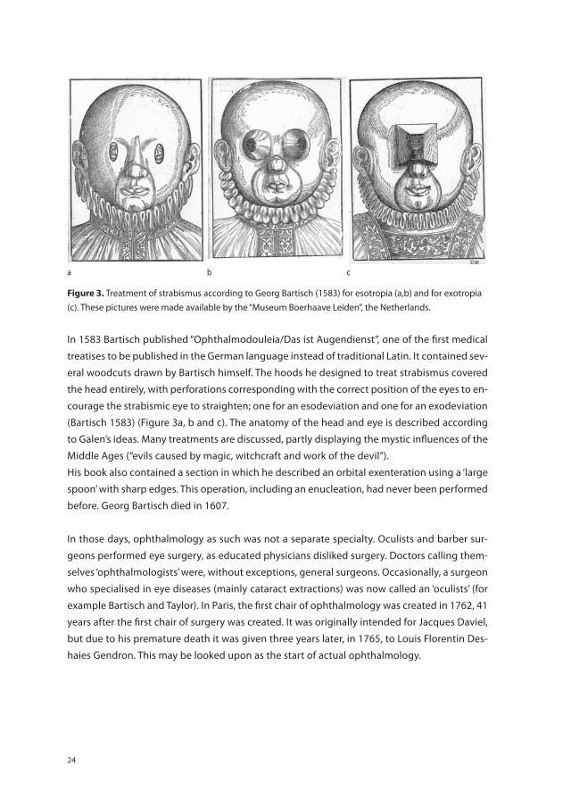
In 1583 Bartisch published “Ophthalmodouleia/Das ist Augendienst”, one of the first medical
treatises to be published in the German language instead of traditional Latin. It contained sev
eral woodcuts drawn by Bartisch himself. The hoods he designed to treat strabismus covered
the head entirely, with perforations corresponding with the correct position of the eyes to en
courage the strabismic eye to straighten; one for an esodeviation and one for an exodeviation
(Bartisch 1583) (Figure 3a, b and c). The anatomy of the head and eye is described according
to Galen’s ideas. Many treatments are discussed, partly displaying the mystic influences of the
Middle Ages (“evils caused by magic, witchcraft and work of the devil”).
His book also contained a section in which he described an orbital exenteration using a ‘large
spoon’ with sharp edges. This operation, including an enucleation, had never been performed
before. Georg Bartisch died in 1607.
In those days, ophthalmology as such was not a separate specialty. Oculists and barber sur
geons performed eye surgery, as educated physicians disliked surgery. Doctors calling them
selves ‘ophthalmologists’ were, without exceptions, general surgeons. Occasionally, a surgeon
who specialised in eye diseases (mainly cataract extractions) was now called an ‘oculists’ (for
example Bartisch and Taylor). In Paris, the first chair of ophthalmology was created in 1762, 41
years after the first chair of surgery was created. It was originally intended for Jacques Daviel,
but due to his premature death it was given three years later, in 1765, to Louis Florentin Des
haies Gendron. This may be looked upon as the start of actual ophthalmology.
3a
Figure 3. Treatment of strabismus according to Georg Bartisch (1583) for esotropia (a,b)
and for exotropia (c). These pictures were made available by the “Museum Boerhaave
Leiden”, the Netherlands.
3b
3c
a b c
Figure 3. Treatment of strabismus according to Georg Bartisch (1583) for esotropia (a,b) and for exotropia (c). These pictures were made available by the “Museum Boerhaave Leiden”, the Netherlands.

25
chap
ter 2
The history of the treatment of amblyopia
charles de Saint-yves
Charles de SaintYves was born in 1677 in MaubertFontaine, France. He started his education
in general surgery when he was 17 years old. Five years later he had specialised in eye diseases
at the general hospital in Paris, a hospital supported by the Countess Françoise Anthénais
Montespan, the mistress of King Louis XIV. In 1711, Charles de SaintYves left the general hos
pital and opened his own Ophthalmology Clinic in Paris where he committed himself fully
to eye diseases. In 1722, he wrote down his experiences in his textbook “Nouveau traité des
maladies de yeux” (Figure 4). Chapter 24 deals with ‘Des yeux louches’ in which he wrote: “One
was sometimes obliged to fully cover the nonstrabismic eye, thereby straightening the stra
bismic eye and so as to be dependent on this eye, it will get used to looking straight ahead”
(De SaintYves 1722). He continued: “When one closes the nonstrabismic eye, the squinting
eye will now look straight ahead and when opening the eye again one now finds a squint in
the eye that was straight before”. Based on this observation, he was one of the first to describe
the cover test. Nowadays, the cover test is an important part of orthoptic practice. It includes
two tests: the coveruncover test and the alternating cover test and is the principle element in
the detection and diagnosis of strabismus. To straighten the eyesight he also recommended
the exercise to “sit the child in front of a mirror and that each eye looks precisely to the pupil
of that eye which corresponds to him in the mirror. In addition, one must also read fine print
and do handicrafts”. His book is clearly written with detailed observations, including cataract
Figure 4. Charles de Saint-Yves was one of the first surgeons who specialised in eye
diseases and wrote detailed observations about all known eye diseases in his book:
“Nouveau traité des maladies des yeux” (1722).
Figure 4. Charles de SaintYves was one of the first surgeons who specialised in eye diseases and wrote detailed observations about all known eye diseases in his book: “Nouveau traité des maladies des yeux” (1722).

26
extractions (he performed 6080 each year). His life’s work shows him to be a dedicated re
searcher and eye specialist. Charles de SaintYves died in 1736.
George comte de Buffon
The person usually credited for the introduction of occlusion of the fixating eye for amblyopia
was the French naturalist and botanist GeorgeLouis Leclerc, Comte de Buffon (Figure 5).
He was born in Montbard (Burgundy, France) in 1707 to rich, middle class parents and inher
ited the title ‘Comte de Buffon et Montbard’ and a large sum of money when his mother died.
This made him financially independent, in those days a necessity for dedicating one’s life to
science. He studied medicine, astronomy and botany at the Angers University. He suffered
from strabismus and poor vision of his squinting eye. According to Buffon, a strabismic eye
was caused by poor vision in one of the eyes, which led to a disruption in binocular vision.
This was contrary to what was generally believed, i.e. that an unequal strength of the muscles
or lack of concordance caused the strabismus. He also rejected the masks as he recognised
they did not have the desired effect. In 1743, in his “Dissertation sur les causes du Strabisme”
he described the weak eye regaining all its strength by occluding the good eye (“reprendre
toutes ses forces”) (De Buffon 1743). Buffon is probably better known for his “Histoire Naturelle
générale et particulière (1749)”, written when he was responsible for the Royal Gardens of the
French King Louis XV. He played an important part in the development of biology as a science.
He was an avowed opponent of Carolus Linnaeus whose taxonomy he described as artificial,
which is probably the reason why Linnaeus denominated an ugly toad as a “Bufonidae”. Buffon
died in April 1788.
Figure 5. George-Louis Leclerc, Comte de Buffon postulated that a strabismic eye was
caused by poor vision in one of the eyes causing a disruption in binocular vision, which
was contrary to what was generally believed at that time (1743).
Figure 5. GeorgeLouis Leclerc, Comte de Buffon postulated that a strabismic eye was caused by poor vision in one of the eyes causing a disruption in binocular vision, which was contrary to what was generally believed at that time (1743).

27
chap
ter 2
The history of the treatment of amblyopia
john Taylor
A new idea, to treat strabismus surgically, was introduced by John Taylor, born in Norwich
(England) in approximately 1703. He started his career as a pharmacist’s student in London
and afterwards moved to the continent where he attended lectures on ophthalmology by
Herman Boerhaave (16681738). He travelled around the major cities in Europe in a carriage
painted with eyes, treating patients with ‘incurable eye diseases’. Taylor carried out cataract
operations, removed blood from inflamed conjunctiva and scars and abraded weak and para
lysed eyes with the convex side of a silver tablespoon. Occasionally, Taylor would even place
the concave side of the spoon over a closed eye and give it a firm push (Figure 6).
Among Taylor’s patients were Johann Sebastian Bach, on whom he operated for cataract, and
Georg Friedrich Händel, both of whom became blind due to complications of the operation.
The most intriguing operation he performed were those on the strabismic eye. Taylor believed
that strabismus was caused by an imbalance between the muscles. By cutting the nerve inner
Figure 6. Chevalier John Taylor travelled around Europe treating ‘incurable eye disease’
and suggested treating strabismus with surgery. However, the technique for strabismus
surgery was not developed until approximately 100 years later.
Figure 6. Chevalier John Taylor travelled around Europe treating ‘incurable eye disease’ and suggested treating strabismus with surgery. However, the technique for strabismus surgery was not developed until approximately 100 years later.

28
vating the strongest muscle, this balance would be restored; he therefore performed a small
cut in the conjunctiva, pretending to cut the nerve and thus so straighten the strabismic eye.
After the procedure he covered the operated eye to demonstrate that the other eye looked
straight ahead. The next day he would cover the other eye, thereby demonstrating the stra
bismic eye now also looked straight ahead. When Taylor and his companions arrived in Rouen,
France, ClaudeNicolas Lecat (or: Le Cat, the famous anatomist and surgeon) took his oppor
tunity to study Taylor’s work so that he might improve his own surgical techniques. Lecat was
not satisfied with the result of Taylor’s operations on the strabismic eye. He argued that it was
unclear which nerve was to be cut in the conjunctiva. Lecat also observed that every time the
better eye was closed, the strabismic eye looked straight ahead and vice versa. He regarded
Taylor as a fraud, invited him to his house for dinner and served him an unexpected dessert: a
human head with its eyes anatomically prepared. It was obvious there were no nerves in the
conjunctiva. Taylor left Rouen the next day. All the eyes he had operated on started squinting
as before (Crone 1992; Taylor 1756).
ThE FIrST STrABISmuS oPErATIonS
Stromeyer, dieffenbach and cunier
It was not until a hundred years later doctors did what Taylor had intended to do: operate
the eye muscles. The initiator was Friedrich Louis Stromeyer from Hannover, Germany. He oc
cupied himself with orthopaedic surgery. In his book “Beiträge zur Operativen Orthopädik”
(1838), he cut the Achilles tendon in the heel in order to treat (straighten) a clubfoot. The same
principle could be applied to strabismus, for which he described a tenotomy of the inner eye
muscle on corpses (Stromeyer 1838). Johann Friedrich Dieffenbach (17921847) was the first
to publish his results, 10 days after operating the medial rectus muscle in Berlin (1839), when
he wrote “Über die Heilung des angeborenen Schielens mittelst der Durchschneidung des
inneren geraden Augenmuskels”. He performed a tenotomy on a 7yearold boy with conver
gent strabismus without anaesthesia (Dieffenbach 1839). Three years later, Dieffenbach had
already operated 1200 patients with strabismus. Unfortunately, total tenotomies often led to
an overcorrection. Meanwhile, in Brussels, Florent Cunier (18121852) also performed a stra
bismus operation claiming to be the first (Cunier/Missotten 2001). However, Dieffenbach was
able to prove that he had performed the same procedure just a few days earlier.
Ludwig Boehm noticed that the visual acuity of the strabismic eye sometimes improved after
the operation. He attributed this effect to the tenotomy itself and presumed that amblyopia
could be cured by an operation (Boehm 1845).

29
chap
ter 2
The history of the treatment of amblyopia
dIScoVEry oF ThE oPhThALmoScoPE And ThE STArT oF modErn oPhThALmoLoGy
Von helmholtz, donders and von Graefe
In 1850, Hermann Ludwig Ferdinand von Helmholtz invented the ophthalmoscope. Born in
Potsdam, Germany in 1821, he was appointed professor of physiology at Königsberg at the
age of 28. He was an important scientist and greatly contributed to modern ophthalmology.
With the ophthalmoscope it was now possible to view the retina (Helmholtz 1851; Keeler
2002). This was revolutionary in eye care and may be considered as the start of modern oph
thalmology. It was confirmed that in most cases of amblyopia the eye was structurally sound,
establishing the functional character of amblyopia.
The physiologist and ophthalmologist Franciscus Cornelis Donders (18181889), a Dutch
scientist, examined thousands of emmetropic and ametropic eyes and recorded the normal
refractive error (Donders 1864). Until then, only three types of errors were known: myopia,
presbyopia and astigmatism. In 1859, Donders described the hypermetropic error, and in
1861 how accommodation was linked to convergence. It was apparent that Donders already
knew about the relationship between accommodation and convergence as early as 1847.
In a postscript to an article written by F.W.C. Krecke on the correction of strabismus using
prisms, Donders hypothesised that convergent strabismus might be related to hyperme
tropia (Krecke 1847). So, as a consequence, the strabismus would often improve when this
refractive error was corrected. While studying in London he visited the World’s Fair in 1851
and became acquainted with the German ophthalmologist Albrecht von Graefe (18281870,
Berlin) (Figure 7).
FIGUREN BIJ HOOFDSTUK 2
Figure 1. Thabit ibn Qurrah ibn Marwan al-Harrani was the first to describe occlusion
therapy for the better eye until the vision in the strabismic eye had returned to normal.
Figure 2. Remains of “Hospice des Quinze-Vingts”, the first institution for the blind
in Paris (1254).
Figure 7. Franciscus Cornelis Donders and Albrecht von Graefe, founders of the
Dutch and German Ophthalmology, at the World’s Fair in London, 1851.
Figure 7. Franciscus Cornelis Donders and Albrecht von Graefe, founders of the Dutch and German Ophthalmology, at the World’s Fair in London, 1851.

30
Albrecht von Graefe was one of the founders of German ophthalmology. Inspired by each oth
er’s work, Donders and Von Graefe became friends. They readily used the Ophthalmoscope to
determine the refractive error and provoked discussions about what came first: strabismus or
amblyopia. This chickenoregg discussion (Bielschowsky, 1926) continued until 1960 (Wiesel
1963a; 1963b; 1965). Donders and Von Graefe argued that amblyopia in strabismic children
could be the result of either ‘not using’ the eye, i.e. functional amblyopia, or of an organic vi
sual impairment (“…wird im Folge von diesem Nichtgebrauche mit physischer Unterdrückung
amblyopisch”) (Donders 1864; von Graefe 1854). Instead, Carl Schweigger and Alfred Graefe
advanced the theory of organic amblyopia (Graefe 1894; Schweigger 1885). In their opinion,
children were born with amblyopia and this caused the strabismus. They also argued that eyes
did not become amblyopic even after a long period of nonuse, for example cataract, and that
the acuity became normal after removal of any obstacle to vision.
Fusion exercises
Darwin, Javal, Worth and Maddox
In England, Erasmus Darwin (17311802) modified Charles de SaintYves’ cover exercises al
most 50 years later (Darwin 1779). Born in Lichfield near Birmingham, he was the first to pre
scribe fusion exercises as a treatment for strabismus. Using a septum, he separated the two
visual fields after which he presented each eye with small coloured pieces of wood to train the
fixation. Once the child was able to fixate equally well with either eye, two pieces of wood were
presented simultaneously to each eye and the child was asked to superimpose them. Erasmus
Darwin worked in close association with other great scientists and highly skilled technicians
such as Josiah Wedgwood, the porcelain manufacturer, James Watt and Joseph Priestley. His
theory of evolution was elaborated on by his grandson Charles Robert Darwin.
In 1897, Louis Emile Javal (18391907) emphasised the use of stereoscopic exercises to treat
strabismus in his “Manuel théorique et pratique du Strabisme” (Javal 1896). Born to a business
man, his parents wished him to study business and economics, hoping he would take over his
father’s coalmining business. In 1865, however, he entered medical school at the University
of Paris. In 1878 he became the director of the Laboratory of Ophthalmology at the University.
He took a particular interest in ophthalmology, probably because his father and sister suffered
from strabismus and he himself from myopic astigmatism. Javal preferred nonsurgical treat
ments for ocular problems, hence his interest in orthoptics. Determined not to let his sister
fall a victim to: “…le massacre des muscles oculaires,” he trained her with his “Stéréoscope à
charnière”. In his manual he advised occlusion of the healthy eye, “occlusion volontaire de l’oeil
meilleur,” as well as the use of the stereoscope to reestablish binocular vision. Even though he
suffered from progressive glaucoma he was able to finish his manual in 1897, after which he
dedicated himself to the further development of Braille together with Louis Braille.

31
chap
ter 2
The history of the treatment of amblyopia
Inspired by the work of Javal, Claud Worth published his first edition of “Squint, its causes,
pathology and treatment” at the turn of the century (Worth 1903). Born in 1869, he made
fundamental contributions to the field of strabismus and became a member of the Ophthal
mological Society of the United Kingdom in 1899. He was an advocate of occlusion therapy
of the nonstrabismic eye and introduced the use of atropine in mild cases of amblyopia, as
an alternative to patching. He recorded the age of onset of the squint, the length of time the
squint had been present before occlusion started and the final visual acuity of the deviating
eye. From these results he developed a ratio: age in months when permanent turn became ap
parent divided by the age in months at which training/treatment began. This ratio indicated
the prognosis, which improved when the ratio approached unity. Worth formulated the the
ory that strabismus was caused by a congenital defect of the fusion mechanism. He therefore
also proposed fusion training and active stimulation of the amblyopic eye using the improved
version of the ‘fusion tubes’ (Priestley Smith 1896), now called the ‘amblyoscope’. However, the
use of the amblyoscope was time consuming and results were not always satisfactory.
Ernest Edmund Maddox made use of the Worth amblyoscope in Bournemouth, England. Born
in Shipton and educated at the University of Edinburgh he received his MD in 1889. He is re
nowned for several inventions: the double prism, the Maddox rod, the Maddox tangent scale
(1898), the Maddox prism verger and the cheiroscope for orthoptic training. Near the end of
his career he developed an interest in orthoptics and the Worth amblyopscope. He found that
he did not have enough time to use the device properly. Instead, he taught his daughter, Mary
Maddox, to use the amblyoscope. She quickly became a professional using this device and from
this she turned her interest to other aspects of orthoptics. In 1929 Mary Maddox opened the
first orthoptic clinic at the Royal Westminster Ophthalmic Hospital in London (Maddox 1931).
reintroduction of occlusion therapy: mastisolverband
Sattler
Occlusion therapy was reintroduced by C.H. Sattler when he published his experiences with
the treatment of amblyopia in Leipzig in 1927 (Sattler 1927). For the treatment of strabismus
related amblyopia he recommended the use of socalled ‘Mastisolverband’. This occluder was
glued to the skin around the eye so as to prevent the child from peeking and remained se
curely fastened there for at least two to three days. This was the first description of the adhe
sive tape patch and as such it meant the reintroduction of occlusion therapy for amblyopia.
He reported that the best results were obtained in children up to the age of six. Of course, full
cooperation and stimulation of the parents was essential and the acuity in the sound eye had
to be checked frequently. Children in whom acuity failed to improve were considered either
not to have applied the ‘Mastisolverband’ properly or not to have applied it at all.

32
Two years later, F. Weckert made use of the children’s spectacles by using a spectacle ‘occluder’
with shields on the side to prevent light incoming from the periphery and to enable the child
to blink behind the occluder more easily (Weckert 1929). The children had to wear these oc
cluders full time. He reported good results using this type of occlusion, which, according to
Weckert, was more readily accepted by the patient than Sattler’s ‘Mastisolverband’.
Arguments for and against occlusion therapy
Poulard, Uhthoff, Stenius, Fuchs and Gifford
At the time, there were many arguments for and against occlusion therapy. The Frenchmen
Poulard shared the opinions of Alfred Graefe. He had never seen any great improvement of
vision in the amblyopic eye with occlusion therapy (Poulard 1921) and could not but conclude
that a deviating eye was caused by amblyopia, rather than amblyopia being caused by the
deviating eye. This opinion he also shared with Uhthoff (Uhthoff 1927). Stenius, however, in
1935, reported excellent results in 2 to 6yearolds with central fixation in their strabismic eye.
He prescribed these children parttime occlusion therapy (1 or 2 hours daily) and encouraged
them to read or draw pictures during the period of occlusion (Stenius 1935). The year after,
Ernst Fuchs wrote in his “Aus meiner augenärztlichen Praxis” that the results he gained with
occlusion therapy did not measure up to the problems parents had to endure when treating
their children (Fuchs 1928). This opinion was supported by Gifford, who thought occlusion
could cause a psychological trauma, leading to disorders such as stammering (Gifford 1935).
Besides these problems, complications of occlusion therapy have also been reported, e.g.
occlusion amblyopia, disruption of binocularity and an increase in the angle of strabismus
(Quéré, et al. 1969). An occasional occurrence of occlusion amblyopia had already been men
tioned by Worth and proven to be usually reversible (Burian 1966; Hardesty 1959). Other stud
ies did not find these adverse effects on the patched eye (Holbach, et al. 1991; Lithander and
Sjöstrand 1991).
Segment occlusion
Bangerter
Segment occlusion was introduced by Alfred Bangerter, St. Gallen, Switzerland in 1953. Self
adhesive synthetic material was applied onto the inside of the spectacles occluding only part
of the glass; what part depended, for instance, on the presence of accommodative or conver
gent strabismus. The occlusion spectacles could also reduce the incoming light by means of a
transparent foil with various densities of gratings (“Bangerter Foil”) (Bangerter 1960). He also
suggested the use of occluders in case of undisciplined children and in warmer weather con
ditions. In his book “Amblyopiebehandlung” (Bangerter 1953) Bangerter described the devel
opment of amblyopia and considered system, consequence, versatility and adjustment to be

33
chap
ter 2
The history of the treatment of amblyopia
the basic principles of his amblyopia treatment. He developed a systematic treatment of the
amblyopic eye according to age, diagnosis and fixation, as well as a prophylactic treatment
of amblyopia. He was a fierce opponent of occluding the better eye in the presence of an ec
centric fixation. In his opinion, this was not only dangerous and futile but could also reinforce
the eccentric fixation (Bangerter 1946; 1953; 1960).
Occlusion therapy would suffice to improve acuity in case of lowgrade amblyopia; however,
highgrade amblyopia, especially amblyopia without fixation or a paracentral fixation, needed
more intensive and direct stimulating measures to achieve increased acuity (Bangerter 1953).
This brings us to the pleopticera.
Pleoptics
Comberg and Cüppers
Pleoptic is also a Greek word and refers to pleion=more and optikos=eyesight. In Rostock in
the northeast of Germany, W. Comberg described an apparatus to train central vision in chil
dren with functional amblyopia (Comberg 1936). The apparatus (1936) was designed to stim
ulate the area of the macula in eyes with eccentric fixation by projecting brightly illuminated
objects onto the fovea.
Bangerter and later Cüppers introduced various other instruments based on these very same
principles: stimulation of the fovea of the amblyopic eye to awaken central fixation.
As he was head of the department, Bangerter had the means and opportunity to pursue his
ideas on how to treat amblyopia the best possible way. He invented over 20 instruments and
after more than a decade of trial and modification, Bangerter constructed the “Pleoptophor”
(Linksz 1961; Priestley 1961; Schlossman 1961); a device that could temporarily blind the ec
centrically fixating area and stimulate the central foveal region.
Two years later, in Cologne, Conrad Cüppers gave a lecture on the pleoptic and orthoptic
treatment of amblyopia and strabismus as performed at the Augenklinik Gießen (Cüppers
1956). He was a strong advocate of occlusion therapy except, like Bangerter, when an ec
centric fixation was present in the amblyopic eye. Cüppers’ great virtue was the development
of the “Visuskop”, an instrument to visualize even the smallest angles of strabismus and de
termine the fixation. He established that onethird of the children treated for amblyopia did
not fixate with their fovea. This assumption led to the development of a different method:
the creation of ‘afterimages’ (‘Nachbilder’) supported by occlusion of the amblyopic eye. The
afterimages were created by a modified ophthalmoscope (called “Euthyskop”) operated by
an experienced practitioner. A circular bright light blinded the retinal periphery, while a circu
lar spot protected the fovea. A negative afterimage was thus created. Now the foveola had a
momentary physiological superiority over the retinal periphery. The patient had to fixate on

34
the centre of the afterimage, thereby regaining central fixation (Cüppers 1961). Cüppers’ idea
was that this technique would change the direction of the principal visual axis from eccentric
to foveal. He emphasised, however, that occlusion therapy was to be preferred in younger
children (he referred to the work of Sattler) and pleoptic therapy in older ones and in those
with eccentric fixation. Orthoptic and pleoptic principles were to be considered as supple
mentary treatments and not as opposites (Cüppers 1967).
For the next decade, pleoptic treatment was a widely used and accepted way of treating am
blyopia with an eccentric fixation. As pleoptic treatment was time consuming and generally
limited to older and more cooperative children, socalled “Sehschulen” (Vision Schools) were
set up where children received treatment for several weeks or even months on end. After the
Second World War, a large number of amblyopic children were admitted.
At that time, various articles reported on the success of this treatment (Bangerter 1960; Cüp
pers 1967; Jablonski and Tomlinson 1979), while others were more skeptical (Koselka, et al.
1991; Schmidt and Stapp 1977) regarding the longterm effectiveness and its economic ad
vantage over conventional occlusion therapy. Soon after, studies reported that pleoptics failed
to produce a significant improvement in the linear visual acuity or a permanent cure in the
majority of amblyopic patients (Fletcher, et al. 1969; von Noorden and Lipsius 1964; Parks and
Friendly 1966; Richter 1960; VéronneauTroutman, et al. 1974). It was even suggested that ple
optics might be dangerous as it could cause permanent monocular diplopia (Campos 1995).
However, it should be acknowledged that pleoptics gave rise to further and greater insight
into the pathophysiology of amblyopia with special emphasis on the amblyopic eye with ec
centric fixation.
The idea that occlusion of the better eye would only reinforce the eccentric fixation (Bangert
er 1953) has not been confirmed. Mackensen et al. demonstrated, via fundus photography,
that occlusion of the better eye did not induce reinforcement of the eccentric fixation. On the
contrary, they found fixation often became foveal with occlusion of the good eye (Mackensen,
et al. 1965).
Penalisation
To “penalise” literally means “to punish” and the term was most probably first used by J.B. Weiss
in 1968 (in previous studies it was described as “atropinisation”) (Weiss and Bourrie 1968). The
use of pharmacological penalisation to treat amblyopia had already been suggested by Worth
in 1903 for children who did not or were too young to cooperate with conventional occlusion
therapy (Worth 1903). Bangerter also used it in the first and second year of life (Bangerter
1960). Generally, it means selectively fogging the image of the nonamblyopic eye by glasses

35
chap
ter 2
The history of the treatment of amblyopia
(optical penalisation) and/or cycloplegia by the daily instillation of drops into the fornix (phar
macological penalisation). This should prevent accommodation and therefore obstruct near
vision. In this way, one eye is used for distance vision and the other eye (the amblyopic eye) for
near vision. Several variations of this technique have been developed (Abraham 1954; John
son and Antuna 1965; Knapp and Capobianco 1956; Pfandl 1958; Pope 1971; 1972; Pouliquen
1964). In 1969, Quéré, Pouliquen, Lavat, Berrondo and Weiss described several techniques of
penalisation (Quéré 1969).
Others who studied the effect of penalisation therapy on amblyopia reported positive results
(Cibis 1974; FoleyNolan, et al. 1997; Kaye, et al. 2002; Lowe 1965; McKenney and Byers 1975;
von Noorden and Milani 1979; North and Kelly 1991; Quéré 1972; Repka and Ray 1993; Si
mons, et al. 1997; Swann and Hunter 1974; Timmerman 1977; Wallman, et al. 1978), either
in combination with occlusion therapy, as maintenance therapy or sometimes as the main
therapy. It was more readily accepted by parents and patients of all age groups and allowed
a reliable assessment of compliance. Quéré observed a decrease in the angle of strabismus
(Quéré 1969) and claimed that penalisation prevented the development of occlusion amblyo
pia (Quéré 1972). Due to the continued use of both eyes, occlusion amblyopia should be less
frequent as compared to occlusion using a patch. However, occlusion amblyopia did occur
and careful monitoring of the patient’s sound and amblyopic eye was necessary (von Noorden
and Milani 1979; North and Kelly 1991; Repka and Ray 1993; Wallman, et al. 1978; Worth 1903).
Aggravation of a (latent) nystagmus, another frequent obstacle to occlusion therapy, could
also be prevented by penalisation, as both eyes were open at all times. Finally, the penalised
eye still received images with low spatial frequency and therefore binocular vision was less
disturbed (FoleyNolan, et al. 1997; Lowe 1965; North and Kelly 1991; Repka and Ray 1993).
Other authors, however, did not find substantiated evidence to support different outcomes
for monocular or binocular vision (Simons, et al. 1997) and were hesitant to prescribe penali
sation in very young children as the negative effect on near vision lasted too long.
A potential disadvantage of penalisation therapy is the effect that image blur or cycloplegia
may have on the development of the refractive error. Both experimental work in animals and
case reports in humans have shown a myopic shift in the blurred eye leading to anisometro
pia (Robb 1977; Wallman, et al. 1978). A second disadvantage would be the daily use of drugs
used over a long period of time. Side effects are mainly related to systemic intoxication: a case
of coma has been reported (Campos, et al. 1991), hypersensitivity to atropine (Lowe 1965;
Pope 1971), iris cysts (Abraham 1954; Knapp and Capobianco 1956) and dilatation of the pupil
causing photophobia (Campos 1997). Moreover, the acuity of the penalised eye must be less
than that of the amblyopic eye, otherwise the patient will continue to use the sound eye de
spite the pharmacologically or optically induced image blur. When using optical penalisation

36
it would also be possible for the child to peek around the glasses or remove them altogether.
It seems reasonable, therefore, to limit penalisation to mild or moderate amblyopia.
other non-conventional treatments
Brinker and Katz: Red-filter treatment
In 1963, following the controversy in treating amblyopia associated with eccentric fixation,
Brinker and Katz reported on the use of a red filter treatment (Brinker and Katz 1963). Their
idea was to totally occlude the nonamblyopic eye and place a red filter on the spectacle
frame in front of the amblyopic eye. The red filter excluded wavelengths shorter than 640 mm
(the filter most often used was Kodak Wratten gelatin red filter No. 92). The rationale for this
method lay in the theory that visual cones are sensitive to stimulation by light from the red
end of the spectrum and visual rods are not. The rodpopulated area of the retina, which is
used for eccentric fixation, is insensitive to this red light. With the nonamblyopic eye occlud
ed and the filter in front of the amblyopic eye, the patient was encouraged to do near work.
After fixation became central, the red filter was removed and conventional treatment for am
blyopia with central fixation was instituted. The results from various studies reported success
rates ranging from 10% to 87.5% (Cowle, et al. 1967; Malik, et al. 1966; Ratiu and Reiter 1966;
Thorleifsson 1966). This range was largely due to differences in duration of treatment and
response was greater when initial amblyopia was less severe. Cowle et al. suggested that in
order to achieve good results, the redfilter treatment should be used for more than 7 months
followed by 5 months of occlusion of the nonamblyopic eye, for a total treatment time of 12
months (Cowle, et al. 1967).
In 1998, a blue filter was suggested instead of the red filter (Metzler, et al. 1998).
Pigassou and Garipuy: Prisms
As mentioned before, the principle of pleoptic treatment stimulated further ophthalmoscopic
studies of amblyopia and precipitated the concept of eccentric fixation. As one result of these
further studies, the use of prisms was introduced for the treatment of amblyopia. In 1966,
Pigassou and Garipuy treated amblyopia with eccentric fixation by means of occlusion of the
better eye and an inverted prism in front of the amblyopic eye (Pigassou and Garipuy 1966a;
1966b; 1967). Other studies showed results that favoured the inverse prism method over con
ventional occlusion therapy and pleoptics (Nawratzki and Oliver 1971).
Other therapies
In 1964, Fernando Losada wrote about functional amblyopia and its treatment with occlusion
therapy and surgically suturing the eyelids (Losada 1964). He especially advocated the neces
sity to suture the eyelid of the better eye in disobedient or uncooperative children in order to
save the vision in their amblyopic eye. He was not the only person favouring this idea. As early

37
chap
ter 2
The history of the treatment of amblyopia
as 1932, F. Weckert had recommended the suturing of the eyelids in noncompliant children
(Weckert 1932).
Less harmful than suturing the eyelids seemed the advice from Fralick in 1943. In very young
children, he applied elbow splints made from tongue blades and adhesives, or plaster cast
arm restraints during occlusion therapy (Fralick 1943; Hiles and Galket 1974). Other methods
tried for the treatment of amblyopia have included eyeball massage (Darier 1904) or a diet of
white wine and veal (Devaux 1912). In 1914, the removal of the adenoids was thought to be
of importance (Adams 1914) and in 1958 the beneficial influence of hypnosis was asserted
(Browning, et al. 1958).
Campbell: CAM Treatment (1978)
In an attempt to improve the acceptance of occlusion therapy by the parents and the child,
Campbell and coworkers developed an apparatus in which highcontrast squarewave grat
ings were rotated slowly in front of the amblyopic eye while the child was performing a task
requiring visual concentration (Banks, et al. 1978; Campbell, et al. 1978). This process took only
7 minutes during which the nonamblyopic eye was occluded. In between the weekly ses
sions, the nonamblyopic eye was not to be occluded. The rationale for using rotating gratings
with different spatial frequencies was that it provided stimulation of the whole range of mo
tion of the amblyopic visual system. However, there was a considerable danger of intractable
diplopia in case the treatment was applied inappropriately and the use of the instrument
ought therefore to be confined to hospitals and clinics. Campbell et al. reported success with
this kind of minimal occlusion therapy in 73% of their patients who achieved acuities of >6/12
after three 7minute sessions. Willshaw et all. reported the same success rate (Willshaw, et all.
1980). In spite of these encouraging results, other authors have not been able to confirm the
value of the apparatus in subsequent studies (Ciuffreda, et al. 1980; Fricker, et al. 1980; Keith,
et al. 1980; Mehdorn, et al. 1981; Schor, et al. 1981; Tytla and LabowDaily 1981).
commEnT
The history of treatment for amblyopia is a rather intriguing one. Throughout the ages, vari
ous methods of treatment have been published, with occlusion of the better eye being the
very first to be described. This method, however, frequently receded into the background
when other types of treatment for amblyopia were tried, e.g. pleoptophor, euthyskop, CAM
treatment, prisms and filters. These new methods became popular for a while, but were aban
doned again, as they were time consuming and less effective. The fluctuations in popularity of
occlusion could be explained by a limited acceptance by children and parents, resulting in low
compliance. Ever since the first prescriptions were written, low compliance was already dis
cerned as a problem affecting the outcome of treatment. It was acknowledged that occlusion

38
was a burden on the family life and that parents needed to be stimulated in order to achieve
good results. Various other efforts to increase compliance included gluing the occluder to the
skin around the eye or even suturing the eyelids in noncompliant children. Instead of imple
menting these radical methods it would be more effective to focus our attention on ways to
increase the acceptance of occlusion therapy by children and parents. For the last decade
it has been possible to measure compliance with occlusion therapy electronically, yielding
exact information on the actual duration of occlusion. This enables us to study the factors
that influence compliance with occlusion therapy, which should be taken into account when
developing new methods for the improvement of compliance.
SEArch STrATEGy
The selection of the literature for this review was based on the Medline database that was
searched from its starting date in 1966, using key words such as amblyopiatreatmenthis
torypenalisationatropineprismspleopticsred filtergratingsCAMocclusion. Parts of the
very early history were obtained from Wolfgang Münchow (“Geschichte der Augenheilkunde”,
1984) and the Picarta database. Articles in any language were considered for inclusion.
AcknowLEdGEmEnT
We thank Prof. R.A. Crone, MD PhD, Prof. G. Kolling, MD PhD and M.V. Joosse, MD PhD for their
advice and contributions.

chap
ter 1
chap
ter 2
chap
ter 3
chap
ter 4
chap
ter 5
chap
ter 6
chap
ter 7
chap
ter 8
chap
ter 9
chap
ter 1
0ch
apte
r 11
Add
enda
Chapter 3
objective survey of the prescription of occlusion therapy for amblyopia

40
ABSTrAcT
Purpose: To identify the variation within and consistency amongst orthoptists when prescrib
ing occlusion therapy for amblyopia in an objective survey.
methods: A questionnaire was designed with five case examples of amblyopic children and
distributed at annual meetings of orthoptists in the Netherlands and Germany. It was filled
out simultaneously within 15 min in complete silence to avoid any exchange of opinions be
tween orthoptists that would reduce variability. For each case the orthoptists were asked to
give their prescription of hours or days of occlusion.
results: The questionnaire was completed by 177 Dutch orthoptists and 227 German orthop
tists. Their prescriptions of occlusion therapy were classified into five main regimens: Part
Time; PartTime Not Every Day; FullTime; Also Occluding the Amblyopic eye; ALTernating and
No Occlusion Therapy. The variation was large: the standard deviation was half the average
prescribed hours of occlusion for each regimen in each of the five cases. All orthoptists were
assigned a rank number for each of the five cases depending on whether their prescription
was above or below average. These five rank numbers were not consistently above or consis
tently below average value per case.
conclusions: The number of prescribed hours of occlusion varied widely per regimen per case.
Orthoptists were not consistently strict or lenient in their prescription of occlusion therapy.

41
chap
ter 3
Objective survey of the prescription of occlusion therapy for amblyopia
InTroducTIon
Treatment for amblyopia with occlusion of the better eye dates back to at least the early
eighteenth century. Charles de SaintYves first described occlusion of the dominant eye to
promote use of the squinting eye in 1722 (De SaintYves 1722). After that, Allen in 1730 and
Comte de Buffon in 1743 recommended occlusion of the good eye to straighten the squinting
one (Allen 1730; De Buffon 1743). Javal in France and Worth (1901) in England advocated the
use of occlusion for amblyopia (Javal 1896; Worth 1901).
Despite the fact that occlusion therapy dates back to the eighteenth century, there are still
few guidelines for prescribing occlusion hours. This is in contrast to the prescription of an
tibiotics or other drug medication, which is done according to protocol and to scientifically
derived standard measures. Age, visual acuity and to a lesser extent diagnosis seem to be the
most important determinants when prescribing a number of hours of occlusion therapy. Mein
and Trimble stated that the lazy eye should be occluded fulltime in the case of strabismic
amblyopia and parttime in case of anisometropic amblyopia followed by fulltime occlusion
if the acuity does not improve sufficiently (Mein, et al. 1991). According to Haase the good
eye should be occluded for a number of days per week corresponding to age of the child in
years, followed by one day of occlusion of the amblyopic eye (Haase 1995). Von Noorden and
Campos stated that the good eye should be occluded fulltime when there is a difference in
visual acuity between the two eyes (Von Noorden, et al. 2002).
As there is no consensus amongst orthoptists, different orthoptists may prescribe very differ
ent hours of occlusion for the same patient. This can lead to confusion amongst parents seek
ing second opinions and result in noncompliance with the therapy (Tan, et al. 2003).
A questionnaire was developed with five case examples of children diagnosed with amblyo
pia in order to try and identify these variations in prescription of occlusion hours amongst
orthoptists, their consistency and their main determinants.
mEThodS
The questionnaire consisted of four example amblyopic cases and one potentially amblyopic
case (Table 1). It was designed to imitate the clinical decisionmaking process in an orthop
tist’s everyday practice.
Cases 1, 2, and 5 were common amblyopic cases with a clear difference in acuity between the
two eyes. Case 1 was a 3yearold with an anisometropia and visual acuity of 5/10 and 5/5 as
measured with a Children’s Picture Chart. Case 2 was a 2yearold with esotropia of the right
eye first noticed 6 months earlier. Case 3 was a 5yearold diagnosed with slight esotropia of
the left eye with eccentric fixation of this eye and mild anisometropia. Case 4 was a 6month
old baby with infantile esotropia of 30° alternating freely. Case 5 was a 3yearold with hyper

42
metropia combined with esotropia of the left eye, an eccentric fixation and a clear difference
in the visual acuity between the two eyes. Case 2, 3, 4 and 5 were children with a strabismic
amblyopia, case 1 was a child with an anisometropic amblyopia.
In March 2001, at the national Spring Meeting of the Union for Dutch Orthoptists in Utrecht,
the questionnaire was put to Dutch orthoptists representing almost all practicing orthoptists
in the Netherlands. In October 2001, at the annual meeting of the Union for German Orthop
tists in Warnemünde, the questionnaire was put to the German orthoptists present. They were
asked to determine their prescription of hours of occlusion (hours per day or days per week)
for each of the five cases within 15 min. Additional instructions for completing the question
naire included the statement that the prescription of glasses was not to be considered. They
were not allowed to discuss or copy the cases and were under strict supervision from the four
researchers present to prevent any exchange of views that might lessen variability. As a result
the questionnaires were completed in silence. After 15 min the completed questionnaires
were collected, also under strict supervision.
Table 1. The questionnaire containing the five sample amblyopic cases. The following instructions were given: Please enter below the number of hours of occlusion therapy you would prescribe in each case. Do not consider the prescription of glasses. Please do not discuss the cases with your neighbour, lest we measure less variability.
case 1 case 2 case 3 case 4 case 5
Age 3 years old 2 years old 5 years old 6 months old 3 years old
diagnosis Anisometropia Esotropia first noticed six months earlier
Microstrabismus, untreated amblyopia
Alternating esotropia
Accommodative esotropia
corneal reflection Straight 20° Right esotropia Slight left esotropia 30° Esotropia 15° Left esotropia
cover test Straight Large right esotropia, no DVD / NL
Slight left esotropia Marked esotropia, alternating, NL, DVD unclear
Large (30 cm) / moderate (5 m) left esotropia
Binocular vision Titmus Fly +, animals ABC +
Titmus Fly
Fixation rE/LE Central / Central Nasal / Central Central / Eccentric nasal Eccentric nasal
Visual acuity rE/LE 5/10 & 5/5 Pictures Cannot maintain fixation, saccadic pursuit, some protest when occl left eye
1.0 / 0.2 EChart Alternates freely 5/6 & 5/15 Pictures
retinoscopy rE/LE +3.5 / +2 +2 / +2 S+1.0=C0.5axis180 / S+1.5=C1.75axis50
+1 / +1 +4 / +4
hours per day
days per week

43
chap
ter 3
Objective survey of the prescription of occlusion therapy for amblyopia
data analysis
A database was created which consisted of four primary items; (1) the number of hours pre
scribed per day, (2) the number of days of occlusion per week, (3) the period of occlusion and
(4) the total number of occlusion hours prescribed per week. Both eyes were itemized this
way. The prescriptions given by each orthoptist were recorded on a casebycase basis in the
database. All the variations in the prescriptions of occlusion therapy could be stored into one
database. From there it was possible to translate them into different regimens of prescribing
occlusion therapy. For the statistical analysis of the database we used the nonparametric
tests (MannWhitney).
To determine whether orthoptists prescribed their hours of occlusion consistently above av
erage or consistently below average, all orthoptists were ranked according to their number
of prescribed hours of occlusion per case. From these rank numbers percentiles were derived
and the standard deviation (SD) of the percentiles was calculated. Due to statistical consider
ations this SD of percentiles could not exceed 28.87%. If occlusion hours were completely ran
domly prescribed with a uniform distribution of the percentiles between 0% and 100%, the
SD of the percentiles would be √1/12(100%−0%)2 =28.87%. The SD would be 0% if orthoptists
prescribed consistently above or consistently below average, i.e. if the strictest orthoptist pre
scribed the most hours of occlusion for each of the five cases and the most lenient orthoptist
prescribed the least hours of occlusion for each of the five cases.
rESuLTS
questionnaire
177 Dutch and 227 German orthoptists filled in the questionnaire. Seven (4%) Dutch orthop
tists and four (2%) German orthoptists failed to complete the questionnaire properly, but in
these cases only one of the five cases on the questionnaire had been left unanswered; there
fore they were included in our analysis. The orthoptists wrote no negative comments on the
forms regarding the sample cases.
regimens
Variation in prescription of occlusion hours was large. In the effort to ascertain variation, con
sistency and determinants in the orthoptists’ prescribing of occlusion, we identified certain
types of regimen that were most frequently prescribed. Within the data analysis process five
typical prescriptions were identified; PartTime Occlusion (PTO), PartTime Occlusion, Not

44
Every Day of the week (PTONED), FullTime Occlusion (FTO), Also Occluding Amblyopic eye
(AOA), ALTernating occlusion (ALT), No Occlusion Therapy (NOT), (Table 2, Figure 1).
Table 2. Definitions of the five prescribed occlusion regimens.
PTO PartTime Occlusion, i.e. the nonamblyopic eye is occluded the same number of hours every day of the week
PTONED PartTime Occlusion Not Every Day of the week, i.e. the nonamblyopic eye is occluded for the same number of hours, followed by days of both eyes open
FTO FullTime Occlusion, i.e. the nonamblyopic eye is occluded for all waking hours every day of the week or the nonamblyopic occluded for all waking hours followed by days when both eyes are open
AOA Also Occluding the Amblyopic eye, i.e. the amblyopic eye is also occluded for any period of time (either parttime or fulltime) in addition to the occlusion of the nonamblyopic eye for any period of time
ALT ALTernating occlusion therapy, i.e. both eyes are occluded alternately for the same number of hours every day, parttime as well as fulltime
NOT No Occlusion Therapy of either eye
Other Pair of spectacles, foil, exercise, more complex regimens
Chapter 3 Figure 1
Figure 1. Median and interquartile range of the prescribed hours of occlusion per day of the nonamblyopic eye per regimen per case. The abscissa represents the five cases and the chosen regimen. The total number of orthoptists prescribing a regimen is given below each whisker. For colour figure please see ‘Colour figures’ on page 195.

45
chap
ter 3
Objective survey of the prescription of occlusion therapy for amblyopia
There was an occasional prescription of a pair of spectacles, foil, inverse occlusion only or of
even more complex regimens. Regimens prescribed by fewer than 12 orthoptists were ex
cluded from analysis.
Because of the different ages of the sample cases, and in order to permit comparison of the
actual hours of occlusion that had been prescribed, the time spent awake by a 6monthold
baby were estimated to be 6 h per day, a 2yearold child, 10 h per day and a 3yearold and
5yearold child, 12 h per day. In this way the prescriptions of days per week were converted
to hours per week.
dutch and German orthoptists’ prescribing behaviour
It became apparent that the prescribing behaviour of Dutch and German orthoptists was
similar in many ways (Table 3). For both countries the preferred occlusion regime was PTO.
Also, the variation in number of prescribed hours for each of the cases was as high among
German orthoptists as it was among their Dutch counterparts. Differences were found in that
FTO and AOA were favoured more often by the German orthoptists. However, the similarities
outweighed the differences; therefore the data derived from the Dutch and German orthop
tists were analysed together.The main result of this study was that for each of the five pre
scribed regimens the SD was half of the averaged prescribed hours of occlusion. This applied
to each of the five sample amblyopic children. In addition, the following three observations
were made:
(1) Orthoptists prescribed significantly more hours of occlusion when prescribing FTO than
when prescribing PTO or PTONED (P < 0.005).
(2) In case 1, 2, 3, and 5, orthoptists who prescribed PTO prescribed significantly more hours
per week than orthoptists who prescribed PTONED. This difference was significant in cases
1, 2, and 5 (P < 0.002).
Table 3. The median of the prescribed hours of occlusion for the nonamblyopic eye per day distributed over the five regimens per case. In brackets are the percentages of Dutch and German orthoptists prescribing that regimen.
Case 1:Anisometropia, 3yrs
Case 2: Esotropia, 2yrs
Case 3: Microstrabismus,5yrs
Case 4: Alternating, 6mo
Case 5: Accomm. eso, 3yrs
Dutch German Dutch German Dutch German Dutch German Dutch German
PTO 3 (72%) 3 (61%) 4 (81%) 4 (45%) 5 (85%) 6 (33%) 1 (7%) 2 (5%) 4 (79%) 4 (41%)
PTONED 2.1 (26%) 2.6 (14%) 2.9 (19%) 2.7 (9%) 5.7 (11%) 5.7 (6%) 0.6 (1%) 0.6 (2%) 2.9 (18%) 2.9 (8%)
FTO 9 (13%) 6.6 (28%) 12 (1%) 10 (32%) 5.1 (1%) 9 (30%)
AOA 8.6 (2%) 10 (10%) 12 (8%) 6.7 (1%) 3.4 (1%) 12 (8%)
ALT 4 (2%) 3.1 (1%) 1 (31%) 1 (26%) 3.4 (1%) 1.1 (2%)
NOT 0 (4%) 0 (1%) 0 (61%) 0 (65%) 0 (1%) 0 (2%)
Other (2%) (4%) (8%) (2%) (20%) (9%)

46
(3) In case 2 (n = 23), 3 (n = 18) and 5 (n = 19) some German orthoptists also prescribed oc
clusion of the amblyopic eye for a period of time. In these cases the nonamblyopic eye
was occluded for significantly more hours than it was by orthoptists who prescribed PTO,
PTONED or FTO (P < 0.005).
consistency in prescription of occlusion hours
To calculate whether orthoptists prescribed their hours of occlusion consistently above aver
age or consistently below average, each orthoptist was assigned a rank number according
to number of prescribed hours per case. From these rank numbers the five percentiles, their
average and their SD were derived. The average SD of the percentiles for all orthoptists was
21%. It would have been 28.87% if occlusion hours had been prescribed completely at ran
dom and 0% if they had been prescribed consistently above or below average (see above,
Data analysis).
dIScuSSIon
By distributing our questionnaire at an annual meeting for orthoptists and preventing an ex
change of opinions we were able to gain an objective insight in the prescriptions of occlusion
therapy for amblyopia.
Whilst analysing the data we were taken aback not only by the amount of variation in the
number of prescribed hours of occlusion, but also by the diversity in the ways of prescrib
ing occlusion therapy. The number of occlusion hours was neither normally nor lognormally
distributed in any of the five example cases. However, during the analysis on a casebycase
basis it became apparent that certain ways of prescribing occlusion therapy, i.e. certain regi
mens, were more prevalent (Table 2). Within these regimens the SD was half of the average
prescribed hours of occlusion. This applied to each of the five sample amblyopic children.
The five example amblyopic cases were chosen in order to make the difference in the pre
scribed hours of occlusion by orthoptists more easily transparent. Cases 1, 2 and 5 were com
mon amblyopic cases, while cases 3 and 4 were more controversial cases. The largest variation
in the prescription was found in case 3, representing a 5yearold child with microstrabismus
and untreated amblyopia. To improve visual acuity long periods of occlusion could have been
thought necessary, although the outcome of the treatment was uncertain. That may have
been the reason why some orthoptists decided not to occlude at all, whereas other orthop
tists prescribed long periods of occlusion.

47
chap
ter 3
Objective survey of the prescription of occlusion therapy for amblyopia
Case 4, a baby with alternating fixation, may not have required any occlusion therapy as there
was no real evidence of amblyopia. However, some orthoptists prescribed ALT with a median
of 1 h every day of the week. This decision to commence with alternating occlusion therapy
may well have been prompted by the desire to prevent the development of an amblyopic eye.
It may also have been influenced by the uncertainty of the diagnosis.
Orthoptists who prescribed FTO prescribed significantly more hours of occlusion in the same
case than orthoptists who prescribed PTO and PTONED. This tendency can be partly explained
by orthoptists opting for the FTO regimen when they wanted to prescribe longer periods of
occlusion. Orthoptists wanting rapid success would select FTO rather than prescribing PTO
or PTONED and waiting patiently. The same difference is seen between PTO and PTONED:
orthoptists who chose PTO prescribed significantly more hours of occlusion in the same case
than those who prescribed PTONED. It can also be partly explained by the assumption that or
thoptists wanting to prescribe shorter periods of occlusion would opt for reducing the num
ber of days per week rather than reducing the number of hours per day.
Orthoptists who prescribed an AOA regimen prescribed significantly more hours of occlusion
than orthoptists who prescribed only occlusion of the nonamblyopic eye, possibly to com
pensate for the hours the amblyopic eye was patched.
In our questionnaire orthoptists were asked to make their prescription on the basis of the
main determinants: age, visual acuity and diagnosis. Not included were further determinants
that may have influenced the results, such as personal experience, the perceived domestic
situation, the possible interference with homework or the waiting time for a followup ap
pointment. The personal attitude of the orthoptists towards the success of the treatment was
also not taken into account when designing the questionnaire.
Based on traditional and educational differences we expected a difference in occlusion pre
scriptions between Dutch and German orthoptists. Indeed, German orthoptists did tend to
prescribe slightly longer hours of occlusion and to prescribe to FTO more often than Dutch
orthoptists. However, the amount of variation in prescribed hours of occlusion was equal in
the Dutch and German orthoptist (Table 3).
Neither consistently strict nor consistently lenient orthoptists of either nationality could be
identified.
This study clearly emphasises the need for prospective studies investigating the relationship
between prescribed occlusion hours and actual occlusion time carried out by the parents
(Loudon, et al. 2003). Secondly, the relationship between the patched hours and the child’s
acuity increase should be established (Stewart, et al. 2002). The findings of such investigations

48
might lead to the development of validated guidelines or protocols for prescribing occlusion
therapy.
If consensus and uniformity could be achieved among orthoptists and ophthalmologists as to
the prescription of occlusion therapy, this would go a long way towards convincing the parent
that a specific regimen of occlusion is the best therapy for the amblyopic eye, thus promoting
compliance.
AcknowLEdGEmEnTS
We thank the 404 orthoptists for their hospitality and filling in the questionnaire. We acknowl
edge René Eijkemans and Caspar Looman for their help with the statistical analysis.

chap
ter 1
chap
ter 2
chap
ter 3
chap
ter 4
chap
ter 5
chap
ter 6
chap
ter 7
chap
ter 8
chap
ter 9
chap
ter 1
0ch
apte
r 11
Add
enda
Chapter 4
Visusevaluierung in einer historischen kohorte von 137 okkludierten
Patienten, 30-35 jahre nach Ende der okklusionsbehandlung
Evaluation of visual acuity in a historic cohort of 137 patients treated for amblyopia
by occlusion 30-35 years ago

50
zuSAmmEnFASSunG
hintergrund: Der Visusverlauf des amblyopen Auges nach Ende der Okklusionsbehandlung
wird unterschiedlich diskutiert. Wir untersuchten eine grosse historische Kohorte, 3035 Jahre
nach der Okklusionsbehandlung.
material und methode: Von 1250 Patienten, die in der Orthoptischen Abteilung des Water
land Krankenhauses in Purmerend zwischen 1968 und 1975 behandelt wurden, wurden 471
Kinder okkludiert, 5%, nach Vergleich mit den örtlichen Geburtsziffern. Wir konnten davon
203 Patienten erneut ausfindig machen, 137 wurden 2003 orthoptisch nachuntersucht. Wir
korrelierten den aktuellen Visus mit der Ursache der Amblyopie (Anisometropie, Strabismus
oder Kombination), dem Alter bei Beginn und Ende der Okklusion, dem Visus bei Beginn und
Ende der Okklusionsbehandlung, der Fixation, dem Binokularsehen, der Refraktion und dem
Refraktionsverlauf.
Ergebnisse: Die Patienten waren bei Beginn der Okklusionsbehandlung 5,4 ± 1,9 Jahre, bei
Ende 7,4 ± 1,7 Jahre und bei der Nachuntersuchung 37 ± 2,7 Jahre alt. Korreliert mit dem
aktuellen Visus waren ein geringer Visus des amblyopen Auges bei Beginn (P < 0,0001) und
Ende der Okklusionsbehandlung (P < 0,0001), eine exzentrische Fixation (P < 0,0001) und die
Ursache der Amblyopie (P = 0,005). Am Ende der Okklusionsbehandlung betrug der Visus der
amblyopen Augen bei Patienten mit StrabismusAmblyopie (n = 98) 0,29 ± 0,3 logMAR und
bei der Nachuntersuchung 0,27 ± 0,3 logMAR. Bei Patienten mit AnisometropieAmblyopie
(>1 D, n = 16) hatte sich der Visus von 0,17 ± 0,23 logMAR auf 0,21 ± 0,23 logMAR und bei Pa
tienten mit Anisometropie und Strabismus als Ursache der Amblyopie (n = 23) von 0,52 ± 0,54
logMAR auf 0,65 ± 0,54 logMAR verschlechtert. Insgesamt hatten 54 (39%) Patienten seit der
Behandlung eine Visusabnahme, davon 18 um mindestens 50% in Bezug zum Visus nach Ende
der Behandlung. 15 dieser 18 Patienten hatten eine Anisohyperopie, die zugenommen hatte.
Schlussfolgerung: Assoziiert mit einer langfristigen Visusabnahme nach Ende der Okklusi
onsbehandlung waren eine kombinierte Ursache (Anisometropie und Strabismus) der Ambly
opie und eine Zunahme der Anisohyperopie.
kurzzusammenfassung
137 Patienten aus einer historischen Kohorte, vor 3035 Jahren wegen Amblyopie okkludiert,
wurden 2003 erneut untersucht. Korreliert mit dem aktuellen Visus waren ein geringer Visus
des amblyopen Auges bei Beginn und Ende der Okklusionsbehandlung, eine exzentrische
Fixation und die Ursache der Amblyopie. Assoziiert mit einer Visusabnahme nach Ende der

51
chap
ter 4
Visusevaluierung in einer historischen Kohorte
Okklusionsbehandlung waren eine kombinierte Ursache (Anisometropie und Strabismus) der
Amblyopie und eine Zunahme der Anisohyperopie.
ABSTrAcT
Purpose: Opinions differ on the course of the visual acuity in the amblyopic eye after cessa
tion of occlusion therapy. This study evaluated visual acuity in a historical cohort treated for
amblyopia with occlusion therapy 3035 years ago.
methods: Between 1968 and 1975, 1250 patients had been treated by the orthoptist in the
Waterland Hospital in Purmerend, the Netherlands. Of these, 471 received occlusion treat
ment for amblyopia, corresponding to 5% of the local birth rate. We were able to contact 203
of these, 137 were orthoptically reexamined in 2003. We correlated the current visual acuity
with the cause of amblyopia, the age at start and end of treatment, the visual acuity at start
and end of treatment, fixation, binocular vision and refractive errors.
results: Mean age at the start of treatment was 5.4 ± 1.9 years old, 7.4 ± 1.7 years old at
the end and 37 ± 2.7 years old at follow up. Current visual acuity in the amblyopic eye was
correlated with a low visual acuity at the start (P < 0.0001) and end of occlusion therapy (P <
0.0001), an eccentric fixation (P < 0.0001), and cause of amblyopia (P = 0.005). At the end of the
treatment, patients with a strabismic amblyopia (n = 98) had a visual acuity in the amblyopic
eye of 0.29 ± 0.3 logMAR, and in 2003 0.27 ± 0.3 logMAR. In patients with an anisometropic
amblyopia (>1 D, n = 16) visual acuity had decreased from 0.17 ± 0.23 logMAR to 0.21 ± 0.23
logMAR. In patients with both strabismic and anisometropic amblyopia (n = 23), visual acuity
had decreased from 0.52 ± 0.54 logMAR to 0.65 ± 0.54 logMAR. Overall, acuity had decreased
in 54 patients (39%) after cessation of treatment. Of these, 18 patients had an acuity decrea
se to less than 50% of their initial acuity. In 15 of these 18 patients anisohypermetropia had
increased.
conclusions: A decrease in visual acuity after cessation of occlusion therapy occurred in pati
ents with a combined cause of amblyopia and an increase in anisohypermetropia.
For an English translation of Chapter 4, please see Appendix.

52
EInLEITunG
In der Literatur wird die Prävalenz von Amblyopie mit 3,25% angegeben (Attebo, et al. 1998).
Die Behandlung der Amblyopie mit Okklusion stellt seit Jahrzehnten eine etablierte Methode
dar. Der Nutzen der Okklusionsbehandlung liegt vor allem in der Vermeidung von Kosten, die
entstehen, wenn Patienten mit unzureichend behandelter Amblyopie im Laufe des Lebens ihr
gutes Auge verlieren (Neubauer and Neubauer 2005). Der Zeitraum, den ein solcher Patient
beidseitig sehbehindert (Visus beidseits >0,3 logMAR) verbringt, verlängert sich etwa von 8
auf 15 ½ Monate (van Leeuwen, et al. 2002).
Mit der Einführung des Screenings konnte Amblyopie früher erfaßt und damit erfolgreicher
behandelt werden, so dass die Zahl der Patienten ohne Lesefähigkeit auf dem amblyopen
Auge reduziert werden konnte. Seit Mütterberatungsstellen in den Niederlanden Kinder auf
Strabismus und Amblyopie testen und den Visus im Alter von 3 sowie 3 ¾ Jahren bestimmen,
ist eine zu spät entdeckte Amblyopie selten. Trotzdem misslingt bei etwa einem Drittel aller
amblyoper Patienten (Jensen and Goldschmidt 1986) die Okklusionsbehandlung (Visus des
amblyopen Auges > 0,3 logMAR). In der Vergangenheit wurde ein Misslingen der Okklusi
onsbehandlung durch zu späte Diagnosestellung (Bowman 1998) verursacht, heutzutage ist
primär die Therapieuntreue (Dorey, et al. 2001; Loudon, et al. 2003) bei der Okklusionsbe
handlung dafür verantwortlich.
Der Langzeiterfolg einer Okklusionsbehandlung wird in der Literatur unterschiedlich ange
geben. Ein Vergleich der Studien ist schwierig aufgrund der unterschiedlichen Patientgrup
pen: Rekrutierung von verschiedenen Kliniken (Woodruff, et al. 1994) oder im Rahmen eines
Vorschulscreenings (Bowman, et al. 1998); aufgrund der underschiedlichen Einslusskriterien:
z.B. nur Patienten, die alle Kontrolltermine einhielten (Levartovsky, et al. 1995; Malik, et al.
1975); und aufgrund verschiedener Behandlungsmethoden: z.B. nur mit Vollokklusion (Scott
and Dickey 1988) oder auch mit Pleoptik, Brillenokklusion oder inverser Okklusion behan
delt (Schröpfer and Meinert 1975; Sparrow and Flynn 1977). Zudem variiert der Zeitpunkt
der Nachuntersuchung nach Ende der Okklusionsbehandlung, Monate (Malik, et al. 1975) bis
maximal zwei Jahrzehnte (Leiba, et al. 2001).
Einige Autoren berichten, dass der Visus nach Ende der Okklusionsbehandlung überwiegend
unverändert blieb (Ohlsson, et al. 2002; Scott and Dickey 1988) oder besser (Leiba, et al. 2001)
wurde, andere Autoren wiesen überwiegend eine geringe Visusabnahme von ein bis zwei
Zeilen nach (Levartovsky, et al. 1995; Schröpfer and Meinert 1975). Negative Faktoren für das
Langzeitergebnis nach abgeschlossener Okklusionsbehandlung waren ein relativ hohes Alter
bei Beginn der Okklusionsbehandlung (Ham, et al. 1985; Levartovsky, et al. 1995; Meyer, et
al. 1991; Stewart, et al. 2004) und ein geringer Startvisus (Meyer, et al. 1991; Scott and Dickey
1988; Sparrow and Flynn 1977; Woodruff, et al. 1994), eine exzentrische Fixation (Ham, et al.

53
chap
ter 4
Visusevaluierung in einer historischen Kohorte
1985; Sparrow and Flynn 1977) und eine kombinierte Ursache der Amblyopie: Strabismus und
Anisometropie (Levartovsky, et al. 1995; Woodruff, et al. 1994).
Wir untersuchten den Visus sowie prognostische Faktoren für den aktuellen Visus nach Ende der
Okklusionsbehandlung in einer Kohorte von 137 Patienten, die vor 3035 Jahren in Waterland,
einer ländlichen Region in Holland, wegen Amblyopie mit Okklusion behandelt wurden. Für
die gesamte Region waren damals nur ein Augenarzt und eine Orthoptistin (HvK, damlas 40%
Arbeitspensum) zuständig. Dieser Orthoptistin wurden von den Mütterberatungsstellen oder
den Hausärzten, die die Kinder auf Amblyopie screenten, alle Amblyopieverdächtigen Kinder
überwiesen. Gemeinsam mit dieser Orthoptistin wurden die Patienten 2003 erneut untersucht.
mATErIAL und mEThodEn
hintergrund
Zum Zeitpunkt der Untersuchung stellte das Waterland Krankenhaus die medizinische Ver
sorgung der Gemeinden Purmerend, Beemster, Waterland, Zeevang, Wormer, (einschliesslich
Edam und Volendam) und Landsmeer (Anteil 50%, weil etwa die Hälfte der Einwohner nach
AmsterdamNord überwiesen wurden) sicher. Ihre Population setzte sich aus Personen zu
sammen, die ursprünglich aus Purmerend oder den angrenzenden Gemeinden stammten so
wie aus einzelnen Familien mit jungen Kindern, welche aus Amsterdam zugewandert waren.
Bestimmung der daten
Es wurden alle Krankenakten von Patienten herausgesucht, die zwischen 1968 und 1975 in
der Orthoptischen Abteilung des Waterland Krankenhauses in Purmerend, kurz oder langfri
stig, behandelt wurden (n = 1250). Bei 471 (38%) Kindern wurde eine Okklusionsbehandlung
durchgeführt. Aus den Krankenakten erhoben wir demografische Angaben der Patienten (Ge
schlecht, Geburtsdatum, Telefonnummer) und orthoptische Daten (Alter bei Beginn und Ende
der Okklusionsbehandlung, Visus des amblyopen Auges bei Beginn und Ende der Okklusions
behandlung, Ursache der Amblyopie).
Behandlung
Eine Amblyopie wurde damals mit Okklusion behandelt, wenn nach einer adäquaten Bril
lenverschreibung bei der folgenden Kontrolluntersuchung noch eine Visusdifferenz von ei
ner Zeile bestand und amblyogene Faktoren vorhanden waren. Konnte kein Visus bestimmt
werden, wurde das Führungs und Fixationsverhalten bei Strabismus beurteilt und bei einem
asymmetrischen Befund eine Okklusionsbehandlung eingeleitet.

54
Damals wurde der Visus monokular bis zum vierten Lebensjahr mit Kinderbildern (Amsterda
mer BilderKarte; Visusbereich 6/30 bis 6/5) getestet. Ab dem vierten Lebensjahr wurde der
Visus mit Landoltringen und etwa ab dem siebten Lebensjahr mit Buchstaben in 6m getestet
(Snellen) und in einem Dezimalwert notiert. Bei diesen zwei Tests war der Abstand zwischen
den Visusstufen linear, der Sehzeichenabstand war grösser als 2,6 Bogenminuten und wurde
als ‚single’ beschrieben. Alle Visuswerte wurden für die Analyse in logMAR umgerechnet (Ta
belle 1).
nachuntersuchung 2003
Von 471 Patienten konnten 203 (43%) Patienten ausfindig gemacht und telefonisch kontak
tiert werden. Diesen Patienten wurde ein Fragebogen zur Beurteilung der Lebensqualität
bei Amblyopie und/ oder Strabismus zugesandt, der Amblyopia & Strabismus Questionnaire
(A&SQ) (van de Graaf, et al. 2004). Die anderen Patienten konnten aus verschiedenen Gründen
nicht kontaktiert werden: Zwei Patienten waren verstorben, bei den anderen Patienten konn
te die Telefonnummer nicht ausfindig gemacht werden, oder sie waren nicht erreichbar. 174
Patienten sendeten den ausgefüllten Fragebogen zurück. Im Herbst 2003 konnten davon 137
Tabelle 1. Umrechnungstabelle Visus in Dezimalen Visus in logMAR
umrechnungstabelle Visus in dezimalen - Visus in logmAr
Visus LogmAr
1,6 0,2
1,25 0,1
1,0 0,0
0,8 0,1
0,63 0,2
0,5 0,3
0,4 0,4
0,32 0,5
0,25 0,6
0,2 0,7
0,16 0,8
0,12 0,9
0,1 1,0
0,08 1,1
0,06 1,2
0,05 1,3

55
chap
ter 4
Visusevaluierung in einer historischen Kohorte
Patienten in der Orthoptischen Abteilung des Waterland Krankenhauses in Purmerend orth
optisch und augenärztlich nachuntersucht werden. Von den 27 Patienten, die den Fragebo
gen zurückgesendet hatten, aber nicht nachuntersucht werden konnten, waren elf nicht er
reichbar, sechs wollten nicht teilnehmen und neun konnten nicht zur Untersuchung kommen,
weil sie hospitalisiert waren, im Ausland weilten oder einen langen Anfahrtsweg hatten.
Wir verfügten über Visusangaben zu drei Zeitpunkten (Beginn, Ende der Okklusionsbehand
lung, sowie 2003, 3035 Jahre nach Ende der Okklusionsbehandlung).
Folgende Untersuchungen wurden bei der Nachuntersuchung durchgeführt: Binokularsehen
wurde mit dem Streifentest nach Bagolini in 6m und 0,4m getestet, Stereosehen mit Titmus
Test, Stereotest nach Lang II und TNOTest. Der Schielwinkel wurde mittels monolateralem
und alternierenden Abdecktest in 6m und 0,3m gemessen sowie eine dissoziierte Vertikaldi
vergenz notiert. Die Refraktion wurde mittels einer Skiaskopie ohne Zykloplegie, jedoch im
abgedunkelten Raum und nachfolgendem subjektiven Abgleich geprüft. Der Visus wurde
mittels Projektor (OCULUS, Medical Workshop Niederlande) mit linearen Visusstufen (Sehzei
chenabstand „single“) monokular in 6m Zahlen gemäss DIN EN ISO 8596 mit bestmöglicher
Korrektion getestet und in Dezimalwerten notiert. Die Lesefähigkeit wurde mit einem nieder
ländischen Lesetext bestimmt, der aus fünf Visusstufen bestand (D=0,5; D=0,8; D=1; D=1,25;
D=2). D=1 entspricht Zeitungsdruckgrösse. Ferner wurde die Fixation ophthalmoskopisch ge
prüft. Es wurden die Vorderabschnitte und der Fundus beurteilt.
Alle Visuswerte, ausser dem Lesetext, wurden für die Analyse in logMAR umgerechnet.
Anisometropie wurde definiert als eine Differenz von mehr als 1 D sphärischem Äquivalent
zwischen dem rechten und linken Auge. Eine Kombination beider Ursachen (Strabismus und
Anisometropie) definierten wir als kombinierte Amblyopie.
Statistische Analyse
Wir haben eine Regressionsanalyse durchgeführt, um Parameter zu bestimmen, welche den
aktuellen Visus am meisten beeinflussen. Um mögliche Störfaktoren auszuschliessen, wurde
eine multivariante Analyse durchgeführt. Für die statistische Analyse wurde SPSS angewandt,
Version 10.0. Als Schwelle für statistische Signifikanz wurde P < 0,05 bestimmt.
ErGEBnISSE
Gesamtgruppe
Die Patientengruppe bestand aus 65 Frauen und 72 Männern. Das durchschnittliche Alter bei
Beginn der Okklusionsbehandlung betrug 5,4 ± 1,9 Jahre, am Ende 7,4 ± 1,7 Jahre. Zwölf (9%)

56
der 137 Kinder wurden bereits vor dem dritten Lebensjahr okkludiert, durchschnittlich ab 2,4
Jahren, bei ihnen bestand ein grosswinkliger Strabismus.
Der durchschnittliche Visus der amblyopen Augen bei Beginn betrug 0,6 ± 0,7 logMAR und
0,3 ± 0,5 logMAR am Ende der Okklusionsbehandlung. 98 Patienten (71%) wiesen eine Stra
bismusAmblyopie auf, 16 Patienten (12%) eine AnisometropieAmblyopie und 23 Patienten
(17%) eine kombinierte Amblyopie.
Zu Beginn der Okklusionsbehandlung hatten 116 Patienten ein Visusunterschied zwischen
dem Führungsauge und dem amblyopen Auge von mindestens 0,5 logMAR. 13 Patienten
hatten ein Visusunterschied zwischen 0,2 und 0,5 logMAR, acht Patienten von < 0,2 logMAR.
Bei fünf Patienten konnten aufgrund des Alters keine Visusangaben erhoben werden, anstatt
dessen hat die Orthoptistin das Führungs bzw. Fixationsverhalten der Patienten beschrie
ben. Bei allen fünf Patienten lag ein Strabismus vor: zwei Patienten hatten nur geringe Folge
bewegungen bei Okklusion des Führungsauges, drei nahmen die Fixation auf, hielten diese
aber nicht. Wir haben ein Visusäquivalent von 0,4 logMAR bzw. 0,2 logMAR angenommen.
Der Visus bei Beginn der Okklusion wurde nur für die Korrelation des Startvisus mit dem ak
tuellen Visus verwendet, jedoch nicht für die prognostischen Faktoren. Diese Gleichsetzung
kann man unterschiedlich interpretieren, es gilt aber zu beachten, dass es sich nur um fünf
Patienten handelt.
Patienten mit StrabismusAmblyopie wurden ab 5,1 ± 1,9 Jahren okkludiert, durchschnittlich
2,3 ± 1,3 Jahre. Patienten mit reiner AnisometropieAmblyopie waren zu Beginn der Okklusi
onsbehandlung 7,2 ± 2,2 Jahre alt und wurden 1,4 ± 1,0 Jahre okkludiert. Patienten mit einer
Tabelle 2. Visus bei Beginn, Ende und 3035 Jahre nach Okklusionsbehandlung sowie Alter bei Beginn der Behandlung, Dauer der Okklusion und Fixationsverhalten nach Ursache der Amblyopie.
FaktorenStrabismus-Amblyopie (n = 98)
Anisometropie-Amblyopie (n = 16)
kombinierte Amblyopie (n = 23)
Visus Beginn okklusion(logmAr)
0,69* ± 0,5 0,53* ± 0,3 0,82* ± 0,54
Visus Ende okklusion (logmAr) 0,29 ± 0,3 0,17 ± 0,23 0,52 ± 0,54Visus nachuntersuchung (logmAr) 0,27 ± 0,3 0,21 ± 0,23 0,65 ± 0,54Alter Beginn okklusion (jahre) 5,1 ± 1,9 7,2 ± 2,2 5,8 ± 0,7dauer okklusion (jahre) 2,3 ± 1,3 1,4 ± 1,0 1,6 ± 1,3Fixation zentral n = 63* n = 16 n = 14 Exzentrisch n = 35* n = 0 n = 9
Sowohl bei der uni als auch multivariaten Analyse waren der Visus des amblyopen Auges bei Beginn der Behandlung (P < 0,0001), die Ursache der Amblyopie (P = 0,001) (Strabismus, Anisometropie, kombinierte Amblyopie) sowie eine exzentrische Fixation (P < 0,0001) bei Strabismus signifikant in Bezug auf den aktuellen Visus in logMAR.*Signifikante Parameter für den Visus 2003 in logMAR bei der Regressionsanalyse.

57
chap
ter 4
Visusevaluierung in einer historischen Kohorte
kombinierten Amblyopie waren 5,8 ± 0,7 Jahre alt und wurden 1,6 ± 1,3 Jahre okkludiert (Ta
belle 2). Das Binokularsehen verbesserte sich in der Gesamtgruppe der Patienten gering
(Abbildung 1).
Visus nach ursache der Amblyopie
Bei Patienten mit kombinierter Amblyopie hatte sich der Visus seit der Behandlung von 0,52
± 0,54 logMAR auf 0,65 ± 0,54 logMAR und bei Patienten mit AnisometropieAmblyopie von
0,17 ± 0,23 logMAR auf 0,21 ± 0,23 logMAR verschlechtert. Bei Patienten mit StrabismusAm
blyopie hatte sich der Visus von 0,29 ± 0,3 logMAR auf 0,27 ± 0,3 logMAR verbessert (Abbil
dung 2). Diese Verbesserung war signifikant (P < 0,001). Der Visus am Führungsauge dieser
Patienten zeigte ebenso eine Zunahme von 0,01 ± 0,15 logMAR auf 0,05 ± 0,07 logMAR und
war auch signifikant (P = 0,04).
Chapter 4
Abbildung 1. Binokularsehen der Gesamtgruppe während der Okklusionsbehandlung und bei der
Nachuntersuchung 2003.
Der Grad von Binokular- und Stereosehen (Abszisse) wurde in sieben Stufen eingeteilt:
1: Lichtschweiftest nach Bagolini negativ, 2: Lichtschweiftest nach Bagolini positiv, 3:
Lichtschweiftest nach Bagolini und Titmus-Stereotest Fliege positiv, 4: mindestens Titmus-Stereotest
Ringe 200” bis 140” positiv, 5: mindestens Titmus-Stereotest Ringe 100” bis 40” positiv, 6: Stereotest
nach Lang oder mindestens TNO-Test Abbildung V (480” & 240”) positiv, 7: TNO-Test Abbildung VI
oder VII (120” – 15”) positiv. Der schwarze Balken zeigt den Grad von Binokularsehen während der
Okklusion in der Gesamtgruppe (n = 137), der graue Balken bei der Nachuntersuchung 2003. Die
Ordinate repräsentiert die Anzahl der Patienten.
Abbildung 1. Binokularsehen der Gesamtgruppe während der Okklusionsbehandlung und bei der Nachuntersuchung 2003. Der Grad von Binokular und Stereosehen (Abszisse) wurde in sieben Stufen eingeteilt: 1: Lichtschweiftest nach Bagolini negativ, 2: Lichtschweiftest nach Bagolini positiv, 3: Lichtschweiftest nach Bagolini und TitmusStereotest Fliege positiv, 4: mindestens TitmusStereotest Ringe 200” bis 140” positiv, 5: mindestens TitmusStereotest Ringe 100” bis 40” positiv, 6: Stereotest nach Lang oder mindestens TNOTest Abbildung V (480” & 240”) positiv, 7: TNOTest Abbildung VI oder VII (120” – 15”) positiv. Der schwarze Balken zeigt den Grad von Binokularsehen während der Okklusion in der Gesamtgruppe (n=137), der graue Balken bei der Nachuntersuchung 2003. Die Ordinate repräsentiert die Anzahl der Patienten.

58
Der Visus des amblyopen Auges zeigte sich bei 14 (10%) Patienten unverändert in Bezug zum
Visus nach Ende der Okklusion. 70 (51%) Patienten wiesen eine Visusverbesserung auf. 54
(39%) Patienten hatten einen Visusabnahme, davon 18 Patienten um 50% und mehr in Bezug
zum Visus des amblyopen Auges nach Ende der Okklusionsbehandlung.
47 (34%) von 137 Patienten hatten am Ende der Okklusionsbehandlung auf dem amblyopen
Auge einen Fernvisus >0,3 logMAR, durchschnittlich 0,48 ± 0,6 logMAR. 55 (40,1%) von 137
Patienten hatten keine Lesefähgkeit (> D=1).
Abbildung 2. Visus des amblyopen Auges, nach Diagnose, bei Beginn der Okklusionsbehandlung und
bei der Nachuntersuchung 2003.
Die Ordinate zeigt den Visus bei der Nachuntersuchung in logMAR, die Abszisse den Visus in
logMAR bei Beginn der Okklusion. Patienten mit einer kombinierten Amblyopie sind dargestellt als
schwarzes Viereckg, Patienten mit Anisometropie-Amblyopie als Dreieck ▲ und Patienten mit
Strabismus-Amblyopie als weisses Viereck □. Die ausgezogene Linie zeigt die Regressionslinie der
kombinierten Amblyopien, die breitgestrichelte Linie die der Anisometropie-Amblyopien und die
feingestrichelte Linie die der Strabismus-Amblyopien.
Sowohl Patienten mit Strabismus-Amblyopie als auch solche mit kombinierter Amblyopie hatten einen
niedrigen Visus bei Beginn der Behandlung, Patienten mit Strabismus-Amblyopie erreichten bei der
Nachuntersuchung, 2003, jedoch einen besseren Visus als diejenigen mit kombinierter Amblyopie.
Abbildung 2. Visus des amblyopen Auges, nach Diagnose, bei Beginn der Okklusionsbehandlung und bei der Nachuntersuchung 2003.Die Ordinate zeigt den Visus bei der Nachuntersuchung in logMAR, die Abszisse den Visus in logMAR bei Beginn der Okklusion. Patienten mit einer kombinierten Amblyopie sind dargestellt als schwarzes Viereck ˙, Patienten mit AnisometropieAmblyopie als Dreieck p und Patienten mit StrabismusAmblyopie als weisses Viereck ß. Die ausgezogene Linie zeigt die Regressionslinie der kombinierten Amblyopien, die breitgestrichelte Linie die der AnisometropieAmblyopien und die feingestrichelte Linie die der StrabismusAmblyopien.Sowohl Patienten mit StrabismusAmblyopie als auch solche mit kombinierter Amblyopie hatten einen niedrigen Visus bei Beginn der Behandlung, Patienten mit StrabismusAmblyopie erreichten bei der Nachuntersuchung, 2003, jedoch einen besseren Visus als diejenigen mit kombinierter Amblyopie.

59
chap
ter 4
Visusevaluierung in einer historischen Kohorte
Patienten mit starker Visusabnahme
Bei 15 von 18 Patienten, die nach Ende der Okklusionsbehandlung eine Visusabnahme um
mindestens 50% in Bezug zum Visus am Ende der Okklusionsbehandlung hatten, bestand
eine Zunahme der Anisohyperopie. Von den 15 Patienten entwickelten neun Patienten bis zur
Nachuntersuchung eine Anisohyperopie von 2,2 ± 1,7 D. Bei sechs Patienten bestand bereits
während der Behandlung eine Anisohyperopie von 2,9 D, die auf 3,5 ± 1,8 D zugenommen
hatte. Bei diesen 18 Patienten nahm das sphärische Äquivalent (Skiaskopiewerte) auf dem
amblyopen Auge von +2,7 D auf +2,8 D zu und auf dem Führungsauge von +1,9 D auf +0,7 D
ab. Nur fünf der 18 Patienten trugen bei der Nachuntersuchung eine adäquate Brille. Von die
sen fünf waren drei Patienten auf dem Führungsauge myop und zwei durchschnittlich +2,75
D hyperop.
Prognostische Faktoren
Die univariate Analyse zeigte eine statistisch signifikante Korrelation zwischen dem aktuellen
Visus und dem Visus bei Beginn der Okklusionsbehandlung (P < 0,001), dem Visus am Ende
der Behandlung (P < 0,001), einer exzentrischen Fixation (P < 0,001) sowie der Ursache (Stra
bismus, Anisometropie oder kombinierte Amblyopie) der Amblyopie (P = 0,005) (Tabelle 2).
Nicht signifikant waren die Dauer der Okklusion (P = 0,622) sowie das Alter bei Beginn (P =
0,320) und Ende der Okklusionsbehandlung (P = 0,119). In der korrigierten Analyse zeigte
sich der Visus bei Beginn der Behandlung signifikant (P < 0,001), die Ursache der Amblyopie
(P = 0,018) und die Fixation (P = 0,004). Patienten mit AnisometropieAmblyopie hatten im
Vergleich zu Patienten mit StrabismusAmblyopie sowie mit kombinierter Amblyopie einen
signifikant besseren Visus bei Beginn der Okklusionsbehandlung. Patienten mit kombinierter
Amblyopie wiesen sowohl bei Beginn der Behandlung, als auch bei der Nachuntersuchung
den niedrigsten Visus auf. Nur Patienten mit StrabismusAmblyopie hatten, im Vergleich zum
Visus am Ende der Behandlung, eine geringe Visusverbesserung. Die Fixation war bei Pati
enten mit Strabismus korreliert mit dem Visus. Es zeigte sich eine Korrelation zwischen der
Visusabnahme und dem Verlust von Binokularsehen (r = 0,38).
Prävalenz der okklusionsbehandlung
Wir wollten festzustellen, ob die historische Kohorte okkludierter Kinder für die damalige Ok
klusionsbehandlung repräsentativ war, bzw. ob ein Selektion Bias aufgetreten sein könnte.
Die Prävalenz von Amblyopie wird mit etwa 3,25% angegeben (Attebo, et al. 1998). Wir erwar
teten eher eine niedrigere Prävalenz zu finden, falls unsere historische Kohorte eine gebiaste
Unterrepräsentation darstellte.

60
Von den 471 okkludierten Kindern waren die meisten Kinder 1965, 1966 und 1967 geboren
(Abbildung 3).
Es waren 66 Kinder im Jahr 1965, 64 im Jahr 1966 und 68 Kinder im Jahr 1967 geboren. In der
gesamten Region des Waterland Krankenhauses wurden in den Jahren 1965, 1966 und 1967
1286, 1328 und 1355 Geburten registriert. Dies würde, unter 19651967 geborenen Kindern,
einer Prävalenz von okkludierten Kindern von etwa 5,0% entsprechen. Nur bei sieben der
137 Kinder, die okkludiert wurden, war die Diagnose im nachhinein diskutabel: Diese Patien
ten hatten einen Visusunterschied von <0,2 logMAR zwischen dem Führungsauge und dem
amblyopen Auge, eine Anisometropie <1 D oder ein alternierendes Schielen, was nicht bei
Anfang der Behandlung festgestellt werden konnte. Nach entsprechender Korrektion betrug
die Prävalenz von okkludierten Kindern etwa 4,7%. Theoretisch wäre es möglich, dass Kinder,
die ausserhalb der Region wohnten, im Waterland Krankenhaus behandelt wurden. Dies kam
in unserer Gruppe aber nicht vor.
Abbildung 3.
Übersicht der Patientenanzahl pro Geburtsjahr für alle im Waterland Krankenhaus okkludierten
Patienten (n = 471, hellgrau) und für die historische Kohorte (n = 137, schwarz). Außerdem sind für die
historische Kohorte Okklusionsbeginn (weiß) und Okklusionsende (dunkelgrau) angegeben. Von den
471 Okkludierten waren 66, 64 und 68 in den Jahren 1965, 1966 und 1967 geboren. Dies entsprach 5,0
% der in der gesamten Region des Waterland Krankenhauses in diesen Jahren registrierten Geburten.
Abbildung 3. Übersicht der Patientenanzahl pro Geburtsjahr für alle im Waterland Krankenhaus okkludierten Patienten (n = 471, hellgrau) und für die historische Kohorte (n = 137, schwarz). Außerdem sind für die historische Kohorte Okklusionsbeginn (weiß) und Okklusionsende (dunkelgrau) angegeben. Von den 471 Okkludierten waren 66, 64 und 68 in den Jahren 1965, 1966 und 1967 geboren. Dies entsprach 5,0% der in der gesamten Region des Waterland Krankenhauses in diesen Jahren registrierten Geburten.

61
chap
ter 4
Visusevaluierung in einer historischen Kohorte
dISkuSSIon
Es war uns möglich, bei 137 Patienten, länger als drei Jahrzehnte nach Ende der Okklusions
behandlung, eine Visusevaluierung durchzuführen. Wir fanden, dass ein niedriger Visus bei
Beginn der Behandlung, eine exzentrische Fixation sowie eine kombinierte Amblyopie am
meisten mit dem bei der Nachuntersuchung erhobenen Visus korrelierten. Die schlechte Aus
gangslage bei einem niedrigen Startvisus des amblyopen Auges und bei einer kombinierten
Ursache der Amblyopie bestätigt die Beobachtung anderer Autoren, acu bei einem weitaus
längeren Beobachtungszeitraum als bisher beschrieben. Eine kombinierte Ursache der Am
blyopie und eine Zunahme der Anisohyperopie waren assoziiert mit einer langfristigen Vi
susabnahme nach Ende der Okklusionsbehandlung.
Der Visus konnte aufgrund des Schulalters der Kinder sowohl bei Beginn und bei Ende der
Okklusion grösstenteils mit Landoltringen getestet werden. Der Visus bei Beginn der Okklu
sion, wobei die grösste Varianz zu erwarten war, wurde lediglich für die Korrelation mit dem
aktuellen Visus verwendet. Bei der Bestimmung der prognostischen Faktoren haben wir Visus
werte bei Ende der Okklusionsbehandlung mit dem aktuellen Visus verglichen, der jedoch mit
unterschiedlichen Visustest bestimmt wurde. Dies kann zu nicht ganz vergleichbaren Werten
führen (Gräf 2004), denn Visusprüfungen mit Optotypen können nur im oberen Visusbereich
gleiche Werte wie der Landoltring erzielen, im unteren Bereich tritt eine leichte Überschät
zung des mit Optotypen gemessenen Visus auf (Rassow and Wang 1999). Trotzdem fanden
wir eine deutliche Signifikanz.
Bei Patienten mit StrabismusAmblyopie trat nach Ende der Okklusionsbehandlung sowohl
im amblyopen Auge als auch im Führungsauge eine geringe Visusverbesserung auf. Wir ver
muten daher einen natürlichen Verlauf der Visusentwicklung. Über die Entwicklung der Füh
rungsaugen wird bisher kaum berichtet, man fand, ohne nach der Ursache der Amblyopie zu
unterscheiden, eine geringe Visusverbesserung (Ohlsson, et al. 2002). Studien, die den natür
lichen Verlauf bei Patienten mit unbehandelter Amblyopie untersucht haben, zeigten sowohl
im Vorschulalter (Simonsz and Preslan 1999), als auch im Erwachsenenalter (Haasse and Wen
zel 1996) eine Verschlechterung im amblyopen Auge auf. Hierbei ist anzumerken, dass eines
der Einschlusskriterium dieser Studie ein niedriger Visus (>0,18 logMAR) war und Kinder mit
nur geringer Visusminderung nicht vorkamen. Es ist daher ein Selektion Bias wahrscheinlich.
Wie andere Autoren (Levartovsky, et al. 1995; Woordruff, et al. 1994) konnten wir keine Kor
relation zwischen dem Alter bei Beginn und Ende der Okklusionsbehandlung und dem 2003
erhobenen Visus des amblyopen Auges nachweisen. Man kann daraus aber nicht schliessen,
dass ein Patient nicht erfolgreicher behandelt werden kann, wenn dieser Patient bereits zu
einem früheren Zeitpunkt okkludiert würde.

62
Das Alter bei Beginn der Okklusionsbehandlung ist in unserem Krankengut relativ hoch,
wahrscheinlich, weil Amblyopiescreening zum Zeitpunkt der Behandlung nicht ganz flächen
deckend durchgeführt wurde. Die Mütterberatungsstellen und, ausserhalb der Stadt, die
Hausärzte führten keine Visusmessung durch, so dass Amblyopie ohne auffälliges Schielen
meist erst im Alter von fünf bis sieben Jahren diagnostiziert wurde. In den Niederlanden wird
heutzutage von den Mütterberatungsstellen flächendeckend mit 3 sowie 3¾ Jahren ein Am
blyopiescreening duchgeführt. Das Durchschnittsalter bei Beginn der Okklusionsbehandlung
bei okkludierten Kindern in Den Haag lag in den Jahren 20012006 bei 4,6 Jahren (Loudon, et
al. 2006).
Bei 15 von 18 Patienten, die nach Ende der Okklusionsbehandlung eine Visusabnahme von
mindestens 50% hatten, bestand eine Zunahme der Anisohyperopie. Wichtig erscheint, dass
nur fünf der 18 Patienten bei der Nachuntersuchung eine Brille trugen. Die anderen gaben
an, die Brille seit ihrem 14./ 15. Lebensjahr nicht mehr zu tragen. Es ist denkbar, dass diese
Patienten bereits kurze Zeit nach Erhalt der neuen Korrektion aufgrund der kontinuierlichen
Zunahme der Anisometropie auf dem amblyopen Auge wieder unscharf sahen und deshalb
die Brille nicht gerne tragen.
Es ist empfehlenswert, Patienten, bei denen eine Anisometropie festgestellt wurde und eine
Zunahme dieser Anisometropie zeigen, häufiger zu kontrollieren und die Brille anzupassen.
In anderen Studien (Attebo, et al 1998; Leiba, et al. 2001; Levartovsky, et al. 1995; PEDIG 2002;
Woodruff, et al. 1994) beträgt der Anteil reiner AnisometropieAmblyopien im Patientengut
etwa 30%, variiert aber zwischen 11% (Leiba, et al. 2001) und 50% (Attebo, et al. 1998). In un
serer Studie waren nur Kinder inkludiert, die okkudiert wurden. Patienten, bei denen die Ani
sometropieAmblyopie ausschliesslich mit einer Brillenkorrektion behandelt wurde, meistens
nur vom Augenzarzt, kamen in unserer Studie daher nicht vor. Ob und zu welchem Zeitpunkt
eine Okklusionsbehandlung, nebst der Verschreibung einer Brillenkorrektion, bei Patienten
mit leichten AnisometropieAmblyopien durchgeführt werden sollte, wird verschieden ge
handhabt.
Erstaunlicherweise fanden wir eine höhere Prävalenz von okkludierten Kindern (4,7%) als die
in anderen Studien genannte Prävalenz der Amblyopie (3,25%) (Attebo, et al. 1998). Wie er
klärt sich diese Diskrepanz? Die Prävalenz der Amblyopie wird in den meisten Studien bei
erwachsenen Patienten mit unbehandelter Amblyopie bestimmt. Aus unserer Studie geht
hervor, dass der Visus nach Ende der Okklusionsbehandlung bei den meisten Patienten leicht
ansteigt, bei einigen aber abnimmt. Die Orthoptistin weiss aber zu Beginn der Behandlung
nicht, bei welchen Kinder, die wegen einer geringen Visusreduktion (>0,1 logMAR) von den

63
chap
ter 4
Visusevaluierung in einer historischen Kohorte
Mütterberatungsstellen oder Kinderärzten überweisen werden, sich der Visus spontan bes
sern wird und ‚überbehandelt’ daher.
dAnkSAGunG
Diese Studie wurde unterstützt von der Foundation Ondersteuning Oogheelkunde ‘sGraven
hage, Foundation Blindenhulp, Ooglijders Foundation.
Spezieller Dank gilt dem Waterland Krankenhaus in Purmerend für die Gastfreundschaft sowie
den Orthoptisten, H.M van Minderhout und J.R. Polling.


chap
ter 1
chap
ter 2
chap
ter 3
chap
ter 4
chap
ter 5
chap
ter 6
chap
ter 7
chap
ter 8
chap
ter 9
chap
ter 1
0ch
apte
r 11
Add
enda
Chapter 5
Electronic recording of occlusion treatment for amblyopia: potential of the new
technology

66
ABSTrAcT
Purpose: Approximately one third of all amblyopic eyes do not reach visual acuity of 20/40 in
spite of occlusion therapy. One of the reasons is a lack of adherence to therapy, which, how
ever could not be quantified in the past. Experience with new devices (Occlusion Dose Moni
tors, ODMs) for electronic recording of occlusion has recently been reported. The aim of the
present study was to evaluate the potential the ODMs developed in the Netherlands. Various
features were tested, including the reliability of the ODM recordings compared to diaries, two
ODMs used simultaneously on one patch, the influence of the ambient temperature, and the
specificity of the recording pattern for measurements on the eye.
methods: The ODMs were taped to the outside of the standard occlusion patch and measured
the temperature difference between their front and back surfaces. Members of the research
group and the families of two patients kept occlusion diaries while using the ODMs. Recorded
and written occlusion periods were compared. Measurements were carried out under various
conditions: patch with one ODM tightly on the eye or detached (allowing peeping); ODMs
taped to various parts of the body; two ODMs simultaneously on one patch; variation of room
temperature.
results: There was good correspondence between the occlusion times recorded by the ODMs
and those from the diaries, as well as between the recordings of two ODMs used simultane
ously on one patch. High ambient temperatures (33°C to 37°C) prevented reliable ODM mea
surements. Measurements on other parts of the body were misclassified with probabilities
between P = 0.099 and P = 0.325 as measurements with the patch tightly on the eye.
conclusions: In spite of some technical limitations, the ODMs provide a chance for reliable
assessment of compliance and therefore objective information on doseresponse function for
occlusion therapy. This will lead to a more evidence based treatment for amblyopia.
The data for this chapter were collected in collaboration with the Department of Ophthalmol
ogy, J.W. Goethe University, Frankfurt am Main, Germany, and the majority of them are part of
the Dr.med thesis of Yaroslava Chopovska.

67
chap
ter 5
Electronic recording of occlusion treatment for amblyopia: potential of the new technology
InTroducTIon
Amblyopia is the most common visual defect of childhood (Attebo, et al. 1998). Introduced
more than 1000 years ago the accepted treatment for children with amblyopia is occlusion
of the nonamblyopic eye, with prescribed regimens between a quarter of an hour and all
waking hours (Loudon, et al. 2004). However, between 26% and 40% of patients do not reach
20/40 acuity in spite of occlusion therapy (Flynn, et al. 1999). One of the reasons is a lack of
adherence to therapy (Dorey, et al. 2001), which could not be quantified in the past. This led
to doubts about the effectiveness of occlusion and to discussions about the justification of
screening for amblyopia (Clarke, et al. 2003; Snowdon and StewartBrown 1997; Williams, et
al. 2002). Continuous objective compliance monitoring could be the key to a more evidence
based treatment for amblyopia.
In 1991, Fielder et al. developed the first prototype electronic occlusion dose monitor (ODM)
(Figure 1a). This prototype was changed into a modified occlusion patch with two miniature
electrocardiogram electrodes attached to its undersurface (measuring patchskin resistance)
and connected with leads to the datalogger which was carried in a shoulder bag (Fielder, et
al. 1995).
The design was modified in 1997 (Figure 1b) (Simonsz, et al. 1999) and again in 2001 and 2002
(Figure 1c, d). A different technical approach, measuring the temperature difference between
the surface of the eye and the ambient temperature, was used. The devices were made smaller
and everything including data storage was built into the ODM. All ODMs were developed in
the public domain.
Although the devices are being used in large amblyopia therapy studies (Loudon, et al. 2004;
Stewart, et al. 2004), details on technical features have not been made available for either of
the technologies. The aim of the present study was to test the potential of the ODM types
2001 and 2002. Although it was assumed that the device might become unreliable in the
summer months (Simonsz, et al. 1999), the respective temperature range is still unclear. Pa
tients and parents might perceive the ODM to be a control and might seek ways of deceiving
the system. While Fielder et al. reported their ODM to reveal episodes of peeping due to the
monitoring of the patchskin contact (Fielder, et al. 1995), this has not yet been tested for the
new temperaturemeasuring devices.
The following aspects of the new technology were tested by two members of the ERPAG study
(ophthalmology departments of the universities of Frankfurt, Germany and Rotterdam, the
Netherlands): reliability of the ODM recordings compared to diaries; two ODMs used simulta
neously on one patch; the temperature range preventing reliable measurements; the specific
ity of the recording pattern for measurements on the eye.
In addition to these quantitative assessments, information was obtained from two families
about the longterm use of the devices.

68
mATErIALS And mEThodS
design and function of the odms
The ODM is composed of a microcontroller, two temperature sensors, a clock and a recharge
able battery (Simonsz, et al. 1999). It is taped to the outside of a standard eye patch using
doublesided Scotch tape. When the patch with the ODM is worn on the eye, the temperature
at the back of the device is higher than at the front. A microcontroller subtracts the tempera
ture measured by the sensor at the front of the ODM from the temperature measured at the
back. The temperature is sampled every 120 seconds. The sensitivity is set at 1/16ºCelsius.
There are no patient operated controls. After a period of recording the data are saved on a
personal computer by means of the docking system.
ODM types 2001 and 2002 (Figure 1c, d) were used. The two differ mainly in size, while the
features mentioned above are the same.
An example for a week’s recording is shown in Figure 2a. By displaying a day’s recording with
better temporal resolution, the exact starting and ending times of occlusion may be deter
mined (Figure 2b).
methods of evaluation of the odm features
Reliability of time recordings
Members of the research groups and the families of amblyopic patients from Frankfurt kept
occlusion diaries while using the ODMs. Recordings of two compliant families were selected
for the purpose of testing the equipment. One child had was prescribed 4 h and the second 12
h of occlusion daily. The diaries and the ODM recordings were evaluated and compared. Prior
to participation, written consent was obtained from the parents after the nature of the tests
Figuren bij Chapter 5
1a 1b 1c 1d
Figure 1. The development of the Occlusion Dose Monitor. a Year 1991: first model developed by
Fielder and Moseley in Birmingham, UK (with permission of M. Moseley). The ODM was a miniature
datalogger, which logs the patch-skin contact resistance every 64 s. b Year 1997: The Academic
Medical Center Amsterdam modified the design into an ODM containing two thermistors connected to
either the front or the back of the ODM (35x23x4 mm, 6 g). c The 2001 type used in the Netherlands
(24x12x3.6 mm, 1.8 g). d The 2002 type used in Germany (31x15x3.5 mm, 2.3 g)
2a
2b
Figure 2. Examples of recordings with the ODM, with measured temperature difference on the
ordinates. a A week’s recording. Abscissa: date of the recording. The temperature difference was about
"0" (0; 0.0625) while the child was not patched (baseline value) and higher during the periods of
1a 1b 1c 1dFigure 1. The development of the Occlusion Dose Monitor. a Year 1991: first model developed by Fielder and Moseley in Birmingham, UK (with permission of M. Moseley). The ODM was a miniature datalogger, which logs the patchskin contact resistance every 64 s. b Year 1997: The Academic Medical Center Amsterdam modified the design into an ODM containing two thermistors connected to either the front or the back of the ODM (35x23x4 mm, 6 g). c The 2001 type used in the Netherlands (24x12x3.6 mm, 1.8 g). d The 2002 type used in Germany (31x15x3.5 mm, 2.3 g). For colour figures please ‘Colour figures’ on page 195.

69
chap
ter 5
Electronic recording of occlusion treatment for amblyopia: potential of the new technology
Figure 2. Examples of recordings with the ODM, with measured temperature difference on the ordinates. a A week’s recording. Abscissa: date of the recording. The temperature difference was about “0” (0; 0.0625) while the child was not patched (baseline value) and higher during the periods of occlusion. The child’s eye was occluded every day except one (12.04.03) for several hours. b A day’s recording. Abscissa: time. The measured temperature difference increased at the beginning of the occlusion and dropped at the end, thus showing the duration of the measurement. Specific features of every measurement are the “mean temperature difference” and the “standard deviation”, which describes the dimension of the temperature fluctuations .
Figuren bij Chapter 5
1a 1b 1c 1d
Figure 1. The development of the Occlusion Dose Monitor. a Year 1991: first model developed by
Fielder and Moseley in Birmingham, UK (with permission of M. Moseley). The ODM was a miniature
datalogger, which logs the patch-skin contact resistance every 64 s. b Year 1997: The Academic
Medical Center Amsterdam modified the design into an ODM containing two thermistors connected to
either the front or the back of the ODM (35x23x4 mm, 6 g). c The 2001 type used in the Netherlands
(24x12x3.6 mm, 1.8 g). d The 2002 type used in Germany (31x15x3.5 mm, 2.3 g)
2a
2b
Figure 2. Examples of recordings with the ODM, with measured temperature difference on the
ordinates. a A week’s recording. Abscissa: date of the recording. The temperature difference was about
"0" (0; 0.0625) while the child was not patched (baseline value) and higher during the periods of
2a
Figuren bij Chapter 5
1a 1b 1c 1d
Figure 1. The development of the Occlusion Dose Monitor. a Year 1991: first model developed by
Fielder and Moseley in Birmingham, UK (with permission of M. Moseley). The ODM was a miniature
datalogger, which logs the patch-skin contact resistance every 64 s. b Year 1997: The Academic
Medical Center Amsterdam modified the design into an ODM containing two thermistors connected to
either the front or the back of the ODM (35x23x4 mm, 6 g). c The 2001 type used in the Netherlands
(24x12x3.6 mm, 1.8 g). d The 2002 type used in Germany (31x15x3.5 mm, 2.3 g)
2a
2b
Figure 2. Examples of recordings with the ODM, with measured temperature difference on the
ordinates. a A week’s recording. Abscissa: date of the recording. The temperature difference was about
"0" (0; 0.0625) while the child was not patched (baseline value) and higher during the periods of
2b

70
was explained fully. All procedures followed the guidelines of the Declaration of Helsinki and
were approved by the medical ethical committee of the University Clinic Frankfurt.
Comparison of recordings with two ODMs used simultaneously
Both groups performed tests placing two ODMs of the same type on one patch together and
sticking the latter to different parts of the body. The mean temperature difference and the
standard deviation of all temperature differences (Figure 2) sampled during each period of
occlusion with both ODMs were compared by means of the PassingBablok regression. The
duration of the occlusion periods was compared.
Influence of the ambient temperature
The investigators carried out measurements with ODM 2002 on the eye varying the room
temperature from 18°C to 38°C to determine the temperature range preventing reliable mea
surements. The relationship between the measured mean temperature difference and the am
bient temperature was evaluated by means of the Pearson regression test.
Specificity of pattern of measurements on the eye
We tested several possible ways of deceiving the system. Measurements were carried out with
ODM 2002 while the patch was tightly on the eye, detached on the eye (peeping), on the
forehead, on the arm covered with a sleeve, on the arm uncovered and in the pocket of the
trousers. Room temperature was between 18°C and 23°C. Parameters “mean temperature dif
ference” and “standard deviation” were compared by means of the Hotelling’sT2 test (method
of multivariate analysis) and the discriminant analysis.
rESuLTS
reliability of the odms compared to diaries
Correspondence between the occlusion times recorded by the ODMs and those from the dia
ries was about 99%, not only for measurements of short duration, but also for those lasting
more than 11 h. The mean time difference between ODM records and the researchers’ diary
was about 0.5 min in both groups. This was due to the sampling rate of the ODM of 2 min.
Only one fifth of the data differed by more than 2 min (maximum 5 min). In both children
the mean time difference between ODM records and the occlusion diary was about 3.5 min,
probably because parents kept less precise records than the researchers.

71
chap
ter 5
Electronic recording of occlusion treatment for amblyopia: potential of the new technology
comparison of recordings with two odms used simultaneously
The difference in data between two ODMs on one patch was minimal. PassingBablok regres
sion revealed highly significant agreement both between the mean temperature differences
(r = 0.93; P< 0.01) and between the standard deviations (r = 0.624; P < 0.01). The mean time
difference between two ODMs on one patch was 0 min (SD = 1.6 min).
Influence of the ambient temperature on the measurements
The ambient temperature had a significant influence on the temperature difference measured
by the ODM: y = 0.0556x + 1.98; P < 0.01; r= 0.966 (Figure 3a).
As expected, the measured temperature difference was “0” when the ambient temperature
approached the temperature 36°C and became negative when the temperature increased fur
ther. High ambient temperatures (33°C to 37°C) prevented reliable ODM measurements (see
Figure 3b for an example).
Is there a specific pattern for the measurement on the eye?
Figure 4 shows an example of the results of the discriminant analysis comparing measure
ments with the patch tightly on the eye with those when the patch was partly detached. The
probability of false classification was P = 0.154. The mean temperature difference tended to
be higher for the measurements with the patch tightly on the eye (P < 0.01, ttest), while the
standard deviation was similar for both data groups (P = 0.885, ttest).
Table 1 summarises the results of all the situations tested.
The rate of misclassification increased from the ODM placed in the trouser pocket (P = 0.099)
to the ODM worn on an uncovered arm (P = 0.325). The researchers were able to discern when
Table 1. Confidence intervals (CI) of the mean temperature differences (TD) and of the standard deviations of temperature differences measured by the ODM on different locations; results of the discriminant analysis (rate of false classification) and Hotelling’sT2 test for the comparison between measurements with the patch put tightly on the eye and measurements made elsewhere.
eye (patch tight)
eye (patch detached)
forehead arm covered arm uncovered
trouser pocket
number of tests 38 23 31 26 25 30
cI of mean Td (°c) 0.6791.130 0.3451.020 0.7501.369 0.4791.154 0.5301.128 0.3500.919
cI of Sd of the Td (°c) 0.0390.199 0.0440.197 0.0510.248 0.0340.240 0.0450.270 0.0390.457
discriminant analysis: rate of false classification
P = 0.154 P = 0.242 P = 0.212 P = 0.325 P = 0.099
hotelling’s-T2 test P < 0.01 P < 0.01 P < 0.01 P < 0.01 P < 0.01

72
occlusion. The child's eye was occluded every day except one (12.04.03) for several hours. b A day’s
recording. Abscissa: time. The measured temperature difference increased at the beginning of the
occlusion and dropped at the end, thus showing the duration of the measurement. Specific features of
every measurement are the "mean temperature difference" and the "standard deviation", which
describes the dimension of the temperature fluctuations
3a
3b
Figure 3. Influence of ambient temperature on ODM measurement. Abscissa: room temperature;
ordinate: mean temperature differences measured by the ODMs. The regression line is shown together
3a
occlusion. The child's eye was occluded every day except one (12.04.03) for several hours. b A day’s
recording. Abscissa: time. The measured temperature difference increased at the beginning of the
occlusion and dropped at the end, thus showing the duration of the measurement. Specific features of
every measurement are the "mean temperature difference" and the "standard deviation", which
describes the dimension of the temperature fluctuations
3a
3b
Figure 3. Influence of ambient temperature on ODM measurement. Abscissa: room temperature;
ordinate: mean temperature differences measured by the ODMs. The regression line is shown together
3b
Figure 3. Influence of ambient temperature on ODM measurement. a Abscissa: room temperature; ordinate: mean temperature differences measured by the ODMs. The regression line is shown together with the confidence interval (95%). Horizontal lines indicate the range of baseline fluctuations. Temperatures between 33°C and 37°C (see vertical line) prevented reliable ODM measurements. b Example of a measurement at 35°C. Abscissa: time; ordinate: temperature difference measured by the ODM. According to the occlusion diary, measurement on the eye started at 19:58 hours and ended at 20:14 hours. It is not possible to distinguish the measurement on the eye from the baseline fluctuations.

73
chap
ter 5
Electronic recording of occlusion treatment for amblyopia: potential of the new technology
parents put the device into their pocket after occluding the child’s eye.
Hotelling’sT2 test (comparing the averages of the variables) yielded significant differences be
tween the measurements with the patch tightly on the eye and the other situations (Table 1).
This is not a contradiction to the results of the discriminant analysis, which explores whether
a specific measurement was on the eye or not.
dIScuSSIon
Our tests proved that the ODM measures the duration of patching with high certainty. Parents’
diaries were in excellent agreement with the objective records. This was not unexpected, as
compliant families were selected.
The correspondence with diaries was as good for the measurements with long duration as for
the shorter ones. Longer duration of the measurement was not a cause for data loss, which
was different from experiences reported with the first ODMs (Fielder, et al. 1995). A different
source of data loss with the ODMs measuring temperature difference was occasional battery
failure.
with the confidence interval (95%). Horizontal lines indicate the range of baseline fluctuations.
Temperatures between 33°C and 37°C (see vertical line) prevented reliable ODM measurements. b
Example of a measurement at 35°C. Abscissa: time; ordinate: temperature difference measured by the
ODM. According to the occlusion diary, measurement on the eye started at 19:58 hours and ended at
20:14 hours. It is not possible to distinguish the measurement on the eye from the baseline fluctuations.
Figure 4. Discriminant analysis of patch tightly on the eye against patch detached on the eye, allowing
peeping. Abscissa: mean temperature difference measured by the ODM; ordinate: SD of temperature
differences measured by the ODM. Dark squares represent the recordings performed with the patch
tightly on the eye, lighter squares those with the patch detached on the eye. The line represents the
optimal separation of the data as calculated by the discriminant analysis. The recordings classified by
discriminant analysis as those being tightly on the eye are to the right of the line, as those being
detached on the eye to its left.
Figure 4. Discriminant analysis of patch tightly on the eye against patch detached on the eye, allowing peeping. Abscissa: mean temperature difference measured by the ODM; ordinate: SD of temperature differences measured by the ODM. Dark squares represent the recordings performed with the patch tightly on the eye, lighter squares those with the patch detached on the eye. The line represents the optimal separation of the data as calculated by the discriminant analysis. The recordings classified by discriminant analysis as those being tightly on the eye are to the right of the line, as those being detached on the eye to its left.

74
We showed the ODM recordings to be reproducible, since the correspondence in data be
tween two ODMs on one patch was highly significant.
We confirmed that the device might become unreliable in summer, showing that tempera
tures between 33°C and 37°C prevent reliable measurements. However, such temperatures
are rare in our climate, and it is hard to imagine that children would choose to spend a lot of
time in conditions that were hardly bearable for the researchers. During very warm periods
in summer both patches and ODMs occasionally came off, which could be another source of
data loss.
Episodes of peeping could be discerned especially well with the devices monitoring patch
skin contact (Fielder, et al. 1995), but in most cases also with the ODMs measuring tempera
ture difference. While the probability of misclassification was low for the ODM carried in the
trouser pockets, it was higher when placed on the arm or forehead. The recognition of cheat
ing could be improved if not only the information about the temperature difference, but also
about the ambient temperature (Figure 3) would be stored. While ODMs measuring tempera
ture difference often allow differentiation of whether the patch was on the eye or not, the
devices recording patchskin contact only provide the information whether the patch was
detached. Obviously neither device is able to detect patching of wrong eye. Hence the ODM
helps to recognise many deceptions but it cannot be used as a lie detector. Cooperation of
patients and parents remains important. However, it is easier for parents to say that the child
does not like the patch than to stick the ODM for weeks somewhere else than to their child’s
eye (Fielder, et al. 1995).
Apart from some technical limitations, ODMs provide objective information on doseresponse
function for occlusion therapy and compliance (Loudon, et al. 2004; Stewart, et al. 2004). Low
acuity increase of the amblyopic eye has been shown to be caused by the lack of adherence
to prescribed therapy (Loudon, et al. 2003). Recent studies monitoring occlusion revealed av
erage compliance of 5060% (Loudon, et al. 2004; Stewart, et al. 2002). This raised concerns
(Fleck, et al. 2003) about the reliability of the occlusion treatment trials without electronic
compliance monitoring. The ATS (Holmes, et al. 2003) and ROTAS (Stewart, et al. 2004) studies
both found that 6 h and 12 h of prescribed occlusion led to similar visual outcomes. However,
electronic monitoring revealed significantly more visual improvement in children receiving
the higher occlusion dose (Stewart, et al. 2004).
Using the ODMs was convenient for the parents. They found the documentation of the occlu
sion duration more bothersome and sometimes forgot it. Carrying the patch with the device
on it was no more inconvenient for children than the patch alone, because the patch itself
was the bothering factor. However, the longterm use of the ODMs was timeintensive for the
researches: to achieve a continuous registration of occlusion, families had to be visited every
week because the capacity of the battery and the memory of the device were limited. Evalu

75
chap
ter 5
Electronic recording of occlusion treatment for amblyopia: potential of the new technology
ation of the record for 1 week took about 30 min. A programmeme for quick and convenient
data evaluation has just been completed (Kosowski, Chopovska, manuscript in preparation).
Higher capacity of the battery and memory would enable use the ODMs on a larger scale.
It would be an important advance if patients could be offered amblyopia therapy that consid
ers established and evidencebased factors influencing the outcome. The ODM may be an
important contribution.
AcknowLEdGEmEnTS
We gratefully acknowledge the Albert von Metzler and Edith von Heyden Foundations and
the association “Augenstern e. V.” for financial support of this study. Thanks are due to Prof.
Ohrloff for continuous support of our research, to Dr. Ackermann from the Biomathematics
Department of the University of Frankfurt for help with statistics and to Valeria Petkova for
research assistance.


chap
ter 1
chap
ter 2
chap
ter 3
chap
ter 4
chap
ter 5
chap
ter 6
chap
ter 7
chap
ter 8
chap
ter 9
chap
ter 1
0ch
apte
r 11
Add
enda
Chapter 6
Electronically measured compliance with occlusion therapy for amblyopia is related to
visual acuity increase

78
ABSTrAcT
Purpose: We set out to determine whether the children who have low compliance (measured
electronically) with occlusion therapy for amblyopia are those with insufficient increase of
visual acuity.
methods: In 14 newly identified amblyopic children (mean age 4.3 ± 1.9 years), compliance
was measured electronically over a period of 1 week, 6 months after the start of occlusion
therapy. Compliance was measured with an Occlusion Dose Monitor (ODM). The measure
ments took place during planned domiciliary visits. The children were diagnosed with aniso
metropia (n = 5), strabismus (n = 4) and anisometropia and strabismus (n = 5). Compliance was
expressed in percentages of the electronically registered time compared with the prescribed
occlusion time. Satisfactory acuity increase following 6 months of occlusion therapy was de
fined on reaching any of the following criteria: acuity increase expressed as a ratio between
acuity of the amblyopic eye and acuity of the good eye more than 0.75, acuity of the amblyo
pic eye exceeding 0.5 as measured on the EChart or LandoltC, or three logMAR lines of acuity
increase.
results: Measured compliance averaged 80% in the eight children who had a satisfactory
acuity increase and 34% in the six children who had an unsatisfactory visual acuity increase.
Children with low acuity increase had statistically significant lower compliance (P = 0.038).
conclusions: The general assumption among orthoptists, that compliance with occlusion
therapy for amblyopia is low in children with insufficient acuity increase, has been validated
by electronic, objective means.

79
chap
ter 6
Relationship between compliance and visual acuity increase
InTroducTIon
Amblyopia is a reduction in best corrected visual acuity without any evidence of an organic
eye disease. It affects up to 3.25% of the children and is the most common eye disorder in
childhood, accounting for over 90% of the visits of children to ophthalmologists and orthop
tists (Moseley, et al. 1997; Sjöstrand and Abrahamsson 1997; Attebo, et al. 1998). As Child
Health Care Centres in The Netherlands check for the presence of strabismus from birth and
measure visual acuity at the age of 3 years, it is rare for amblyopia to go undetected (Lantau, et
al. 1999). The primary treatment for children with amblyopia is to occlude the better eye, with
regimens ranging from a few hours of occlusion per day to all waking hours. Approximately
a third of the children diagnosed do not attain visual acuity of 0.5 and are therefore unable
to read with the amblyopic eye (Jensen and Goldschmidt 1986). Some amblyopic children
in one study the proportion was estimated at 0.175% (Tommila and Tarkkanen 1981) will
loose the function of the better eye later in life. Low acuity increase of the amblyopic eye is
frequently caused by lack of compliance with prescribed therapy. This was confirmed in a
recent study by Dorey et al., which indicated that when offering occlusion on an inpatient
supervised basis for a period of five days to patients with low compliance, an acuity increase
of three lines could be measured in the amblyopic eye during these five days (Dorey, et al.
2001).
In 1991 Fielder and Moseley developed an Occlusion Dose Monitor (ODM), enabling the elec
tronic measurement of compliance with the prescribed occlusion regime for the treatment
of amblyopia (Fielder, et al. 1994; 1995). Stewart et al. are currently investigating the relation
ship between prescribed hours of occlusion and the increase in visual acuity (doseresponse
relationship) with the ODM. They concluded that occlusion therapy was most effective in the
first few weeks of treatment. Moreover, the acuity increase is positively related to the actual
hours of occlusion and age of the patient; children younger than five years showed a greater
improvement (Moseley, et al. 2001; Stewart, et al. 2002).
In 1996 and 1997 we were able to reduce the ODM to the size of a coin (Simonsz, et al. 1997)
and it was used in a first pilot study. In that study compliance was found to be moderate in
many cases. In some children compliance was measured four to seven times over a 2year
period. Interestingly, the patterns of noncompliance remained the same for one child and
were apparently case specific (Simonsz and Polling 2001). In 1999 a second pilot study was
conducted to determine whether the ODM could be used in a clinical setting. In the clinic
of the Sophia Children’s Hospital Rotterdam the orthoptists distributed the ODM to patients
whose compliance was thought to be low. Parents, however, interpreted the ODM as a ‘liede
tector’, resulting in a breakdown in the relationship between them and orthoptist (Simonsz,
et al. 1999).
In this present study, compliance was measured in all newly identified amblyopic children
over a period of 1 week, 6 months after the start of occlusion therapy. The purpose was to

80
determine whether the children whose acuity had not improved sufficiently after six months
of patching were indeed the children with low compliance.
mEThodS And mATErIALS
Subjects
For the period July to December 2000 all newly identified amblyopic children visiting the
Sophia Children’s Hospital in Rotterdam were registered. On their first visit to the clinic, age,
diagnosis, acuity, ocular alignment, binocular vision, fixation, cycloplegic refraction and ther
apy were documented. Acuity was measured every 34 months using the Amsterdam Picture
Chart (APK) and, in older children, the EChart or the LandoltC. Compliance was measured
electronically over a time period of 1 week, 6 months after the start of occlusion therapy.
Parents were first contacted by telephone and were asked to give full oral informed consent
during a subsequent domiciliary visit. The parents were provided with information on the
exact purpose of the study, how the device worked and were shown exactly how to apply the
patch with the ODM on the eye. Any questions put forward by the parents were answered. The
ODM was collected after 1 week. In total, 19 children were recruited. However, the ODM failed
Table 1. Characteristics of 14 patients in whom compliance was measured: in particular, the acuity in the amblyopic eye before and after 6 months of occlusion therapy and the method used to measure visual acuity (APK=Amsterdam Picture Chart).
No Age (yrs)
Diagnosis Prescribed occl per day
Percentage compliance
Acuity ambl eye before occl
Acuity ambl eye after 6 mo.
Method
1 5.5 anisometropia & strabismus 1 h 100% 0.5 1.25 LandoltC2 5.3 anisometropia & strabismus 30 min 87% 0.25 1 EChart3 4.5 anisometropia 4.5 h 98% 0.1 1 APK4 5 anisometropia 1 h 81% 0.63 1 APK5 2 strabismus 30 min 83% 0.5 0.9 APK6 6.7 anisometropia & strabismus 3 days/week 51% 0.32 0.63 APK7 2.5 strabismus 1 h 84% 0.5 0.7 APK8 1.5 strabismus 1 h 100% 0.1 0.8 APK9 4.9 anisometropia 2 h 5% 0.63 1.25 APK10 5 anisometropia & strabismus 4 h 2% 0.15 0.5 EChart11 2 strabismus 15 min 41% 0.7 0.7 APK12 6.7 anisometropia 1 h 9% 0.03 0.17 APK13 6.4 anisometropia 1 h 80% 0.78 1.25 LandoltC14 1.9 anisometropia & strabismus 1 day/week 19% 0.3 0.5 APK

81
chap
ter 6
Relationship between compliance and visual acuity increase
in two cases due to technical difficulties and in two other children reliable visual acuity could
not be obtained. Also, one parent experienced problems with occluding her child’s eye due
to the child’s other difficulties (Down syndrome). Thus, 14 reliable recordings were obtained.
These fourteen children (mean age 4.3 ± 1.9 years) had diagnoses of anisometropia (n = 5),
strabismus (n = 4) and anisometropia and strabismus (n = 5). Prescribed occlusion time varied
from 15 min per day to 3 full days of occlusion per week (Table 1). The children were assigned
a number corresponding to their date of entry into the study.
occlusion dose monitor (odm)
Compliance was determined by means of the ODM. The ODM measured 35x23x4 mm and
weighed 6 g. The back of the ODM was fixed to the front of the patch with doublesided ad
hesive tape. At 3min intervals the ODM registered the temperature difference between the
front and back of the device. Applying the patch with the ODM to the child’s eye resulted in
a typical temperature difference for as long as the patch remained on the eye; the difference
was zero the rest of the time. After 1 week the ODM was collected and the computerised read
out provided information on when and for how long the child had worn the patch.
outcome variables
Compliance was defined as the number of hours of patch wear per week (as measured by the
ODM) divided by the number of hours prescribed by the orthoptist. In this way compliance
could be expressed as a percentage.
Sufficient acuity increase after 6 months of occlusion therapy was defined as attainment of
any one of the following criteria: a ratio between acuity of the amblyopic eye and acuity of
the good eye of more than 0.75 using the APK, acuity of the amblyopic eye exceeding 0.5 as
measured on the EChart or LandoltC or three LogMAR lines of increase in acuity. If the acuity
increase of the child fulfilled any of these criteria, he/she was categorized as having sufficient
acuity increase.
rESuLTS
Three cases selected arbitrarily from our study serve to illustrate our method.
Case 3
A 4.5yearold patient with anisometropia was prescribed 4.5 h of occlusion every day. The
patient had an acuity increase in the amblyopic eye from 0.1 to 1.0 at 6 months after the start
of occlusion therapy, measured with the APK. Acuity in the good eye was 1.0. His recordings

82
showed that occlusion was carried out for 4.4 h per day on average, resulting in a compliance
of 98%.
Case 6
A 6.7yearold girl with anisometropia and strabismus was prescribed 3 days of occlusion per
week. Acuity in the amblyopic eye improved from 0.32 to 0.63. Recordings showed only 1.5
days of occlusion was managed, compliance averaging 51%.
Case 8
A 1.5yearold patient with strabismus was prescribed 1 h of occlusion daily. Figure 1 illus
trates the occlusion routine as measured by the ODM.
From the recordings compliance was 100%; the patient wore the patch every day late in the
afternoon for 1 h, except for one day. To compensate for this the mother had her child wear
the patch for 2 h the next day. The flutter in the ODM reading was caused by minimal tempera
ture fluctuations around the threshold value. The acuity in the patient’s amblyopic eye had
improved from 0.1 to 0.8 at 6 months after commencing the occlusion therapy.
In the 14 children in the study acuity was measured at each visit to the orthoptist. The acu
ity increase was assessed according to the given criteria. Based on these criteria the children
were divided into two groups, those with satisfactory and those with unsatisfactory acuity
increase after 6 months of occlusion therapy. These criteria were based upon our clinical ex
perience and may be arbitrarily chosen.
Figuren bij Chapter 6.
Figure 1. One-and-a-half-year-old patient with Cohen Syndrome, craniosynostosis, infantile esotropia;
1h of occlusion every day. The ordinate represents the temperature difference between the front and
back of the ODM. The ODM is set at 1260 mV, meaning no temperature difference between the front
and back of the ODM. The abscissa represents the corresponding days; vertical lines indicate midnight.
The child wore the patch every day (rectangular peaks), except for one, so she wore a patch for 2 h the
following day.
Figure 1. Oneandahalfyearold patient with Cohen Syndrome, craniosynostosis, infantile esotropia; 1h of occlusion every day. The ordinate represents the temperature difference between the front and back of the ODM. The ODM is set at 1260 mV, meaning no temperature difference between the front and back of the ODM. The abscissa represents the corresponding days; vertical lines indicate midnight. The child wore the patch every day (rectangular peaks), except for one, so she wore a patch for 2 h the following day.

83
chap
ter 6
Relationship between compliance and visual acuity increase
The electronically measured compliance was compared in the two groups. In the children
who achieved satisfactory acuity increase after 6 months of occlusion (n = 8), average compli
ance was found to be 80% (SD 32%). In the children who did not attain a satisfactory acuity
increase after 6 months of patching (n = 6), average compliance was found to be 34% (SD
31%), (Figure 2).
Compliance was significantly lower in those children who did not reach a sufficient acuity in
crease according to the criteria than in those who did reach a sufficient acuity increase (Mann
Whitney; P = 0.038).
During this study we came upon several factors influencing the outcome, which will be con
sidered further in the discussion section. However, one confounding factor was a 5yearold
patient with anisometropia, whose compliance was 5% yet achieved an improvement of acu
ity in the amblyopic eye 0.63 to 1.25. He was prescribed 2 h of occlusion therapy every day
and, simultaneously, a pair of spectacles. This was the only patient with no prior adaptation to
the prescribed spectacles. If this patient were omitted from the analysis the significance of the
relationship between acuity increase and compliance would increase to P < 0.01.
In contrast to the general assumption, children with low acuity at the start of the occlusion ther
apy did not necessarily have low compliance. Of the seven children whose acuity was less than
Chapter 6
Figure 2. Graphic representation of the relationship between acuity increase during treatment and
electronically measured compliance. The ordinate represents the ratio between visual acuity in the
amblyopic eye and the acuity in the good eye. The abscissa represents the months after the start of
occlusion therapy. On the right: patient number corresponding to the actually measured compliance in
percentages in parenthesis. Lines with a square ■ represent patients with high compliance; lines with a
triangle ▲represent low compliance, corresponding to the visual acuity increase after 6 months of
occlusion therapy.
Figure 2. Graphic representation of the relationship between acuity increase during treatment and electronically measured compliance. The ordinate represents the ratio between visual acuity in the amblyopic eye and the acuity in the good eye. The abscissa represents the months after the start of occlusion therapy. On the right: patient number corresponding to the actually measured compliance in percentages in parenthesis. Squares represent patients with high compliance; circles moderate compliance; triangles represent low compliance, corresponding to the visual acuity increase after 6 months of occlusion therapy. For colour figure please see ‘ Colour figures’ on page 196.

84
0.5 at the start of occlusion therapy, three attained a sufficient acuity increase their compliance
ranged from 87% to 100%. In the other four children with low acuity at the start of the occlusion
therapy compliance ranged from 2% to 51% and the acuity increase was insufficient.
dIScuSSIon
This study has demonstrated that there is a statistically significant relationship between acuity
increase and compliance measured by objective means. Children with insufficient improve
ment in the acuity of the amblyopic eye were indeed the children who hardly ever wore their
patch or not at all. Although our study population was relatively small we were able to identify
several factors, either confounding or otherwise affecting compliance, that influenced this
relationship.
Firstly, an outlier in the data was a patient with anisometropia (case number 9), with a compli
ance rate of 5%, yet an acuity increase from 0.63 to 1.25 as measured with the APK. This patient
had been prescribed a pair of spectacles, however, in addition to 2 h of occlusion therapy per
day. This supports Fielder’s argument that first the spectacles should be prescribed, followed
by occlusion therapy 34 months later, if still necessary (Moseley, et al. 1997). In this study this
patient was the only patient who had been prescribed occlusion without prior adaptation to
the prescribed spectacles.
Secondly, patient number 10 visited four orthoptists in different hospitals and received four
different prescriptions for occlusion tine. After the last of these four visits, to the orthoptist in
our hospital, she was prescribed 4 h of occlusion every day. Compliance in this case was 2%.
Apparently, inconsistent prescription of occlusion hours is not conducive for compliance.
Thirdly, in two cases full days of occlusion were prescribed: in case number 6, 3 days of occlu
sion per week and in case number 14, 1 day. Here compliance was found to be relatively low:
51% and 19%, respectively. This low compliance may, of course, be coincidental, but could
also be the result of the lack of a daily routine with the occlusion therapy.
A fourth parameter influencing the outcome was age and acuity of the patient at the start of
therapy. The acuity increase was slightly higher in younger patients and was also higher in the
children who started with very low acuity. This finding was confirmed by Stewart et al. (Mose
ley, et al. 2001; Stewart, et al. 2002).
Finally, it became apparent that the prescription of occlusion, even in similar cases, varies
considerably among orthoptists and may have varied among the orthoptists treating the chil
dren in this study. In order to identify the variation, and the determining factors in the pre
scribing of occlusion therapy, a questionnaire was presented to 177 Dutch and 227 German
orthoptists. This questionnaire comprised the cases of five sample amblyopic children. The
orthoptists were asked to decide their prescribed hours of occlusion for the five cases within

85
chap
ter 6
Relationship between compliance and visual acuity increase
10 min simultaneously. They were not allowed to discuss the cases with each other. The survey
showed a wide variation in prescribed hours of occlusion in each of the five cases (Loudon,
et al. 2002). As a result there is difficulty in expressing compliance in the form of percentages,
because there is no standardisation of prescription for occlusion therapy.
All the parents were aware that a recording of the occlusion was being made. This could influ
ence compliance, in a sense that compliance would be better in the week of electronic mea
surement. This argument is not confirmed by the data, however; compliance was moderate
in many cases, despite the fact that the parents were fully informed. It seems that the child’s
presumably unvarying dislike of the patch plays an important part.
Compliance was measured for only 1 week because of the considerable risk of losing the rath
er expensive ODM device at the home or outside in the playground: in one case the device
ended up in the washing machine and tumble dryer. The final outcome of visual acuity of the
amblyopic eye was not likely to be influenced by 1 week of better compliance out of 13 weeks
of treatment, however.
Recently the effectiveness of occlusion therapy for amblyopia has been questioned by Snow
don & StewartBrown (Snowdon and StewartBrown 1997). They maintain that occlusion
therapy has not yet been subjected to formal controlled trials and that much of the improve
ment in acuity could be spontaneous and unrelated to the therapy. However, they may have
overlooked the possibility that the lack of evidence for the efficacy of occlusion is due to low
compliance rather than to ineffectiveness of the treatment.
Currently we are performing a randomised controlled trial in The Hague among 200 newly
identified amblyopic children to investigate ways of enhancing compliance costeffectively
and identifying certain predictors for noncompliance.
AcknowLEdGEmEnTS
We thank Ir. M.J.C. Eijkemans and Prof.Dr. T. Stijnen for their help with the statistical analysis.
We acknowledge A. Fielder and M. Moseley, Academic Unit of Ophthalmology, University of
Birmingham Medical School, United Kingdom, for their advice and help with the development
of the Occlusion Dose Monitor in 1996. The Occlusion Dose Monitor was developed in the
public domain by C. Romijn, J. van Leeuwen, P. Heeman and R. Voorn in 1996 and 1997 in the
Academical Medical Center, Amsterdam, The Netherlands (Simonsz, et al. 1997).


chap
ter 1
chap
ter 2
chap
ter 3
chap
ter 4
chap
ter 5
chap
ter 6
chap
ter 7
chap
ter 8
chap
ter 9
chap
ter 1
0ch
apte
r 11
Add
enda
Chapter 7
Predictors and a remedy for non-compliance with amblyopia therapy in children
measured with the occlusion dose monitor

88
ABSTrAcT
Purpose: Noncompliance is one of the limiting factors in the success of occlusion therapy
for amblyopia. Electronic monitoring was used to investigate predictors of noncompliance
and, in a prospective randomised clinical trial, determined the effectiveness of an educational
programme.
methods: Compliance was measured electronically during one week every three months
in 310 newly diagnosed amblyopic children. The family’s demographic parameters and the
child’s clinical parameters were assessed for their influence on the level of compliance. In ad
dition to standard orthoptic care, children were randomised to receive an educational car
toon story, reward stickers, and an information sheet for the parents (intervention group), or
a picture to colour (reference group). These and the electronic device were distributed during
home visits by researchers. The primary outcome measure was the percentage of compliance
(actual/prescribed occlusion time) in the two groups. The secondary outcome measure was
the influence of demographic and clinical factors on compliance.
results: Compliance was associated with parental fluency in the national language, country
of origin, level of education, and initial visual acuity of the child. During the first oneweek
measurement period children in the intervention group had better compliance than the refer
ence group had (78% ± 32% vs. 57% ± 40%; P < 0.0001), and fewer children were not occluded
at all (3 vs. 23 in the reference group; P < 0.0001). This difference remained throughout the
study period.
conclusions: Poor parental fluency in the national language, a low level of education and
were predictors for low compliance. Education, primarily aimed at the child, improved poor
acuity at the start of treatment were predictors for low compliance. Education, primarily aimed
at the child, improved compliance and reduced the number of children who did not comply
with occlusion at all.
Additional data for this chapter were collected at the Department of Ophthalmology,
J.W.Goethe University, Frankfurt am Main, Germany as part of the Dr.med thesis of Larisa
Pepler and at the Department of Ophthalmolgy of the University of Leicester, Great Britain
by Musarat Awan.

89
chap
ter 7
Predictors and a remedy for noncompliance with amblyopia therapy
InTroducTIon
Amblyopia, with a prevalence of 3.5% (Attebo, et al. 1998; Brown, et al. 2000; Vinding, et al.
1991), is loss of visual function, usually unilaterally, caused by strabismus, anisometropia and/
or visual deprivation. It is commonly treated with patching of the nonamblyopic eye, prefer
ably before the age of 6. Approximately one third of the affected children do not reach visual
acuity of 6/12 in the amblyopic eye (Jensen and Goldschmidt 1986; Vinding, et al. 1991), there
by increasing the risk of bilateral visual impairment due to loss of vision in the nonamblyopic
eye (Chua and Mitchell 2004; Fronius, et al. 2005; Rahi, et al. 2002) and decreasing quality of
life in adulthood (van de Graaf, et al. 2004). Treatment effectiveness was questioned in a report
published by Snowdon and StewartBrown in 1997 (Snowdon and StewartBrown 1997) that
contributed to the setup of five randomised controlled trials (RCTs) (Awan, et al. 2005; Clarke,
et al. 2003; PEDIG 2002; 2003a; 2003b). These produced evidence of treatment effectiveness.
Many other studies have suggested that one of the factors influencing outcome with treat
ment is the level of compliance (Dorey, et al. 2001; Fielder, et al. 1994; Flynn and Cassady 1978;
Lithander and Sjöstrand 1991; Simmers, et al. 1999; Simons and Preslan 1999; Simonsz, et al.
1999; Smith, et al. 1995; Stewart, et al. 2004; 2005; Woodruff 1995). Low compliance was found
to be associated with social deprivation (Smith, et al. 1995) and lower attendance rates were
reported among amblyopic patients from more deprived areas (Bowman, et al. 1996).
In general, compliance is referred to as the degree of correspondence between the recom
mendations from the healthcare provider and the patients’ actual dosage (Urquhart 1992).
Poor compliance decreases the effectiveness of treatment and increases costs to the health
care system (Cleemput and Kesteloot 2002). Especially in pediatric patients, low levels of com
pliance have been found across a range of treatments and are an important obstacle to pro
viding effective healthcare in longterm treatments (Dunbar 1983; Farley, et al. 2003; Milgrom,
et al. 1996; Parrish 1986).
Reported rates of compliance depend on the mode of measurement: pill count, patients’ re
port, appointmentkeeping behavior, blood tracers or electronic measurements, for example
(Cramer, et al. 1989; Kass, et al. 1986; de Klerk and van der Linden 1996; Urquhart and de Klerk
1998). Since the development of the Occlusion Dose Monitor (ODM) by Fielder and Moseley
(Fielder, et al. 1994), compliance with occlusion therapy for amblyopia can be measured elec
tronically. By means of the ODM, it has been demonstrated that, generally, compliance with
amblyopia treatment is low and treatment success is related to the level of compliance (Awan,
et al. 2005; Loudon, et al. 2003; Simonsz, et al. 1999; Stewart, et al. 2004; 2005). Therefore, pro
grammes designed to improve compliance are being distributed, and it has been concluded
that increasing parents’ understanding of the disease may contribute to improved compliance
(Goransson, et al. 1998; Newsham 2002). However, no study has provided evidence of whether
compliance can be improved in concurrence with the electronic monitoring of that compli
ance. The primary purpose of this study was, by means of electronic monitoring, to assess the

90
effectiveness of a newly developed educational cartoon story explaining to the child, without
text, the rationale for treatment, combined with reward stickers and an information sheet for
the parents. As a secondary purpose, it identified demographic and clinical factors determin
ing compliance.
mEThodS
Patient selection
Children were recruited from four clinics by six treating orthoptists in The Hague between July
2001 and December 2003. The study area consisted of The Hague (approximately 442,000 in
habitants), the third largest city in The Netherlands. It is a very ethnically and culturally diverse
city with 58% of the population having Dutch nationality, 10% of Surinam, 6% of Turkish, and
5% of Moroccan nationality; and 21% of other nationalities. Additional children were recruited
in Frankfurt am Main (Germany) and in Leicester (United Kingdom).
Included were all children with newly diagnosed amblyopia (i.e., never treated for amblyo
pia) with an interocular difference in visual acuity of at least 0.2 logMAR (logarithm of the
minimum angle of resolution) that persisted after 6 weeks of spectacle correction, strabismus
and/or anisometropia (>1.0 D), or a deprivation in the absence of additional ocular or neuro
logical diseases.
Exclusion criteria were previous treatment for amblyopia, a neurological disorder, other eye
disorders, and diminished acuity due to medication, brain damage or trauma. The Ethics Com
mittee of Erasmus University Rotterdam and the boards of the participating clinics approved
the protocol and informed consent forms. Written informed consent by the parents or guard
ian was a prerequisite for participation. The research adhered to the tenets of the Declaration
of Helsinki.
Standard orthoptic care
Eligible children received a routine ophthalmic examination and explanation of diagnosis and
treatment by the treating orthoptist. Binocular vision, ocular motility, cycloplegic refraction,
and visual acuity were assessed. For this study, the charts used to assess visual acuity depend
ed on the age: children aged 2.5 to 4 years: Amsterdam Picture Chart (uncrowded, linear op
totypes; Medical Workshop, Oculus, Groningen, The Netherlands); children aged 4 to 5 years:
Echart (uncrowded, linear optotypes; Medical Workshop, Oculus); children aged 5 and older:
LandoltC (uncrowded; linear; Medical Workshop, Oculus). In children younger than 2.5 years,
or otherwise unable to cooperate with the visual acuity tests, decimal equivalences were used
for the following categories: far eccentric fixation and no pursuit when looking monocularly,

91
chap
ter 7
Predictors and a remedy for noncompliance with amblyopia therapy
fierce protest when covering the better eye, 0.1; eccentric fixation and hardly any pursuit
when looking monocularly, considerable protest when covering the better eye, 0.3; cannot
maintain fixation and saccadic pursuit when looking monocularly, some protest when cover
ing the better eye, 0.5; prefers fixation with the other eye and almost smooth pursuit when
looking monocularly, 0.7; alternating freely and smooth pursuit when looking monocularly,
no dominance found in 4PD baseout tests; 0.9.
The severity of amblyopia was expressed as the ratio (in decimals) between the acuity in the
amblyopic eye and the better eye, to minimize the influence of possible differences in testing
conditions. The acuity data were expressed as decimal scores.
Anisometropic amblyopia was defined as an amblyopia in the presence of anisometropia
>1.0D of spherical equivalent or >1.5D difference in astigmatism in any meridian, with no
measurable heterotropia at distance or near fixation. Spectacles were prescribed in case of
anisometropia >1.0D, astigmatism >1.5D, hypermetropia (spheric equivalent) >1.5D. Occlu
sion therapy commenced after a minimum of 6 weeks’ adaptation to the spectacles.
Few guidelines exist when prescribing a certain number of occlusion hours; therefore, in this
study, the duration of occlusion (number of hours per day) for the first prescription was stan
dardized in a focus group consisting of the treating orthoptists, as experts in the field. They
were given example cases of persons in whom diagnosis, visual acuity, and age varied and
were asked to prescribe the number of occlusion hours per day. Diagnosis proved to be of
little importance and the relationship between the two other parameters (visual acuity and
age) could be represented by: 6.63 * ratio acuity amblyopic eye/acuity better eye + 0.5 * age
(years) + 4.97. For example, for a 3yearold child with an acuity ratio of 0.6, the number of
hours would then be: 6.63 * 0.6 + 0.5 * 3 + 4.97, which equals ≈ 2.5 hours of occlusion per day.
A table was then developed from which the orthoptists could directly read off the daily num
ber of occlusion hours to be prescribed. It was not possible to standardize subsequent pre
scriptions of occlusion therapy, as the treating orthoptists wished to proceed on an individual
basis, dependent on treatment success. All children were seen by the treating orthoptist for
routine assessment every 3 to 4 months. This schedule continued independently of the elec
tronic recordings of compliance. After each examination, the treating orthoptist completed
and forwarded a standard examination form to the research center. Occlusion therapy was
terminated when the interocular difference in visual acuity was one logMAR line or less on
two consecutive visits to the orthoptist.
The researchers visited the participating orthoptists at their clinic 10 times over a 1year pe
riod (January 2002January 2003). The total time spent on taking patient history, examining
the child, and explaining the diagnosis and treatment was recorded per child. A distinction
was made between children who visited the orthoptist for the first time and followup visits.

92
randomisation
After the first visit to the treating orthoptist, parents of children with newly diagnosed am
blyopia were contacted by telephone by the researchers to obtain verbal consent and an ap
pointment for a home visit. Before the home visit, each patient was randomised to either the
intervention group or the reference group, using permuted blocks of length six. That is, when
six children were randomised, three were allocated to the intervention group and three to
the reference group, in a random order. Children in the intervention group received the edu
cational programme; children in the reference group received a picture to colour. During the
home visit, the researcher explained the nature and possible consequences of the study and
use of the ODM, and full written informed consent was obtained.
Compliance was measured with the ODM, in all children during an entire week every 3
months. It was attached to the front of the occlusion patch with doublesided adhesive tape
and measured the temperature difference between the front and back of the ODM every 2
minutes. The device has been extensively tested, including the ability to differentiate between
measurements on the eye and on other parts of the body, and has been found satisfactorily
reliable (Chopovska, et al. 2005). In case of an unsuccessful recording due to battery failure (no
data at all or less than 7 days of data), a broken or lost ODM, the oneweekmeasurement with
the ODM was repeated the subsequent week.
The study was designed as a prospective, single blind, randomised clinical trial. The researcher
who made the home visits was aware of randomization. The treating orthoptist, however, was
blind to group assignment, and randomization took place after the first visit to the orthoptist.
Time spent on each home visit was approximately equal for children in both groups – that is,
45 minutes for a first home visit and 10 minutes for a following visit, and all parents were given
the same explanation. The distribution of the ODM via home visits minimized the number of
withdrawals from the study: home visits continued even when the parents failed to attend
their clinic appointments.
Assessment of demographic and clinical factors affecting compliance
The socioeconomic and ethnic background of the families was assessed via a 23item ques
tionnaire registering the highest level of education of either parent (scores from 1 to 5, with
1 representing “no education” and 5 “university”); the mother’s native country; material fac
tors including housing and employment status; religion; family structure; age of the parents;
and the parents’ marital status. The highest level of education of either parent indicated the
socioeconomic status. This questionnaire was filled in during the first home visit. The mothers’
fluency in the national language was rated by the researchers (scores from 1 to 5, with 1 repre

93
chap
ter 7
Predictors and a remedy for noncompliance with amblyopia therapy
senting “not speaking the national language [Dutch, German, or English] at all”; 3, “moderate
fluency”; and 5, “excellent fluency” [see also legend Table 1]).
The clinical factors studied included age at start of treatment, cause of amblyopia, visual acu
ity at start of treatment, binocular vision, and sex.
Intervention protocol
Intervention group
Children received the educational cartoon story, a calendar with reward stickers, and a one
page information sheet for the parents.
The cartoon story was designed for this study by two artists specializing in art for sick chil
dren (José Vingerling and Gerard de Bruyne) together with two specialists from the Municipal
Health Service, The Hague (Karen Bree and Nicole Goedee). The educational programme was
developed considering the most efficient way to transmit the message and whether to target
the parent or the child. It was designed as a cartoon story, without text, as most of the children
treated for amblyopia are too young to read. The cartoon depicts the orthoptic examination
of a preschool child, subsequent patching therapy, and the reasons for therapy seen from the
perspective of the child. As no animal figures were included, the children were more able to
identify themselves with the child depicted in the story. The cartoons could not be linked to a
certain ethnic or cultural group. A sheet containing general information about amblyopia and
its treatment for the parents was made in eight languages and distributed with the cartoon
story only during the first home visit.
Reference group
Children were given a picture to colour (e.g., Mickey Mouse or Winnie the Pooh) that was also
considered a reward, but did not contain the educational message.
At the three monthly home visits, the ODM was delivered and children were given the next
episode of the cartoon story (intervention) or a different picture to colour (reference). No spe
cific instructions were given for the use of the cartoon story, the calendar with stickers, and
the information sheet or, in case of the reference group, the picture to colour. To determine
whether the material was used, parents were asked to fill out a short questionnaire assessing
duration and frequency of usage of the cartoon story and the picture to colour during the
intervening period.
outcome measures and statistical analysis
Calculation of the sample size was based upon previous compliance studies of other diseas
es that used electronic monitoring to measure compliance (Cramer, et al. 1989; Kass et al.
1986; de Klerk and van der Linden 1996; Urquhart and de Klerk 1998). These results showed

94
a normally distributed relationship between the number of patients and the percentage of
compliance with a maximum at approximately 80%90%, an average of 68% and a SD of 22%.
To reach an effect size of 3%, 2 x 150 was necessary.
The main outcome was the level of compliance in the two treatment groups, defined as the
actual occlusion time measured with the ODM divided by the prescribed occlusion time and
expressed as a percentage. This calculation was made for each child separately for each week
of measurement with the ODM. The recruitment of patients continued for 30 months. On av
erage, a child received three electronic measurements (ranging from one to a maximum of
seven measurements) during, on average, 8 months of treatment. The effect of the educa
tional programme on compliance was determined in a multilevel analysis in which all avail
able weeks of ODM monitoring were used, corrected for the differences in the number of
observations during followup. This analysis was used to estimate compliance of all included
children throughout the treatment. The difference in percentages of compliance in the two
treatment groups was assessed with leastsquares regression analysis. Testing for an unequal
distribution of baseline characteristics between the randomization groups was performed
with logistic regression (Wald test).
The second research question was the influence of demographic and clinical factors on the
level of compliance. We used leastsquares regression analysis on the data from the first entire
oneweek measurement with the ODM only, because it was certain that at this moment the
number of occlusion hours had been prescribed according to protocol and that the orthop
tist was unaware of randomization. The results of the univariate analysis are presented first;
the influence of potential confounding is corrected for in the multivariate analysis. P < 0.05
indicated statistical significance. The percentage of the variation in compliance that could be
ascribed to the different factors involved was defined as the ratio of the percentage of the
variance explained by the model, with and without the factor in question.
It would have been desirable to have final visual acuity as an outcome measure; however, the
statistical noise between compliance and visual acuity increase, as measured in current or
thoptic practice, would have led to the treatment groups’ becoming excessively large. There
fore, the primary outcome measure was restricted to electronically measured compliance.
Children were included in the analysis when they had received at least one entire one
week measurement. When parents refused further participation in the study after the first
oneweekmeasurement, they were asked their consent as to whether the previous ODM
measurement(s) and their child’s baseline characteristics could be used for analysis. Only the
data of the children, whose parents agreed remained in the study (n = 6; intervention group;
n = 5; reference group). Children who moved out of the area and consequently received no
further ODM measurements were included in the analysis.

95
chap
ter 7
Predictors and a remedy for noncompliance with amblyopia therapy
Figuren bij Chapter 7.
Figure 1. The study design. Recruitment of children continued for 30 months (July 2001-December
2003), and compliance was monitored 1 week every 3 months. The 6-month follow-up with the ODM
finished in June 2004. It follows that the first children included received most measurements; on
average, a child received 3 one-week measurements.
Figure 1. The study design. Recruitment of children continued for 30 months (July 2001December 2003), and compliance was monitored 1 week every 3 months. The 6month followup with the ODM finished in June 2004. It follows that the first children included received most measurements; on average, a child received 3 oneweek measurements.

96
Tabl
e 1.
Bas
elin
e ch
arac
teris
tics
of th
e in
clud
ed c
hild
ren.
num
ber o
f chi
ldre
n (%
)m
ean
com
plia
nce
%
Char
acte
ristic
In
terv
entio
n gr
oup
Refe
renc
e gr
oup
Inte
rven
tion
grou
p Re
fere
nce
grou
pTo
tal (
N=31
0)
Over
all c
ompl
ianc
e78
5768
Gend
er
Mal
e 77
(50)
93 (6
0)72
5764
Fem
ale
78 (5
0)62
(40)
8358
72
Age
yea
rs
<462
(40)
60 (3
9)80
6372
4<6
71 (4
6)56
(36)
7752
66
6>22
(14)
39 (2
5)74
5562
Age
§4.
5 ±
1.6
4.8
± 2.
3
Caus
e of
ambl
yopi
a
Aniso
met
ropi
a76
(49)
64 (4
1)76
5767
Stra
bism
us41
(27)
47 (3
0)86
6374
Stra
bism
us &
Ani
som
etro
pia
19 (1
2)27
(18)
6752
58
Depr
ivat
ion
3 (2
)2
(1)
8049
72
Othe
r16
(10)
15 (1
0)77
5763
Num
ber o
f occ
lusio
n ho
urs p
er d
ay §
3:22
± 1
:36
3:00
± 1
:38
Visu
al ac
uity
ratio
† ** 0
<0.2
522
(14)
15 (1
0)65
4558

97
chap
ter 7
Predictors and a remedy for noncompliance with amblyopia therapy
0.25
<0.
551
(33)
36 (2
3)75
5868
0.5
<0.7
540
(26)
56 (3
6)75
5070
0.75
1.0
42 (2
7)48
(31)
8872
79
Coun
try o
f orig
in **
Nativ
es77
(50)
67 (4
4)79
6472
Turk
ey11
(7)
25 (1
6)59
4550
Mor
occo
19 (1
2)14
(9)
8348
68
Surin
am10
(7)
13 (8
)85
7177
Othe
r38
(25)
36 (2
3)80
5367
Fluen
cy n
atio
nal la
ngua
ge ~
*
Exce
llent
85 (5
5)74
(48)
7864
72
Good
17 (1
1)16
(10)
8171
76
Mod
erat
e17
(11)
17 (1
1)87
5270
Poor
10 (6
)17
(11)
6948
56
None
26 (1
7)31
(20)
7143
56
High
est l
evel
of e
duca
tion
*
Unive
rsity
30 (1
9)20
(13)
7865
73
High
er e
duca
tion
44 (2
8)43
(28)
8062
71
Seco
ndar
y edu
catio
n49
(32)
53 (3
4)80
5164
Prim
ary e
duca
tion
26 (1
7)35
(22)
7662
67
None
6 (4
)4
(3)
7834
60
§ Pl
usm
inus
val
ues
are
mea
ns ±
SD
. †
Deg
ree
of a
mbl
yopi
a w
as e
xpre
ssed
as
the
ratio
(in
deci
mal
s) b
etw
een
the
visu
al a
cuity
in th
e am
blyo
pic
eye
and
the
bett
er e
ye.
~ Fl
uenc
y in
the
natio
nal l
angu
age
subj
ectiv
ely
asse
ssed
by
a re
sear
cher
. ‘Exc
elle
nt’ e
qual
s a
nativ
e sp
eake
r. ‘G
ood’
was
non
nat
ive,
but
flue
nt. ‘M
oder
ate’
equa
ls
unde
rsta
ndab
le a
nd a
ble
to e
ngag
e in
con
vers
atio
n. ‘P
oor’
equa
ls s
carc
ely
fluen
t, an
d ‘n
one’
equa
ls th
e na
tiona
l lan
guag
e w
as n
ot s
poke
n at
all.
* Ch
arac
teris
tics
sign
ifica
ntly
affe
ctin
g co
mpl
ianc
e af
ter u
niva
riate
ana
lysi
s (P
< 0
.05)
.**
Cha
ract
eris
tics
sign
ifica
ntly
affe
ctin
g co
mpl
ianc
e af
ter m
ultiv
aria
te a
naly
sis
(P <
0.0
5).

98
rESuLTS
Study population
Of the 418 recruited children, 310 (74%) were eligible for analysis (Figure 1). Of these, 20 chil
dren were recruited in Frankfurt (18 participated) and 4 in Leicester. There was no difference in
frequency of withdrawal of participants between the clinics. Throughout the study, 12 children
from the intervention group no longer attended their appointments in the clinic; of those, 5
also refused further study participation. In all, parents of 13 children refused further study par
ticipation. In the reference group, 17 children no longer attended their appointments in the
clinic; of those, 8 also refused further study participation. In all, parents of 19 children refused
further study participation. During the study, four children (two from either group) were lost
to followup (moved out of the area).
The birth rate in The Hague was approximately 5,071 children in the year during which most
of the children in the study were born (i.e., 1997). In this study, 394 were registered for a period
of 2.5 years, indicating that 3.1% of the children born were included in this study.
In case the ODM was lost or broken or the recording failed, the oneweekmeasurement was
repeated the subsequent week. During the study period, 13 parents lost the ODM and 11
broke it (e.g., tore it apart when removing the doublesided adhesive tape), and in 29 cases,
the ODM had incomplete data. Because of measurement failure, 15 parents refused further
participation, but 40 children received a measurement the subsequent week.
Table 1 depicts the baseline characteristics of the included children according to their ran
domization; both randomization groups were comparable for the baseline characteristics, in
cluding the number of prescribed occlusion hours per day by the orthoptist. The mean age of
the included children was 4.6 ± 2.0 years; 56% were boys. Amblyopia was associated with an
isometropia in 140 children (mean age, 5.3 ± 1.9), with strabismus in 88 (mean age, 3.5 ± 1.9)
and with both anisometropia and strabismus in 46 children (mean age, 4.7 ± 1.9). Five children
had deprivation amblyopia (mean age, 3.9 ± 1.7). In 31 children, a difference in visual acuity
of only 0.2 logMAR at baseline was found: the treating orthoptist had commenced occlusion
therapy, although anisometropia was mild (≤1D), and there was no strabismus in these cases
(mean age, 4.9 ± 1.2).
Study outcome
Predictors of compliance
This analysis was performed on the entire study population using the data obtained from the
first oneweekmeasurement, as the two treatment groups were comparable for the different
baseline characteristics (Wald test for logistic regression).

99
chap
ter 7
Predictors and a remedy for noncompliance with amblyopia therapy
Fluency in the national language by the mother was a significant predictor of the level of
compliance (P = 0.018). Mean compliance ranged from 56% in the group who did not speak
the national language at all to 72% in the group who spoke it excellently (Table 1, righthand
column). Univariate analysis also demonstrated the country of origin of the mother to be sig
nificant (P = 0.018). The highest level of education attained by either parent was also signifi
cant (P = 0.021). Mean compliance ranged from 60% in the group with no education to 73% in
the group with an academic education.
The child’s visual acuity at the start of treatment was the only significant clinical factor (P =
0.033). Mean compliance ranged from 58% in children with the lowest visual acuity to 79% in
children with the highest visual acuity. Compliance was not significantly related to the degree
of binocular vision (P = 0.667), the type of amblyopia (P = 0.219), gender (P = 0.057) and age at
start of treatment (although the latter was borderline significant: P = 0.050).
After multivariate analysis the child’s visual acuity (P = 0.031) and country of origin remained
significant (P = 0.035), level of education and fluency in the national language were not se
lected. However, the correlation between country of origin and fluency in the national lan
guage was too strong to be able to separate the effects of either variable (P < 0.001). Adjusted
analysis for the country of origin demonstrated that per 10% visual acuity increase or de
Figuren bij Chapter 7.
Figure 1. The study design. Recruitment of children continued for 30 months (July 2001-December
2003), and compliance was monitored 1 week every 3 months. The 6-month follow-up with the ODM
finished in June 2004. It follows that the first children included received most measurements; on
average, a child received 3 one-week measurements.
Figure 2. Mean compliance (%) throughout the study for the intervention group and the reference group. The ordinate represents compliance (%), and the abscissa is the time after start of treatment. Solid regression line: compliance in the intervention group (expressed as y = b + a * x: compliance [%] = 80 – 0.85 * treatment duration); dashed line: compliance in the reference group (compliance [%] = 55 – 2.35 * treatment duration). Symbols represent individual children. There was considerable superimposition of data, especially at the 0 compliance level.
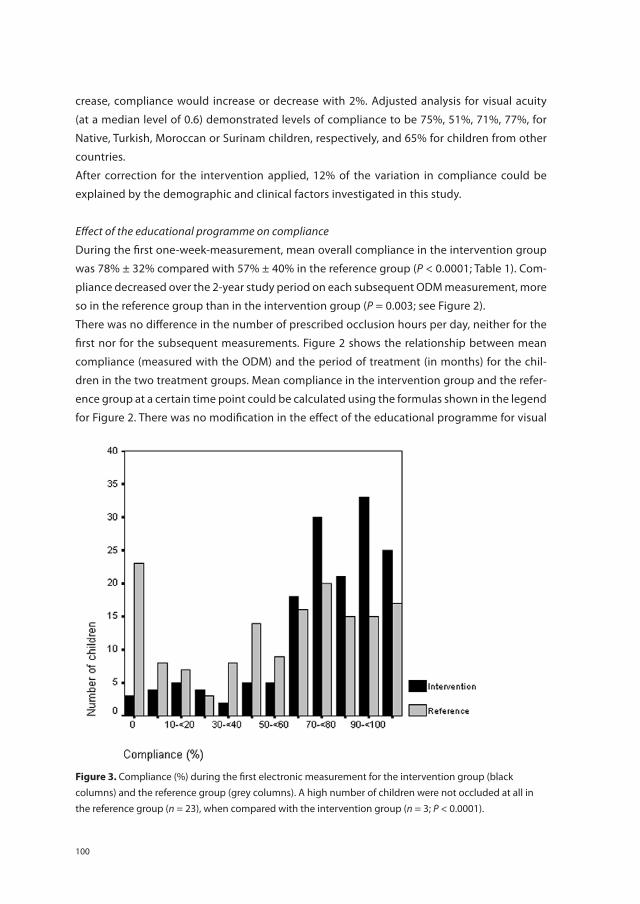
100
crease, compliance would increase or decrease with 2%. Adjusted analysis for visual acuity
(at a median level of 0.6) demonstrated levels of compliance to be 75%, 51%, 71%, 77%, for
Native, Turkish, Moroccan or Surinam children, respectively, and 65% for children from other
countries.
After correction for the intervention applied, 12% of the variation in compliance could be
explained by the demographic and clinical factors investigated in this study.
Effect of the educational programme on compliance
During the first oneweekmeasurement, mean overall compliance in the intervention group
was 78% ± 32% compared with 57% ± 40% in the reference group (P < 0.0001; Table 1). Com
pliance decreased over the 2year study period on each subsequent ODM measurement, more
so in the reference group than in the intervention group (P = 0.003; see Figure 2).
There was no difference in the number of prescribed occlusion hours per day, neither for the
first nor for the subsequent measurements. Figure 2 shows the relationship between mean
compliance (measured with the ODM) and the period of treatment (in months) for the chil
dren in the two treatment groups. Mean compliance in the intervention group and the refer
ence group at a certain time point could be calculated using the formulas shown in the legend
for Figure 2. There was no modification in the effect of the educational programme for visual
Chapter 7
Figure 3. Compliance (%) during the first electronic measurement in the intervention group (■) and the
reference group (□). A high number of children did not use occlusion at all in the reference group (n =
23), when compared with the intervention group (n = 3; P < 0.0001).
Figure 3. Compliance (%) during the first electronic measurement for the intervention group (black columns) and the reference group (grey columns). A high number of children were not occluded at all in the reference group (n = 23), when compared with the intervention group (n = 3; P < 0.0001).

101
chap
ter 7
Predictors and a remedy for noncompliance with amblyopia therapy
acuity of the child (P = 0.59), country of origin (P = 0.57) or fluency in the national language
(P = 0.48).
There was considerable variation in the level of compliance between the children and within
each child, as indicated by the ODM measurements. The SD of mean compliance during the
first oneweekmeasurement of all children (n = 310) was 37%. The mean of all the standard
deviations of mean compliance in each child during that week was 28%.
In the intervention group, three children (2%) were not occluded at all during the first ODM
measurement (mean age, 4.3 ± 0.8) compared with 23 (15%; mean age 4.6 ± 2.0) in the refer
ence group (P < 0.0001, χ2; Figure 3). Children who were not occluded at all and children who
did were comparable for clinical parameters. However, there were differences in the level of
parental education, country of origin and fluency in the national language.
The questionnaire designed to estimate the usage of the educational programme or the pic
ture to colour showed that the cartoon story was used by 87% of the families. Mean time was
25 minutes per week (range, 5180; number of observations, 123). The calendar and stickers
were used by 80% of the families. Mean time was 10 minutes (range, 215; number of observa
tions, 123). The information sheet was read by 67% of the parents. Mean time was 6 minutes
(range, 210; number of observations, 123).
In the reference group the colouring picture was coloured by 71% of the children. Mean time
was 12 minutes (range, 520; number of observations, 131).
In 28 patients, the researchers recorded time spent on explaining the diagnosis and treatment
during the first visit to the orthoptist. It averaged 234 seconds in nonnative patients, and 416
seconds in native patients. During followup visits to the orthoptist these averages were 116
seconds and 233 seconds, respectively (62 patients).
dIScuSSIon
This study identified the following demographic parameters as primary predictors for non
compliance: parental fluency in the national language, level of education and the country
of origin. Although country of origin was selected in the multivariate model to be most sig
nificant, we cannot exclude the possibility that fluency in the national language and level of
education (being significantly correlated with the country of origin) may be the causal factors.
The most important clinical parameter was the initial visual acuity of the amblyopic eye. The
educational programme featuring the cartoon story that explained without words the ratio
nale for treatment to the child significantly improved compliance throughout the study, limit

102
ing in particular the number of children who were not occluded at all (3 in the intervention
group vs. 23 in the reference group). The electronic monitoring performed by the researchers,
who were working independently from the treating orthoptists as they distributed the ODMs
during home visits, enabled us to detect these striking differences between the two treatment
groups.
The study was designed as a singleblind, randomised clinical trial. The treating orthoptists
were unaware of randomization, but the researcher distributing the educational programme
or the picture to colour was not. Efforts were made to ensure that time spent explaining the
study was equal in both groups. Randomization took place after the first visit to the orthoptist.
Main results of this study were based on the data obtained from the first ODM measurement,
thereby excluding any possible biases of a treating orthoptist later on in the study.
As compliance was measured in the week after the home visit, it would be reasonable to ex
pect that compliance would be higher in the week it was monitored, and we cannot exclude
that the ODM itself acted as an intervention. This bias applied to children in both groups. The
study found that compliance was moderate despite the fact that parents knew that compli
ance was being monitored. This finding was also made in one of the previous pilot studies, in
which compliance was monitored for longer periods (Simonsz, et al. 2001). For this reason, it
was decided that compliance would be measured on a regular basis: 1 week every 3 months,
also making the study more feasible for both parents and the researchers. Mean compliance
in our reference group during the measurements was similar to that reported by the MOTAS
(Monitored Occlusion Treatment of Amblyopia Study) Cooperative, who objectively moni
tored occlusion for a longer consecutive period (Stewart, et al. 2004).
Visual acuity at the start of treatment was the clinical parameter most significantly correlated
with compliance. This finding is in agreement with other studies (Flynn and Cassady 1978;
Lithander and Sjöstrand 1991; Smith, et al. 1995; Stewart, et al. 2004; 2005) and is explained
by the fact that the acceptance of the patch is less when acuity is low.
The decrease in compliance during treatment, as found in our study, is partly due to a selec
tion bias: children with low visual acuity at the start of treatment were less compliant, there
fore wore the patches for a longer period, and consequently were recorded more often. The
influence of treatment age on compliance has been a point of debate (Flynn and Cassady
1978; Stewart, et al. 2004; 2005); our study found the treatment age to be borderline signifi
cant, with younger children tending to have better compliance.
Having established this large study group of more than 300 children, the research group in
tends to proceed to an analysis of their visual acuity outcome. For this, the children’s final

103
chap
ter 7
Predictors and a remedy for noncompliance with amblyopia therapy
acuity will be assessed in a standardized fashion by the research orthoptist after the children
have completed their patching treatment.
Apparently, native parents received more lengthy explanations at diagnosis and treatment
than nonnative parents did. Several factors may have contributed to this observation (e.g.,
cultural background, language skills, confidence, assertiveness, education, interests). Also, in
The Netherlands, the time an orthoptist can spend on a patient varies and is sometimes lim
ited to 15 minutes for a new patient.
Although occlusion therapy for amblyopia has been the primary treatment for centuries
(Loudon, et al. 2005; von Noorden and Campos 2002) and clinically is an effective treatment,
its success is limited, by, among other factors, noncompliance (Awan, et al. 2005; Dorey, et
al. 2001; Loudon, et al. 2004; Stewart, et al. 2004; 2005). In this study, we confirmed, using
electronic monitoring, that lack of understanding of the disease and treatment in pediatric
medicine are obstacles that can be remedied by an educational programme: the cartoon
story, reward stickers and an information sheet. Important demographic predictors for low
compliance included poor parental fluency in the national language, country of origin and
a low level of education. Education, primarily aimed at the child, improved compliance and
reduced the number of children who did not comply with occlusion at all.
AcknowLEdGmEnTS
The authors thank Bregje van Dijk, Ellen van Minderhout, Marleen Vermeulen, Roberta Delle
Site, Janine van Zon, Eline Deelstra, Jorrien Bouma, Maurits Joosse, Hans Büller, Larisa Pepler,
Yaroslava Chopovska, Iris Bachert, Angelika Cordey, Regine Neukam, Heike Bartz, Birgit Her
rmann, Marlies Murtro, Dr. Marc Lüchtenberg, Prof. Alina Zubcov, Dr. Claudia Kuhli, Dr. Richard
Harrad, Dr. David Newsham and Prof. Irene Gottlob for their advice, help with data collection
and efforts in recruiting the children.
The Occlusion Dose Monitor was developed at the department for Medical Technical Develop
ment at the Academical Medical Center, Amsterdam, the Netherlands in 19961997 as a public
domain project.


chap
ter 1
chap
ter 2
chap
ter 3
chap
ter 4
chap
ter 5
chap
ter 6
chap
ter 7
chap
ter 8
chap
ter 9
chap
ter 1
0ch
apte
r 11
Add
enda
Chapter 8
The influence of parental attitudes and behaviour on compliance with amblyopia therapy and the effect of an educational
programme

106
ABSTrAcT
Purpose: In the context of a randomised clinical trial, in which the effect of an educational
programme on compliance with occlusion therapy was determined, this study focussed on
the psychological reasons for failure to comply after 6 months of therapy.
methods: In 310 children with newly diagnosed amblyopia compliance was measured elec
tronically during one week, every three months. Children were randomly allocated to receive
an educational programme (intervention group), or a picture to colour (reference group). Af
ter 69 months of treatment, during the third measurement, parents completed a 72item
questionnaire based on the Protection Motivation Theory to assess their beliefs about am
blyopia and occlusion treatment. Parents with compliance less than 20%, received a separate
semistructured interview with indepth questions that was based on differences in answers
from the 72item questionnaire between the group with compliance <20% and the compliant
group. The measures were distributed via homevisits by researchers.
results: 150 parents had reached the third measurement and 149 completed the question
naire. A high degree of distress was most predictive for poor compliance (P < 0.001), followed
by increased vulnerability (P = 0.014), increased stigma (P = 0.017) and poor logistics of treat
ment (P = 0.044). Twentyeight (64%) parents who patched less than 20% cooperated with
a semistructured interview. This showed lack of knowledge, family distress and logistical
problems to be the primary reasons for noncompliance. The educational programme posi
tively influenced the level of distress (P = 0.038), logistics of treatment (P = 0.016) and parental
knowledge (P = 0.015).
conclusions: Distress, poor parental knowledge and difficulties implementing the treatment
into the daily routine seem to be the predominant causes for noncompliance with occlusion
therapy for amblyopia, part of which were obviated by the educational programme.

107
chap
ter 8
The influence of parental attitudes and behaviour on compliance
InTroducTIon
Amblyopia is the most frequent cause of visual deficit in childhood (Attebo, et al. 1998; Brown,
et al. 2000). The condition is usually unilateral and caused by strabismus, anisometropia and/
or visual deprivation. It is commonly treated with patching of the fellow eye preferably before
the age of six. Adults with insufficiently treated amblyopia are at an increased risk of bilateral
visual impairment due to loss of vision in the fellow eye (trauma or other eye diseases), and
a decreased quality of life in adulthood (Chua, et al. 2004; Fronius, et al. 2005; van de Graaf,
et al. 2004; Rahi, et al. 2002). The factor most frequently quoted to influence the visual acuity
is degree of compliance (Cobb, et al. 2002; Dorey, et al. 2001; Lithander and Sjöstrand 1991;
Loudon, et al. 2003; Simmers, et al. 1999; Simons and Preslan 1999; Smith, et al. 1995; Stewart,
et al. 2004; 2005). The development of the Occlusion Dose Monitor (ODM) in 1991 enabled
objective monitoring of compliance (Fielder, et al. 1994) and it was found that the degree of
compliance averaged 5060% (Loudon, et al. 2006; Simonsz, et al. 1999; Stewart, et al. 2004;
2005), which is also found in studies that investigated compliance with prescriptions of medi
cine (Wright 1993). Since it has been shown that treatment is effective (Awan, et al. 2005;
Clarke, et al. 2003; PEDIG 2002; 2003a; 2003b; Stewart, et al. 2004; 2005), efforts are underway
to identify determinants of poor compliance and ways to improve it (DixonWoods, et al. 2006;
Goransson, et al. 1998; Loudon, et al. 2006; Newsham 2002; Smith, et al. 1995; Tripathi, et
al. 2002). The emotional impact of occlusion therapy (DixonWoods, et al. 2006; Hrisos, et al.
2004; Packwood, et al. 1999; PEDIG 2003) and poor parental understanding (Newsham 2002)
were factors that affect parental implementation of occlusion therapy.
A model employed by Searle et al. based on Protection Motivation Theory (PMT) proved to
be useful to understand psychosocial factors involved with noncompliance with occlusion
therapy (Norman, et al. 2003; Searle, et al. 2000; 2002). PMT is based on two behavioural adap
tations that occur when an individual faces a health threat. The first is threat appraisal, which
focuses on the source of the threat: the individual’s perception of the severity and vulnerabil
ity to the threat. The second is coping appraisal, which evaluates one’s ability to address the
threatened danger: that the individual is able to successfully perform an action (selfefficacy)
and that this action is effective (response efficacy), both increasing the probability of an adap
tive response (compliance with treatment). Both appraisal pathways contribute to protective
motivation, followed by a decision to take action or not: protective behaviour. PMT has pro
vided a coherent framework for exploring the impact and determinants of patching and it was
concluded that PMT was predictive of compliance intentions and behaviour (Rogers 1975).
In a large randomised clinical trial, the ERPAG study, we previously found that poor parental
fluency in the national language, a low level of education and poor acuity at start of treat
ment were predictors for low compliance, and, also that the educational programme, which

108
explained the reasons for therapy to the child, was very effective in improving compliance and
reducing the number of children who were not occluded at all (Loudon, et al. 2006).
Herein, we provide reasons for failure or success to comply with occlusion therapy evaluated
after 69 months of receiving treatment, using a questionnaire based on PMT with additional
domains on knowledge and logistics of treatment, and give further insight into circumstances
leading to noncompliance. In addition, the effect of the educational programme on the rea
sons for failure to comply with therapy was investigated.
mEThodS
The design of this prospective randomised clinical trial and CONSORT statement has been
reported in detail in a prior publication (Loudon, et al. 2006) and is summarised as follows:
between July 2001 and December 2003, children were recruited from four clinics by six treat
ing orthoptists in The Hague; additional children were recruited in Frankfurt (Germany). All
children with newly diagnosed amblyopia, with an interocular difference in visual acuity of at
least 0.2 logMAR, strabismus and/or anisometropia (>1.0 Dioptres (D)), or a deprivation in the
absence of additional ocular or neurological diseases, were eligible. The severity of amblyopia
was expressed as the ratio (in decimals) between the acuity in the amblyopic eye and the
better eye and expressed as decimal scores. Compliance was measured electronically in all
children during an entire week, every three months, using the Occlusion Dose Monitor (ODM)
(Chopovska, et al. 2005; Fronius, et al. 2006).
Included children were randomised to either the intervention group or the reference group.
Children in the intervention group received the educational programme: a cartoon story
without text, depicting the orthoptic examination of a preschool child, subsequent patch
ing therapy and the reasons for therapy seen from the perspective of the child, designed to
improve compliance. Folded in the back of the cartoon story was a sheet containing general
information about amblyopia, its treatment and certain guidelines for the parents, available
in eight languages. Children in the reference group received a picture to colour (e.g. Winnie
the Pooh) that was also considered a reward, but did not contain the educational message.
All parents were visited at their home by the researchers to explain nature and possible con
sequences of the study and to distribute the ODM with either the educational programme or
the picture to colour. All children received standard orthoptic care. Occlusion was prescribed
according to a standardised protocol developed for this study and representative of current
orthoptic practice (Loudon, et al. 2006). The family’s socioeconomic status was ascertained
using a 23item questionnaire.
The Ethical Committee of Erasmus University Rotterdam and the boards of the participating
clinics approved the protocol and informed consent forms. Written informed consent by the

109
chap
ter 8
The influence of parental attitudes and behaviour on compliance
parents or guardian was a prerequisite for participation. The research adhered to the tenets of
the Declaration of Helsinki.
Patching Success questionnaire
To identify reasons for failure to comply with occlusion therapy, an inventory was made using
a 72item questionnaire (‘Patching Success Questionnaire’ – PSQ), which was based on com
ponents of the PMT, 69 months after the start of treatment, during the third homevisit.
The PMT items were scored on a 5point response domain (strongly disagree/strongly agree).
Reliable measures were identified through factor analyses and internal reliability analyses:
Cronbach’s α (Norman, et al. 2003; Searle, et al. 2000; 2002). Measures of the variables were
constructed by averaging responses to individual items such that high scores indicated high
degrees on the variable of interest. Protection Motivation was assessed using three items
(α=0.86). Perceived severity of the disease was assessed using six items (α=0.89). Perceived
vulnerability was assessed using six items (α=0.86). Response efficacy was assessed using five
items (α=0.83). Selfefficacy was assessed using four items (α=0.88). The perceived distress ex
perienced by parents when patching their child was assessed using eight items (α=0.90), the
extent to which patching may interfere or prohibit the child from engaging in every day ac
tivities was assessed using five items (α=0.62) and the stigma attached to wearing a patch was
assessed using five items (α=0.78). In addition to the PMT items, information was collected on
parental knowledge of disease (3 items, all on a 5point scale; e.g. ‘In general, a lazy eye is an
eye that sees less clearly than the other’) and treatment (2 items on a 4point scale; e.g. ‘The
aim of patching therapy would be: equal vision sufficient vision to read a book decrease
in the squint not to wear glasses later in life’) and logistic of treatment (4 items on a 5point
scale; e.g. ‘I find it difficult fitting patching into my daily routine’). The questionnaire also con
tained 10 questions about the families’ experience with the eye patch (e.g. ‘after removing the
eye patch my child’s skin is red and/or painful’).
Identification of non-compliers
To define the group of noncompliers, data of the first oneweek ODM measurement were
analysed. A cutoff point at compliance of 20% was chosen, which included not only the non
patchers, but also the lowfrequencypatchers (i.e. the eye was occluded less than twice a
week). As expected, a strong association between compliance and frequency of patching was
found (Figure 1a & 1b): when compliance was less than 20%, the frequency of patching was
less than three times a week (sensitivity 0.84, specificity 1.00).
Parents from the noncompliant group were contacted by phone and an appointment for
an interview during a separate home visit was made. In case of poor fluency in the national
language by the parents, an interpreter was present.

110
Statistical analysis
Descriptive statistics and crosstabs were used to calculate the means, standard deviations and
intercorrelations of each domain of the questionnaire. These results were then compared to
the results of the questionnaire reported by Norman et al. (2003). Linear regression analysis
was used to predict protection motivation and compliance (both as dependent, continuous
variables). The independent variables were: severity, vulnerability, response efficacy, distress,
prohibition, stigma, selfefficacy, knowledge, logistics and protection motivation (scored as
continuous variables).
The nonparametric MannWhitney test was used to determine the effect of the educational
programme on the domains of the ‘Patching Success Questionnaire’. Fvalues and βvalues
were calculated. The Fvalue is a test statistic that tests for differences in the variance between
two groups; F=1 indicates no difference in variance between two groups. The percentage of
the variation in compliance and motivation to patch their child that could be ascribed to the
different factors involved was defined as the ratio of the percentage of the variance explained
by the model with, and the model without the factor in question. Analysis was both Univariate
and Multivariate. Descriptive statistics were applied to analyse the completed semistructured
interviews. P < 0.05 was considered statistically significant.
Figuren bij Chapter 8.
1a 1b
Figure 1a. A scatter plot displaying the relationship between the frequency of patching per week and
electronically measured compliance during the first ODM measurement, the black line indicating the
chosen cut-off point of 20% compliance. 1b. A histogram presenting compliance during the first ODM
measurement, with 10% intervals.
Figure 2. Flowchart illustrating the study design. After the first one-week measurement, children who
patched less than 20% were identified as non-compliant. Between November 2004 and March 2005,
the non-compliant group received a separate semi-structured interview with more in-depth questions
Figure 1a. A scatter plot displaying the relationship between the frequency of patching per week and electronically measured compliance during the first ODM measurement, the black line indicating the chosen cutoff point of 20% compliance. 1b. A histogram presenting compliance during the first ODM measurement, with 10% intervals.
1a 1b

111
chap
ter 8
The influence of parental attitudes and behaviour on compliance
rESuLTS
The study design was prospective, recruiting children for a period of 30 months. On average
a child received three electronic measurements during, on average, 8 months of treatment.
Therefore, not all children had reached the moment of the third measurement, the moment
the questionnaire was distributed. After 69 months of treatment, the 150 patients who re
ceived the third measurement were asked to complete the questionnaire, 75 in the interven
tion group and 75 in the reference group (Figure 2). The PSQ was completed for 149 parents
during the third homevisit. Mean age was 4.6 years (SD 1.9) and 53% were boys; the clinical
and demographic characteristics were similar to those reported for the full study cohort (n =
310) (Loudon, et al. 2006).
Figuren bij Chapter 8.
1a 1b
Figure 1a. A scatter plot displaying the relationship between the frequency of patching per week and
electronically measured compliance during the first ODM measurement, the black line indicating the
chosen cut-off point of 20% compliance. 1b. A histogram presenting compliance during the first ODM
measurement, with 10% intervals.
Figure 2. Flowchart illustrating the study design. After the first one-week measurement, children who
patched less than 20% were identified as non-compliant. Between November 2004 and March 2005,
the non-compliant group received a separate semi-structured interview with more in-depth questions
Figure 2. Flowchart illustrating the study design. After the first oneweek measurement, children who patched less than 20% were identified as noncompliant. Between November 2004 and March 2005, the noncompliant group received a separate semistructured interview with more indepth questions that was based on differences in answers from the Patching Success Questionnaire between the compliant and the noncompliant group.

112
Patching Success questionnaire
The mean scores and standard deviations per PMT domain as observed in this study differed
from the scores found by Norman et al., for the domains: protection motivation, severity, vul
nerability, degree of distress and stigma barrier (Table 1). The intercorrelations between the
domains of the PMT, however, were comparable to those reported by Norman et al. (2003).
Stepwise backward selection demonstrated that the motivation to patch their child (PM) was
high when selfefficacy (β = 0.51; P < 0.001), response efficacy (β = 0.30; P < 0.001) and severity
(β = 0.08; P = 0.048) were also high, all three domains explaining 54% of the variation in pro
tection motivation (adj. R2). Stepwise backward selection showed that compliance increased
when parents were well capable of implementing the occlusion therapy into their daily rou
tine (logistics; β = 6.1; P = 0.044) and when they replied that their child’s eye sight would
get worse if left untreated (β = 9.3; P = 0.014). Compliance decreased when the two barriers
distress and stigma increased (β = 22; P < 0.001 and β = 6.0; P = 0.017, respectively). The four
domains explained 22% of the variation in compliance (adj. R2).
Visual acuity was significantly and negatively correlated with the domains severity (r = 0.37;
P < 0.001) and the degree of distress (r = 0.17; P = 0.007). Age of the children and cause of
amblyopia were not correlated with any of the domains, neither was gender.
Poor fluency in the national language was positively related to stigma (r = 0.37; P < 0.001), a
higher degree of distress (r = 0.45; P < 0.001) and severity (r = 0.5; P < 0.001); and negatively
related to knowledge (r = 0.22; P = 0.009) and selfefficacy (r = 0.18; P = 0.03). Level of educa
tion was positively correlated with knowledge (r = 0.20; P = 0.018); and negatively correlated
with the domains severity (r = 0.23; P = 0.005); degree of distress (r = 0.29; P < 0.001); and
stigma (r = 0.21; P = 0.009).
The clinical and socioeconomic variables studied, explained 11% of the variance in protection
motivation and 12% of the variation in compliance.
relationship between Patching Success questionnaire and compliance, and the effect of the educational programme
Univariate analysis of the variance demonstrated that an increased degree of distress caused
by occlusion therapy negatively infl uenced compliance. However, a clear effect of the educa
tional programme was found (F = 4.15; P = 0.038), in a sense that children and their parents
who received the programme suffered less distress than the reference group (Table 2).
Compliance increased when parents found it easier to implement occlusion therapy into their
daily routine (logistics). The implementation was significantly better in parents who had re
ceived the educational programme (F = 5.97; P = 0.016). Parental scores on the domain ‘knowl
edge about treatment’ were significantly higher when they had received the educational pro

113
chap
ter 8
The influence of parental attitudes and behaviour on compliance
gramme (F = 6.1; P = 0.015). This, however, did not affect compliance. Compliance increased
significantly when parents were highly motivated to patch their child, when they thought
their child’s eye sight would be vulnerable when left untreated, when parents were able to
adhere to the prescription given by the orthoptist, and decreased when their child was pro
hibited in his activities by the patch. The educational programme showed no demonstrable
effect on these domains.
Group of non-compliers
After the first oneweek ODM measurement 44 children had electronically measured compli
ance below 20% (8 intervention group; 36 reference group), of whom 33 could be contacted,
while 11 (25%) could not be contacted with the available address or telephone numbers. 28
(64%) were interviewed at their home (5 intervention group; 23 reference group), 5 (11%)
would no longer cooperate. The interviews were undertaken by two interviewers (LC and SdV)
who wrote extensive reports of each visit. The time spent on these homevisits was approxi
mately 30 minutes (range 2045 min).
The compliant and noncompliant group were compared concerning the clinical and socio
economic factors and the answers given to the ‘Patching Success Questionnaire’. The follow
ing significant differences using ANOVA were found: visual acuity at onset (F = 3.78; P = 0.049),
fluency in the national language (F = 6.64; P = 0.01), country of origin (F = 3.85; P = 0.047), per
ceived distress (F = 12.51; P = 0.001), selfefficacy (F = 9.32; P = 0.003), protection motivation (F
= 12.12; P = 0.001), logistics of treatment (F = 15.1; P = 0.001) and knowledge about amblyopia
and its treatment (F = 8.13; P = 0.005). The differences found were in favour of the compliant
group. These differences were used to develop a semistructured interview with 33 additional
focussed questions based on these domains, including 6 questions specifically for the child.
Since compliance was related to fluency in the national language and as communication is an
Table 1. Mean scores and standard deviation per domain for the study population in The Hague and Bristol; * Pvalue indicating a significant difference in mean scores.
Scales The Hague (n =149) Bristol (n =151) P value
PM 4.25 ± 0.68 4.5 ± 0.85 *<0.001
Severity 3.02 ± 0.93 3.57 ± 0.82 *<0.001
Vulnerability 3.2 ± 0.74 3.49 ± 1.00 *<0.001
Response efficacy 4.36 ± 0.5 4.33 ± 0.78 0.252
Distress barrier 2.19 ± 0.84 2.75 ± 1.04 *<0.001
Prohibit barrier 2.07 ± 0.95 1.94 ± 1.05 0.101
Stigma barrier 2.48 ± 0.83 2.85 ± 0.95 *0.001
Selfefficacy 4.04 ± 0.86 3.86 ± 1.2 0.767

114
important part in compliance (Loudon, et al. 2006), questions regarding communication and
relationship with the therapist in general were also included.
The semistructured nature of the interview made it possible to thoroughly question the par
ents about amblyopia and its treatment. The reports were objectified by scoring them for
gauging criteria done by the two researchers separately. These criteria were compiled through
analyses of the survey. Based on the literature (DiMatteo 2004) and the results of the question
naire, three main parties involved in the occlusion treatment could be distinguished: i.e. child,
carer and therapist. Conclusively, 12 gauging criteria were drawn up, 4 for each party involved
(Figure 3). These criteria refer to reasons for total noncompliance.
The analysis of the interviews was then conducted by scoring each report of the interview on
the abovementioned 12 criteria. This was done on a scale from 1 (not present) to 5 (domi
nantly present or behaviour defining). The two independent scores were analysed with the
weighed kappatest to test if the two scores differed significantly.
All parents interviewed were under the impression they had sufficient knowledge regarding
the treatment, however, objectively, only 14% had sufficient knowledge. Also, a high degree
of distress and logistical problems were prevalent. Parents did not regard occlusion therapy to
be an effective treatment. Interviewed children replied they were often teased by their peers
whilst wearing the patch (43%, of whom 29% suffered severe teasing), which decreased their
selfesteem. A third of the parents answered they had received incomplete information from
the treating orthoptist and that there was little time during the visit to discuss their problems.
18% of the parents thought that the orthoptist did not sufficiently address the child when
explaining the eye condition and its treatment.
The following significant correlations (using Pearson’s chisquare) were found: time and at
tention spent by therapists to problems regarding the occlusion therapy were correlated with
Table 2. β, F and Pvalues of a univariate analysis of the effect of the PSQ domains on compliance. * Indicates a significant influence.
Scales β F P value
PM 16.4 14.1 *<0.001
Severity 3.1 0.8 0.358
Vulnerability 10.9 6.9 *0.009
Response efficacy 5.7 0.6 0.364
Distress barrier 17 24.2 *<0.001
Prohibit barrier 8.4 6.8 *0.01
Stigma barrier 5.9 2.5 0.116
Selfefficacy 12.8 13.4 *<0.001
Knowledge disease 0 0 0.998
Knowledge treatment 2 0.4 0.546
Logistics of treatment 11.1 13.9 *<0.001

115
chap
ter 8
The influence of parental attitudes and behaviour on compliance
clarity of information, possibility to ask questions and childorientation of the visits. Parental
knowledge about amblyopia and its treatment and the perceived feeling of invasiveness of
therapy were correlated to response efficacy. Confidence in the response efficacy was corre
lated with time and attention paid by therapists to problems encountered during patching,
clarity of information given and possibility to ask questions.
dIScuSSIon
In the context of a prospective, randomised, clinical trial in which the effect of an educational
programme on compliance was investigated, this study provided reasons for failure to comply
with occlusion therapy after 69 months of treatment. We used a 72item questionnaire based
on the PMT components with added domains on knowledge and logistics of treatment and
separately interviewed parents who did not occlude at all. In addition, the effect of the educa
tional programme on the domains of the questionnaire was determined.
In concordance with Searle, et al.(2002) and Norman, et al.(2003) the degrees of response effi
cacy and protection motivation were high, indicating that most parents thought eye patching
to be an effective treatment for their child’s amblyopic eye and were motivated to patch their
child as prescribed by the orthoptist. More than 50% of the variation in protection motivation
could be ascribed to the following three domains: selfefficacy, response efficacy and sever
ity; thus parents’ motivation to patch their child increased when they thought patching to be
an effective treatment, when they were able to patch their child as recommended by the or
thoptist and when they thought their child’s eye condition to be severe. This finding was also
that was based on differences in answers from the Patching Success Questionnaire between the
compliant and the non-compliant group.
Figure 3. 12 criteria subdivided into the carer, the child and the therapist.
Figure 3. 12 criteria subdivided into the carer, the child and the therapist.

116
found in other studies which applied PMT in relation to compliance with therapy in amblyopia
and other conditions (Abraham, et al. 1994; Flynn, et al. 1995; Fruin, et al. 1991; Norman, et al.
2003; Rippertoe and Rogers 1987; Rudman, et al. 1999; Searle, et al. 2002; van der Velde and
van der Pligt 1991). As the parents in The Hague were given the same questionnaire as the
parents in Bristol, it would be interesting to compare their mean scores. For several domains
the parents in The Hague scored lower, probably explained by the difference in population
and country.
Univariate analysis demonstrated that compliance was influenced by protection motivation,
vulnerability, distress and prohibition barrier, selfefficacy and logistics of treatment. How
ever, when corrected for potentially confounding factors no effect of protection motivation
on compliance could be found: though parents may be motivated to patch their child, this
did not influence compliance. Compliance decreased most drastically when parents reported
a high degree of distress. This was also found in other studies (Hrisos, et al. 2004; Norman, et
al. 2003; Packwood, et al. 1999; PEDIG 2003; Parkes 2001; Searle, et al. 2002). However, fami
lies who were given the educational programme that explained the occlusion therapy to the
child, suffered less distress and had better compliance. The educational programme had a
similar positive effect on parental knowledge of treatment and logistics of treatment. This
finding is in agreement with Newsham’s (Newsham 2002). Somewhat surprisingly, knowledge
of the disease itself was not affected by the educational programme; perhaps it was still un
clear for the parents, or they found amblyopia itself not interesting or serious enough.
The age of the child, depth of amblyopia, gender and cause of amblyopia showed little rela
tionship with the overall scores. Poor visual acuity at treatment onset caused parents to con
sider the amblyopia as a serious eye condition and increased the degree of distress.
Parents who spoke the native language excellently, had better knowledge about the treat
ment, were confident in their ability to patch their child and were of the opinion that amblyo
pia was a serious eye condition; they also suffered less distress and stigma.
The questionnaire and interview were carried out on one occasion. It might have been in
teresting to, in a prospective design, also have distributed the questionnaire and conducted
the interview immediately after the first visit to the orthoptist for prediction and to compare
outcomes and any changes in opinions.
When analysing the group who did not occlude at all, it was apparent that there was a clear
discrepancy of subjective knowledge and the measured knowledge. This indicated that many
parents were ignorant of their lack of knowledge, therefore could not give feedback or put
forward any questions, thus leaving the orthoptist unaware of the parents’ problem. For a
small group of parents the orthoptist was felt as the main reason for failing to patch: when
there was no attention paid to problems encountered during patching, no possibility to ask
questions, insufficient or unclear information supplied, parents were most likely to become
steadfast noncompliers. A number of parents indicated that visits to the orthoptist should
be more childfocused. Children were poorly involved in the communication between parent

117
chap
ter 8
The influence of parental attitudes and behaviour on compliance
and orthoptist and consequently did not understand the reason they had to be patched. The
interviews were taken at their home, to overcome the problem of dropouts who no longer
visited the clinic. The interview was open framed, rather than with multiplechoice questions.
This open framework allowed a focused, conversational, twoway communication.
This study demonstrated that occlusion therapy for amblyopia is difficult to implement, is ac
companied by a high degree of distress and is hampered by poor knowledge and understand
ing by the parents and therapist. If knowledge and understanding is improved, this might di
minish the effect of the other determinants found in this study. The educational programme, a
cartoon story explaining without text the reasons for therapy to a 4year old and the informa
tion sheet for the parents, partly reduced the influence of these determinants and increased
the acceptance of occlusion therapy.
AcknowLEdGmEnTS
The authors thank Bregje van Dijk, Ellen van Minderhout, Marleen Vermeulen, Roberta Delle
Site, Janine van Zon, Eline Deelstra, Jorrien Bouma, Maurits Joosse, Hans Büller, Larisa Pepler,
Yaroslava Chopovska, Iris Bachert, Angelika Cordey, Regine Neukam, Heike Bartz, Birgit Her
rmann, Marlies Murtro, Dr. Marc Lüchtenberg, Prof. Alina Zubcov and Dr. Claudia Kuhli for their
advice, help with data collection and efforts in recruiting the children.
The Occlusion Dose Monitor was developed at the department for Medical Technical Develop
ment at the Academical Medical Center, Amsterdam, the Netherlands in 19961997 as a public
domain project.


chap
ter 1
chap
ter 2
chap
ter 3
chap
ter 4
chap
ter 5
chap
ter 6
chap
ter 7
chap
ter 8
chap
ter 9
chap
ter 1
0ch
apte
r 11
Add
enda
Chapter 9
Physiological and mechanical properties of the eye patch: influence on compliance and
parental satisfaction

120
ABSTrAcT
Purpose: To evaluate the physiological and mechanical properties of the eye patch and their
influence on compliance as part of the prospective randomised clinical trial identifying risk
factors for noncompliance with occlusion therapy for amblyopia.
methods: For a period of 30 months all children with newly diagnosed amblyopia in The
Hague were registered. Compliance was measured during 1 week every 3 months with the
Occlusion Dose Monitor (ODM) that was distributed during homevisits. All children received
standard orthoptic care by the treating orthoptist. After 6 months of treatment, parents com
pleted a questionnaire evaluating comfort, size and adhesion of the patch. In the study, four
brands of patches were used: Opticlude, Orthopad, ProOphta and Beiersdorf. On one occa
sion the following physiological and mechanical properties were measured: breathing capa
bility at 23°C (73°F) and 33°C (91°F), resistance to water penetration, opacity and strength of
adhesion to the skin.
results: 149 Parents completed the questionnaire, of whom 62% used Opticlude, 24% Ortho
pad, 11% ProOphta and 3% used Beiersdorf. The brand of patch prescribed was correlated to
the orthoptist only (P = 0.005). Compliance was neither related to the brand (P = 0.179), nor
to the use of different colours or designs of patches (P = 0.639). It was, however, related to the
use of stickers (P = 0.028). Parental satisfaction was moderate. Answers given by the parents
matched the measured physiological and mechanical properties of the eye patch. There were
large differences in the properties of the eye patch, especially in opacity and strength of adhe
sion to the skin. In all brands of patches the breathing capability was minimal.
conclusions: When prescribing a certain brand the orthoptist needs to consider the wide va
riety in physiological and mechanical properties of the patch. Further study into these proper
ties is necessary; especially water permeability and opacity of the eye patch require improve
ment since children often wear them for a longer period of time. This could contribute to
increasing parental satisfaction and consequently may improve compliance.

121
chap
ter 9
Physiological and mechanical properties of the eye patch
InTroducTIon
Amblyopia is the commonest visual defect in children with a prevalence of approximately
3.5% (Attebo, et al. 1998). The condition may be partially or completely treated preferably
before the age of 6. Traditionally, treatment involves occlusion of the nonamblyopic eye with
an adhesive patch applied directly to the skin around the eye, thereby forcing the use of the
amblyopic eye. Occlusion therapy has been the mainstay of treatment for centuries. However,
it frequently receded into the background when other types of treatment for amblyopia were
tried. In 1927 it was reintroduced by C.H. Sattler in Leipzig (Sattler 1927). His occluder, called
“Mastisolverband”, was attached to the skin around the eye with strong adhesive tape and
remained securely fastened for at least two or three days. Acceptance of this patch was poor
as it caused localised skin irritation in almost all children. Nowadays, a wide selection in brand
of patches is available, with patches varying in size, colour, elasticity, and gadgets or gifts
distributed by the manufacturers. Whether or not a patch remains glued to the skin depends
on a number of factors, e.g., skin type, the child’s activities while wearing the patch and ad
hesive strength of the patch. The eye patches are mostly made of nonwoven materials. Some
manufactures assert their brand of patch is hypoallergen, indicating special glue was used to
reduce the occurrence of allergies and itching. Nevertheless, it has been reported that parents
dislike the cosmetic appearance of their child wearing an eye patch. They argue that wearing
the eye patch is uncomfortable, causes irritation to the skin and leads to considerable distress
for the child, sometimes even outweighing any benefits from improvement in vision (Hsiros,
et al. 2004; Norman, et al. 2003; Packwood, et al. 1999; Parkes 2001; PEDIG 2003; Snowdon and
StewartBrown 1997). To date, no study has investigated the physiological and mechanical
properties of various brands of eye patches and their effect on compliance. Therefore, as part
of a randomised clinical trial designed to investigate a method to improve compliance and to
identify certain risk factors for noncompliance, this study investigated the properties of the
eye patches, focussed on the orthoptists’ choice in prescribing a particular brand, the correla
tion with compliance and parental satisfaction.
mEThodS
The design of this prospective trial has been reported in detail in a prior publication and is
summarised as follows (Loudon, et al. 2006): between July 2001 and December 2003, all chil
dren with newly diagnosed amblyopia in The Hague were recruited from four clinics by six
treating orthoptists; additional children were recruited in Frankfurt (Germany). Eligible chil
dren received a routine orthoptic examination and explanation of diagnosis and treatment
after which a standard examination form was completed and forwarded to the research cen
tre. The duration of occlusion (number of hours per day) for the first prescription had been

122
Tabl
e 1.
The
10
item
que
stio
nnai
re a
s gi
ven
to th
e pa
rent
s. Ea
ch it
em h
ad 5
resp
onse
cho
ices
, exc
ept f
or q
uest
ions
7 a
nd 9
, whi
ch h
ad 2
resp
onse
cho
ices
. n.a
.= n
ot
appl
icab
le: a
t the
tim
e of
the
stud
y Pr
oO
phta
and
Bei
ersd
orf d
id n
ot h
ave
colo
ured
pat
ches
ava
ilabl
e. *
The
Pv
alue
indi
cate
s a
sign
ifica
nt d
iffer
ence
in a
nsw
ers.
Ques
tions
:Op
ticlu
de (N
=92)
Orth
opad
(N=3
5)Pr
oOp
hta (
N=17
)Be
iers
dorf
(N=5
)P
valu
e *
N (%
)N
(%)
N (%
)N
(%)
The
patc
h on
my c
hild
’s ey
e is
0.44
muc
h to
o bi
g3
(3)
01
(6)
0
too
big
1 (1
)2
(6)
2 (1
2)0
abou
t rig
ht87
(95)
33 (3
4)13
(76)
5 (1
00)
too
smal
l1
(1)
01
(6)
0
muc
h to
o sm
all
00
00
The
patc
h st
ays o
n m
y chi
ld’s
eye
*0.0
27
stro
ngly
agre
e19
(21)
5 (1
4)3
(18)
3 (6
0)
agre
e so
mew
hat
55 (6
0)23
(66)
9 (5
3)2
(40)
neith
er ag
ree
or d
isagr
ee12
(13)
5 (1
4)2
(12)
0
disa
gree
som
ewha
t5
(5)
2 (6
)2
(12)
0
stro
ngly
disa
gree
1 (1
)0
1 (6
)0
The
patc
h st
icks t
o m
y chi
ld’s
skin
0.68
5
muc
h to
o w
ell
4 (4
)3
(9)
1 (6
)1
(20)
too
wel
l25
(27)
5 (1
4)4
(23)
3 (6
0)
abou
t rig
ht59
(65)
26 (7
4)9
(53)
1 (2
0)
not s
trong
ly en
ough
4 (4
)1
(3)
2 (1
2)0
not a
t all
00
1 (6
)0
Afte
r rem
ovin
g th
e pa
tch
my c
hild
’s sk
in is
red
and/
or p
ainf
ul0.
162
neve
r17
(18.
5)11
(31)
8 (4
6)0
occa
siona
lly23
(25)
10 (2
9)3
(18)
0
som
etim
es34
(37)
8 (2
3)3
(18)
3 (6
0)
quite
ofte
n13
(14)
4 (1
1)3
(18)
1 (2
0)
alm
ost a
lway
s5
(5.5
)2
(6)
01
(20)

123
chap
ter 9
Physiological and mechanical properties of the eye patch
To w
hat e
xten
d di
d th
e pa
tch
rem
ain
on th
e ey
e0.
216
The
patc
h re
mai
ned
on th
e ey
e72
(78)
28 (8
0)10
(59)
3 (6
0)
The
ends
of t
he p
atch
hav
e be
com
e lo
ose
18 (2
0)5
(14)
6 (3
5)2
(40)
The
patc
h re
mai
ned
on th
e ey
e on
ly ju
st2
(2)
2 (6
)1
(6)
0
The
patc
h fe
ll off
00
00
I was
invo
lved
in th
e de
cisio
n ab
out t
he ty
pe o
f pat
ch0.
058
stro
ngly
agre
e8
(9)
3 (9
)1
(6)
1 (2
0)
agre
e so
mew
hat
18 (2
0)13
(36.
5)3
(18)
1 (2
0)
neith
er ag
ree
or d
isagr
ee22
(24)
13 (3
6.5)
6 (3
5)3
(60)
disa
gree
som
ewha
t29
(31)
3 (9
)5
(29)
0
stro
ngly
disa
gree
15 (1
6)3
(9)
2 (1
2)0
Wha
t col
our p
atch
did
you
use
skin
colo
ured
, ski
p ne
xt q
uest
ion
42 (4
6)11
(31)
17 (1
00)
5 (1
00)
colo
ured
pat
ches
50 (5
4)24
(69)
00
Beca
use
of th
e di
ffere
nt co
lour
s, pa
tchi
ng m
y chi
ld b
ecam
e ea
sier
stro
ngly
agre
e9
(17)
5 (2
1)n.
a.n.
a.
agre
e so
mew
hat
28 (5
2)14
(58)
neith
er ag
ree
or d
isagr
ee10
(5)
2 (8
)
disa
gree
som
ewha
t3
(6)
3 (1
3)
stro
ngly
disa
gree
00
Did
you
use
pict
ures
and/
or st
icker
s whe
n pa
tchi
ng yo
ur ch
ild0.
319
no, s
kip
next
que
stio
n26
(28)
15 (4
3)5
(29)
2 (4
0)
yes
66 (7
2)20
(57)
12 (7
1)3
(60)
Beca
use
of th
e pi
ctur
es an
d/or
stick
ers,
patc
hing
my c
hild
bec
ame
easie
r0.
733
stro
ngly
agre
e13
(20)
5 (2
5)5
(42)
1 (2
0)
agre
e so
mew
hat
31 (4
7)9
(45)
4 (3
3)3
(60)
neith
er ag
ree
or d
isagr
ee18
(27)
2 (1
0)2
(17)
1 (2
0)
disa
gree
som
ewha
t3
(5)
4 (2
0)1
(8)
0
stro
ngly
disa
gree
1 (1
)0
00

124
standardised and prescribed according to the protocol representative of current orthoptic
practice (i.e., 6.63 * ratio acuity amblyopic eye/acuity better eye + 0.5 * age (years) + 4.97).
After the first visit to the orthoptist, parents were contacted by the independent researchers
who explained the nature and possible consequences of participating in the study and use
of the Occlusion Dose Monitor (ODM) (Fielder, et al. 1994) during a homevisit. Full written in
formed consent was obtained. Compliance was measured with the ODM in all children during
a period of 1 week, every 3 months (Chopovska, et al. 2005; Fronius, et al. 2006).
In the Netherlands four brands of patches are available: 3M OpticludeTM, Orthopad Masteraid®
(Regular and Simpathy), ProOphta® or Beiersdorf® (Elastopad and CoverletS), all of which
had also been prescribed in the study. The orthoptists were free in their choice to decide what
brand of patch they prescribed. After completing the study, in December 2005, the treating
orthoptists received a semistructured interview by the researcher. During the open part they
were asked to motivate their choice for prescribing a particular brand of patch. During the
structured part they were asked if their choice was influenced by the age of the children, the
number of prescribed occlusion hours, their idea of compliance with treatment, and by gad
gets or gifts distributed by the manufacturers. In addition, the pharmacies in The Hague were
contacted by telephone to document if all patches were in stock, and if not, which brand they
did have in stock.
questionnaire
Parental experience with the eye patch was evaluated via a 10item questionnaire; each item
had 4 or 5 response choices (Table 1, lefthand column). The questions were developed to
gether with the Consumers’ Organisation, the Netherlands who published a report on all
available brands of plaster (July 2001) (Consumers’ Organisation the Netherlands 2001). The
parents completed this questionnaire during the third homevisit after approximately 69
months of treatment.
Physiological and mechanical properties of the patch
Firstly, breathing capability was tested at 23ºC (73ºF) and 33ºC (91ºF): the patches were glued
on top of small plastic pots, each containing 20 grams of water. The pots were completely
sealed off by the patch; the only way of ventilation was through the patch. They were left on
a rocking table in a stove. The various patches were tested simultaneously at a temperature of
23ºC (73ºF) and a humidity of 30%. The same procedure was followed at a temperature of 33ºC
(91ºF) and a humidity of 22%. To make sure these conditions were constant; a separate digital
thermometer was used to measure the temperature and humidity. After 24 hours the amount
of water left in the plastic pot was measured. The breathing capability, or ‘water vapour re
sistance’ (Ret), was calculated using: m² * Pa / W (m = π * r² * time (seconds); Pa = saturated

125
chap
ter 9
Physiological and mechanical properties of the eye patch
(water) vapour pressure, depending on temperature and humidity; W=difference in amount
of water (grams) after a certain time, with 1 gram of water equalling 2430 Joule). The Ret is
classified as follows: an Ret > 40: ‘uncomfortable’, e.g. raincoat and has a restricted wearing
time; 20 < Ret ≤ 40: ‘somewhat comfortable’; or Ret ≤ 20, which equals ‘comfortable’.
Secondly, the resistance to water penetration was tested: 1 drop of water was placed on the
centre of the patch and time necessary for the drop to be absorbed was measured with a
stopwatch. Material is labelled waterresistant when the drop of water is not absorbed within
1 minute.
Thirdly, the opacity of the patches was tested. All patches were glued to a fluorescent lamp of
18 Watt. We measured the amount of light transmitted through the centre and at the side of
the patch. 100% light transmission equals ‘no patch present’.
Finally, the strength of adhesion of the patch to the skin was tested using the ‘maximum force
grab method’. The patches were stuck to the skin and the force necessary to remove the patch
was measured (expressed in Newton).
The breathing capability was tested at the laboratory of the ErasmusMC University Medical
Center Rotterdam; the other tests were performed at the Netherlands Organisation for Ap
plied Scientific Research (TNO), Textile Industry Enschede. All tests were performed by the
researcher (SL) under supervision of coauthor AW.
In addition to the tests, the elasticity of the eye patches, hygiene, available sizes and distrib
uted gifts per brand, was noted.
Statistical analysis
Chisquare test (Pearson Chisquare in crosstabs) was used to determine the influence of age,
gender, number of prescribed occlusion hours, cause of amblyopia and socioeconomic status
on the orthoptists’ choice in prescribing a brand of patch.
Compliance was measured during 1 week, every 3 months. It was defined as the actual occlu
sion time measured with the ODM divided by the prescribed occlusion time and expressed
as a percentage. Differences in compliance between the four groups of patches was assessed
using leastsquares regression analysis, with compliance as the dependent (continuous) vari
able and the brand of patch as the independent (categorical) variable, using data obtained
during the first ODM measurement.
To calculate differences in answers to the questionnaire between the four groups, the answers
were first transformed into rank numbers and then imported in the linear regression analysis
with the rank number as the dependent variable and the brand of patch as the independent
variable.
Whether the answers influenced compliance was also assessed with linear regression analysis.
P < 0.05 indicated statistical significance.

126
rESuLTS
In the randomised clinical trial, 310 children were eligible for analysis, mean age was 4.6 years
(SD 2.0), and 56% were boys. At the start of the study 165 children used Opticlude (53.2%),
100 children used Orthopad (32.3%), 26 children used ProOphta (8.5%), and 19 children used
Beiersdorf (6%), (Table 2). An 8% deviation from a random prescription was found.
Overall mean compliance, as measured during the first ODM measurement, was 68% (range
0100%; SD 38%). Compliance was not significantly influenced by the brand of patch (P =
0.179). However, the highly skewed distribution of the prescribed brand of patches prevented
a reliable statistical analysis.
questionnaire
After 6 months of treatment 150 children participated in the study, of whom 149 parents
completed the questionnaire. At that time in the study, 92 children used Opticlude (62%), 35
children used Orthopad (24%), 17 children used ProOphta (11%), and 5 children used Bei
ersdorf (3%). Results from the questionnaire are shown in Table 1. The one question to which
parents responded significantly different was: “The patch stays on my child’s eye: strongly
agree – strongly disagree” (P = 0.027). To calculate whether the answers given by the parents
were predictors for compliance we used linear regression analysis; only when the parents re
sponded positively to the use of stickers, their compliance was significantly better (P = 0.028;
r = 0.22).
Table 2. Gender, mean age, prescribed occlusion hours and compliance (%) of the 310 children in our study, according to the eye patch used by the parents.
Opticlude Orthopad ProOphta Beiersdorf n = 165 n = 100 n = 26 n = 19Characteristic
Gender Male (%) 61 53 44 39 Female (%) 39 47 56 61
Mean age years 4.7 4.5 4.1 5.5
Mean prescribed hours of occlusion per day 2:57 3:23 2:51 3:41
Overall compliance (%) 71 62 75 73

127
chap
ter 9
Physiological and mechanical properties of the eye patch
Physiological and mechanical properties of the patch
The results of the water vapour resistance (expressed as Ret) test showed that none of the
Table 3. Physiological and mechanical properties, flexibility, hygiene, sizes, and distributed gadgets or gifts of the 4 eye patches.
Brand/Type*1) Breathing capability (Ret)
*2) Opacity centre of patch
*3) Waterresistant
Max. Force to remove patch from skin (N)
Flexibility/ Elasticity
Packed a piece
Size availability Gifts
23°C 33°C
3M Opticlude 3.2 broadwise only, very limited
nomini, midi, maxi
Sponge Bob
skin coloured 64.1 34.9 89% > 1 min
blue 79% > 1 min
red 82% > 1 min
green 78% > 1 min
Orthopad 5.9 broadwise only
yes mini, maxiGift voucher
Regular skin 21.8 15.2 49% 1015 sec
Regular white 54% > 1 min
Simpathy red 42% < 1 sec
Simpathy black/white 51%
black: 10 sec; white: > 1 min
Simpathy blue 43%
ProOphta 30.1 14.5 62% > 1 min 2.6 lengthways only
no 1 size -
Beiersdorf 8.8 yes mini, maxi Disney DVD or Video Elastopad 53.9 29.7 19% > 1 min
broadwise only
Elastopad lite 39.9 21.8 29% > 1 minbroadwise only
Coverlet S 55.7 33.3 > 1 min very flexible in all directions
Coverlet S with sticker 70.8 37.8
*1) Ret: water vapour resistance and classified as follows: Ret > 40 = uncomfortable (e.g. raincoat) and has a restricted wearing time 20 < Ret < 40 somewhat comfortableRet < 20 = comfortable to wear*2) Opacity: 100% equals ‘no patch present’*3) Material is labelled waterresistant when the drop of water is not absorbed within 1 minute

128
various brands of patches were ‘comfortable’ to wear at a temperature of 23˚C (73ºF) (= Ret <
20; Table 3). Beiersdorf and Opticlude were actually ‘very uncomfortable’ to wear (Ret > 40). At
a temperature of 33ºC (91ºF) and a humidity of 22%, ProOphta and Orthopad were ‘comfort
able’ to wear and Beiersdorf and Opticlude were ‘somewhat comfortable’.
All brands, except Orthopad, were waterresistant.
Only one brand was able to eliminate more that 70% of the light transmitted by a fluorescent
lamp (Beiersdorf ), whereas other brands eliminated 50% (Orthopad), 48% (ProOphta) and
20% (Opticlude) of the light.
To remove the patches from the skin an average maximum force of 8.8 Newton (Beiersdorf ),
5.9 N (Orthopad), 3.2 N (Opticlude) and 2.6 N (ProOphta) was needed (Figure 1).
The measured physiological and mechanical properties matched the answers given by the
parents on the questionnaire, e.g. Beiersdorf patches were the most difficult to remove from
the skin (using the maximum force grab method), in correspondence with all parents (strong
ly) agreeing to the question that the patch remained on their child’s eye.
The brand of patch prescribed was not significantly influenced by the age (P = 0.428), gender
(P = 0.147) or the socioeconomic status (P= 0.072) of the children, the cause of amblyopia (P =
Chapter 9
Figure 1. To remove the patches form the skin an average maximum force of 8.8 Newton (Beiersdorf –
Elastopad), 5.9 N (Orthopad), 3.2 N (Opticlude) and 2.6 N (Pro-Ophta) was needed.
Figure 1. To remove the patches from the skin an average maximum force of 8.8 Newton (Beiersdorf ), 5.9 N (Orthopad), 3.2 N (Opticlude) and 2.6 N (ProOphta) was needed. For colour figures please see ‘Colour figures’ on page 197.

129
chap
ter 9
Physiological and mechanical properties of the eye patch
0.093) of the number of prescribed hours of occlusion (P = 0.923). It was, however, significantly
related to the prescribing orthoptist (P = 0.005). Reasons for the six orthoptists to prescribe a
particular brand included: (1) parent and child preference for the various motifs and coloured
patches (n = 6); (2) Opticlude was the only brand that had a medium size available (n = 3);
(3) personal experience (n = 2); and (4) out of habit (n = 1). Opticlude, Orthopad and Beiers
dorf distributed gifts for the children when they handed in 2 or 3 barcodes from the box of
patches.
The survey conducted of the pharmacies, showed 10% had all brands in stock, 5% had none,
29% Opticlude, 33% Opticlude and Orthopad and 14% had three brands in stock. In case chil
dren had been prescribed a patch that was not in stock, 19% of the pharmacies changed the
orthoptists’ prescription into the prescription of a patch they did have in stock.
dIScuSSIon
This study evaluated the physiological and mechanical properties of the eye patch and the
influence on compliance and parental satisfaction. In addition, reasons for prescribing a cer
tain brand of patch by the orthoptist were recorded. The tests performed on the eye patches
demonstrated large differences in these properties between the four brands. The breathing
property of all patches was minimal at room temperatures; some patches could be compared
to wearing a raincoat in the sun and would therefore clearly be more suited only when oc
cluding for shorter periods of time. There was also considerable difference in strength neces
sary to remove the patch from the skin. As a consequence, when prescribing occlusion for a
longer period of time, one might consider to prescribe the patch that was the most difficult to
remove from the skin. No patch was able to eliminate 100% of the light, however, it is unclear
whether the patch must exclude all light and form, or that it is sufficient to exclude form, but
allow the passage of some light. The differences in properties did not seem to influence the
orthoptists’ choice in prescription. There was a clear preference to prescribe Opticlude patch
es, though when testing them they were not of a higher quality. The orthoptists alleged that
overall they left it to the parent and child to decide and because Opticlude had the largest
assortment this would thus increase the change of choosing an Opticlude patch. Moreover,
differences in publicity and Public Relations of the patch manufacturers might also influence
the decision to prescribe a particular brand of patch.
Consequently, the high prevalence of Opticlude patches in our study population made it dif
ficult to statistically analyse differences in answers given by the parents. No correlation could
be found between the electronically measured compliance and the brand of patch or the use
of the different coloured patches. In addition, the answers given by the parents were com
parable to the measured physiological and mechanical properties of the patch. Parents were
moderately satisfied with the eye patches. Therefore, as children often wear the eye patches

130
for longer periods of time, it seems reasonable to expect orthoptists to take comfort of wear
into consideration when prescribing a certain brand of patch and for manufacturers to spend
more time and effort on improving the properties of their patches.
AcknowLEdGEmEnTS
The authors thank Bregje van Dijk, Ellen van Minderhout, Marleen Vermeulen, Roberta Delle
Site, Janine van Zon, Eline Deelstra, Jorrien Bouma, Hans Büller, Larisa Pepler, Yaroslava Chop
ovska, Iris Bachert, Angelika Cordey, Regine Neukam, Heike Bartz, Birgit Herrmann, Marlies
Murtro, Dr. Marc Lüchtenberg, Prof. Alina Zubcov and Dr. Claudia Kuhli for their help with data
collection and efforts in recruiting the children.
The Occlusion Dose Monitor was developed at the department for Medical Technical Devel
opment at the Academical Medical Center, Amsterdam, The Netherlands in 19961997 as a
public domain project.

chap
ter 1
chap
ter 2
chap
ter 3
chap
ter 4
chap
ter 5
chap
ter 6
chap
ter 7
chap
ter 8
chap
ter 9
chap
ter 1
0ch
apte
r 11
Add
enda
Chapter 10
Account of the study population in The hague: prevalence of occluded children,
causes of amblyopia and final visual acuity


133
chap
ter 1
0
Account of the study poulation in The Hague
AccounT oF ThE STudy PoPuLATIon In ThE hAGuE: PrEVALEncE oF occLudEd chILdrEn, cAuSES oF AmBLyoPIA And FInAL VISuAL AcuITy
This chapter gives an account of three aspects of our study population in The Hague area. First
of all, we determined the prevalence of occluded children in The Hague and we considered
to what extent our study group represented a true coverage of the amblyopic children. Sec
ondly, the diagnoses given to the children and the causes of amblyopia in our study group
are analysed. Finally, the visual outcome of the children in our study group is evaluated and
the possibility of having visual acuity as primary outcome measure instead of compliance is
discussed.
Prevalence of occluded children
Estimates of amblyopia’s prevalence in the literature have ranged from 0.5% to 5.3%. This
broad range is caused by differences in the approach to studying the prevalence. Studies have
differed in terms of study population (e.g. preschool children, 6year old children, and mili
tary recruits), the applied examination methods and the visual acuity criterion used to diag
nose amblyopia. In addition, these criteria are different at the start of occlusion treatment, the
end of the occlusion treatment and in adulthood. This study defined amblyopia at the start of
occlusion treatment as follows:
A difference of visual acuity between the sound eye and the amblyopic eye of >2 logMAR
lines or acuity <20/40 in the amblyopic eye.
Strabismic amblyopia: Amblyopia in the presence of a heterotropia at distance and/or near
fixation, with anisometropia of 1.00 D or less of spherical equivalent.
Anisometropic amblyopia was defined as an amblyopia in the presence of anisometropia
>1.0D, with no measurable heterotropia at distance or near fixation.
Combined mechanism amblyopia: Amblyopia in the presence of any heterotropia at dis
tance and/or near fixation and anisometropia >1.00 D spherical equivalent or >2D difference
in astigmatism in any meridian, which persists after at least 4 weeks of spectacle correction.
Our study population encompassed all children with newly diagnosed amblyopia in the mu
nicipality of The Hague (with the exception of the areas Voorburg, Leidschendam, Wassenaar,
Nootdorp, Ypenburg and Rijswijk). The Hague, being the third largest city in the Netherlands,
harbours four main ophthalmology clinics with six treating orthoptists. Between July 2001
and December 2003, the six treating orthoptists recruited children who had been diagnosed
with amblyopia, using the above mentioned criteria, and had consequently been prescribed
occlusion therapy. In this study 361 children with newly diagnosed amblyopia were registered
for a period of 2.5 years, which equals 144 children per year. Also included in this number are

134
the families who refused study participation and those who could not be contacted before
the second visit to the orthoptist and were therefore excluded from the analysis.
The birth rate in the defined area of The Hague averaged 4,262 children / year in the years
during which most of the children in the study were born (i.e. 1997, 1998 and 1999). Thus,
the prevalence of occluded children in The Hague would be 144 / 4,262, which equals 3.38%.
However, in 25 children the cause of amblyopia was undetermined. When correcting for these
children, the prevalence of occluded children would be 3.15%.
We analysed the coverage of the eligible children in our defined area of The Hague, and it be
came apparent that not all orthoptists had recruited the same number of children per work
ing hour. In some cases this could be explained by the fact that in one clinic several children
had already been prescribed occlusion therapy by an ophthalmologist, therefore, these chil
dren could not be included in the study. We calculated an estimate of the eligible number of
children, based on the number of children recruited, divided by the number of working hours
per recruiting orthoptist. This estimate recruitment ratio was 10 newly diagnosed amblyopic
children per 4 working hours per orthoptist per year, resulting in a total estimate of 230 eli
gible children per year in our defined study area of The Hague. In our study 144 children had
been included, implying 63% coverage. Based on the 230 eligible children per year, the preva
lence of occluded children in our defined study area of The Hague would be 230 / 4,262, which
equals 5.4%. As discussed in Chapter 4, the prevalence of occluded children in the population
is higher than the prevalence of amblyopia in adulthood (defined as acuity >0.3 logMAR).
This is possibly caused by overtreatment of amblyopia in childhood, because after cessation
of occlusion therapy (at the age of app. 8) the visual acuity may increase or decrease until
adulthood. The orthoptist is unaware in which child the visual acuity will increase or decrease
and will therefore treat all children that have any chance of reaching an acuity of 0.5 or less
in adulthood.
cause of amblyopia
Of the 361 occluded children in The Hague, the diagnoses were marked on the standard ex
amination form by the treating orthoptists. In our study 35 possible diagnoses could be given.
The cause of amblyopia was assessed after cessation of occlusion therapy by the researchers.
We thought it important to register for each patient all the diagnoses of the amblyopia; there
fore a distinction between the diagnoses and the cause of amblyopia was made.
The cause of amblyopia in the 361 occluded children was assessed as anisometropia in 132
children (37%), strabismus in 81 (22%), both strabismus and anisometropia in 42 (12%), hyper
metropia in 12 (3%), both strabismus and hypermetropia in 18 (5%), both anisometropia and
hypermetropia in 8 (2%), astigmatism in 27 (7%), both strabismus and astigmatism in 9 (3%),
and deprivation in 7 (2%). The cause of amblyopia in 25 children (7%) was undetermined.

135
chap
ter 1
0
Account of the study poulation in The Hague
In the 361 children the orthoptists marked a diagnosis a total of 701 times, varying from 132
children who had only one diagnosis marked, to one child who had six different diagnoses.
The three most frequent diagnoses in our study population were anisometropia > 1D (n = 156;
22%), hypermetropia > 3D (n = 145; 21%) and astigmatism (n = 135; 19%). Infantile esotropia
was diagnosed in 21 children (3%), accommodative esotropia in 35 children (5%) and micro
strabismus in 64 children (9%).
On further analysis of the children with the diagnosis hypermetropia, 33 children were also
diagnosed with a microstrabismus, 20 with an accommodative esotropia and 10 with an in
fantile esotropia. In 42 children with anisometropia, strabismus was also present, mainly mi
crostrabismus (n = 27). This also applies to the children with astigmatism: in a quarter of these
children (n = 36) strabismus was present, mainly microstrabismus (n = 23).
When analysing the children with the diagnoses microstrabismus, half of these children also
had hypermetropia (n = 34), 27 an eccentric fixation, 27 an anisometropia, 23 astigmatism
and 4 a latent nystagmus. In half of the children with an infantile esotropia, hypermetropia
was present (n = 10), 6 children also had astigmatism, 5 a latent nystagmus and 4 had aniso
metropia.Figuren bij Chapter 10.
Figure 1.a. The distribution of the 10 possible causes of amblyopia per diagnose. Each bar represents a
diagnosis. In our study population 23 diagnoses were given by the treating orthoptist and more than one
diagnosis may be given to one child. Each bar was subdivided into the cause of amblyopia (assessed by
the researchers), e.g. the diagnosis ‘late-onset esotropia’ was present in the following three causes of
amblyopia: strabismus, strabismus & anisometropia and strabismus & hypermetropia.
Figure 1.a. The distribution of the 10 possible causes of amblyopia per diagnose. Each bar represents a diagnosis. In our study population 23 diagnoses were given by the treating orthoptist and more than one diagnosis may be given to one child. Each bar was subdivided into the cause of amblyopia (assessed by the researchers), e.g. the diagnosis ‘lateonset esotropia’ was present in the following three causes of amblyopia: strabismus, strabismus & anisometropia and strabismus & hypermetropia. For colour figure please see ‘Colour figures’ on page 198.

136
Figure 1a shows the distribution of the 10 causes of amblyopia per diagnosis, of the 361 oc
cluded children in The Hague. In our study population 23 out of the possible 35 diagnoses
were given by the treating orthoptist. The diagnoses given by the treating orthoptist may not
necessarily be the only cause of amblyopia. For example, of the 21 children who were given
the diagnosis ‘infantile esotropia’ (Figure 1a, bar on the lefthand side), only in 14 of those, the
infantile esotropia, i.e., strabismus was indeed the cause of amblyopia. In 3 out of those 23
children, a combined anisometropia and strabismus was the cause of amblyopia; in 3 children
the cause of amblyopia was strabismus and hypermetropia and one child had diagnosis ‘in
fantile esotropia’, but had amblyopia caused by both strabismus and astigmatism.
Figure 1b shows the distribution of the 23 diagnoses given to the children in our study popu
lation per cause of amblyopia. As mentioned before, a child may have more than one diag
nosis. For example, in the 81 children in whom the amblyopia was caused by strabismus, 108
diagnoses were given: see bar on the lefthand side. In 42 children the amblyopia was caused
Figure 1.b. The distribution of the 23 diagnoses in our study population per cause of amblyopia. Each
bar represents 1 of the 10 causes of amblyopia, which was subdivided in the diagnoses given in our
study population. One child may have more than one diagnosis, but only one cause of amblyopia. For
example, anisometropia was assessed as the cause of amblyopia in 132 children; however, 6 children
with anisometropic amblyopia also had the diagnosis ‘esophoria’ and 11 ‘exophoria’. These diagnoses
were not the main cause of amblyopia. The number above the bar corresponds with the number of
children whose cause of amblyopia is represented in that bar.
Figure 1.b. The distribution of the 23 diagnoses in our study population per cause of amblyopia. Each bar represents 1 of the 10 causes of amblyopia, which was subdivided in the diagnoses given in our study population. One child may have more than one diagnosis, but only one cause of amblyopia. For example, anisometropia was assessed as the cause of amblyopia in 132 children; however, 6 children with anisometropic amblyopia also had the diagnosis ‘esophoria’ and 11 ‘exophoria’. These diagnoses were not the main cause of amblyopia. The number above the bar corresponds with the number of children whose cause of amblyopia is represented in that bar. For colour figure please see ‘Colour figures’ on page 199.

137
chap
ter 1
0
Account of the study poulation in The Hague
by strabismus and anisometropia; in this group a diagnoses was given 142 times to these
children.
This manner of visualisation was chosen to facilitate a comparison with other studies that
investigated causes of amblyopia.
Visual acuity as treatment outcome
When designing the study it was intended to have the visual acuity of the children as pri
mary outcome measure. However, the referees objected to this outcome measure because of
the statistical noise between compliance and visual acuity increase, as measured in current
orthoptic practice. When measuring visual acuity in young children the testability and co
operation is often limited. Consequently, when having visual acuity as the primary outcome
measure, the treatment groups would need to be excessively large. Therefore, the primary
outcome measure was restricted to electronically measured compliance. Now, we contem
plated whether visual acuity could have been used as primary outcome measure as we have
included almost three times the primary target (which was 110 patients).
Visual acuity was assessed in children whose occlusion treatment was either ‘completed’ by
the orthoptists or ‘terminated’ by the parents, and in children who had reached the age of
six, using standard protocols (n = 203). The independent research orthoptist tested the best
corrected visual acuity using LandoltC chart 2.6’. At least 3 out of 5 optotypes had to be an
swered correctly per line. The luminance was measured during the tests. This ranged from 160
cd/m² to 320 cd/m², which is in accordance with the norm. To correct for a difference in testing
distance, a correction factor was applied (e.g. when the distance to the chart was 5.7m, a cor
rection factor of 1.14 was applied).
Figure 2 shows the relationship between visual acuity of the amblyopic eye (logMAR) at the
start of treatment, at the end of treatment (both measured by the treating orthoptist) and
the moment the acuity was assessed in a standardised fashion (measured by the treating or
thoptist), per category compliance. Visual acuity as measured by the orthoptists at the end
of treatment was comparable to that measured by the research orthoptist in a standardised
fashion at followup. There was no significant difference in age of the children between the
four categories of compliance. The graph illustrates that children with poor visual acuity in
their amblyopic eye at treatment onset have low compliance. Despite their low compliance,
visual acuity improves to an average of 0.7 (= 0.21 logMAR). This supports the idea that visual
acuity improves with little patching, but could also be due to the natural course of visual acu
ity development.
In general, having established this large study group of over 300 children, it was possible
to provide an evaluation of visual acuity outcome, despite the critical note of the referees.
However, we feel that testability and cooperation of young children in the current everyday

138
orthoptic practise may be too unreliable a variable and unsuitable as primary outcome mea
sure for our study design. Nevertheless, when applying standard protocols, e.g. one very ex
perienced orthoptist using only one acuity test (all logMAR scale) for all children in the same
consultation room with sufficient testing time available, to measure visual acuity at treatment
onset, during treatment and at end of treatment, visual acuity could be used as primary out
come measure.
Chapter 10 Figure 2
Appendix Figure 1
Figure 2. The relationship between visual acuity of the amblyopic eye (logMAR) at the start of treatment as measured by the treating orthoptist, at the end of treatment and the moment the acuity was assessed in a standardised fashion by the research orthoptist, per category compliance. The red line represents patients with poor compliance, the green line with high compliance. The graph illustrates that despite a low level of compliance, visual acuity improves.

chap
ter 1
chap
ter 2
chap
ter 3
chap
ter 4
chap
ter 5
chap
ter 6
chap
ter 7
chap
ter 8
chap
ter 9
chap
ter 1
0ch
apte
r 11
Add
enda
Chapter 11
General discussion and future prospects


141
chap
ter 1
1
General discussion and future prospects
GEnErAL dIScuSSIon And FuTurE ProSPEcTS
Amblyopia (a lazy eye) is the most common eye disorder in children. It is usually treated with
patching of the nonamblyopic eye preferably before the age of 6. Since Child Health Care
Centres in the Netherlands check for strabismus in infants and measure visual acuity at age
3, untreated amblyopia has become a rarity. Nevertheless, one third of the affected children
do not reach visual acuity of 6/12 in their amblyopic eye (acuity necessary to read) primarily
caused by noncompliance: children and/or their parents do not patch the nonamblyopic eye
as prescribed by the orthoptist. This increases the risk of bilateral visual impairment due to
loss of vision in the nonamblyopic eye and decreases their quality of life in adulthood.
To date, no study has provided information on which factors influence compliance and wheth
er compliance with occlusion therapy can be improved, in concurrence with the electronic
monitoring of that compliance. Therefore, all children with newly diagnosed amblyopia in The
Hague were registered in order to determine the effect of clinical, socioeconomic and psy
chometric factors on compliance. To investigate whether compliance could be improved, an
educational programme was developed: included children were randomised to receive either
the educational programme (intervention group) or a picture to colour (reference group). The
main findings of our research will be discussed and the clinical relevance and future prospects
will be considered.
occlusion therapy
Occlusion therapy for amblyopia has been the mainstay treatment for centuries (Chapter 2).
In current orthoptic practice, prescribed occlusion hours range from 15 minutes per day to all
waking hours (Tan, et al. 2003). Nevertheless, few guidelines for the prescription exist. Differ
ent orthoptists prescribe very different regimens of prescriptions for the same patient and no
consistently strict or consistently lenient orthoptists could be identified (Chapter 3). Our study
in The Hague found a borderline significant relationship between the number of occlusion
hours prescribed by the orthoptist and the actual hours patched by the parents: when the
number of prescribed hours increased, compliance decreased and the variation in compliance
was larger (P = 0.058; r = 0.512) (Loudon, et al. 2004). On average, when 6 hours/day had been
prescribed, the actual dose received was 3 hours/day. These findings were confirmed by Awan
et al. (Awan, et al. 2005). The relationship between the actual hours of patching and the visual
acuity improvement was objectively documented by Stewart et al. (Stewart, et al. 2004). They
found that 82% of the improvement was achieved during the first 6 weeks of treatment, and 2
hours/day had a similar effect as 6 hours/day, although the children with a higher number of
occlusion hours attained a successful outcome level more rapidly. The authors suggested that
occlusion regimens of 1 hour/day would have a similar effect on the visual acuity outcome as
2 hours/day or more (Stewart, et al. 2005); a result also found by the recent Amblyopia Treat

142
ment Study who compared the visual acuity outcome of 2 hours/day with 6 hours/day (PEDIG
2003). In addition, it seems as though the doseresponse appears to be saturated at a level of
100 cumulative hours of occlusion (Stewart, et al. 2005). These findings all emphasise the idea
that when compliance is poor, or is expected to be poor, a more flexible approach towards
treatment and less intensive patching regimens could offer a solution.
Group at risk for non-compliance
Several studies have provided evidence that the initial visual acuity of the amblyopic eye is a
prognostic factor of outcome of treatment (Hiscox, et al. 1992; Lithander and Sjöstrand 1991;
Smith, et al. 1995; Stewart, et al. 2005). Children with severe amblyopia have significantly
greater residual amblyopia at the end of the treatment than children with mild or moderate
amblyopia. This is not unexpected, since we found the initial visual acuity of the amblyopic
eye to be the most important clinical parameter for compliance: poor visual acuity caused
poor compliance with treatment, explained by the fact that the acceptance of the patch is less
when acuity is low (Chapter 7).
In our study population in The Hague the following socioeconomic parameters as predictors
for noncompliance were identified: poor parental fluency in the national language, a low
level of education and the country of origin. Although country of origin was selected in the
multivariate model to be most significant, we cannot exclude the possibility that fluency in
the national language and level of education may be the causal factors (both being strongly
correlated with the country of origin). Nonetheless, we were able to conclude that families
from Surinam, who were either native speakers or otherwise fluent in the Dutch language,
showed the same level of compliance as the Dutch families. Therefore, in practice, the fluency
in the national language appeared to be the most important factor. In concordance, time
spent on explaining the diagnosis and treatment was shorter in patients who were scarcely
fluent or did not speak the national language at all, compared to patients who were native or
good speakers of the national language. Several factors may have contributed to this obser
vation, e.g. language skills, confidence, assertiveness, education, interests, etc. Patients who
lack the confidence to converse in the national language might be inhibited to ask questions,
assuming they understand the care givers’ explanation of disorder and treatment. Also, in the
Netherlands, the time an orthoptist can give a patient varies and is sometimes limited to 15
minutes for a new patient, allowing the orthoptist very little extra time for those less fluent in
the native language.
Another common theme of studies into factors affecting compliance with occlusion therapy
is that eye patching causes considerable distress for both the family and the child, sometimes
with adverse psychological impact on the child. However, outcomes of behavioural measure
did not suggest any adverse effect on the child’s general wellbeing either during or after

143
chap
ter 1
1
General discussion and future prospects
cessation of treatment (Hrisos, et al. 2004; Parkes 2001). A model employed by Searle et al.
based on Protection Motivation Theory (PMT) by Rogers in 1983 proved useful to understand
psychosocial factors involved with noncompliance (Norman, et al. 2003; Rogers 1975, 1983;
Searle, et al. 2000; 2002). They concluded that many parents experienced distress related to
patching therapy and also, that past compliance behaviour was predictive for future compli
ance behaviour. In our study in The Hague we used this PMT questionnaire to identify the
most important psychometric factors influencing compliance. We found the prohibition bar
riers ‘degree of distress’ and ‘stigma’ to negatively influence compliance: when the child cried,
got upset or was teased while wearing the patch, parents were afraid this might have a nega
tive influence on their relationship with their child and consequently the patch would come
off. An increased ability to ‘implement the occlusion therapy into their daily routine’ positively
influenced compliance, and finally, the ‘vulnerability’ of their child’s eye condition also influ
enced compliance: when parents were of the opinion that if the eye was left untreated it
would affect their child’s abilities later in life, compliance increased (Chapter 8). DixonWoods
et al. interviewed 25 parents whose child was being treated with occlusion therapy for the am
blyopic eye (DixonWoods, et al. 2006). They described many parents experiencing tensions
due to the treatment and were likely to noncomply when no or insufficient improvement in
acuity was found. Therefore, they recommended that interventions applied to improve com
pliance should focus on strategies that parents often already applied themselves.
Improving compliance
Current compliance enhancing programs, aimed at the parents, have been proven to be effec
tive in increasing their understanding of the disease (Goransson, et al. 1998; Newsham 2002).
The existing programs aimed at the children contain animal figures that wear eye patches and
have exciting adventures. José Vingerling, an artist specialised in art for sick children, thought
it was necessary to develop a fully selfexplanatory cartoon written from the perspective of a
child. The child follows the same course of events as children do in real life: the ophthalmolog
ic examination and the subsequent occlusion therapy. Hence, this made it easier for them to
identify themselves with the child in the cartoon. The programme was intended for children
from all ethnic, social and cultural backgrounds and was not gender specific. Furthermore, it
had no text, as most of the children treated for amblyopia are too young to read. Objective
monitoring of compliance in the amblyopic children in The Hague revealed mean compliance
to be 78% in the group who received our educational programme as compared to 57% in the
group who did not (reference group). Compliance decreased over the 2year study period on
each subsequent electronic measurement, more so in the reference group than in the inter
vention group, partly due to a selection bias: children with low visual acuity at the start of
treatment were less compliant, were therefore patched for a longer period of time and, con
sequently, were monitored more often. There was considerable variation within and between

144
the children; we found past compliance behaviour to be an unreliable predictor for future
compliance behaviour. The most striking finding of our study was the number of children who
did not occlude at all: 3 in the intervention group as compared to 23 in the reference group
(Chapter 7). At present, these children are expensive in terms of diagnoses and treatment and
in the future, in case the vision in their better eye is lost or decreases. Children who were not
occluded at all and children who were, were comparable for clinical parameters. However,
there were differences in the level of parental education, country of origin and fluency in
the national language. On further examination of the children in whom compliance was less
than 20%, it was apparent that there was a clear discrepancy of objective knowledge and the
measured knowledge of the parents. This indicated that parents were unaware of their lack of
knowledge, therefore could not give feedback or put forward any questions, thus leaving the
orthoptist oblivious to the parents’ problem. For a small group of parents that what happened
at the orthoptists’ was the main reason for failing to patch: when no attention could be paid
to problems encountered during patching, no questions could be asked or answered and only
poor information was supplied, parents were most likely to become noncompliers. A number
of parents indicated that visits should be more childfocused. Children were poorly involved
in the communication between parent and orthoptist and consequently did not understand
the reason they had to be patched. However, the children and families who received the edu
cational programme had better knowledge of the treatment, suffered less distress, and were
more able to implement the treatment into their daily routine (Chapter 8).
Therefore, we suggested that the educational programme, explaining reasons for therapy to
the child, partially obviated the poor communication between parent and orthoptist.
Future prospects
The research described in this thesis focussed on a method to improve compliance with occlu
sion therapy for amblyopia and to identify certain riskfactors for noncompliance. We aimed
to treat the included children according to standard orthoptic care in The Netherlands. How
ever, performing housevisits and measuring compliance with an ODM the child had to wear
on the eye patch during one week, every three months, are not part of standard orthoptic
care. Our method had the advantage of the researchers being seen as working independently
from the treating orthoptists and, also, that children who no longer visited the clinic remained
in the study, but naturally may have affected compliance. For future studies it would be desir
able to have visual acuity as primary outcome measure. This would also preclude the problem
that in our study compliance was measured only one week out of a possible 12.
The educational programme employed in this study and the riskfactors identified should be
made widely available. Future research should therefore focus on the implementation and
distribution of the educational programme in the current orthoptic practice. Furthermore, the

145
chap
ter 1
1
General discussion and future prospects
orthoptists should be made aware of, and be able to identify the group most at risk for non
compliance and take the appropriate action.
Since the most significant factors that affect compliance were not disease specific (i.e. com
pliance was found only to be influenced by the initial visual acuity of the amblyopic eye), it
seems validated that subsequent research should focus on the development of an education
al programme for other enduring treatments for young children, based on the same principles
as the programme used in this thesis.
In this thesis the influence of clinical, socioeconomic and psychometric factors on compliance
was calculated. The analysis demonstrated that 12% of the variation in compliance could be
explained by the socioeconomic and clinical factors. Another 22% of the variation in com
pliance was explained by the psychological variables. These present results suggest that ad
ditional factors need to be included in further studies into compliance behaviour models.
Eventually, we aim to proceed to the development of a structural model, exploring the rela
tionships between the factors influencing compliance and the final visual acuity outcome.


chap
ter 1
chap
ter 2
chap
ter 3
chap
ter 4
chap
ter 5
chap
ter 6
chap
ter 7
chap
ter 8
chap
ter 9
chap
ter 1
0ch
apte
r 11
Add
enda
References


149
References
rEFErEncES
Abraham CS, Sheeran P, Abrams D, Spears R. 1994. Exploring teenagers adaptive and maladaptive thinking in relation to the threat of HIV infection. Psychology and Health 9:25372.
Abraham SV. 1954. Intraepithelial cysts of the iris. Am J Ophthalmol 37:32731.
Adams CF. 1914. Adenoids as a factor in amblyopia. Ophthalmol Chicago. 10:2647.
Allen J. 1730. Synopsis Universae Medicinae Practicae. Westenius Amsterdam.
Ansons AM, Davis H. 2001. Diagnosis and management of ocular motility disorders. Amblyopia.3rd Edition Blackwell Science Ltd, p 213.
Attebo K, Mitchell P, Cumming R, Smith W, Jolly N, Sparkes R. 1998. Prevalence and causes of amblyopia in an adult population. Ophthalmol 105:1549.
Awan M, Proudlock FA, Gottlob I. 2005. A randomized controlled trial of unilateral strabismic and mixed amblyopia using occlusion dose monitors to record compliance. Invest Ophthalmol Vis Sci 46:14359.
Bangerter A. 1946. Behandlung der Amblyopie. Ophthalmologica. 111:220.
Bangerter A. 1953. Amblyopiebehandlung 1 Aufl. Karger, Basel.
Bangerter A. 1960. Die Okklusion in der Pleoptik und Orthoptik. Klin Monatsbl Augenheilkd. 136:30531.
Banks RV, Campbell FW, Hood C. 1978. A neurophysiological approach to the treatment of amblyopia. J Physiol. 275: 16P.
Bartisch G. 1583. Ophthalmodouleia/Das ist Augendienst. Stoeckel, Dresden.
Bielschowsky A. 1926. Zur Frage der Amblyopia ex anopsia. Klin Monatsbl Augenheilkd 77:30214.
Birch EE, Stager DR, Berry P, Everett ME. 1990. Prospective assessment of acuity and stereopsis in amblyopic infantile esotropes following early surgery. Invest Ophthalmol Vis Sci 31:75865.
Boehm L. 1845. Das Schielen und der Sehnenschnitt in seinen Wirkungen auf Stellung und Sehkraft der Augen. Duncker & Hamblot, Berlin.
Bowman RJC, Williamson TH, Andrews RG, Aitchison TC, Dutton GN. 1998. An inner city preschool visual screening programme: long term visual results. Br J Ophthalmol 82:5438.
Bowman RJC, Bennett HGB, Houston CA, Aitchinson TC, Dutton GN. 1996. Waiting times for and attendance at paediatric ophthalmology outpatient appointments. BMJ 313:12446.
Brinker WR, Katz SL. 1963. New and practical treatment of eccentric fixation. Am J Ophthalmol 55:10335.
Brown SA, Weih LM, Fu CL, Dimitrov P, Taylor HR, McCarty CA. 2000. Prevalence of amblyopia and associated refractive errors in an adult population in Victoria, Australia. Ophthalmic Epidemiol 7:24958.
Browning CW, Quinn LH, Crasilneck HG: 1958. The use of hypnosis in suppression amblyopia of children. Am J Ophthalmol 46:5367.
Buch H, Vinding T, La Cour M, Nielsen NV. 2001. The prevalence and causes of bilateral and unilateral blindness in an elderly urban Danish population. The Copenhagen City Eye Study. Acta Ophthalmol Scand 79:4419.
Buffon de. 1743. Dissertation sur la cause de strabisme on des yeux louches. Memoires de l’academie royale des sciences Paris.
Burian HM. 1966. Occlusion amblyopia and the development of eccentric fixation in the occluded eyes. Am J Ophthalmol 62:8537.
Campbell FW, Hess RF, Watson PG, Banks R. 1978. Preliminary results of a physiologically based treatment of amblyopia. Br J Ophthalmol 62:74855.
Campos E. 1995. Amblyopia: Major Review. Surv Ophthalmol 40:2339.

150
Campos EC, Chiesi C, Rozzi N. 1991. Intossicazione da atropina in corso di terapia antiambliopica. G It Ortotte Tec Strum Oft 8:1858.
Campos EC. 1997. Future directions in the treatment of amblyopia. The Lancet 349:1190.
Ching FC, Parks MM, Friendly DS. 1986. Practical management of amblyopia. J Pediatr Ophthalmol 23: 126.
Chopovska Y, Loudon SE, Cirina L, Zubcov A, Simonsz HJ, Lüchtenberg M, Fronius M. 2005. Electronic recording of occlusion treatment for amblyopia: potential of the new technology. Graefes Arch Clin Exp Ophthalmol 243:53944.
Chua B, Mitchell P. 2004. Consequences of amblyopia on education, occupation, and long term vision loss. Br J Ophthalmol 88:111921.
Cibis L. 1974. Penalization, treatment of ARC and amblyopia. Am Orthopt J 25:7984.
Ciuffreda KJ, Goldner K, Connelly R. 1980. Lack of positive results of a physiologically based treatment of amblyopia. Br J Ophthalmol. 64:60712.
Ciuffreda KJ, Levi D, Selenow A. 1999. Amblyopia: basic and clinical aspects. Oxford: ButterworthHeinemann.
Clarke MP, Wright CM, Hrisos S, Anderson JD, Henderson J, Richardson SR. 2003. Randomised controlled trial of treatment of unilateral visual impairment detected at preschool vision screening. BMJ 327:12516.
Cleemput I, Kesteloot K. 2002. Economic implications of noncompliance in health care. The Lancet 359:212930.
Cobb CJ, Russell K, Cox A, MacEwen CJ. 2002. Factors influencing visual outcome in anisometropic amblyopes. Br J Ophthalmol 86:127881.
Cole RBW. 1959. The problems of unilateral amblyopia. A preliminary study of 10,000 national health patients. BMJ. 1:202.
Cole SR, Beck RE, Moke PS, Celano MP, Drews CD, Repka MX, Holmes JM, Birch EE, Kraker RT, Kip KE; Pediatric Eye Disease Investigator Group. 2001. The amblyopia treatment index. J AAPOS 5:2504.
Comberg W. 1936. Ein Gerät zur Unbung des zentralen Sehens bei funktioneller Schwachsichtigkeit. Zentralbl Ophthalmol 36:369.
Consumers’ Organisation the Netherlands. July 2001. Pleisters. 49:668.
Corpus Hippokratikum. 1895. Sämmtliche Werke, Hrsg. R. Fuchs, 3Bd. Verlag, Lüneburg, München.
Cowle JB, Kunst JH, Philpotts AM. 1967. Trial with redfilter in the treatment of eccentric fixation. Br J Ophthalmol 51:165.
Cramer JA, Mattson RH, Prevey ML, Scheyer RD, Ouellette VL. 1989. How often is medication taken as prescribed? A novel assessment technique. JAMA 261:32737.
Crawford MLJ, Noorden GK von, Meharg LS, Rhodes JW, Harwerth RS, Smith EL 3rd, Miller DD. 1983. Binocular neurons and binocular functions in monkeys and children. Invest Ophthalmol Vis Sci 24:4915.
Crone RA. 1992. Licht, kleur, ruimte: de leer van het zien in historisch perspectief. Bohn Stafleu Van Lochum 103125.
Cunier F. 2001. Sur la myotomie appliquée au traitement du strabisme. Translated by L Missotten. Strabismus 9;17995.
Cüppers C. 1956. Moderne Schielbehandlung. Klin Monatsbl Augenheilkd 129:579604.
Cüppers C. 1961. Grenzen und Möglichkeiten der pleoptischen Therapie. In Hollwich F ed, volume 38: 168

151
References
Cüppers C. 1967. Ergebnisse der pleoptischen Therapie unter besonderer Berücksichtigung der Dauerresultate. Doc Ophthalmol 23:249547.
Darier. 1904. Amblyopie aus Nichtgebrauch durch Massage von 1/50 auf 1/3 gebessert. Klin Monatsbl Augenheilkd 42:1002.
Darwin E. 1779. A new case in squinting. Philos Transact Roy Soc London 68,I,86.
Daw NW. 1998. Critical periods and amblyopia. Arch Ophthalmol 116:5025.
de Klerk E, van der Linden SJ. 1996. Compliance monitoring of NSAID drugtherapy in ankylosing spondylitis, experiences with an electronic monitoring device. Br J Rheumatol 35:605.
Devaux. 1912. Un cas d’amblyopie gueri par le régime de la viande et du vin blanc. J Ophthalmol Provinc 9.
Dieffenbach JF. 1839. Über die Heilung des angeborenen Schielens mittelst Durchschneidung des innern geraden Augenmuskels. Med Zeit Verein Heilk Preussen 8, 227.
DiMatteo RM. 2004. Review. The role of effective communication with children and their families in fostering adherence to pediatric regimens. Patient Educ Couns 55:33944.
DixonWoods M, Awan M, Gottlob I. 2006. Why is compliance with occlusion therapy for amblyopia so hard? A qualitative study. Arch Dis Child 91:4914.
Donders FC. 1864. On the anomalies of accommodation and refraction of the eye. London: The new Sydenham society. Vol.22.
Dorey SE, Adams GGW, Lee JP, Sloper JJ. 2001. Intensive occlusion therapy for amblyopia. Br J Ophthalmol 85:3103.
Dunbar JM. 1983. Compliance in pediatric populations: a review. Pediatric and adolescent behavioral medicine. McGrath PJ and Taylor CB, editors. New York: Spectrum; 3:11545.
Epelbaum M, Milleret C, Buisseret P, Dufier JL. 1993. The sensitive period for strabismic amblyopia in humans. Ophthalmol 100:3237.
Farley J, Hines S, Musk A, Ferrus S, Tepper V. 2003. Assessment of adherence to antiviral therapy in HIVinfected children using the Medication Event Monitoring System, pharmacy refill, provider assessment, caregiver selfreport, and appointment keeping. J Acquir Immune Defic Syndr 33:21118.
Fawcett SL, Wang YZ, Birch EE. 2004. The critical period for susceptibility of human stereopsis. Invest Ophthalmol Vis Sci 46:5215.
Fielder AR, Irwin M, Auld R, Cocker KD, Jones HS, Moseley MJ. 1994. Compliance monitoring in amblyopia therapy. The Lancet 343:547.
Fielder AR, Irwin M, Auld R, Cocker KD, Jones HS, Moseley MJ. 1995. Compliance in amblyopia therapy: objective monitoring of occlusion. Br J Ophthalmol 79:5859.
Fleck BW. 2003. Amblyopia therapy. Br J Ophthalmol 87:2556.
Fletcher MC, Silverman SJ, Boyd J, Callaway M. 1969. Biostatistical Studies: Comparison of the Management of Suppression Amblyopia by Conventional patching, Intensive Hospital Pleoptics and Intermittent Office Pleoptics. Am Orthopt J 19:407.
Flynn JT, Cassady JC. 1978. Current trends in amblyopia therapy. Ophthalmol 85:42850.
Flynn JT, Woodruff G, Thompson JR, Hiscox F, Feuer W, Schiffman J, Corona A, Smith LK. 1999. The therapy of amblyopia: an analysis comparing the results of amblyopia therapy utilizing two pooled data sets. Trans Am Ophthalmol Soc 97:37390.
Flynn ME, Lyman RD, PrenticeDunn S. 1995. Protection motivation theory and adherence to medical treatment regimens for muscular dystrophy. J of Soc and Clin Psychol 14:6175.
FoleyNolan A, McCann A, O’Keefe M: 1997. Atropine penalisation versus occlusion as the primary treatment for amblyopia. Br J Ophthalmol. 81:547.

152
Fralick FB. 1943. The treatment of amblyopia. Am J Ophthalmol 26:11958.
Fricker SJ, Kuperwaser MC, Stromberg AE, Goldman SG. 1980. A study of a modified “stripe therapy” for amblyopia. Invest Ophthalmol Vis Sci 19 (ARVO abstracts): 143.
Fronius M, Chopovska Y, Nolden J, Loudon SE, Luchtenberg M, Zubcov A, Pepler L. 2006. Occlusion treatment for amblyopia: assessing the performance of the electronic occlusion dose monitor. Strabismus 14:6570.
Fronius M, Cirina L, Cordey A, Ohrloff C. 2005. Visual improvement during psychophysical training in an adult amblyopic eye following visual loss in the contralateral eye. Graefes Arch Clin Exp Ophthalmol 243:27880.
Fruin DJ, Pratt C, Owen N. 1991. Protection motivation theory and adolescents’ perceptions of exercise. J of Appl Soc Psychol 22:5569.
Fuchs E. 1928. Aus meiner augenärtzlichen Praxis. Festschrift der UnivAugenklinik in Zagreb.
Gifford SR. 1935. Some notes on the treatment of strabismus. Br J Ophthalmol 19:148151.
Goransson A, Dahlgren LO, Lennerstrand G. 1998. Changes in conceptions of meaning, effects and treatment of amblyopia. A phenomenographic analysis of interview data from parents of amblyopic children. Patient Educ Couns 34:21325.
Graaf van de ES, Sterre van der GW, Polling JR, Kempen van H, Simonsz B, Simonsz HJ. 2004. Amblyopia & Strabismus Questionnaire: design and initial validation. Strabismus 12:18193.
Graefe A v. 1854. Beiträge zur Physiologie und Pathologie der schiefen Augenmuskuln. Arch. Ophthalmol (Graefe) 1,I,1.
Graefe A. 1854. Schwachsichtigkeit bei Schielenden und Behandelung des Schielens mittels stereoskop. Übungen GraefeSaemisch Hb ges Augenheilkd, vol 6. Leipzig, Engelmann: 110115; 142.
Gräf M. 2004. Strategien der Visusbestimmung. Klin Monatsbl Augenheilkd 221:55765.
Haase W, Wenzel F. 1996. The natural course of untreated functional amblyopia: Does it progress between childhood and adulthood? Binoc Vis Strab Quart 12:1724.
Haase W. 1995. Amblyopie. In Kaufmann H., ed. Strabismus, 2nd ed. Stuttgart: Ferdinand Enke Verlag, 283395.
Ham O, Claramunt M, Diasz T. 1985. Strabismic amblyopia final results of occlusion treatment in 205 cases. Binocular Vision 1:1959.
Hardesty HH. 1959. Occlusion amblyopia. Report of a case. Arch Ophthalmol 63:314.
Helmholtz H. 1851. Beschreibung eines Augenspiegels zur Untersuchung der Netzhaut im lebenden Auge. Jean Renaud, Berlin.
Helveston EM. 1963. The incidence of amblyopia ex anopsia in young adult males in Minnesota in 19621963. Am J Ophthalmol 60:7582.
Hiles DA, Galket RJ. 1974. Plaster cast arm restrains and amblyopia therapy. J Ped Ophthalmol 11:1512.
Hiscox FN, Strong N, Thompson JR, Minshull C, Woodruff G. 1992. Occlusion for amblyopia: a comprehensive survey of outcome. Eye 6:3004.
Holbach HT, Noorden GK von, Avilla CW. 1991. Changes in esotropia after occlusion therapy in patients with strabismic amblyopia. J Pediatr Ophthalmol Strabismus 28:612.
Holmes JM, Kraker RT, Beck RW, Birch EE, Cotter SA, Everett DF, Hertle RW, Quinn GE, Repka MX, Scheiman MM, Wallace DK; Pediatric Eye Disease Investigator Group. 2003. A randomized trial of prescribed patching regimens for treatment of severe amblyopia in children. Ophthalmol 110:207587.
Hsiros S, Clarke MP, Wright CM. 2004. The emotional impact of amblyopia treatment in preschool children. Ophthalmol 111:15506.

153
References
Jablonski M, Tomlinson E. 1979. A new look at pleoptics. Ophthalmol 86:21124.
Javal E. 1896. Manuel théoretique et practique du strabisme. Paris, Masson, 9197.
Jensen H, Goldschmidt E. 1986. Visual Acuity in Danish schoolchildren. Acta Ophthalmol 64:18791.
Johnson DS, Antuna J. 1965. Atropine and miotics for treatment of amblyopia. Am J Ophthalmol 60:88991.
Juttmann RE. 2001. The Rotterdam AMblyopia Screening Effectiveness Study (RAMSES): compliance and predictive value in the first 2 years. Br J Ophthalmol 85:13325.
Kass MA, Gordon M, Meltzer DW. 1986. Can ophthalmologists correctly identify patients defaulting from pilocarpine therapy? Am J Ophthalmol 101:52430.
Kass MA, Gordon M, Morley RE, Meltzer DW, Goldberg JJ. 1987. Compliance with topical timolol treatment. Am J Ophthalmol 103:18893.
Kass MA, Meltzer D, Gordon M, Cooper D, Goldberg J. 1986. Compliance with topical pilocarpine treatment. Am J Ophthalmol 101:51523.
Kaye SB, Chen SI, Price G, Kaye LC, Noonan C, Tripathi A, Ashwin P, Cota N, Clark D, Butcher J. 2002. Combined optical and atropine penalization for the treatment of strabismic and anisometropic amblyopia. J AAPOS 6:28993.
Keeler CK. 2002. The ophthalmoscope in the lifetime of Hermann von Helmholtz. Arch Ophthalmol 120:194201.
Keith CG, Howell ER, Mitchell DE, Smith S. 1980. Clinical trial of the use of rotating grating patterns in the treatment of amblyopia. Br J Ophthalmol 64:597606.
Knapp P, Capobianco NM. 1956. Use of miotics in esotropia. Am Orthopt J 6:406.
Koselka PU, Mikkola T, Laatikainen L. 1991. Permanent results of pleoptic treatment. Acta Ophthalmol Scand 69:3944.
Krecke FWC. 1847. Over eene nieuwe soort van brillen voor scheelzienden. Nederlandsch Lancet 22745.
Lantau VK, Juttmann RE, van der Maas PJ. 1991. State of the Rotterdam Amblyopia Screening Evaluation Study (RAMSES). Trans IX Orthoptic Cong, Stockholm, p3941.
Leeuwen van R. 2002. Altersabhängige Prävalenz von bilateralen Sehbehinderungen bei unbehandelten Amblyopen aufgrund einer prospektiven BevölkerungsKohortstudie. Poster, 9. Meeting BielschowskyGesellschaft für Schielforschung, Heidelberg.
Leiba H, Shimshoni M, Oliver M, Gottesman N, Levartovsky S. 2001. Longterm followup of occlusion therapy in amblyopia. Ophthalmol 108:15525.
Levartovsky S, Oliver M, Gottesman N, Shimshoni M. 1995. Factors affecting long term results of successfully treated amblyopia: initial visual acuity and type of amblyopia. Br J Ophthalmol 225–8.
Linksz A. 1961. Theory of pleoptics. Int Ophthalmol Clin 1. 74785.
Lithander J, Sjöstrand J. 1991. Anisometropic and strabismic amblyopia in the age group 2 years and above: a prospective study of the results of treatment. Br J Ophthalmol 75:1116.
Losada F. 1964. Consideaciones sobre la amblyopia funcional y su tratamiento por la oclusion o la blefarorrafia. Arch Soc Hisp Am 24:56886.
Loudon SE, Fronius M, Looman CWN, Awan M, Simonsz B, van der Maas PJ, Simonsz HJ. 2006. Predictors and a remedy for noncompliance with amblyopia therapy in children measured with the Occlusion Dose Monitor. Invest Ophthalmol Vis Sci 47:4393400.
Loudon SE, Polling JR, Simonsz B, Simonsz HJ. 2004. Objective survey of the prescriptions of occlusion therapy for amblyopia. Graefes Arch Clin Exp Ophthalmol 242:73640.

154
Loudon SE, Polling JR, Simonsz HJ. 2003. Electronically measured compliance with occlusion therapy for amblyopia is related to visual acuity increase. Graefes Arch Clin Exp Ophthalmol 241: 17680.
Loudon SE, Simonsz B, Joosse MV, Fronius M, Awan M, Newsham D, Harrad RA, Simonsz HJ. 2004. Electronic Recording of Patching for Amblyopia Study: predictors for noncompliance. Ann Meet Abstr Progr Planner (www.arvo.org) Ass Res Vision & Ophthalmol. EAbstract 4991.
Loudon SE, Simonsz HJ. The history of the treatment of amblyopia. Strabismus 2005;13(2):93106.
Loudon SE, Verhoef BL. Polling JR, Simonsz HJ. 2002. Amblyopic children with low compliance measured electronically have insufficient acuity increase. Ann Meet Abstr Progr Planner (www.arvo.org) Ass Res Vision & Ophthalmol. EAbstract 4696.
Lowe RF. 1965. Atropine treatment for amblyopia ex anopsia. Br Orthopt J 22:3542.
Mackensen G, Kröner B, Postic G. 1965. Zur Änderung der exzentrischen Fixation unter der Okklusionsbehandlung. Klin Monatsbl Augenheilkd 147:21330.
Maddox M. 1931. The orthoptic treatment of strabismus. Trans Ophthal Soc UK 51:296307.
Malik SR, Virdi PS, Goel BK. 1975. Followup results of occlusion and pleoptic treatment. Acta Ophthalmol (Copenh) 53:6206.
Malik SRK, Gupta AK, Choudry S. 1966. The redfilter treatment of eccentric fixation. Am J Ophthalmol 67:58690.
Massie H. 1965. Fixing eye for occlusion: survey of 1,000 cases of patients receiving occlusion of the fixing eye. Trans Ophthalmol Soc Aust 24:3946.
McKenney S, Byers M. 1975. Aspects and results of penalization treatment. Am Orthopt J 25:859.
Mehdorn E, Mattheus S, Schuppe A, Klein U, Kommerell G. 1981. Treatment for amblyopia with rotating gratings and subsequent occlusion therapy: a controlled study. Int Ophthalmol 3:1616.
Mein J, Trimble R. 1991. Diagnosis and management of ocular motility disorders. 2nd ed. Oxford: Blackwell Scientific.
Metzler U, Ham O, Flores V, Claramunt M, Sepulveda C, Casanova D. 1998. Blue filter amblyopia treatment protocol for strabismic amblyopia: a prospective comparative study of 50 cases. Binocul Vis Strabismus Q 13:2418.
Meyer E, Mizrahi E, Perlman I. 1991. Amblyopia success index – a new method of quantitative assessment of treatment efficiency, application in a study of 473 anisometropic amblyopic patients. Binocular Vision Quarterly 6:8390.
Milgrom H, Bender B, Ackerson L, Bowry P, Smith B, Rand C. 1996. Noncompliance and treatment failure in children with asthma. J Allergy Clin Immunol 98:10517.
MintzHittner HA, Fernandez KM. 2000. Successful amblyopia therapy initiated after age 7 years. Arch Ophthalmol 118:153541.
Mitchell DE. 1991. The longterm effectiveness of different regimes of occlusion on recovery from early monocular deprivation in kittens. Philosophical Trans R Soc Lond Series 333:5179.
Moseley MJ, Fielder AR. Irwin M, Jones HS, Auld RJ. 1997. Effectiveness of occlusion therapy in ametropic amblyopia: a pilot study. Br J Ophthalmol 81:95661.
Moseley MJ, Fielder AR, Stewart CE, Stephens DA, The MOTAS Cooperative. 2001. Design of the Monitored Occlusion Treatment for Amblyopia Study (MOTAS) Invest Ophthalmol Vis Sci 42:S399.
Nawratzki I, Oliver M. 1971. Eccentric fixation managed with inverse prism. Am J Ophthalmol 71:54952.
Neubauer AS, Neubauer S. 2005. Kosteneffektivität von Screening auf Amblyopie. Klin Monatsbl Augenheilkd 222:1106.
Newsham D. 2002. A randomised controlled trial of written information: the effect on parental nonconcordance with occlusion therapy. Br J Ophthalmol 86:04.

155
References
Noorden GK von, Campos EC. 2002. Binocular Vision and Ocular Motility, 6th Edition. Mosby Inc. St. Louis Chapter 14:24797.
Noorden GK von, Lipsius RMC. 1964. Experiences with pleoptics in 58 patients with strabismic amblyopia. Am J Ophthalmol 58:4151.
Noorden GK von, Milani JB. 1979. Penalization in the treatment of amblyopia. Am J Ophthalmol 88:5118.
Noorden GK von. 1967. Classification of Amblyopia. Am J Ophthalmol 63:238.
Noorden GK von. 1985. Amblyopia: A multidisciplinary approach (Proctor Lecture). Invest Ophthalmol Vis Sci 26:170416.
Noorden von GK, Campos EC. 2002. Binocular Vision and Ocular Motility, 6th Edtion. Mosby Inc. St. Louis. Chapter 24:53758.
Noorden von GK, Campos EC. 2002. Principles of Nonsurgical Treatment, 6th ed. St. Louis: CV Mosby, Inc, 53758.
Norell SE, Granstrom PA. 1980. Selfmedication with pilocarpine among outpatients in a glaucoma clinic. Br J Ophthalmol 64:13741.
Norman P, Searle A, Harrad RA, Vedhara K. 2003. Predicting adherence to eye patching in children with amblyopia: An application of protection motivation theory. Br J Health Psychology 8:6782.
North RV, Kelly ME. 1991. Atropine occlusion in the treatment of strabismic amblyopia and its effect on the nonamblyopic eye. Ophthalmic Physiol Opt 11:1137.
Ohlsson J, Baumann M, Sjostrand J, Abrahamsson M. 2002. Long term visual outcome in amblyopia treatment. Br J Ophthalmol 86:114851.
Packwood EA, Cruz OA, Rychwalski PJ, Keech RV. 1999. The Psychosocial Effects of Amblyopia Study. J AAPOS 3:157.
Parkes LC. 2001. An investigation of the impact of occlusion therapy on children with amblyopia, its effect on their families, and compliance with treatment. Br Orthopt J 58:307.
Parks MM, DS Friendly. 1966. Treatment of eccentric fixation in children under four years of age. Am J Ophthalmol 61:3958.
Paroisse Saint Antoine des QuinzeVingts. Histoire. http://perso.wanadoo.fr/saqv/hop_histoire.htm
Parrish JM. 1986. Parent compliance with medical and behavioral recommendations. Child health behavior: A behavioral pediatrics perspective. Krasnegor NA, Arasteh JD, Cataldo MF, editors. New York: Wiley 453501.
Paulos von Aegina. 1914. Des besten Arztes. Sieben Bücher. Translated and edited by I. Berendes. Verlagsbuchhandlung vormals E.J. Brill. Leiden.
Pfandl E. 1958. Ein neuer Weg zur Verhinderung der Ausbildung einer anomalen retinalen Korrespondenz bei Strabismus convergens concomitans. Acta XVIII Concilium Ophthalmologicum Belgica. 1:825.
Pigassou R, Garipuy J. 1966. Essai d’Interprétation du Mécanisme d’Action du Traitement de la fixation excentrique par le prisme calibré et l’occlusion totale. Ann. Oculist (Paris) 201:2708.
Pigassou R, Garipuy J. 1966. Traitement Excentrique de la Fixation Strabique par le Port d’un Prisme et l’Occlusion. Bull. Mém. St’é Française Ophth 79:36782.
Pigassou R, Garipuy J. 1967. Treatment of Eccentric Fixation. J Ped Ophthalmol 4:3542.
Pope LJ. 1971. Treatment of eccentric fixation without occlusion. Br Orthoptic J 28:7782.
Poulard M. 1921. Amblyopie par strabisme. Ann Ocul 158:95100.
Pouliquen P. 1964. Sur correction optique et angle strabique. Bull Soc Ophthalmol Fr 64:7425.
Pouliquen P. 1972. Zum Problem der Penalisation. Klin Monatsbl Augenheilkd 161:1309.
Priestley BS. 1961. Pleoptic instruments and methods. Int Ophthalmol Clin 1. 787801.

156
Quéré MA, Rossazza C, Delplace MP. 1969. Les techniques de pénalisation à la période préorthoptique du strabisme convergent. Bull Soc Ophthalmol Fr 69:3659.
Quéré MA. 1972. Die Methoden der Penalisation in der Behandlung des Strabismus convergens. Klin Monatsbl Augenheilkd 161:1406.
Rahi JS, Logan S, Timms C, RussellEggitt I, Taylor D. 2002. Risk, causes, and outcomes of visual impairment after loss of vision in the nonamblyopic eye: a populationbased study. The Lancet 360:597601.
Rassow B, Wang Y. 1999. Anschluß von BuchstabenOptotypen an den LandoltRing für verschiedene Bereiche der Sehschärfe. Klin Monatbl Augenheilkd 215:119126.
Ratiu E, Reiter E. 1966. Results obtained with the red filter method in the treatment of amblyopia with eccentrische fixation. J Ped Ophthalmol 3:2830.
Repka MX, Ray JM. 1993. The efficacy of optical and pharmacological penalization. Ophthalmol 100:76975.
Richter S. 1960. Erfahrungsbericht über pleoptische und orthoptische Behandlungsergebnisse. Klin Monatsbl Augenheilkd 137:15560.
Rippertoe PA, Rogers RW. 1987. Effects of components of protection motivation theory on adaptive and maladaptive coping with a health threat. J of Personality and Social Psychology 52:596604.
Robb RM. 1977. Refractive errors associated with hemangiomas of the eyelids and orbit in infancy. Am J Ophthalmol 83:528.
Rogers RW. 1975. A protection motivation theory of fear appeals and attitude change. J of Psychology 91:93114.
Rogers RW. 1983. Cognitive and psychological processes in fear appeals and attitude change: A revised theory of protection motivation. In B.L. Cacioppo, L.L. Petty & D. Shapiro (Eds), Social Psychophysiology: A Sourcebook. London: Guilford Press.
Rudman LA, Gonzales MH, Borgida E. 1999. Mishandling the gift of life: Noncompliance in renal transplant patients. J of Soc and Appl Psychol 29:83451.
SaintYves de C. 1722. Des yeux louches. Noveau traité des maladies des yeux. Paris: PierreAugustin le Mercier 118123.
Sattler CH. 1927. Erfahrungen über die Beseitigung der Amblyopie und die Wiederherstellung des binokularen Sehakts bei Schielenden. Z Augenheilkund 63:1937.
Schlossman A. 1961. Prognosis, management and results of pleoptic treatment. Int Ophthalmol Clin 1. 82940.
Schmidt D, Stapp M. 1977. Uber die Wirkung der Euthyskop und Okklusionsbehandlung beim Konvergenzschielen. Klin Monatsbl Augenheilkd 171:10516.
Schor C, Gibson J, Hsu M, Mah M. 1981. The use of rotating grating patterns for the treatment of amblyopia: a clinical trial. Am J Optom Physiol Opt 58:9308.
Schröpfer HD, Meinert K. 1975. Spätergebnisse in der Amblyopiebehandlung. Klin Monatsbl Augenhkd 166:31520.
Schweigger C. 1885. Amblyopie und Amaurose. Handbook der Augenheilkd, ed. 5. Berlin, Hirschwald 50827.
Scott WE, Dickey CF. 1988. Stability of visual acuity in amblyopic patients after maturity. Graefe’s Arch Clin Exp Ophthalmol 226:1547.
Searle A, Norman P, Harrad RA, Vedhara K. 2002. Psychosocial and clinical determinants of compliance with occlusion therapy for amblyopic children. Eye 16:1555.

157
References
Searle A, Vedhara K, Norman P, et al. 2000. Compliance with eye patching in children and its psychosocial effects: A quantative application of protection motivation theory. Psychology, Health and Medicine 4354.
Simmers AJ, Gray LS, McGraw PV, Winn B. 1999. Functional visual acuity loss in amblyopia and the effect of occlusion therapy. Invest Ophthalmol Vis Sci 40:285971.
Simons K, Gotzler KC, Vitale S. 1997. Penalization versus parttime occlusion and binocular outcome in treatment of strabismic amblyopia. Ophthalmol 104:215660.
Simons K, Preslan M. 1999. Natural history of amblyopia untreated owing to lack of compliance. Br J Ophthalmol 83:5827.
Simons K. 1996. Preschool vision screening: rationale, methodology and outcome. Surv Ophthalmol 41:330.
Simonsz HJ, Polling JR, Voorn R, van Leeuwen J, Meester H, Romijn C, Dijkstra BG. 1999. Electronic monitoring of treatment compliance in patching for amblyopia. Strabismus 7:12333.
Simonsz HJ, Polling JR. 2001. Clinical use of electronic monitoring of patch wearing in amblyopia. Ann Meet Abstr Progr Planner (www.arvo.org) Ass Res Vision & Ophthalmol. EAbstract 399.
Simonsz HJ. 2001. Call for collaborators on compliance measurements in amblyopia patching.
Sjöstrand J, Abrahamsson M. 1997. Prevention of amblyopia and the concept of cure. Eur J Ophthalmol 7:1219.
Smith LK, Thompson JR, Woodruff G, Hiscox F. 1995. Factors affecting treatment compliance in amblyopia. J Pediatr Ophthalmol Strabismus 32:98101.
Snowdon SK, StewartBrown SL. 1997. Preschool Vision Screening: Results of a Systematic Review. York Publishing Services Ltd.: University of York. NHS Centre for Reviews and Dissemination.
Sparrow JC, Flynn JT. 1977. Amblyopia: a longterm followup. J Pediatr Ophthalmol 14:3336.
Stenius S. 1935. Sehübungen bei Schielamblyopie und ihre Ergebnisse. Zentralbl Ophthalmol 33:683.
Stewart CE, Fielder AR, Moseley MJ, Stephens DA, MOTAS Cooperative. 2002. Doseresponse relationship for occlusion therapy for the treatment of amblyopia: initial results from the MOTAS Study. Ann Meet Abstr Progr Planner (www.arvo.org). Ass Res Vision & Ophthalmol. EAbstract 2939.
Stewart CE, Fielder AR, Stephens DA, Moseley MJ. 2002. Design of the Monitored Occlusion Treatment of Amblyopia Study (MOTAS). Br J Ophthalmol 86:9159.
Stewart CE, Fielder AR, Stephens DA, Moseley MJ. 2005. Treatment of unilateral amblyopia: factors influencing visual outcome. Invest Ophthalmol Vis Sci 46:315260.
Stewart CE, Moseley MJ, Fielder AR, Stephens DA, ROTAS Cooperative. 2004. Optimisation of the doseresponse of occlusion therapy for amblyopia: the ROTAS study. 2002 Ann Meet Progr Planner (www.arvo.org). Assoc Res Vision Ophthalmol. EAbstract 2579.
Stewart CE, Moseley MJ, Stephens DA, Fielder AR, MOTAS Cooperative. 2004. Treatment doseresponse in amblyopia therapy: The Monitored Occlusion Treatment of Amblyopia Study (MOTAS). Invest Ophthalmol Vis Sci 45:304854.
Stromeyer GFL. 1838. Beiträge zur Operativen Orthopädik. Helwing, Hannover.
Swann AP, Hunter CD. 1974. A survey of amblyopia treated by atropine occlusion. Br Orthopt J 31:659.
Tan JHY, Thompson JR, Gottlob I. 2003. Differences in the management of amblyopia between European countries. Br J Ophthalmol 87:2916.
Taylor J. 1756. Dissertazione sopra l’arte di ristabilire la sana posizione degli occhi, prodotta da un vizio conosciuto sotto nome di strabismo. Malatesta, Mailand.
Thabit Ibn Qurrah. 1991. Vision and perception. Translated by MZ Wafai. Riyadh, SaudiArabia. Obiekan Publishing House.

158
The Pediatric Eye Disease Investigator Group. 2002. A randomized trial of atropine vs. patching for treatment of moderate amblyopia in children. Arch Ophthalmol 120:26878.
The Pediatric Eye Disease Investigator Group. 2002. The clinical profile of moderate amblyopia in children younger than 7 years. Arch Ophthalmol 120:2817.
The Pediatric Eye Disease Investigator Group. 2003. A randomized trial of patching regimens for treatment of moderate amblyopia in children. Arch Ophthalmol 121:60311.
The Pediatric Eye Disease Investigator Group. 2003. A randomized trial of prescribed patching regimens for treatment of severe amblyopia in children. Ophthalmol 110:207587.
The Pediatric Eye Disease Investigator Group. 2003. Impact of patching and atropine treatment on the child and family in the Amblyopia Treatment Study. Arch Ophthalmol 121:162532.
The Pediatric Eye Disease Investigator Group. 2004. A randomized trial of atropine regimens for treatment of moderate amblyopia in children. Ophthalmol 111:207685.
The Pediatric Eye Disease Investigator Group. 2005. Twoyear followup of a 6months randomized trial of atropine vs. patching for treatment of moderate amblyopia in children. Arch Ophthalmol 123:14957.
Theodore FH, Johnson RM, Miles NE, Bonser WH. 1946. Causes of impaired vision in recently inducted soldiers. Arch Ophthalmol 31:30912.
Thorleifsson H. 1966. Red filter in the treatment of eccentric fixation. Acta Ophthal 44:57.
Timmerman GJMEN. 1977. The results of penalization. Doc Ophthalmol 42:3859.
Tommila V, Tarkkanen A. 1981. Incidence of loss of vision in amblyopia after loss of the good eye. Br J Ophthalmol 65:5257.
Tripathi A, O’Donnell N, Holden R, Kaye L, Kaye SB. 2002. Occlusion therapy for the treatment of amblyopia: Letting the parents decide. Ophthalmologica 216:4269.
Tytla ME, LabowDaily LS. 1981. Evaluation of the CAM treatment for amblyopia: a controlled study. Invest Ophthalmol Vis Sci 20:4006.
Uhthof W. 1927. Zur “Schielamblyopie”. Klin Monatsbl Augenheilkd 78:45360.
Urquhart J, Klerk de E. 1998. Contending paradigms for the interpretation of data on patient compliance with therapeutic drug regimens. Stat Med 17:25167.
Urquhart J. 1992. Ascertaining how much compliance is enough with outpatient antibiotic regimens. Postgrad Med J 68 Suppl 3:S49S59.
Urquhart J. 2002. The odds of the three nons when an aptly prescribed medicine isn’t working: noncompliance, nonabsorption, nonresponse.Br J Pharmacol 54:21220.
Velde van der FW, Pligt van der LS. 1991. Aidsrelated healthbehaviour: Coping, protection motivation and previous behaviour. J of Behavioural Medicine 14:42951.
VéronneauTroutman S, Schudel Dayanoff S, Stohler T, Clahane AC. 1974. Conventional occlusion vs. pleoptics in the treatment of amblyopia. Am J Ophthalmol 78:117120.
Vinding T, Gregersen E, Jensen A, Rindziunski E. 1991. Prevalence of amblyopia in old people without previous screening and treatment. Acta Ophthalmol 69:7968.
Wallman J, Turkel J, Trachtman J. 1978. Extreme myopia produced by modest change in early visual experience. Science 201:124950.
Weckert F. 1929. Zur Bekampfung der Schielamblyopie. Klin Monatsbl Augenheilkd 82:240241.
Weckert F. 1932. Neue Wege zur Bekämpfung der Schielamblyopie. Klin Monatsbl Augenheilkd 88:8189.
Weiss JB, Bourrie F. 1968. Pénalisation de l’œil dominant. Ann Ocul 201:82731.

159
References
Wiesel TN, Hubel DH. 1963. Effects of visual deprivation on morphology and physiology of cells in the cat’s lateral geniculate body. J Neurophysiol 26:97893.
Wiesel TN, Hubel DH. 1963. Singlecell responses in striate cortex of kittens deprived of vision in one eye. J Neurophysiol 100217.
Wiesel TN, Hubel DH. 1965. Comparison of the effects of unilateral and bilateral eye closure on cortical unit responses in kittens. J Neurophysiol 28:102940.
Williams C, Northstone K, Harrad RA, Sparrow JM, Harvey I, ALSPAC Study Team. 2002. Amblyopia treatment outcomes after screening before or at age 3 years: follow up from randomised trial. BMJ 324:154951.
Willshaw HE, Malmheden A, Clarke J, Williams A, Dean L. 1980. Experience with the CAM vision stimulator: preliminary report. Br J Ophthalmol 64:33941.
Woodruff G, Hiscox F, Thompson JR, Smith LK. 1994. Factors affecting the outcome of children treated for amblyopia. Eye 8:62731.
Woodruff G. 1995. Amblyopia: could we do better? BMJ 310:115354.
Worth C. 1901. Squint: its causes, pathology and treatment. London: John Bale, Sons & Danielsson.
Worth CA. 1903. Squint: its causes, pathology and treatment. 1st Edition John Bale, Sons&Danielsson.
Wright EC. 1993. Noncomplianceor how many aunts has Mathilda? The Lancet 342:90913.


chap
ter 1
chap
ter 2
chap
ter 3
chap
ter 4
chap
ter 5
chap
ter 6
chap
ter 7
chap
ter 8
chap
ter 9
chap
ter 1
0ch
apte
r 11
Add
enda
Summary / Samenvatting


163
Summary / Samenvatting
SummAry
In this thesis the following questions concerning the occlusion treatment for amblyopia using
an eye patch, were addressed: guidelines for prescribing a certain number of occlusion hours,
the course of visual acuity after cessation of occlusion therapy, and the relationship between
compliance and visual acuity increase. Then it was determined which factors influenced com
pliance with occlusion therapy for amblyopia and whether compliance could be improved.
Compliance is an important factor for treatment success. It is generally referred to as the de
gree of correspondence between the patients’ dosage or behaviour and recommendations
from the health care provider. Poor compliance decreases the effectiveness of treatment and
increases costs to the health care system. Amblyopia (a lazy eye, prevalence 3.25%) is defined
as a diminished acuity in one eye. It is caused by strabismus, anisometropia and/or visual
deprivation. By occluding the nonamblyopic eye for several hours per day, preferably before
the age of six, a permanent deterioration in the acuity of the amblyopic eye can be prevented.
Despite screening at the Child Health Care Centres and an effective treatment by the orthop
tist, the amblyopia persists in approximately a third of the affected children, primarily caused
by noncompliance.
Since the development of the Occlusion Dose Monitor (ODM) by Fielder and Moseley in 1991,
compliance with occlusion therapy for amblyopia can be measured electronically and there
fore objectively. The design of the FielderODM was modified by the Medical Technical De
partment of the Academic Medical Center Amsterdam. It measures the temperature differ
ence between the front and the back of the ODM every two minutes and is attached to the
front of the patch on the eye by double sided tape.
In the introduction, chapter 1, the objective of this thesis is discussed and special attention is
given to previous studies of other (eye) diseases that used electronic monitoring to measure
compliance.
chapter 2 provides an historical overview of the discussion, which has continued for the past
three centuries, as to the optimal treatment for amblyopia. The first treatment described for
amblyopia, as found in the literature, was the eye patch. This was described by Charles de
SaintYves in Paris in 1722, and not by the person usually credited for the introduction of oc
clusion therapy: George Comte de Buffon (1743). Besides the eye patch, various other treat
ment methods have been ventured (i.e. atropine treatment).
Few guidelines exist when prescribing a certain number of occlusion hours and the treat
ment is more or less ‘practice based’. Consequently, for a child with a given age and acuity,
the prescribed occlusion hours may vary from a few minutes per day to all waking hours. An

164
inventory of the variation in the prescription of occlusion therapy is described in chapter 3.
A questionnaire was designed with five case examples of amblyopic children. It was complet
ed by 404 orthoptists simultaneously and in complete silence during two national orthoptic
meetings in the Netherlands and Germany. The variation in the number of prescribed hours of
occlusion was large. In addition, the orthoptists were not consistently strict or lenient in their
prescription of occlusion therapy.
There is a worldwide discussion on the subject of the course of the visual acuity after ces
sation of occlusion therapy at the age of approximately eight years. chapter 4 describes a
historical cohort of 137 patients who were treated for their amblyopia 30 years ago and were
orthoptically reexamined in 2003. In general, in most patients the acuity in the amblyopic eye
had increased after cessation of occlusion therapy. However, when the amblyopia was caused
by both strabismus and anisometropia, or when the anisohypermetropia had increased dur
ing the years, acuity in the amblyopic eye had decreased.
chapter 5 considers the reliability and limitations of the ODM. The ODM was found satisfacto
rily reliable to be able to differentiate between measurements on the eye and on other parts
of the body. However, the ODM placed elsewhere on the head (i.e. the forehead) and a high
ambient temperature (> 33°C) prevented a reliable ODM measurement.
chapter 6 describes a pilot study that preceded the study in The Hague. This study confirmed
the assumption that a higher level of compliance was correlated with a better improvement
in visual acuity.
chapters 7 to 10 describe the main study of this thesis, in which risk factors for noncompli
ance are analysed and the effect of a newly developed educational programme is assessed.
The study was designed as a prospective randomised clinical trial. For 30 months all children
with newly diagnosed amblyopia were registered in The Hague, with additional children from
Frankfurt and Leicester. Compliance was measured electronically with the ODM. The ODM was
distributed by means of home visits by the researchers after the child had been randomised to
either the intervention or control group. Children in the intervention group received the new
ly developed educational programme designed to improve compliance. It was developed by
two artists who are specialised in art for sick children: José Vingerling and Gerard de Bruyne.
The programme consisted of a cartoon story without text that explained to the child why it
should patch, a calendar with stickers and an information sheet for the parents. Children in
the control group received a picture to colour that was also considered a reward, but did not
contain the educational message. Because of the home visits and not distributing the ODM in
the outpatient clinic, the researchers were seen as working independently from the treating

165
Summary / Samenvatting
orthoptist. Therefore, the ODM was less likely to be interpreted as a lie detector and, also, that
noncompliant patients or patients who no longer visited the clinic remained in the study.
All parents, in both groups, completed the socioeconomic questionnaire. After six months
of treatment they completed a second questionnaire assessing possible reasons for failure or
success of treatment.
chapter 7 evaluates which clinical and socioeconomic factors influence compliance and the
effect of the educational programme on compliance. The most important socioeconomic
factors included parental fluency in the national language, country of origin and the level of
education. However, the correlation between country of origin and fluency in the national
language was too strong to be able to detect the effect of either variable. Nonetheless, it was
found that children from mothers born in Surinam, who were either native speakers or oth
erwise fluent in the Dutch language, showed the same or even higher levels of compliance
as the children from mothers born in the Netherlands. Therefore, the fluency in the national
language appeared to be the most important factor. The most important clinical factor was
the initial visual acuity of the child: children with low visual acuity were less compliant.
Children who received the educational programme had better compliance than the control
group had (78% vs. 57%), and fewer children were not occluded at all (3 vs. 23 in the control
group). The educational programme, explaining reasons for therapy to the child, partially ob
viated the main risk factors for noncompliance with occlusion therapy for amblyopia.
In chapter 8 the most important reasons for failure or success of occlusion therapy are as
sessed by means of a questionnaire. It was concluded that a high level of distress caused
by occlusion therapy, poor parental knowledge about disease and treatment and difficulties
implementing the treatment into the daily routine were strongly correlated with noncompli
ance.
In the study in The Hague, four brands of patches were used: Opticlude, Orthopad, ProOphta
and Beiersdorf. chapter 9 describes a study into the properties of the eye patch, parental
satisfaction and the influence of the different patches on compliance. There were large differ
ences in the properties of the eye patch, especially in opacity and strength of adhesion to the
skin. In all brands of patches the breathing capability was minimal. These differences however,
did not influence the orthoptists’ choice in prescribing a certain brand of patch; one orthoptist
mainly prescribed one brand of patch, without taking into account, for example, the occlu
sion duration or skin type. Compliance was not influenced by the brand of patch. In general,
parental satisfaction was moderate.
chapter 10 gives an account of to what extent the study group represented a true coverage
of the amblyopic children in The Hague and describes the diagnoses given to the children and

166
the causes of amblyopia. Finally, the possibility of having visual acuity as primary outcome
measure instead of electronically measured compliance is discussed. The visual outcome of
the children was evaluated. This analysis demonstrated that children who patched less than a
quarter of the prescribed occlusion time showed practically no improvement in visual acuity
in the amblyopic eye.

167
Summary / Samenvatting
SAmEnVATTInG
In dit proefschrift worden de volgende punten ten aanzien van de behandeling van het luie
oog met de afplakpleister aan de orde gesteld: richtlijnen voor het voorschrijven van de duur
van het afplakken, het verloop van de gezichtsscherpte na het beëindigen van het afplakken,
en de relatie tussen de therapietrouw en verbetering van de gezichtsscherpte. Vervolgens
wordt de vraag gesteld welke factoren van invloed zijn op de therapietrouw bij de afplakbe
handeling en wordt er gekeken of de therapietrouw verbeterd kan worden.
Therapietrouw is één van de belangrijkste factoren voor een succesvolle behandeling. Over
het algemeen wordt therapietrouw gedefinieerd als de mate waarin de patiënt zich houdt aan
de voorgeschreven dosering of gedragsregels van een zorgverlener. Therapieontrouw leidt
niet alleen tot een verminderde effectiviteit van de behandeling, maar ook tot een verhoging
van de kosten ervan. Amblyopie (lui oog, prevalentie 3,25%) is een verminderde gezichts
scherpte in één van beide ogen, wat ontstaat door een onderbreking van de normale visu
ele ontwikkeling. De belangrijkste oorzaken zijn scheelzien, een refractie afwijking (ongelijke
plussterkte in de bril) of, meer zeldzaam, aangeboren staar. Door het goede oog enkele uren
per dag vóór de leeftijd van zes jaar af te plakken door middel van een oogpleister kan een
permanente achteruitgang in de gezichtsscherpte van het luie oog voorkomen worden. On
danks het screenen op een lui oog op het Consultatiebureau en een effectieve behandeling
ingezet door de orthoptist, blijft ongeveer één derde van deze kinderen een lui oog houden,
waarvan de hoofdoorzaak therapieontrouw is.
Sinds de introductie van de Occlusion Dose Monitor (ODM) door Fielder en Moseley in 1991
kan de therapietrouw bij de afplakbehandeling van kinderen met een lui oog elektronisch
gemeten worden. De ODM is door de afdeling Medisch Technisch Onderzoek van het Acade
misch Medisch Centrum aangepast en kleiner gemaakt. Het meet iedere twee minuten het
temperatuurverschil tussen de voorkant en de achterkant en wordt met dubbelzijdig plak
band aan de buitenkant van de oogpleister mee op het oog geplakt.
In de inleiding, hoofdstuk 1, wordt de vraagstelling behandeld en wordt nader inge
gaan op eerdere studies die elektronisch de therapietrouw bij de behandeling van andere
(oog)aandoeningen onderzocht hebben.
In hoofdstuk 2 wordt een historisch overzicht gegeven van een al meer dan drie eeuwen
durende discussie over de beste behandelmethode van een lui oog. De eerst beschreven be
handeling voor het luie oog was de afplakpleister. Deze werd voor het eerst beschreven door
Charles de SaintYves in Parijs in 1722 en niet, zoals door velen wordt gedacht door de George
Comte de Buffon (1743). Tal van andere behandelmethoden worden besproken waarbij de
pleister niet gedragen hoeft te worden (bijv. Atropine behandeling).

168
Er zijn nauwelijks richtlijnen voor het voorschrijven van het aantal uren per dag dat een kind
afgeplakt moet worden gegeven een bepaalde gezichtsscherpte en leeftijd. Voor paramedi
sche behandelingen bestaan over het algemeen nauwelijks richtlijnen en is de behandeling
vaak ‘practice based’. In hoofdstuk 3 wordt een studie beschreven waarin deze variatie in
het voorschrift van de orthoptist werd geïnventariseerd. Tijdens twee nationale orthoptisten
vergaderingen, in Nederland en in Duitsland, werd een enquête met vijf standaard amblyope
kinderen door alle 404 aanwezige orthoptisten tegelijkertijd en in stilzwijgen ingevuld. De
variabiliteit in het plakvoorschrift was enorm. Bovendien waren er geen orthoptisten die con
sequent streng voorschreven en orthoptisten die consequent nietstreng voorschreven.
Wereldwijd is het al jaren een punt van discussie wat het verloop is van de gezichtsscherpte
na het beëindigen van de afplakbehandeling rond ongeveer het achtste levensjaar. In hoofd-
stuk 4 wordt een historisch cohort beschreven wat 30 jaar geleden de afplakbehandeling
heeft ondergaan en in 2003 opnieuw orthoptisch onderzocht is. Over het algemeen was de
gezichtsscherpte in het luie oog bij de meeste mensen na het stoppen van de behandeling
toegenomen. Echter, als het luie oog veroorzaakt werd door zowel scheelzien als een onge
lijke brilsterkte of als er gedurende de jaren een toename was van het verschil tussen de bril
lenglazen, was er juist een daling van de gezichtsscherpte in het luie oog.
In hoofdstuk 5 worden de betrouwbaarheid en de beperkingen van de ODM, het elektroni
sche apparaatje, uiteengezet. De ODM werd voldoende betrouwbaar gevonden om onder
scheid te kunnen maken tussen de locatie op het oog of elders op het lichaam. Alleen wan
neer de ODM ergens anders op het hoofd werd geplakt (bijv. voorhoofd) en er sprake was van
een hoge omgevingstemperatuur (> 33°C) was een betrouwbare meting vrijwel onmogelijk.
In hoofdstuk 6 wordt een pilotstudie beschreven, die vooraf ging aan het onderzoek in Den
Haag, waarin bevestigd werd dat een betere therapietrouw gecorreleerd was met een snellere
stijging van de gezichtsscherpte.
hoofdstuk 7 tot en met 10 zijn gewijd aan de hoofdstudie van dit onderzoek, waarbij risi
cofactoren voor therapieontrouw zijn geanalyseerd en het effect van een nieuw ontwikkeld
educatief programma is onderzocht. Het was een prospectief, gerandomiseerde studie waar
bij gedurende 30 maanden alle kinderen in Den Haag, inclusief enkele kinderen in Frankfurt
en Leicester, die voor het eerst een afplakbehandeling ondergingen meededen. De therapie
trouw werd elektronisch gemeten met de ODM. Deze werd rondgebracht door middel van
huisbezoeken door de onderzoeker, nadat het kind was ingedeeld in de interventie groep of
de controle groep. Kinderen in de interventie groep kregen het speciaal, door twee kunstena
ren gespecialiseerd in kunst voor het zieke kind (José Vingerling en Gerard de Bruyne) voor de
studie ontwikkelde educatieve programma, bedoeld om de therapietrouw te verbeteren. Het

169
Summary / Samenvatting
programma bestond uit een tekstloos stripverhaal, zonder dierfiguren, gericht op het kind,
dat aan het kind uitlegt waarom er geplakt moest worden. Daarnaast kregen de kinderen
een kalender met stickers en de ouders een informatiefolder, beschikbaar in zes talen, over
de aandoening en de behandeling. De kinderen in de controle groep kregen een kleurplaat
die volledig los stond van de behandeling, maar wel door de kinderen werd ervaren als een
beloning. De onderzoekers werden door de ouders en kinderen als onafhankelijk van de be
handelende orthoptist gezien. Dit kwam mede doordat de onderzoekers via huisbezoeken de
metingen verricht hebben en niet in het ziekenhuis op de polikliniek. Hierdoor werd de ODM
niet door de ouders als een leugendetector gezien en bleven ook therapieontrouwe mensen
en de mensen die niet meer op controle in het ziekenhuis kwamen toch in de studie.
Alle ouders, zowel in de interventie als in de controle groep, vulden tijdens het eerste huis
bezoek een sociaaleconomische enquête in. Na zes maanden behandeling werd een tweede
enquête ingevuld waarin redenen voor het falen of succesvol zijn van de behandeling werden
geïnventariseerd.
hoofdstuk 7 beschrijft welke klinische en sociaaleconomische factoren van invloed waren
op de therapietrouw en het effect van het educatieve programma op de therapietrouw. De
belangrijkste factoren voor therapieontrouw waren de vaardigheid van de ouders in de Ne
derlandse taal, het land van herkomst en de hoogst genoten opleiding van een van beide ou
ders. Echter, de correlatie tussen het land van herkomst en de vaardigheid in de Nederlandse
taal was te sterk om het aparte effect van één van beide parameters te bepalen. Wel werd er
gevonden dat kinderen van moeders geboren in Suriname, met een goede beheersing van
de Nederlandse taal, net zo een goede therapietrouw hadden of zelfs beter, dan de kinderen
van moeders geboren in Nederland. Dit lijkt de indruk te bevestigen dat de vaardigheid in de
Nederlandse taal de belangrijkste rol speelt. De belangrijkste klinische factor voor de thera
pietrouw was de gezichtsscherpte in het luie oog van het kind bij het begin van de behande
ling: hoe slechter de gezichtsscherpte hoe slechter er geplakt werd.
De kinderen die het educatieve programma met het stripverhaal kregen hadden een betere
therapietrouw dan de kinderen in de controle groep (respectievelijk 78% en 57%). Het meest
opvallende verschil was het aantal kinderen dat helemaal niet afplakte: 3 in de interventie
groep en 23 in de controle groep.
Het educatieve programma, wat aan het kind uitlegt waarom hij of zij geplakt moet worden,
lijkt de belangrijkste redenen voor therapieontrouw te ondervangen.
In hoofdstuk 8 is achteraf, door middel van een vragenlijst, gekeken wat de belangrijkste
redenen zijn waarom de afplakbehandeling wel of niet succesvol verlopen is. Hieruit bleek dat
een hoge mate van stress in het gezin, gebrek aan kennis over de aandoening en behande
ling en moeite met de dagelijkse implementatie van de afplakbehandeling sterk gecorreleerd
waren met de therapieontrouw.

170
In de studie in Den Haag werd gebruik gemaakt van vier merken afplakpleister: Opticlude,
Orthopad, ProOphta en Beiersdorf. In hoofdstuk 9 wordt een onderzoek beschreven waarin
aandacht is besteed aan de kwaliteit van de afplakpleister, de tevredenheid van de ouders
erover en de invloed van de pleistersoort op de therapietrouw. Er werden grote verschillen
gevonden tussen de pleistermerken wat betreft de eigenschappen. Vooral in lichtdoorlaat
baarheid en plakkracht waren de verschillen aanzienlijk. In alle pleisters was het ademende
vermogen minimaal. Deze verschillen in eigenschappen lieten zich echter niet vertalen in het
voorschrift van de orthoptist; een orthoptist schreef voornamelijk één merk voor zonder reke
ning te houden met bijvoorbeeld de duur van het afplakken of het huidtype.
Er kon niet aangetoond worden dat de therapietrouw gecorreleerd was met een merk pleister.
Over het algemeen waren de ouders matig tevreden over de door hen gebruikte pleister.
In hoofdstuk 10 wordt een verantwoording gegeven van in hoeverre de studie populatie
in Den Haag representatief is voor de kinderen in Den Haag met een lui oog en worden de
resultaten van de verschillende oorzaken van het luie oog (scheelzien, verschil in brilsterkte,
etc.) weergegeven. Tot slot wordt besproken of, achteraf gezien, ook de stijging in gezichts
scherpte van de kinderen als uitkomstmaat voor de studie gebruikt had kunnen worden, in
plaats van elektronisch gemeten therapietrouw. Uit deze analyse kon geconcludeerd worden
dat kinderen die minder dan een kwart van de voorgeschreven uren hadden afgeplakt, vrijwel
geen stijging hadden in de gezichtsscherpte van het luie oog.

171
dankwoord
Onderzoek doen is boeiend, verrijkend en leuk. Toch is een woord van dank zeker nodig, want
promotieonderzoek doe je nooit alleen en hoewel alleen mijn eigen naam op de voorkant
van dit proefschrift staat, hebben veel mensen aan het onderzoek bijgedragen. De interesse
en betrokkenheid van iedereen heeft mij altijd goed gedaan, daarvoor mijn hartelijke dank. In
het bijzonder wil ik graag de volgende mensen bedanken.
Mijn promotor en begeleider, Prof. dr. H.J. Simonsz. Beste Huib, je vroeg of ik kon borduren en
of ik al een richting voor verdere specialisatie had gekozen. Vervolgens leerde jij mij zodanig
naar ogen kijken waardoor ik zeker wist dat ik in die richting verder wilde. Je gedrevenheid,
passie voor het vak en voor onderzoek waren voor mij reden genoeg om na een bespreking
weer met goede moed verder te gaan. Waar ook ter wereld en op welk tijdstip dan ook, ik
mocht je altijd bellen: “The sky is the limit”. Zeer veel dank voor dit alles! Ook je hulp bij het
opzetten van een onderzoek in Boston waardeer ik zeer.
Prof.dr. G. van Rij, mijn andere promotor, hartelijk bedankt voor de mogelijkheid die ik gekre
gen heb om deel te nemen aan het onderzoek, uw interesse daarin en begeleiding daarbij.
Voor de juiste woorden op die zo cruciale momenten. Daarnaast wil ik u bedanken om na het
onderzoek de opleiding tot oogarts te mogen volgen.
De leden van de kleine commissie, Prof.dr. Passchier en Prof.dr. van der Maas, hartelijk bedankt
voor het beoordelen van het manuscript en de discussies die op vele plaatsen terug te vinden
zijn. Dr. Merrick Moseley, thank you very much for critically reading the manuscript and the
pleasant discussions we had during the sunny conferences.
Ich möchte mich auch bei Frau Dr. Fronius bedanken für Ihre Gastfreundlichkeit während mei
nes Aufenthaltes im Goethe Institut Frankfurt, Deutschland und für die konstruktive Zusam
menarbeit. Vielen Dank für die kritische Beurteilung des Manuskriptes.
De mensen van de afdeling Medisch Technisch Onderzoek van het AMC in Amsterdam, in
het bijzonder Ruud Voorn en Hugo Molenaar; dankzij jullie technisch vernuft en doorzett
ingsvermogen hebben we als één van de eersten de therapietrouw op een zo grote schaal
elektronisch kunnen meten. Zeer hartelijk dank daarvoor.
Ik zou geen resultaten hebben kunnen verzamelen zonder ook de grote inzet van de orthop
tisten in Den Haag: Ellen van Minderhout, Marleen Vermeulen, Roberta Delle Site, Janine van
Zon, Eline Deelstra en Jorrien Boumavan Wondergem. Hartelijk bedankt voor al jullie hulp,

172
jullie rekrutering was overweldigend! Dr.Maurits Joosse, de oogarts, bedankt voor je hulp en
je relativerende vermogen als het even niet liep zoals gepland.
Grote dank gaat uit naar alle kinderen en hun familieleden in Den Haag die aan het onderzoek
hebben meegewerkt.
Gerard de Bruyne en José Vingerling, jullie originele ideeën en creativiteit mondde uit in het
stripverhaal met stickers en mede daardoor is deze studie een succes geworden. Hartelijk
dank daarvoor. De omslag van het proefschrift is prachtig.
Bregje van DijkVerhoef, mijn fietsmaatje in Den Haag. Mede dankzij jouw grote inspannin
gen hebben we het onderzoek in Den Haag kunnen opzetten. Het memorabele fietstochtje
naar Stompwijk zal mij altijd bijblijven.
Brigitte SimonszTóth, mijn automaatje in Den Haag. Herzlichen Dank voor het voortzetten
van het onderzoek in Den Haag en je orthoptische expertise bij de analyse van de database.
Dank ook voor de gezelligheid bij jullie thuis met Annika en het goede eten, kortom: het
Zwitserleven gevoel!
Caspar Looman, ik kon altijd even bij je binnenlopen voor een statistische vraag. Hartelijk be
dankt voor je input en correcties, de manuscripten zijn er significant beter door geworden!
Graag wil ik mijn directe collega’s van “de 16e” bedanken voor de collegialiteit en de prettige,
ontspannen sfeer op de afdeling. Het afwisselen van de coschappen met het onderzoek en
vice versa was niet altijd even gemakkelijk, maar dankzij jullie kon ik altijd snel mijn draai weer
vinden. Emine, samen AiO zijn schept een band! Dat was super! Walter, onze figuur deskundi
ge en grootste afnemer van het legaladdictivestimulantapparaat: Douwe Egbert’ s Senseo!
Het succesvol afronden van je promotie moet nu zeker lukken! Michael, we hebben ongeveer
even lang op de 16e mogen bivakkeren, bedankt voor je gezelligheid en je traktatie op de
swirl ijsjes. Jan Roelof, bedankt voor je swingende bijdrage op de donderdagochtenden. Eliza
beth, de woensdag begon pas echt als jij er ook was, there was never a dull moment. Hartelijk
bedankt voor je interesse en het lezen van mijn abstracts. Angela, ik ben erg blij dat jij met
de behaalde resultaten een nieuw project gaat uitvoeren. Heel veel succes daarmee, samen
met Elizabet, go for it girl! Hanneke en Jolanda veel succes met het oogmelanomenonderzoek.
Dominiek (ook een beetje van de 16e), heel veel succes met je onderzoek en ik kijk er naar uit
om je als collega beneden te kunnen begroeten.
Sander Schutte, mister sjoette, je technische blik op het artszijn opende voor mij een tot nu
toe onbekende wereld. Ik respecteer je ongedwongenheid en openheid over tal van zaken.
Je gezelligheid en hulp bij de voorbereidingen voor congressen en op de congressen zelf
maakte alles een stuk leuker. Heel veel succes met je promotie, en ik hoop dat ik later je uitvin
dingen in de praktijk kan brengen.

173
Dankwoord
De (oud)assistenten en stafleden van de oogheelkunde, bedankt voor jullie interesse en kri
tische vragen tijdens alle besprekingen. Joan Boller en Nicole van Basten, bedankt voor jul
lie snelle reactie, hulp en organisatorische talenten als ik weer eens hulpeloos over de gang
struinde.
Jacqueline Willemse, bedankt voor het wijzen van de goede weg als ik dan weer op een zij
spoor beland was.
Ylian Liem. Lieve Yls, je enthousiasme, betrouwbaarheid, systematiek en betrokkenheid heb
ik vanaf de eerste dag dat ik je leerde kennen altijd bewonderd. Mensen als jij zijn schaars en
moeten gekoesterd worden. Veel succes met je onderzoek en de opleiding, en de opmerking:
“So you’re not a statistician then?” houden we in gedachten!
Mijn paranimfen, Jan Roelof Polling en Emine Kiliç. Jan Roelof, jij was het die mij introduceerde
in ‘de wereld van de orthoptie’ en ‘de wereld van de wintersport’. Je was van het begin af aan
betrokken bij het onderzoek. Niet alleen bij de uitvoering ervan, maar ook voor de mentale
ondersteuning kon ik altijd op je rekenen. Je mening en hulp zijn voor het tot een succesvol
einde brengen van het onderzoek absoluut onontbeerlijk geweest. Heel veel dank daarvoor.
Emine, zowel op de 16e, als op congressen, als thuis voor de buis of achter de laptop, was het
altijd goed toeven met jou! Je optimisme, kritische en nuchtere kijk op de zaken hebben zeker
ook aan mijn ontwikkeling een positieve bijdrage geleverd. Ik verheug me op de dag dat ik
samen met jou artsassistent mag zijn en ik hoop nog lang met je te mogen samenwerken.
Mijn vrienden, dichtbij en ver weg, bedankt voor de momenten van ontspanning en jullie
interesse in de vorderingen van het onderzoek. Ik kom nu weer uit mijn grot.
Mum & Dad, thank you for giving me roots and wings! You deserve a whole thesis of thanks
and respect. The never ceasing support, encouragement and love have played a very central
part in who I am and what I have accomplished. This book is for you, I hope you like it!
Stephanie & Oliver, mijn zus en broer, bij jullie kon ik altijd gewoon mijn verhaal kwijt. Dank
jullie wel daarvoor!
Allerliefste Gerard, I am always on the lookout for a way to tell you how special you are!
Oneindig veel dank voor al je hulp, je geduld, je humor en bovenal je liefde.
Next stop: Boston!


175
About the author
Sjoukje Loudon was born in Huddersfield in the United Kingdom on the 30th October 1978. In
1982 she moved with her family to the Netherlands. She graduated from the “Professor Zee
man” Secondary School in 1997 and started her medical study at the ErasmusMC University
Medical Center Rotterdam that same year. In her third year she spent a onemonth clinical
exchange at the First Internal Medicine Department in Kurume University Hospital in Japan.
During her studies she participated in arranging the international clinical exchanges and was
president of the local committee of the International Federation of Medical Students’ Associa
tions (IFMSA). In 2003 she was secretary of the Organising Committee of the IFMSA Confer
ence “Medical Ethics” and August Meeting 2003. After obtaining her master’s degree in July
2001, she started the research described in this thesis at the Department of Ophthalmology;
supervisors were Prof. dr. G. van Rij and Prof. dr. H.J. Simonsz. In 2005 she received the ‘Jonkers
Prize’ for the most innovative research leading to important new insights in the field of or
thoptics. In February 2007, she will become a medical doctor.
After this PhD research she will participate in a research project at the Department of Ophthal
mology, Children’s Hospital Boston, USA, supervised by Prof. dr. D.G. Hunter.
In January 2008 she will start her training as an ophthalmologist at the Department of Oph
thalmology ErasmusMC University Medical Center Rotterdam (head Prof. dr. G. van Rij).


177
List of publications
S.E. Loudon, J.R. Polling, H.J. Simonsz Electronically measured compliance with occlusion ther-
apy for amblyopia is related to visual acuity increase. Graefes Arch Clin Exp Ophthalmol 2003
Mar;241(3):17680
S.E. Loudon, J.R. Polling, B. Simonsz, H.J. Simonsz Objective survey of the prescription of occlu-
sion therapy for amblyopia. Graefes Arch Clin Exp Ophthalmol 2004 Sep;242(9):73640
S.E. Loudon, H.J. Simonsz The history of the treatment of amblyopia.
Strabismus 2005 Jun;13(2):93106
Y. Chopovska, S.E. Loudon, L. Cirina, A. Zubcov, H.J. Simonsz, M. Luchtenberg, M. Fronius Elec-
tronic recording of occlusion treatment for amblyopia: potential of the new technology. Graefes
Arch Clin Exp Ophthalmol 2005 Jun;243(6):53944
S.E. Loudon, M. Fronius, C.W.N. Looman, M. Awan, B. Simonsz, P.J. van der Maas, H.J. Simonsz
Predictors and a remedy for non-compliance with amblyopia therapy in children measured with
the Occlusion Dose Monitor. Invest Ophthalmol Vis Science 2006 Oct;47(10):4393400
B. SimonszTóth, S.E. Loudon, H. van Kempendu Saar, E.S. van de Graaf, J.H. Groenewoud,
H.J. Simonsz Visusevaluierung in einer historischen Kohorte von 137 okkludierten Patienten, 30-
35 Jahre nach Ende der Okklusionsbehandlung / Evaluation of visual acuity in a historic cohort of
137 patients treated for amblyopia by occlusion 30-35 years ago. Accepted for publication in Klin
Monatsbl Augenheilkd 2006
S.E. Loudon, L. Chaker, S. de Vos, M. Fronius, J. Passchier, R.A. Harrad, C.W.N. Looman, B. Simon
sz, H.J. Simonsz Effect of an educational programme on attitudes and behaviour with occlusion
therapy and reasons for total non-compliance. (submitted)
S.E. Loudon, A.W. Wypekma, C.W.N. Looman, M. Fronius, B. Simonsz, H.J. Simonsz Physiological
properties of the eye patch and influence on compliance with occlusion therapy. (submitted)
S.E. Loudon, J.R. Polling, H.J. Simonsz A preliminary report about the relation between visual acu-
ity increase and compliance in patching therapy for amblyopia. Strabismus 2002 Jun;10(2):79
82

178
S.E. Loudon Charles de Saint-Yves (1677-1736). Strabismus 2005 Sep;13(3):1434
M. Fronius, Y. Chopovska, J. Nolden, S.E. Loudon, M. Luchtenberg, A. Zubcov, L. Pepler Occlu-
sion treatment for amblyopia: assessing the performance of the electronic occlusion dose monitor.
Strabismus 2006 Jun;14(2):6570

chap
ter 1
chap
ter 2
chap
ter 3
chap
ter 4
chap
ter 5
chap
ter 6
chap
ter 7
chap
ter 8
chap
ter 9
chap
ter 1
0ch
apte
r 11
Add
enda
Appendix / Colour figures
Evaluation of visual acuity in a historic cohort of 137 patients treated for amblyopia
by occlusion 30-35 years ago
(English translation chapter 4)


181
Evaluation of visual acuity in a historic cohort
InTroducTIon
Treatment of amblyopia with occlusion therapy has been a long established method of treat
ment. The value of occlusion therapy can be found mainly in the prevention of costs which
may arise when patients with insufficiently treated amblyopia lose their dominant eye later in
life (Neubauer and Neubauer 2005).
The time such a patient spends with a diminished visual acuity in both eyes (acuity both eyes
>0.3 logMAR) increases from 8 months to 15.5 months (van Leeuwen, et al. 2002).
With the introduction of screening, amblyopia could be detected at an earlier age, making
treatment more effective, so that the number of patients who were unable to read with the
amblyopic eye could be reduced. Since health centres in the Netherlands test children for
strabismus and amblyopia and measure the acuity at the age of 3 and 3 years and 9 months
an undetected amblyopia rarely happens.
In spite of this, the occlusion therapy fails in almost one third of all patients with amblyopia
(acuity in the amblyopic eye >0.3 logMAR) (Jensen and Goldschmidt 1986).
The literature quotes a prevalence of amblyopia of 3.25% (Attebo, et al. 1998). Formerly, failure
of the occlusion therapy was thought to be due to a late diagnose (Bowman 1998), nowadays
noncompliance with occlusion therapy (Dorey, et al. 2001; Loudon, et al. 2003) is held primar
ily responsible. The longterm success of occlusion therapy varies according to literature. Most
studies include groups of patients with varying inclusion criteria: recruitment of patients from
different clinics (Woodruff, et al. 1994) or in the context of preschool screening (Bowman,
et al. 1998), only patients who attended all check up appointments (Levartovsky, et al. 1995;
Malik, et al. 1975), who had total occlusion treatment only (Scott and Dickey 1988) or were
also treated with pleoptics, spectacle occlusion or inverse occlusion (Schröpfer and Meinert
1975; Sparrow and Flynn 1977). In addition, the moment for the followup examination after
cessation of the occlusion therapy varies from a couple of months (Malik, et al. 1975) up to a
maximum of twenty years (Leiba, et al. 2001).
Some authors suggested that the acuity more or less stayed the same after cessation of the oc
clusion therapy (Ohlsson, et al. 2002; Scott and Dickey 1988), or improved (Leiba, et al. 2001),
whilst other authors demonstrated a slight decrease in acuity of one to two lines (Levartovsky,
et al. 1995; Schröpfer and Meinert 1975).
Factors negatively influencing the course of visual acuity after cessation of therapy, included
a relatively high age (Ham, et al. 1985; Levartovsky, et al. 1995; Meyer, et al. 1991; Stewart, et
al. 2004) and a low start acuity (Meyer, et al. 1991; Scott and Dickey 1988; Sparrow and Flynn
1977; Woodruff, et al. 1994), an eccentric fixation (Ham, et al. 1985; Sparrow and Flynn 1977)
and a combined cause, strabismus and anisometropia, for the amblyopia (Levartovsky, et al.
1995; Woodruff, et al. 1994).

182
We examined the current visual acuity and analysed the influence of certain factors on the
current acuity after cessation of occlusion therapy in a total of 137 patients, who, 3035 years
ago, were treated for amblyopia with occlusion therapy in Waterland, a rural region in Holland.
At that time only one ophthalmologist and one orthoptist (less than 0.5 Fte) provided for that
region. All children suspected of amblyopia were referred to this orthoptist (HvK) by health
care centres and general practitioners who screened the children for amblyopia. Together
with this orthoptist these patients were examined once more in 2003.
mATErIAL And mEThodS
Background
At the time of the research the Waterland hospital took care of the medical service to the
townships of Purmerend, Waterland, Zeevang, Wormer, Edam and Volendam. The population
consisted of people originating from these areas and families with young children who had
come to live there from Amsterdam.
data
All files from patients who had been treated in the orthoptic department of the Waterland
hospital in Purmerend, short term or long term, between 1968 and 1975 were analysed (n =
1250). 471 (38%) of these children received occlusion therapy. From the files we derived the
demographic data of the patients (sex, date of birth, telephone number) and orthoptic data
(age at start and end of occlusion therapy, acuity at start and end of occlusion therapy and
cause of the amblyopia, fixation, binocular vision, ocular motility and cycloplegic refraction).
Treatment 30 years ago
Amblyopia was treated with occlusion therapy when, after an adequate spectacle prescrip
tion, an acuity difference of one line together with other amblyogenic factors, was present.
Up to the age of four, acuity was tested monocularly with pictures (Amsterdam Picture Chart;
acuity range 6/30 to 6/5). From the age of four, acuity was tested with LandoltC and from
about the age of seven Letters (Snellen) were used and noted down in decimals.
In both these tests the distance between the lines of the chart was linear; the distance be
tween the optotypes was larger than 2.6”and was described as ‘single’. All visual acuity out
comes were converted into logMAR (Table 1).

183
Evaluation of visual acuity in a historic cohort
Follow- up re-examination in 2003
Of the 471 patients, 203 (42%) could be traced and contacted by telephone. These patients
were sent a questionnaire to establish their quality of life with amblyopia and/or strabismus;
the amblyopia & strabismus questionnaire (A&SQ) (van de Graaf, et al. 2004). The other pa
tients could, for different reasons, not be reached: two patients had died, of the other patients
telephone numbers could not be traced or they could not be contacted. 174 patients returned
the completed questionnaire.
In the autumn of 2003, 137 of them could be reexamined by the orthoptist and the ophthal
mologist at the orthoptic department of the Waterland hospital in Purmerend.
Of the 174 patients who had returned the questionnaire, 27 patients could not be reexam
ined, 11 could not be contacted, six refused participation and nine could not attend as they
were in hospital, abroad or had a long way to travel.
We now had acuity measurements taken at three different moments in time (at the start of
occlusion therapy, end of occlusion therapy and in 2003, 3035 years after cessation of the
occlusion therapy).
The following parameters were determined at the followup examination: binocular vision
was tested using the Bagolini at 6m and 0.4m distance, stereo vision using the TitmusFly
Table 1 . Table of conversion: acuity in decimals to acuity in logMAR.
conversion tableacuity in decimals - acuity in logmAr
Visus LogmAr
1.6 0.2
1.25 0.1
1.0 0.0
0.8 0.1
0.63 0.2
0.5 0.3
0.4 0.4
0.32 0.5
0.25 0.6
0.2 0.7
0.16 0.8
0.12 0.9
0.1 1.0
0.08 1.1
0.06 1.2
0.05 1.3

184
test, the stereotest from Lang II and TNOTest. The angle of the strabismus was measured by
means of the coveruncover and alternating cover test at 6m and 0.3m distance.
Cycloplegic refraction was tested by means of retinoscopy without atropine. It was tested,
however, in a darkened room and according to subjective equations.
Visual acuity was tested by means of a projector (OCULUS, Medical Workshop, the Nether
lands) with linear acuity lines monocularly at 6m numbers analogue DIN EN ISO 8596 with the
best possible spectacle correction and noted in decimals.
The reading ability was determined by means of a Dutch reading text that consisted of five
acuity gradations (D=0.5; D=0.8; D=1; D=1.25; D=2) D=1 is similar to newspaper print size.
In addition fixation was tested. The macula and front segment were assessed.
All acuity outcomes, the reading text test excepted, were converted into logMAR for the analy
sis. Anisometropia was defined as a difference of more than 1D spherical equivalent between
the right eye and the left. A combination of both causes (strabismus and anisometropia) was
defined as combined amblyopia.
Statistical analysis
We applied a regression analysis to determine the influence of the measured parameters on
current visual acuity. The influence of potential confounding is corrected for in the multivari
ate analysis. SPSS, version 10.0 was used for the statistical analysis. P < 0.05 indicated statisti
cal significance.
rESuLTS
Study population
Of the 137 patients, 65 were female. Mean age at start of occlusion therapy was 5.4 ± 1.9 years
and 7.4 ± 1.7 years at the end of treatment. Of the 137 children twelve (9%) were occluded
before the age of three (mean age 2.4 years); all had amblyopia caused by strabismus.
Amblyopia was associated with strabismus in 98 patients (71%), with anisometropia in 16
patients (12%) and with both anisometropia and strabismus in 23 patients (17%).
Mean acuity in the amblyopic eye at start of treatment was 0.6 ± 0.7 logMAR and 0.3 ± 0.5
logMAR at the end of treatment.
At the start of occlusion therapy, 116 patients had an acuity difference of at least 0.5 logMAR
between the dominant and the amblyopic eye. 13 patients had an acuity difference of 0.20.5
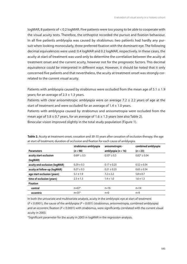
185
Evaluation of visual acuity in a historic cohort
logMAR, 8 patients of < 0.2 logMAR. Five patients were too young to be able to cooperate with
the visual acuity tests. Therefore, the orthoptist recorded the pursuit and fixation behaviour.
In all five patients amblyopia was caused by strabismus: two patients had hardly any pur
suit when looking monocularly, three preferred fixation with the dominant eye. The following
decimal equivalences were used: 0.4 logMAR and 0.2 logMAR, respectively. In these cases, the
acuity at start of treatment was used only to determine the correlation between the acuity at
treatment onset and the current acuity, however not for the prognostic factors. This decimal
equivalence could be interpreted in different ways. However, it should be noted that it only
concerned five patients and that nevertheless, the acuity at treatment onset was strongly cor
related to the current visual acuity.
Patients with amblyopia caused by strabismus were occluded from the mean age of 5.1 ± 1.9
years; for an average of 2.3 ± 1.3 years.
Patients with clear anisometropic amblyopia were on average 7.2 ± 2.2 years of age at the
start of treatment and were occluded for an average of 1.4 ± 1.0 years.
Patients with amblyopia caused by strabismus and anisometropia were occluded from the
mean age of 5.8 ± 0.7 years, for an average of 1.6 ± 1.3 years (see also Table 2).
Binocular vision improved slightly in the total study population (Figure 1).
Table 2. Acuity at treatment onset, cessation and 3035 years after cessation of occlusion therapy, the age at start of treatment, duration of occlusion and fixation for each cause of amblyopia.
Parametersstrabismus-amblyopia (n = 98)
anisometropic-amblyopia (n = 16)
combined amblyopia (n = 23)
acuity start occlusion(logmAr)
0.69* ± 0.5 0.53* ± 0.3 0.82* ± 0.54
acuity end occlusion (logmAr) 0.29 ± 0.3 0.17 ± 0.23 0.52 ± 0.54
acuity at follow-up (logmAr) 0.27 ± 0.3 0.21 ± 0.23 0.65 ± 0.54
age start occlusion (years) 5.1 ± 1.9 7.2 ± 2.2 5.8 ± 0.7
time of occlusion (years) 2.3 ± 1.3 1.4 ± 1.0 1.6 ± 1.3
Fixation central n=63* n=16 n=14
eccentric n=35* n=0 n=9
In both the univariate and multivariate analysis, acuity in the amblyopic eye at start of treatment (P < 0.0001), the cause of the amblyopia (P = 0.001) (strabismus, anisometropia, combined amblyopia) and an eccentric fixation (P < 0.0001) with strabismus, were significantly correlated with the current visual acuity in 2003.*Significant parameter for the acuity in 2003 in logMAR in the regressionanalysis.

186
relationship between visual acuity and the cause of amblyopia
Since the cessation of occlusion therapy visual acuity had deteriorated from a mean of 0.52 ±
0.54 to 0.65 ± 0.54 logMAR in patients with a combined cause of amblyopia and from a mean of
0.17 ± 0.23 to 0.21 ± 0.23 logMAR in patients with an anisometropic amblyopia.
Visual acuity had improved from 0.29 ± 0.3 to 0.27 ± 0.3 logMAR in patients with strabismic
amblyopia (Figure 2). This improvement was significant (P < 0.001). The acuity of the dominant
eye of these patients had also improved from 0.01 ± 0.15 to 0.05 ± 0.07 logMAR, this was also
significant (P = 0.04).
In general, in 14 patients (10%) the acuity in the amblyopic eye was unchanged. In 70 patients
(51%) acuity had improved. In 54 patients (39%) acuity had deteriorated, of which 18 patients
had an acuity loss of >50% compared to acuity at the end of treatment.
Of the 137 patients, 47 (34%) had an acuity in the amblyopic eye at distance (6m) of > 0.3 log
MAR lines at the end of treatment, on average 0.48 ± 0.6 logMAR.
Chapter 10 Figure 2
Appendix Figure 1
Figure 1. Binocular vision of the total study population during occlusion treatment and at the time of the followup reexamination in 2003.The degree of binocular vision (abscissa) was arranged into seven categories 1: Bagolini negative, 2: Bagolini positive, 3: Bagolini and TitmusStereotest Fly positive, 4: lowest TitmusStereotest circle 200”140” positive, 5: lowest TitmusStereotest circle 100”40” positive, 6: Stereotest from Lang or lowest TNOTest figure V (480” & 240”) positive, 7: TNOTest figure VI or VII (120”15”) positive. The black bars show the degree of binocular vision during the occlusion treatment (n=137), the grey bars at the time of the followup reexamination. The ordinate represents the number of patients.

187
Evaluation of visual acuity in a historic cohort
Of the 137 patients, 55 (40%) were unable to read with the amblyopic eye (>D=1).
Patients with profound deterioration in visual acuity
18 Patients had an acuity loss >50% compared to acuity at the end of treatment. Of these,
15 showed an increase of anisohypermetropia. Of these 15 patients, nine had developed an
anisohypermetropia of 2.2 ± 1.7D. In six patients, an anisohypermetropia had already been
present (mean was 2.9D), which had increased to 3.5 ± 1.8D.
Appendix Figure 2
Figure 2. Acuity in the amblyopic eye at treatment onset and at followup reexamination in 2003, for each cause of amblyopia.The ordinate shows the acuity at the time of the followup reexamination in logMAR lines, the abscissa shows the acuity in logMAR lines at treatment onset. Patients with a combined amblyopia are represented by a black square ß, patients with anisometropicamblyopia by a triangle p and patients with strabismicamblyopia by a white square ˙. The solid regression line represents the combined amblyopias, the dashed regression line those of the anisometropicamblyopias and the dotted line those of the strabismicamblyopias.Both patients with strabismicamblyopia and those with combined amblyopia had a poor acuity at treatment onset, patients with strabismicamblyopia, however, had a better acuity at the followup reexamination in 2003.

188
In the 18 patients the spherical equivalent of the amblyopic eye had increased from +2.7D
to +2.8D. The spherical equivalent had decreased from +1.9D to +0.7D in the dominant eye.
Only five of the 18 patients wore adequate spectacles at the time of the followup reexamina
tion. Three of these five patients were myopic in the dominant eye, and two were on average
+2.75D hyperopic.
correlations
The univariate analysis showed a statistically significant correlation between the current vi
sual acuity and the acuity at the start of the occlusion therapy (P <0.001), an eccentric fixation
(P <0.001) and the cause of the amblyopia (strabismus, anisometropia or combined cause), (P
= 0.005) (see also table 2). No significant influence could be found for the duration of occlu
sion therapy (P = 0.622), the age at start of occlusion treatment (P = 0.320) and at the end of
occlusion treatment (P = 0.119).
When correcting for potentially confounding factors in the multivariate analysis, the visual
acuity at the start of treatment, the cause of the amblyopia and the fixation remained signifi
cant (P < 0.001; P = 0.018; P = 0.004, respectively).
Prognostic factors
Patients with anisometropic amblyopia had, compared to patients with strabismic amblyopia
and patients with combined amblyopia, a significantly better visual acuity at the start of the
occlusion therapy. Patients with combined amblyopia had the poorest acuity at the start of
treatment and also at the followup reexamination. Only patients with strabismic amblyopia
showed a slight acuity improvement, compared to acuity at the end of the treatment.
In patients with strabismus, the fixation was correlated with the acuity. A correlation could be
found between the decrease in acuity and the loss of binocular vision (r = 0.38).
Prevalence of occlusion therapy
We aimed to establish whether the historical cohort was representative of the number of chil
dren receiving occlusion therapy at that time; i.e. if any selection bias had occurred. The preva
lence of amblyopia has been quoted to be approximately 3.25% (Attebo, et al. 1998). It was
expected to find a lower prevalence rate, as our historical cohort was comprised of a biased
underrepresentation. Of the 471 occluded children, 66, 64 and 68 children were born in 1965,
1966 and 1967, respectively (Figure 3). In the whole of the Waterland hospital region 1286,
1328 and 1355 births were registered in 1965, 1966 and 1967, respectively. This would concur
with a prevalence of 5.0% occluded children.

189
Evaluation of visual acuity in a historic cohort
Figure 3.
Overview of the total number of patients for their year of birth: all patients occluded in the Waterland
hospital (n = 471, light grey) and of the historical cohort (n = 137, black). Also for the historical cohort
start of occlusion (white) and cessation of occlusion (dark grey) has been given. Of the 471 occluded
children 66, 64 and 68 were born in 1965, 1966 and 1967, respectively. This corresponded to 5.0% of
the registered births in the total region of the Waterland hospital in those years.
Figure 3. Overview of the total number of patients for their year of birth: all patients occluded in the Waterland hospital (n=471, light grey) and of the historical cohort (n=137, black). Also for the historical cohort start of occlusion (white) and cessation of occlusion (dark grey) has been given. Of the 471 occluded children 66, 64 and 68 were born in 1965, 1966 and 1967, respectively. This corresponded to 5.0% of the registered births in the total region of the Waterland hospital in those years.
In hindsight, the amblyopia could not be confirmed in seven of the 137 children. These pa
tients had an acuity difference of <0.2 logMAR lines between the dominant and the amblyopic
eye, an anisometropia <1D, and/or an alternating strabismus.
After the necessary correction, the prevalence of occluded children was approximately 4.7%.
Theoretically, it could be possible that children living outside the region were also treated in
the Waterland hospital. This, however, was not the case in our group.
dIScuSSIon
For this study we were able to establish an evaluation of the visual acuity in 137 patients more
than thirty years after the cessation of occlusion therapy. We found that a poor acuity in the
amblyopic eye at the start of treatment, an eccentric fixation and a combined cause of ambly
opia were correlated with the current visual acuity measured at the followup reexamination.
That a poor visual acuity in the amblyopic eye at start of treatment and the cause of amblyo
pia were predictors for a poor outcome has also been described by other authors, even when
a lengthier period of observation time was examined. A combined cause of amblyopia and an

190
increase of the anisohypermetropia were, after 30 years, associated with a decrease in visual
acuity after the end of the occlusion therapy.
In most of the children, due to the school age, acuity could be tested using the LandoltC
chart, both at the start and the end of treatment. The acuity at treatment onset, in which the
largest variation was to be expected, was merely used in the analysis of the correlation with
the current acuity. When analysing the prognostic factors the acuity at the end of treatment
was used. The acuity was determined with different acuity charts. This could cause the out
comes to be not entirely comparable (Gräf 2004), as acuity tests with optotypes can only score
similar outcomes as when using LandoltC, when acuity is higher. When acuity is poor, a slight
overrating of the acuity will occur when it is measured using optotypes (Rassow and Wang
1999). Nevertheless, we found a strong significance that could be even stronger, when acuity
is tested in equal testing conditions.
After cessation of occlusion therapy a slight improvement of the acuity was found in both the
amblyopic eye and the dominant eye in patients with strabismic amblyopia. We presumed
this to be the natural development of the acuity. Information is scarce concerning the devel
opment of the acuity in the dominant eye. In general, when not taking into account the cause
of amblyopia, a slight improvement of the acuity was found (Ohlsson, et al. 2002). Studies of
the natural course of the acuity in patients with untreated amblyopia, showed a worsening
of the acuity in the amblyopic eye both at preschool age (Simonsz and Preslan 1999) and in
adulthood (Haasse and Wenzel 1996).
It should be taken into account, however, that an inclusion criteria for the study was poor
acuity (>0.18 logMAR lines; n = 18) and that children with a slight decrease in acuity were
excluded, making a selection bias probable.
In agreement with other studies (Levartovsky, et al. 1995; Woordruff, et al. 1994) we were un
able to demonstrate a correlation between the age at treatment onset, the age at the end
of treatment and the current visual acuity in 2003. However, it could not be concluded that
a patient may not be treated more successfully if occlusion in this patient had started at an
earlier age.
In our study group the age at the start of the occlusion therapy was relatively high, probably
caused by the fact that, at that time, screening for amblyopia had not altogether been prop
erly introduced. With the exception of the cities, health centres and general practitioners did
not carry out acuity measurements, causing amblyopia without a notable strabismus to be
first diagnosed at the age of 57 years. Nowadays, in the Netherlands, the Child Health Care
Centres screen for the presence of strabismus and measure visual acuity at the age of three.
The current mean age at treatment onset is 4.6 years (Loudon, et al. 2006).

191
Evaluation of visual acuity in a historic cohort
Fifteen of the 18 patients, in whom acuity had decreased >50% after the cessation of the oc
clusion therapy, had an increase of the anisohypermetropia. At the time of the followup re
examination it seemed important to remark that only five of these were wearing spectacles or
contact lenses. They indicated they stopped wearing the spectacles from the age of 14/15. It
may be possible that, after the latest spectacle correction, these patients started to see out of
focus again with their amblyopic eye, because of the increase in anisometropia and, for that
reason, did not like to wear the spectacles.
It is therefore recommendable to check patients that had anisometropic amblyopia and show
an increase of the anisometropia more often and to adjust the spectacles accordingly.
In other studies (Attebo, et al 1998; Leiba, et al. 2001; Levartovsky, et al. 1995; PEDIG 2002;
Woodruff, et al. 1994), amblyopia caused by a pure anisometropia is approximately 30%, vary
ing, however, between 11% (Leiba, et al. 2001) and 50% (Attebo, et al. 1998). In our study, only
the children who received occlusion therapy for their amblyopic eye were included. Therefore,
children who had been treated for their anisometropic amblyopia using a spectacle correc
tion were not included.
Surprisingly, we found the prevalence of occluded children (4.7%) to be higher than the prev
alence’s of amblyopia mentioned in other studies (3.25%), (Attebo, et al. 1998). How could this
discrepancy be explained? The prevalence of amblyopia in most studies is determined among
adult patients with untreated amblyopia. Our study demonstrated that in most patients the
acuity after the cessation of the occlusion therapy improves slightly; in only few it deterio
rates. At the start of occlusion therapy the orthoptist is unaware which children, who have
been referred by health centres or paediatricians because of a slight reduction in acuity (>0.1
logMAR), will have a spontaneous improvement of acuity and may therefore, overtreat.


193
Appendix 2
Appendix 2: educational cartoon programme

194
The educational programme, developed by José Vingerling and Gerard de Bruyne, to improve compliance with occlusion therapy for amblyopia. It consisted of a cartoon story without words, explaining to the child why he or she should patch. There were no animal figures included, making it easier for the child to identify him or herself with the child in the story. It was bolstered by an immediate reward system (calendar and stickers). © Vingerling & De Bruyne.
The educational programme, developed by José Vingerling and Gerard de Bruyne, to improve
compliance with occlusion therapy for amblyopia. It consisted of a cartoon story without
words, explaining to the child why he or she should patch. There were no animal figures
included, making it easier for the child to identify him or herself with the child in the story. It
was bolstered by an immediate reward system (calendar and stickers). © Vingerling & De
Bruyne.

195
colour figures
chAPTEr 1
chAPTEr 3
Figuren bij Chapter 5
1a 1b 1c 1d
Figure 1. The development of the Occlusion Dose Monitor. a Year 1991: first model developed by
Fielder and Moseley in Birmingham, UK (with permission of M. Moseley). The ODM was a miniature
datalogger, which logs the patch-skin contact resistance every 64 s. b Year 1997: The Academic
Medical Center Amsterdam modified the design into an ODM containing two thermistors connected to
either the front or the back of the ODM (35x23x4 mm, 6 g). c The 2001 type used in the Netherlands
(24x12x3.6 mm, 1.8 g). d The 2002 type used in Germany (31x15x3.5 mm, 2.3 g)
2a
2b
Figure 2. Examples of recordings with the ODM, with measured temperature difference on the
ordinates. a A week’s recording. Abscissa: date of the recording. The temperature difference was about
"0" (0; 0.0625) while the child was not patched (baseline value) and higher during the periods of
1a 1b 1c 1dFigure 1. a the first model of the ODM developed by Prof. Alistair Fielder and Dr. Merrick Moseley in London, United Kingdom b: in 1997 the department of Medical Technical Development at the Academic Medical Center Amsterdam modified the design and made it smaller: it now measures temperature difference between the front and the back c & d the ODM as used in the study in The Hague: it weighs 1.8 g and measures 24 x 12 x 3.6 mm.
Colour Figures
Chapter 3
Figure 1. Median and interquartile range of the prescribed hours of occlusion per day of the non-
amblyopic eye per regimen per case. The abscissa represents the five cases and the chosen regimen.
The total number of orthoptists prescribing a regimen is given below or above each whisker.
Figure 1. Median and interquartile range of the prescribed hours of occlusion per day of the nonamblyopic eye per regimen per case. The abscissa represents the five cases and the chosen regimen. The total number of orthoptists prescribing a regimen is given below each whisker.

196
chAPTEr 6
Chapter 6
Figure 2. Graphic representation of the relationship between acuity increase during treatment and
electronically measured compliance. The ordinate represents the ratio between visual acuity in the
amblyopic eye and the acuity in the good eye. The abscissa represents the months after the start of
occlusion therapy. On the right: patient number corresponding to the actually measured compliance in
percentages in parenthesis. Blue lines represent patients with high compliance; red lines represent low
compliance, corresponding to the visual acuity increase after 6 months of occlusion therapy.
Figure 2. Graphic representation of the relationship between acuity increase during treatment and electronically measured compliance. The ordinate represents the ratio between visual acuity in the amblyopic eye and the acuity in the good eye. The abscissa represents the months after the start of occlusion therapy. On the right: patient number corresponding to the actually measured compliance in percentages in parenthesis. Blue lines represent patients with high compliance; red lines represent low compliance, corresponding to the visual acuity increase after 6 months of occlusion therapy.

197
Colour figures
chAPTEr 9
Chapter 9
Figure 1. To remove the patches form the skin an average maximum force of 8.8 Newton (Beiersdorf –
Elastopad), 5.9 N (Orthopad), 3.2 N (Opticlude) and 2.6 N (Pro-Ophta) was needed.
Figure 1. To remove the patches from the skin an average maximum force of 8.8 Newton (Beiersdorf ), 5.9 N (Orthopad), 3.2 N (Opticlude) and 2.6 N (ProOphta) was needed.

198
chAPTEr 10
Chapter 10
Figure 1.a. The distribution of the 10 possible causes of amblyopia per diagnose. Each bar represents a
diagnosis. In our study population 23 diagnoses were given by the treating orthoptist and more than one
diagnosis may be given to one child. Each bar was subdivided into the cause of amblyopia (assessed by
the researchers), e.g. the diagnosis ‘late-onset esotropia’ was present in the following three causes of
amblyopia: strabismus, strabismus & anisometropia and strabismus & hypermetropia.
Figure 1.a. The distribution of the 10 possible causes of amblyopia per diagnose. Each bar represents a diagnosis. In our study population 23 diagnoses were given by the treating orthoptist and more than one diagnosis may be given to one child. Each bar was subdivided into the cause of amblyopia (assessed by the researchers), e.g. the diagnosis ‘lateonset esotropia’ was present in the following three causes of amblyopia: strabismus, strabismus & anisometropia and strabismus & hypermetropia.

199
Colour figures
Chapter 10
Figure 1.b. The distribution of the 23 diagnoses in our study population per cause of amblyopia. Each
bar represents 1 of the 10 causes of amblyopia, which was subdivided in the diagnoses given in our
study population. One child may have more than one diagnosis, but only one cause of amblyopia. For
example, anisometropia was assessed as the cause of amblyopia in 132 children; however, 6 children
with anisometropic amblyopia also had the diagnosis ‘esophoria’ and 11 ‘exophoria’. These diagnoses
were not the main cause of amblyopia. The number above the bar corresponds with the number of
children whose cause of amblyopia is represented in that bar.
Figure 1.b. The distribution of the 23 diagnoses in our study population per cause of amblyopia. Each bar represents 1 of the 10 causes of amblyopia, which was subdivided in the diagnoses given in our study population. One child may have more than one diagnosis, but only one cause of amblyopia. For example, anisometropia was assessed as the cause of amblyopia in 132 children; however, 6 children with anisometropic amblyopia also had the diagnosis ‘esophoria’ and 11 ‘exophoria’. These diagnoses were not the main cause of amblyopia. The number above the bar corresponds with the number of children whose cause of amblyopia is represented in that bar.