Topography of Visual Features in the Human Ventral Visual ...

Improved Disease Classification in Chest X-rays withTransferred Features from Report Generation
Yuan Xue, Xiaolei Huang?
College of Information Sciences and Technology,The Pennsylvania State University, University Park, PA
Abstract. Radiology includes using medical images for detection and diagnosisof diseases as well as guiding further interventions. Chest X-rays are commonlyused radiological examinations to help spot thoracic abnormalities or diseases,especially lung-related diseases. However, the reporting of chest x-rays requiresexperienced radiologists who are often in shortage in many regions of the world.In this paper, we first develop an automatic radiology report generation system.Due to the lack of large annotated radiology report datasets and the difficulty ofevaluating the generated reports, the clinical value of such systems is often lim-ited. To this end, we train our report generation network on the small IU ChestX-ray dataset then transfer the learned visual features to classification networkstrained on the large ChestX-ray14 dataset and use a novel attention guided featurefusion strategy to improve the detection performance of 14 common thoracic dis-eases. Through learning the correspondences between different types of featurerepresentations, common features learned by both the report generation and theclassification model are assigned with higher attention weights and the weightedvisual features boost the performance of state-of-the-art baseline thoracic diseaseclassification networks without altering any learned features. Our work not onlyoffers a new way to evaluate the effectiveness of the learned radiology report gen-eration network, but also proves the possibility of transferring different types ofvisual representations learned on a small dataset for one task to complement fea-tures learned on another large dataset for a different task and improve the modelperformance.
1 Introduction
Deep learning has shown its strength in various types of computer vision tasks such asclassification and detection in the last decade. With the rapid development of advancedalgorithms and the availability of more annotated datasets, potential applications ofdeep learning in radiology have become possible and desired by the public. Amongsuch applications, the report generation networks [11, 14, 26] and the disease classifica-tion networks [23, 24, 27, 19] are two popular trends. An automated or computer-aidedradiology reporting system can provide preliminary findings to radiologists to assistwith report writing; A disease classification network can help detect potential abnor-malities and diseases shown in the examinations. Both systems can help reduce theworkload of expert radiologists as well as make up for staff shortage.
? Xiaolei Huang is the corresponding author. Her email address is: [email protected]

2 Yuan Xue, Xiaolei Huang
Current report generation methods mainly follow the encoder-decoder architecturewhich has been widely used for image captioning [22, 25, 17]. In the radiology report,the Impression and the Findings can be generated based on the interpretation of im-ages without prior examinations or patient history, which makes the generation tasks aperfect fit for deep learning models. Impression can be a single-sentence conclusion ordiagnosis, and findings can be a coherent paragraph containing multiple sentences thatdescribe the radiologist’s observations and findings regarding both normal and abnor-mal features in the images. While the impression generation can often be handled byan image captioning algorithm, a findings paragraph consists of multiple sentences thustraditional captioning algorithms designed for generating single-sentence description nolonger apply. For findings generation, state-of-the-art radiology report generation mod-els use either a hierarchical architecture [11, 14] to generate different sentences basedon different topic vectors, or a recurrent architecture [26] to generate one sentence ata time whereby the following sentence is conditioned upon the content of its preced-ing sentence. Combining the merits of both, we propose a modified recurrent attentionmodel where a succeeding sentence is generated based upon both the prior sentenceand a global topic vector encoded by the generated impression. The recurrent attentionmechanism guarantees the model to generate diverse and coherent sentences, where theglobal topic vector forces the model to produce sentences supporting the conclusion ordiagnosis.
Although some accomplishments have been made in radiology report generation,there are still several issues remaining unsolved. First, training a general and robust re-port generation system on a small training set is impractical, as the ground truth reportprovided in the training set can be biased towards the radiologist’s personal style. Evenfor the same image, different radiologists can produce entirely different written reportsas they only provide information that they think might be important to the potential ref-erees. More importantly, evaluation of the generated report is difficult. Any automaticmetrics based on the overlap between the prediction and ground truth cannot capturethe words describing negation and uncertainty and lack measurement of semantic simi-larity, while human evaluation requires extra efforts by human experts and can be error-prone. While the generated report learned by the decoder model can be biased towardsthe training data, the learned visual features from the encoder model are expected to bemore general and robust. Otherwise they are not able to provide enough information tothe decoder model to produce diverse sentences describing different regions. Therefore,we introduce a novel evaluation of the trained chest X-ray report generation network,where we only take the encoder part of the model as a visual feature extractor, and trans-fer these learned features to be used in another large chest X-ray dataset and evaluatetheir performance for thoracic disease classification.
Traditional transfer learning benefits from training with a large-scale dataset thentransferring the learned knowledge or model weights to another dataset that is typicallymuch smaller for better initialization and faster convergence. However, transfers froma small dataset to a large dataset with cross-task generalization are under-investigated,but such transfers have great research and clinical value since some annotations likecomplete radiology reports are expensive to obtain due to the difficulty of labeling orprivacy concerns. In this work, taking the learned image encoder from the report gen-

Title Suppressed Due to Excessive Length 3
eration model, we apply the extracted visual features to another chest X-ray dataset fordisease classification. We combine the features from the report generator and the fea-tures extracted by a well-trained disease classification network to form an attention map,where common features learned by both networks are emphasized with higher weights.Then we apply the attention map to the original feature learned by the classificationnetwork and re-train the transition layer for new predictions. The transferred visual fea-tures along with the attention guided feature fusion shows considerable improvementsover a baseline classification network, and even achieve further improvements over thestate-of-the-art CheXNet [19] without changing any visual features. Our work not onlyoffers a new way to evaluate the effectiveness of learned radiology report generationnetwork, but also proves the possibility of transferring different types of visual repre-sentation learned on a small dataset for one task to complement features learned onanother large dataset for a different task and improve the model performance.
2 Related WorkRadiology Report Generation. As a joint application of Computer Vision (CV) andNature Language Processing (NLP) in healthcare, the automatic radiology report gener-ation task has recently attracted considerable attention. Following the encoder-decoderarchitecture and attention mechanism in image captioning [22, 25, 17], several reportgeneration works have been proposed. To generate long and coherent reports, previousworks [11, 14] use a hierarchical architecture [13, 1] to generate a sequence of encodedtopic vectors, then each sentence in the report is generated conditioned on the topicvector; Xue et al. [26] utilizes a recurrent attention model and brings the contextualinformation into the loop when predicting next words and sentences. Li et al. [14] com-bines a retrieval module and a generation module to either select a phrase from thetemplate database or generate a new sentence. However, such models still suffer fromthe bias of training set, and automatic evaluation cannot capture the words describingnegation and uncertainty in the predicted report. Taking the merits of previous meth-ods, we modify the recurrent attention model [26] and introduce a global topic vectorto guide the generation of findings. Instead of only evaluating the predicted report, wetransfer the learned visual features to the ChestX-ray14 dataset and use an attentionguided feature fusion scheme to help improve the disease classification performance.
Thoracic Disease Classification. Wang et al. [23] introduced the ChestX-ray14 datasetwhich is currently the largest public repository of radiographs. They also reported aweakly supervised multi-label thoracic disease classification and localization frame-work in their paper. Yao et al. [27] leveraged inter-dependencies among different pathol-ogy labels in chest X-rays via LSTMs to improve the disease classification performance.In [19], state-of-the-art disease classification result is reported using a DenseNet-121 [10]backbone; their trained model gets impressive results and exceeds the average radiolo-gist performance on the pneumonia detection task.
A different approach for thoracic disease classification trains the model with multi-ple tasks to improve classification result. Li et al. [15] presents a unified network thatsimultaneously improves classification and localization with the help of extra boundingboxes indicating disease location. Wang et al. [24] uses a CNN-RNN model to generate
4 Yuan Xue, Xiaolei Huang
a radiology report and encodes the generated report directly to improve the classifica-tion result. Different from our model, their report generation and classification modelsare trained on the same dataset where the ground truth disease labels and reports are in-herently correlated since the labels are text-mined from the original report. Since thereis no large dataset with annotated radiology report available to the public, it is hard toextend their framework to other domains and tasks.
Transfer Learning for Medical Imaging. While multi-task learning can benefit fromsharing features between different tasks to get more general features, transfer learningcan leverage the learned features from a source domain to improve the performance ina target domain. Traditional transfer learning use knowledge or model weights learnedfrom tasks for which a large amount of annotated data is available in tasks where onlya limited amount of labeled data is available. The large dataset can help generalizethe model as training on small datasets can lead to overfitting. Transfer learning hasbeen widely adopted in deep learning such as initializing models with weights pre-trained on ImageNet [4] and has shown performance improvements for medical imageclassification [20, 9].
However, in medical applications, the amount of labeled data is typically low sousing features learned from other medical domains directly is often impractical. Toevaluate the visual features learned by our report generator and explore the possibilityof transferring visual features learned from a small set to a larger set and improvingthe cross-task generalization, we propose an attention based feature fusion scheme tomodify the already trained classification model. Instead of averaging features in all re-gions as average pooling in standard models [7, 10], our transfer learning model assignsmore weights to the features coexisting in both original feature space and transferredfeature space through an attention guided feature fusion mechanism. In this way, fea-tures learned in a small set can also help generalize the model for another domain ortask.
3 Methodology
3.1 Radiology Report Generator
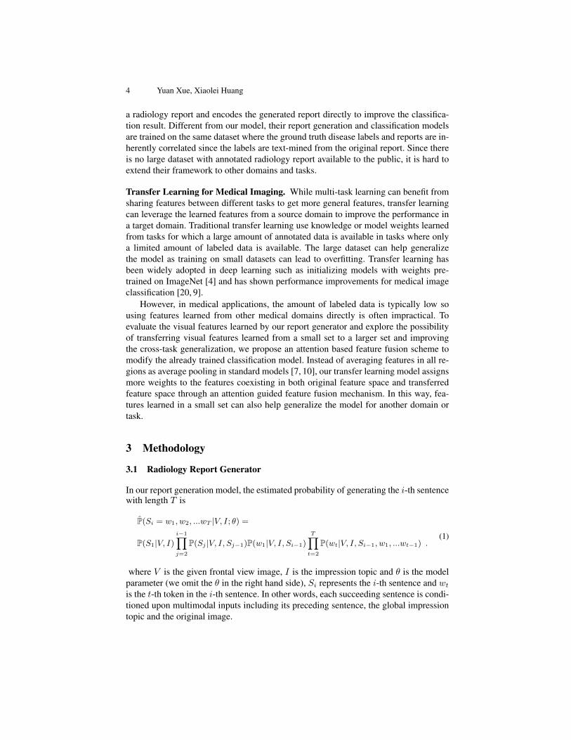
In our report generation model, the estimated probability of generating the i-th sentencewith length T is
P̂(Si = w1, w2, ...wT |V, I; θ) =
P(S1|V, I)i−1∏j=2
P(Sj |V, I, Sj−1)P(w1|V, I, Si−1)T∏t=2
P(wt|V, I, Si−1, w1, ...wt−1) .(1)
where V is the given frontal view image, I is the impression topic and θ is the modelparameter (we omit the θ in the right hand side), Si represents the i-th sentence and wt
is the t-th token in the i-th sentence. In other words, each succeeding sentence is condi-tioned upon multimodal inputs including its preceding sentence, the global impressiontopic and the original image.
Title Suppressed Due to Excessive Length 5
Input Image
Image En
coder
Impression Decoder
Sentence Encoder
Impression
Sentence Decoder
Next Sentence in Findings
Attention Model Semantic FeaturesVisual Features
Impression: No acute disease.
Findings: The heart is normal in size. The mediastinum is unremarkable. The lungs are clear.
Topic Encoder
Classifier
Feature Attention
Classification Result
Pathology LabelAtelectasis 0Cardiomegaly 0Effusion 0
Hernia 0
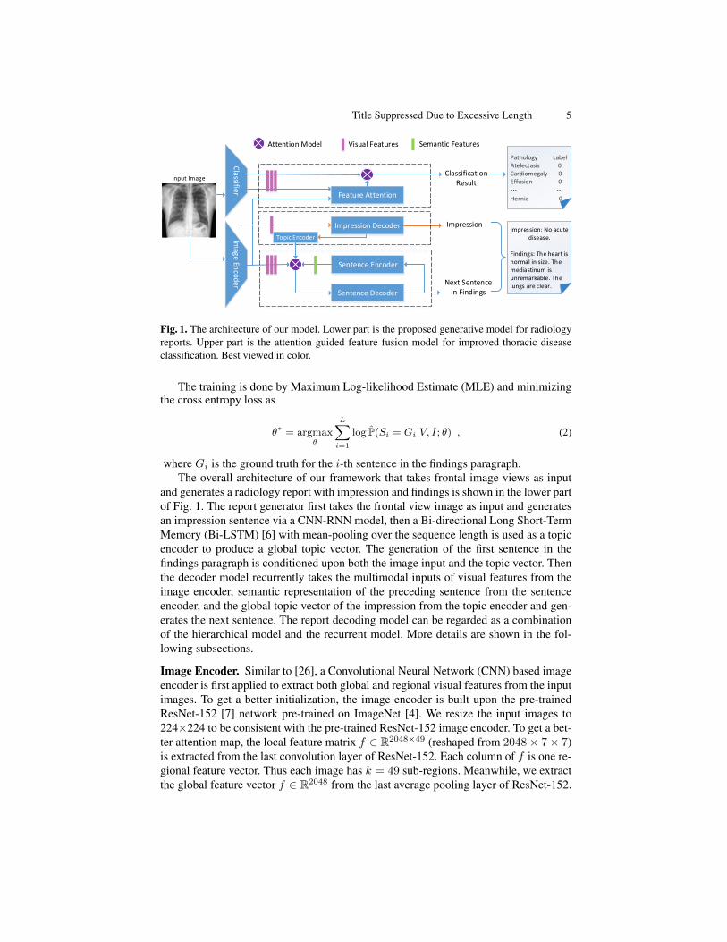
Fig. 1. The architecture of our model. Lower part is the proposed generative model for radiologyreports. Upper part is the attention guided feature fusion model for improved thoracic diseaseclassification. Best viewed in color.
The training is done by Maximum Log-likelihood Estimate (MLE) and minimizingthe cross entropy loss as
θ∗ = argmaxθ
L∑i=1
log P̂(Si = Gi|V, I; θ) , (2)
where Gi is the ground truth for the i-th sentence in the findings paragraph.The overall architecture of our framework that takes frontal image views as input
and generates a radiology report with impression and findings is shown in the lower partof Fig. 1. The report generator first takes the frontal view image as input and generatesan impression sentence via a CNN-RNN model, then a Bi-directional Long Short-TermMemory (Bi-LSTM) [6] with mean-pooling over the sequence length is used as a topicencoder to produce a global topic vector. The generation of the first sentence in thefindings paragraph is conditioned upon both the image input and the topic vector. Thenthe decoder model recurrently takes the multimodal inputs of visual features from theimage encoder, semantic representation of the preceding sentence from the sentenceencoder, and the global topic vector of the impression from the topic encoder and gen-erates the next sentence. The report decoding model can be regarded as a combinationof the hierarchical model and the recurrent model. More details are shown in the fol-lowing subsections.
Image Encoder. Similar to [26], a Convolutional Neural Network (CNN) based imageencoder is first applied to extract both global and regional visual features from the inputimages. To get a better initialization, the image encoder is built upon the pre-trainedResNet-152 [7] network pre-trained on ImageNet [4]. We resize the input images to224×224 to be consistent with the pre-trained ResNet-152 image encoder. To get a bet-ter attention map, the local feature matrix f ∈ R2048×49 (reshaped from 2048× 7× 7)is extracted from the last convolution layer of ResNet-152. Each column of f is one re-gional feature vector. Thus each image has k = 49 sub-regions. Meanwhile, we extractthe global feature vector f ∈ R2048 from the last average pooling layer of ResNet-152.
6 Yuan Xue, Xiaolei Huang
The image encoder is fine tuned on the frontal view chest X-rays so that the learnedvisual features can be transferred to another classification task.
Report Decoder. The decoder network is responsible for generating both the impres-sion and the findings paragraph. The impression decoder first takes the global visualfeatures learned by the image encoder as input and a single layer Long Short-TermMemory (LSTM) [8] is used for sentence decoding. Following [2] , we adopt a Bi-LSTM that reads the generated impression in two opposite directions along with a meanpooling over each dimension of the hidden units to get a global topic vector. The sen-tence decoder takes regional visual features and the topic vector to generate the firstsentence. After that, each sentence is generated by taking regional visual features, thepreviously generated sentence and the topic vector as a multimodal input. The sentencedecoder is a stacked 2-layer LSTM. All hidden and embedding dimensions are fixedto 512 for all of the LSTM models discussed herein. Both regional and global visualfeatures are converted into channel dimension 512 to match the embedding size beforebeing fed into any LSTMs. The regional visual features are converted as input to thesentence decoder. The global topic vector is used as the initialization of the sentencedecoder, while the learned encoding of the preceding sentence is combined with the vi-sual representations to generate an attention map. The attention weights for the regionalvisual features are computed as follows:
a = Watt((v; s1k)) + batt , (3)
where v ∈ R512×49 are the regional visual features learned by the image encoder,s ∈ R512×1 represents the encoding of the preceding sentence, 1k ∈ R1×49 is a vectorwith all ones. v and s1k are concatenated along the embedding dimension as the inputto the attention network. Watt ∈ R1×1024, and batt ∈ R1×49 are parameters of theattention network. Then the weights are normalized over all regions to get the attentiondistribution and applied to the original visual representation as:
vatt =
k∑i=1
exp(ai)∑i exp(ai)
vi , (4)
where i refers to the i-th region in the regional visual representation.The generation process is repeated until an empty sentence is generated or a max-
imum number of sentences is reached, which indicates the end of the paragraph. Ourmodel combines the recurrent and hierarchical architecture in the decoder network.While the recurrent attention mechanism forces the model to focus on different regionsof the input image to generate more diverse sentences and keep intra-paragraph coher-ence, the global topic vector adds an additional constraint so that the generated sen-tences can support the theme of the entire report. An example of the generated reportby our report generator is shown in Fig. 2.
Our proposed report generator is trained by the Adam optimizer [12]. The initiallearning rate is set to be 1e-4, and learning rate decay is 0.5 for every 5 epochs. Thebatchsize is 32 for training. During inference, the greedy search is adopted for generat-ing words and sentences in every timestep. The maximum number of sentences is set tobe 7. Although the impression decoder and the findings decoder can be trained jointly,we separate the training process to get more diverse results. The final model transferredto the classification model for feature fusion is trained on all training data. Although

Title Suppressed Due to Excessive Length 7
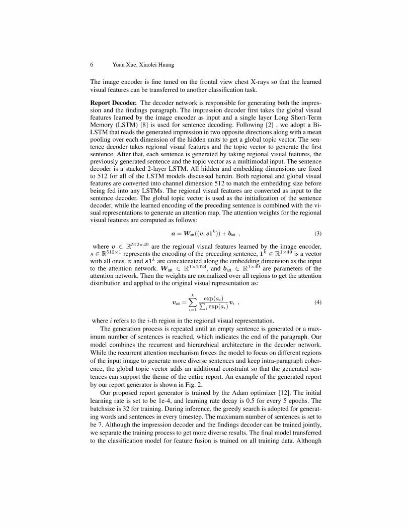
Original ReportPredicted ReportInput ImageFindings: The cardiac contours are normal. The lungs are clear. There is no pleural effusion or pneumothorax. There is no focal air space opacity to suggest a pneumonia. Degenerative changes of the thoracic spine.Impression: No acute cardiopulmonary finding.
Findings: Heart size and mediastinal contour normal. Lungs are clear. Pulmonary vascularity normal. No pleural effusions or pneumothoraces. Minimal degenerative changes thoracic spine.
Impression: No acute cardiopulmonary process.
Fig. 2. An example report generated by our model in comparison with the original written reportprovided by the radiologist. The notable words in bold font in the findings indicate that our modelis capable of capturing some of the abnormalities in the input image.
the generated report can be biased towards the training data, we believe that the learnedvisual features should be more general than the semantic features and can be transferredto other domains.
3.2 Thoracic Disease Classifier and Attention Guided Feature Fusion
The basic thoracic disease classification network is a CNN that takes frontal view ra-diographs as input and generates multiple disease labels. We first train two differentbaseline classification models to get the visual features. We start from the ResNet-18model and later move to the DenseNet-121 model with more layers. For the ResNetbaseline, the final fully connected layer is replaced with one that has 14 outputs for 14disease categories, after which we apply a sigmoid nonlinearity as in [19]. The trainingis done by minimizing the binary cross entropy loss. The weights of the network areinitialized with weights from models pre-trained on ImageNet [4]. We pick the modelwith the lowest validation loss as the final model. After the training is completed, wediscard the transition block including the final pooling layer, the final fully connectedlayer and the sigmoid nonlinearity to keep only the local visual features. Then, we runthe image encoder of our report generator on the classification dataset. The extractedlocal visual features serve as a high-level linguistic abstract of the input images here.Both the local visual features from the classification network and the report generationnetwork with k = 49 sub-regions are then fed into the feature attention module. Thefeature attention module can be interpreted as:
a′= W
′att((v
r;vc)) + b′att , (5)
where vr are the regional visual features from the report generator, vc are the visualfeatures from the original classification network. vr and vc are concatenated along thechannel/embedding dimension as the input to the feature attention network. Watt andbatt are parameters of the attention network. Then the modified visual representation iscomputed as:
vcatt =
k∑i=1
exp(a′i)∑
i exp(a′i)vci , (6)
where i refers to the i-th region in the regional visual representation. The detailed pro-cess is shown in the upper part of Fig. 1. Local features from the image encoder and theclassification model are concatenated and fed into the feature attention module. Aftercalculating an attention map over all regions, we apply the attention weights to the orig-inal visual features learned from the classification network. We fix the visual features

8 Yuan Xue, Xiaolei Huang
and re-train the transition block to get a new classification prediction. While visual fea-tures remain unchanged, the feature attention module can discover the correspondencesbetween features trained on two separate domains and tasks, and emphasize more on thefeatures that coexist in both representations. We replace the average pooling operationin the original classification model with learned attention weights for feature fusion.Compared with average pooling, the attention guided feature fusion can achieve bettergeneralization and get potentially higher classification accuracy without re-training thefeature extraction model or changing any learned visual features.
To better illustrate the strength of our feature attention module, we also applythe attention guided feature fusion on the state-of-the-art CheXNet [19] which uses aDenseNet-121 backbone model. The training process is similar to the ResNet-18 base-line model; the only difference is that the output channel dimension in DenseNet-121is 1024 while it is 512 in ResNet-18. Since the CheXNet is the state-of-the-art modeland already gets very high accuracy on the ChestX-ray14 [23] dataset, it is hard toget further improvements. We did experiments that performed a naive concatenationof the two feature representations, but failed to get any significant improvements overthe original classification model. Using our attention guided feature fusion mechanism,however, we are able to achieve improvements (as shown in Table 2) with the help ofvisual features learned on a small report generation dataset and thus demonstrate theeffectiveness of our model. More details are explained in Section 4.
4 Experiments
Our report generator is trained on the Indiana University Chest X-Ray Collection [3].The IU X-ray dataset contains 3,955 radiology reports from 2 large hospital systemswithin the Indiana Network for Patient Care database, and 7,470 associated chest X-rays from the hospitals picture archiving systems. Each report is associated with a pairof images which are the frontal and lateral views, and contains comparison, indication,findings, and impression sections. Since the transferred visual features will be utilizedin the ChestX-ray14 dataset containing only frontal view X-rays, we further filter outreports without frontal view images or without complete sections of findings and im-pression, resulting in 3,331 reports with associated frontal view images.
For data preprocessing, we tokenize and lowercase all the words that appear morethan twice in the findings and impression sections of all reports and obtain 1,357 uniquewords. To mitigate the small size of the dataset and generalize our model, dropout of 0.2is added in both the encoder and decoder networks. Moreover, considering that imagesin the IU X-ray and ChestX-ray14 datasets can look different, we further resize theoriginal input images to size 256× 256 then randomly crop them to size 224× 224 andrandomly change the brightness, contrast and saturation of input images with rate 0.1 fordata augmentation. All images are normalized based on the mean and standard deviationof images in the ImageNet training set. To provide some insights to the performance ofour report generator, we report BLEU [18], METEOR [5], ROUGE [16] and CIDEr [21]scores as for image captioning tasks and in previous works [11, 14, 26]. The automaticevaluation results reported in Table. 1 are done on the test set with 300 randomly pickedreports. We tokenize and lowercase all words in both the predicted report and the ground

Title Suppressed Due to Excessive Length 9
Table 1. Evaluation of generated reports on our test set using BLEU, METEOR, ROUGE andCIDEr metrics. For findings generation, we compare our model with two baseline models [26,14]. We also provide a comparison with one baseline model [26] for the impression and findingsgeneration.
Data Method BLEU1 BLEU2 BLEU3 BLEU4 METEOR ROUGE CIDEr
FindingsRecurrent-Attn [26] 0.441 0.320 0.231 0.181 0.220 0.366 0.243HRGR-Agent [14] 0.438 0.298 0.208 0.151 - 0.322 0.343
Ours 0.477 0.332 0.243 0.189 0.223 0.380 0.320
Findings + Recurrent-Attn [26] 0.465 0.332 0.244 0.190 0.224 0.480 0.495Impression Ours 0.489 0.340 0.252 0.195 0.230 0.478 0.565
truth report, and all punctuation are considered as independent tokens. We compare withbaseline model [26] on both the findings generation and the combination of findingsand impression. The result of [14] is from the original paper and they use differenttrain/test split only for findings generation; baseline model of [26] is re-trained usingour train/test split with only frontal view radiographs. During the evaluation, we observethat higher scores do not necessarily indicate better generation performance and reportswith more sentences describing normal findings typically get higher scores but maymiss more crucial abnormalities. The combination results of findings and impressionalways get higher scores since most impression sentences are normal such as “no acutecardiopulmonary findings”. Due to the small size of the training dataset and the natureof the radiology report generation problem, we believe that metrics designed for imagecaptioning are not appropriate evaluations for report generation, as they cannot capturethe words describing negation and uncertainty in the report. Even for the CIDEr [21]which has shown a better correlation with human judgments than other metrics forcaptioning tasks, it is based on term frequency-inverse document frequency (TF-IDF).However, the corpus of radiology reports is very different from other text corpora soCIDEr may not work as well as in other tasks. Thus, our goal is not to evaluate ourmodel with these metrics and compare directly with other models. Rather, the resultsare only illustrated to show that our report generation model is among the state-of-the-arts and should be able to learn meaningful features by the image encoder. Thelearned visual features are formally evaluated on the ChestX-ray14 dataset via featuretransferring and feature fusion.
ChestX-ray14 [23] is currently the largest public repository of radiographs con-taining 112,120 frontal view chest X-rays of 30,805 unique patients. Each image isannotated with up to 14 different thoracic pathology labels text-mined from the associ-ated report. The labels are expected to have accuracy higher than 90%. The dataset israndomly split into training (70%), validation (10%), and test (20%) sets as in previouswork on ChestX-ray14 [23, 27, 19]. The data preprocessing is the same as in the reportgeneration model, including the random crop and color jitter for data augmentation andconsistent with the training of the report generator. Training of both classification mod-els and the feature fusion are done via the Adam optimizer [12] and minimizing thebinary cross entropy (BCE) loss. The initial learning rate is also set to be 1e-4, andlearning rate decay is 0.5 every time the validation loss has not been decreased for 5epochs. The batchsize is 32 for training. We first train two baseline classification net-

10 Yuan Xue, Xiaolei Huang
Table 2. Comparison of AUCs of ROC curve for classification of 14 disease categories in ChestX-ray14 test set. R18 and D121 represent the ResNet-18 and the DenseNet-121 baseline models,respectively. TF denotes the model with transferred features and attention guided feature fusion.Note that the state-of-the-art baseline model of CheXNet [19] with DenseNet-121 backbone isre-implemented and re-trained since we use different data preprocessing.
Pathology [23] [24] [27] [15] R18 R18-TF D121[19]∗ D121-TFAtelectasis 0.716 0.732 0.772 0.80 0.797 0.819 0.814 0.822
Cardiomegaly 0.807 0.844 0.904 0.81 0.895 0.884 0.902 0.892Effusion 0.784 0.793 0.859 0.87 0.874 0.875 0.878 0.881
Infiltration 0.609 0.666 0.695 0.70 0.694 0.709 0.706 0.710Mass 0.706 0.725 0.792 0.83 0.817 0.813 0.838 0.841
Nodule 0.671 0.685 0.717 0.75 0.732 0.789 0.782 0.794Pneumonia 0.633 0.72 0.713 0.67 0.766 0.770 0.774 0.767
Pneumothorax 0.806 0.847 0.841 0.87 0.857 0.852 0.845 0.870Consolidation 0.708 0.701 0.788 0.80 0.795 0.810 0.806 0.813
Edema 0.835 0.829 0.882 0.88 0.89 0.898 0.895 0.898Emphysema 0.815 0.865 0.829 0.91 0.883 0.905 0.910 0.922
Fibrosis 0.769 0.796 0.767 0.78 0.821 0.844 0.838 0.851Pleural Thickening 0.708 0.735 0.79 0.772 0.763 0.780 0.779 0.788
Hernia 0.767 0.876 0.914 0.77 0.923 0.929 0.951 0.946Average 0.738 0.772 0.802 0.80 0.822 0.834 0.837 0.842
works: ResNet-18 [7] and CheXNet [19] using a DenseNet-121 [10] backbone, thenevaluate the disease classification performance of the feature fusion, using the area un-der ROC curve (AUC) score for 14 different diseases. During feature fusion, all visualfeatures learned by the image encoder of report generator and by the original classifi-cation model are fixed to ensure the improvements are not from the re-training of themodel. Table 2 illustrates the per-class and average AUCs comparison of 14 diseases onthe test set. Unlike [19], random horizontal flipping is not implemented as we believethe input frontal view chest X-rays are not symmetrical (e.g., cardiac abnormalities al-ways appear in the left side of the chest) and it is not reasonable to flip the input images.For baseline classification models, we change the last fully connected layer accordinglyto fit the number of categories which is 14 for the ChestX-ray14 dataset.
As we can see in Table 2, the ResNet-18 model with transferred features and at-tention guided feature fusion outperforms the baseline ResNet-18 classification modelconsiderably on almost all diseases except for Mass. Remind that we do not re-train anyof the visual features learned by the original classification network and the performanceboost comes from better utilization of the learned features. To better illustrate the effec-tiveness of feature transferring and the attention guided feature fusion, we also applyour model to the state-of-the-art CheXNet [19]. With different data preprocessing, were-train the CheXNet with a DenseNet-121 backbone as a baseline model. After featurefusion, we observe clear improvements on 12 diseases except for Cardiomegaly andHernia, without altering any visual features learned by the original CheXNet. More-over, our DenseNet-121 model with transferred features achieves highest AUC scoreson 11 out of 14 classes, and has the highest average AUC score among all methods.The experimental results show that the image encoder in the report generator indeed

Title Suppressed Due to Excessive Length 11
learned meaningful features during training, and the attention guided feature fusion iscapable of improving the classification result through a better utilization of features andan emphasis on features that generalize across tasks.
5 Conclusion
In summary, we have proposed an improved recurrent attention model for radiologyreport generation along with an attention guided feature transfer and feature fusionmodel for thoracic disease classification. The report generation model is first trainedon a small chest X-ray dataset with written reports provided by radiologists, then thelearned visual representations including some high-level abstract of the input radio-graphs are transferred to a larger chest X-ray dataset with multiple disease labels. Thefeatures are combined under the guidance of a feature attention module. After apply-ing the attention weights to the features extracted by the original classification model,we successfully improve the disease classification result without changing any visualfeatures on the state-of-the-art baseline classification network. The experimental resultson ChestX-ray14 dataset demonstrate that the proposed transferring of visual represen-tations learned for different tasks on different datasets and the attention guided featurefusion can improve the model performance even on a large dataset. We believe that,by utilizing feature representations from different domains or tasks in a complemen-tary manner, such feature transfer and fusion models have great potential and can beextended to other medical imaging applications where training data are limited so as togeneralize the original model and enhance performance.
References
1. Chatterjee, M., Schwing, A.G.: Diverse and coherent paragraph generation from images.arXiv preprint arXiv:1809.00681 2 (2018)
2. Conneau, A., Kiela, D., Schwenk, H., Barrault, L., Bordes, A.: Supervised learning ofuniversal sentence representations from natural language inference data. arXiv preprintarXiv:1705.02364 (2017)
3. Demner-Fushman, D., Kohli, M.D., Rosenman, M.B., Shooshan, S.E., Rodriguez, L., An-tani, S., Thoma, G.R., McDonald, C.J.: Preparing a collection of radiology examinations fordistribution and retrieval. Journal of the American Medical Informatics Association 23(2),304–310 (2015)
4. Deng, J., Dong, W., Socher, R., Li, L.J., Li, K., Fei-Fei, L.: Imagenet: A large-scale hier-archical image database. In: Computer Vision and Pattern Recognition, 2009. CVPR 2009.IEEE Conference on. pp. 248–255. IEEE (2009)
5. Denkowski, M., Lavie, A.: Meteor universal: Language specific translation evaluation for anytarget language. In: Proceedings of the ninth workshop on statistical machine translation. pp.376–380 (2014)
6. Graves, A., Schmidhuber, J.: Framewise phoneme classification with bidirectional lstm andother neural network architectures. Neural Networks 18(5-6), 602–610 (2005)
7. He, K., Zhang, X., Ren, S., Sun, J.: Deep residual learning for image recognition. In: 2016IEEE Conference on Computer Vision and Pattern Recognition (CVPR). pp. 770–778. IEEE(2016)

12 Yuan Xue, Xiaolei Huang
8. Hochreiter, S., Schmidhuber, J.: Long short-term memory. Neural computation 9(8), 1735–1780 (1997)
9. Hoo-Chang, S., Roth, H.R., Gao, M., Lu, L., Xu, Z., Nogues, I., Yao, J., Mollura, D., Sum-mers, R.M.: Deep convolutional neural networks for computer-aided detection: Cnn archi-tectures, dataset characteristics and transfer learning. IEEE transactions on medical imaging35(5), 1285 (2016)
10. Huang, G., Liu, Z., van der Maaten, L., Weinberger, K.Q.: Densely connected convolutionalnetworks. In: 2017 IEEE Conference on Computer Vision and Pattern Recognition (CVPR).pp. 2261–2269. IEEE (2017)
11. Jing, B., Xie, P., Xing, E.: On the automatic generation of medical imaging reports. arXivpreprint arXiv:1711.08195 (2017)
12. Kingma, D.P., Ba, J.: Adam: A method for stochastic optimization. arXiv preprintarXiv:1412.6980 (2014)
13. Krause, J., Johnson, J., Krishna, R., Fei-Fei, L.: A hierarchical approach for generatingdescriptive image paragraphs. In: 2017 IEEE Conference on Computer Vision and PatternRecognition (CVPR). pp. 3337–3345. IEEE (2017)
14. Li, C.Y., Liang, X., Hu, Z., Xing, E.P.: Hybrid retrieval-generation reinforced agent for med-ical image report generation. arXiv preprint arXiv:1805.08298 (2018)
15. Li, Z., Wang, C., Han, M., Xue, Y., Wei, W., Li, L.J., Li, F.F.: Thoracic disease identificationand localization with limited supervision. arXiv preprint arXiv:1711.06373 (2017)
16. Lin, C.Y.: Rouge: A package for automatic evaluation of summaries. Text SummarizationBranches Out (2004)
17. Lu, J., Xiong, C., Parikh, D., Socher, R.: Knowing when to look: Adaptive attention via avisual sentinel for image captioning. In: 2017 IEEE Conference on Computer Vision andPattern Recognition (CVPR). pp. 3242–3250. IEEE (2017)
18. Papineni, K., Roukos, S., Ward, T., Zhu, W.J.: Bleu: a method for automatic evaluation ofmachine translation. In: Proceedings of the 40th annual meeting on association for computa-tional linguistics. pp. 311–318. Association for Computational Linguistics (2002)
19. Rajpurkar, P., Irvin, J., Zhu, K., Yang, B., Mehta, H., Duan, T., Ding, D., Bagul, A., Langlotz,C., Shpanskaya, K., et al.: Chexnet: Radiologist-level pneumonia detection on chest x-rayswith deep learning. arXiv preprint arXiv:1711.05225 (2017)
20. Tajbakhsh, N., Shin, J.Y., Gurudu, S.R., Hurst, R.T., Kendall, C.B., Gotway, M.B., Liang,J.: Convolutional neural networks for medical image analysis: Full training or fine tuning?IEEE transactions on medical imaging 35(5), 1299–1312 (2016)
21. Vedantam, R., Zitnick, C.L., Parikh, D.: Cider: Consensus-based image description evalua-tion. In: 2015 IEEE Conference on Computer Vision and Pattern Recognition (CVPR). pp.4566–4575. IEEE (2015)
22. Vinyals, O., Toshev, A., Bengio, S., Erhan, D.: Show and tell: A neural image caption gen-erator. In: 2015 IEEE Conference on Computer Vision and Pattern Recognition (CVPR). pp.3156–3164. IEEE (2015)
23. Wang, X., Peng, Y., Lu, L., Lu, Z., Bagheri, M., Summers, R.M.: Chestx-ray8: Hospital-scalechest x-ray database and benchmarks on weakly-supervised classification and localizationof common thorax diseases. In: 2017 IEEE Conference on Computer Vision and PatternRecognition (CVPR). pp. 3462–3471. IEEE (2017)
24. Wang, X., Peng, Y., Lu, L., Lu, Z., Summers, R.M.: Tienet: Text-image embedding networkfor common thorax disease classification and reporting in chest x-rays. In: Proceedings ofthe IEEE Conference on Computer Vision and Pattern Recognition. pp. 9049–9058 (2018)
25. Xu, K., Ba, J., Kiros, R., Cho, K., Courville, A., Salakhudinov, R., Zemel, R., Bengio, Y.:Show, attend and tell: Neural image caption generation with visual attention. In: Internationalconference on machine learning. pp. 2048–2057 (2015)
Title Suppressed Due to Excessive Length 13
26. Xue, Y., Xu, T., Long, L.R., Xue, Z., Antani, S., Thoma, G.R., Huang, X.: Multimodal recur-rent model with attention for automated radiology report generation. In: International Con-ference on Medical Image Computing and Computer-Assisted Intervention. pp. 457–466.Springer (2018)
27. Yao, L., Poblenz, E., Dagunts, D., Covington, B., Bernard, D., Lyman, K.: Learn-ing to diagnose from scratch by exploiting dependencies among labels. arXiv preprintarXiv:1710.10501 (2017)