Improved Axonal Regeneration Responses in the Injured ...
18
133 서 론 , 1) . , , , 2) , , , , 1,3) . 4) , , , , , , 5) . Improved Axonal Regeneration Responses in the Injured Sciatic Nerve of Rats by Danggui Treatment Hong Soon-Sung, Oh Min-Seok Dept. of Oriental Rehabilitation Medicine, College of Korean Medicine, Daejeon University, Korea. Objective: This study was performed to examine Danggui (DG, Angelica gigas Nakai)’s potential activity for promoting axonal regeneration in the injured peripheral nerve. Methods: Using the sciatic nerve in the rats, DG extract 5 (10 / in 0.5% saline) was dripped into the injury site of the nerve. Results: DG treatment facilitated axonal elongation responses in the distal portion to the injury site. GAP-43 protein levels were upregulated by DG treatment in the injured nerve and also in the DRG, suggesting the induction of GAP-43 expression at gene expression level after nerve injury. Phospho-Erk1/2 protein levels were upregulated in the injured nerve area and also in the DRG, suggesting retrograde transport of phospho-Erk1/2 protein from the injury area to the cell body. Cdc2 protein levels were slightly upregulated by DG treatment. DG treatment increased the number of non-neuronal cells in the distal portion to the injury site. Conclusions: The present data suggest that DG is effective for enhanced axonal regrowth after sciatic nerve injury. Key Words : Danggui (DG, Angelica gigas Nakai), sciatic nerve, axonal regeneration 2008 3 14 2008 5 9 : , 2 1136 (Tel:+82-42-470-9136, Fax:+82-42-470-9008, E-mail:[email protected]) 홍순성, 오민석 Original Article 29 2 (2008 5 ) J Korean Oriental Med 2008;29(2):133-150
Transcript of Improved Axonal Regeneration Responses in the Injured ...

133
서 론
,
1).
,
, , 2),
,
, ,
1,3).
4),
, , , ,
, 5).
Improved Axonal Regeneration Responses in the Injured Sciatic Nerve of Rats by Danggui Treatment
Hong Soon-Sung, Oh Min-Seok
Dept. of Oriental Rehabilitation Medicine, College of Korean Medicine, Daejeon University, Korea.
Objective: This study was performed to examine Danggui (DG, Angelica gigas Nakai)’s potential activity for promoting axonal regeneration in the injured peripheral nerve.
Methods: Using the sciatic nerve in the rats, DG extract 5 (10 / in 0.5% saline) was dripped into the injury
site of the nerve.
Results: DG treatment facilitated axonal elongation responses in the distal portion to the injury site. GAP-43 protein
levels were upregulated by DG treatment in the injured nerve and also in the DRG, suggesting the induction of
GAP-43 expression at gene expression level after nerve injury. Phospho-Erk1/2 protein levels were upregulated in the injured nerve area and also in the DRG, suggesting retrograde transport of phospho-Erk1/2 protein from the injury
area to the cell body. Cdc2 protein levels were slightly upregulated by DG treatment. DG treatment increased the
number of non-neuronal cells in the distal portion to the injury site.
Conclusions: The present data suggest that DG is effective for enhanced axonal regrowth after sciatic nerve injury.
Key Words : Danggui (DG, Angelica gigas Nakai), sciatic nerve, axonal regeneration
2008 3 14 2008 5 9
: , 2 1136
(Tel:+82-42-470-9136, Fax:+82-42-470-9008,
E-mail:[email protected])
홍순성, 오민석
Original Article
29 2 (2008 5 )J Korean Oriental Med 2008;29(2):133-150

29 2 (2008 5 )
134
6)
, rat
,
,
.
재료 및 방법
1. 실험재료
1)
7
Sprague-Dawley(SD, , Korea)
30 . 1
.
, 22 2
4 , 50±10% ,
(12
/ ) .
2)
(Angelica gigas Naka)
.
3)
PVDF membrane
(Pall Corporation, USA), anti-rabbit IgG(Santa
Cruz Biotech, USA), anti-GAP-43 antibody(H-100,
rabbit polyclonal, Santa Cruz Biotech, USA),
anti-Cdc2 antibody(Santa Cruz Biotech, USA),
anti-β -tubulin antibody(Tuj-1, Covarice, USA),
anti-p-Erk antibody(Santa Cruz Biotech, USA),
anti-neurofilament200 antibody(NF-200, N52, Sigma,
USA), anti-actin antibody(MP Biomedicals, USA),
rho-damin-goat anti-rabbit antibody(Molecular Probes,
USA), carbocyanine dye l, l'-dioctodecyl-3 ,3 ,3'
,3' tetramethylindocarbo-cyanine perchlorate(DiI;
dimethylsulfixide 3%, Sigma, USA), poly-L-ornithine
(0.1 / , Sigma, USA), laminin(0.02 / ,
Collaborate Research, USA), DMEM medium
(Gibco, USA), type XI collagenase(2500 U/ ,
Sigma, USA) .
( , Korea), rotary
vaccum evaporator(Büchi B-480, Switzerland),
freeze dryer(EYELA FDU-540, Japan), CO2
incubator(Forma Scientific Co., USA), clean
bench(Vision Scientific Co., Korea), autoclave
(Sanyo, Japan), micro-pipet(Gilson, France), water
bath(Vision Scientific Co., Korea), vortex mixer
(Vision Scientific Co., Korea), blood glucose
meter(Lifescan, USA), spectro-photometer(Shimazue,
Japan), centrifuge(Sigma, USA), deep-freezer(Sanyo,
Japan), thermocycler system(MWG Biotech, Ger-
many), ice-maker(Vision Scientific Co., Korea),
homogenizer(OMNI, USA), plate shaker (Lab-Line,
USA), i-solution software(Image & Microscope
Technology Goleta, Canada), Kodak scientific
imaging film(Eastman Kodak Co., USA),
(Nikon E-600, Kawasaki, Japan)
.
2. 방 법
1)
54 g 2,000
3
6 g
, (-84 )
.
(356)
1 : rat
135
2)
3
.
(Control),
( ),
(
) .
(10 / in 0.5% ) 5
pipette
,
. 0.5% 5
.
3) 7)
. ketamine(80 / )
rompun(5 / )
(intraperitoneal injection) ,
(30×20 )
.
30 1
, 30 .
,
heating pad
36 37 .
4) Western blot analysis
137 mM NaCl, 2.7 mM KCl,
10mM Na2PO4, 2 mM KH2PO4(pH 7.4)
PBS 50 200 triton lysis
buffer(20 mM tris, pH 7.4, 137 mM NaCl, 25
mM β-blycerophosphate, pH 7.14, 2 mM sodium
pyrophosphate, 2 mM EDTA, 1 mM Na3VO4,
1% triron X-100, 10% glycerol, 5 / leupeptin,
5 / aprotinin, 3 µM benzamidine, 0.5 mM
DTT, 1 mM phenylmethylsulfonyl fluoride)
. sample
, 10
western blot analysis .
anti-glu 4 antibody .
Membrane proteins 12% SDS-PAGE(1.5 M
trizma base, 10% sodium dodecyl sulfate, 30%
acrylamide, 10% ammonium sulfate, TEMED)
PVDF membrane
. antibody
3% BSA, 0.1% tween 20
TBS buffer membrane 1
4 16 .
membrane wash-ing rat glut
4 C-terminal polyclonal antibody
blocking buffer(1× TBS buffer, 3% BSA, 0.1%
tween-20) 1:1000
30 . membrane
anti-rabbit IgG horser-
adish peroxidase 1:1000
30
. western blotting detection
system membrane
Kodak scientific imaging film
.
5) Hoechst
-20 cryostat
20
. (double immunofluo-
rescence staining) , 4% parafo-
rmaldehyde, 4% sucrose PBS
45 .
(357)

29 2 (2008 5 )
136
blocking buffer 4
1 . 1
anti-neurofilament200 antibody(NF-200),
anti-GAP-43 antibody, anti-Cdc2 antibody, anti-β
-tubulin antibody(Tuj-1), anti-p-Erk antibody
. 2.5% BSA, 2.5% horse
serum blocking buffer 1:600
4 16
. 1
PBST , 2.5% BSA, 2.5%
horse serum blocking buffer
fluorescein-goat anti-mouse rhodamin-goat anti-
rabbit antibody 1:100 1
30 2 . 2
3 PBST(phosphate-buffered
saline with 0.1% triton ×100) . Hoechst
2 0.25%
Hoechst 33258 PBST
PBST .
sample ,
Adobe photoshop
(version 5.5) green red
.
photoshop program layer blending mode options
.
3. 통계 처리
data student's t-test8)
. p 0.05
.
성 적
1. GAP-43 단백질 유도 생성의 변화
1) 3 western
blot analysis
3
, GAP-43
,
.
actin
(Fig. 1).
CTL SAL DG
GAP-43
Actin
Fig. 1.
(358)

1 : rat
137
2) 3
3
NF-200 ,
, 1
, 3
, 1
3
.
NF-200
GAP-43
, 1 3
GAP-43
(Fig. 2).
3) 7
7
NF-200 ,
7 ,
10
, GAP-43
CTL
SAL
1: Injury site
2: 1mm distal
3: 3mm distal
1: Injury site
2: 1mm distal
3: 3mm distal
DG
Fig. 2.
(359)

29 2 (2008 5 )
138
10
.
4) 3
3
, GAP-43
,
.
Tuj-1
(Fig. 4).
DG
SAL
Fig. 3.
CTL
DG
SAL
Fig. 4.
(360)
1 : rat
139
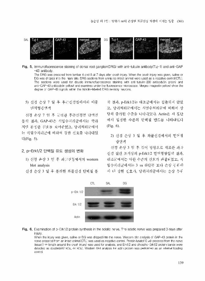
5) 7
7
, GAP-43
,
(Fig. 5).
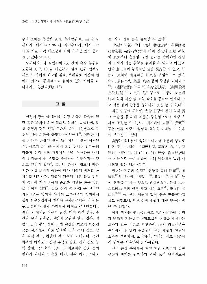
2. p-Erk1/2 단백질 유도 생성의 변화
1) 3 western
blot analysis
3
, p-Erk1/2
,
. Actin
(Fig. 6).
2) 3
3
p-Erk1/2 ,
,
3
,
SAL DG
Fig. 5.
CTL SAL DG
p-Erk 1/2
Erk 1/2
Actin
Fig. 6.
(361)

29 2 (2008 5 )
140
3
(Fig. 7).
3) 7
7
p-Erk1/2 ,
3
,
3
(Fig. 8).
CTL
SAL
DG
Injury site 3 mm distal
Fig. 7.
SAL
DG
Injury site 3 mm distal
Fig. 8.
(362)

1 : rat
141
4) 3, 7
3
p-Erk1/2 ,
,
(Fig. 9A).
7
p-Erk1/2 ,
,
(merged image)
(Fig. 9B).
3 7
p-Erk1/2 7 3
.
3. Cdc2 단백질 유도 생성의 변화
1) 7 western
blot analysis
SAL
DG
SAL
DG
A B
Fig. 9.
CTL
Cdc2
SAL DG
Actin
Fig. 10.
(363)

29 2 (2008 5 )
142
7
, Cdc2 ,
. Actin
(Fig. 10).
2) 3, 7
3
Cdc2 ,
3
,
3
,
.
7
Cdc2 ,
3
(Fig. 11).
4. 비신경 세포 증식 효과
1) 3 Hoechst
3
Hoechst
, 0.1
450±83 , 1, 3
0.1
758±67 ,
780±79
(p<0.05).
CTL
SAL
DG
Injury site 3mm distal
3 days post crushInjury site 3mm distal
7 days post crush
Fig. 11.
(364)

1 : rat
143
,
,
,
,
(Fig. 12).
2) 7 Hoechst
7
Hoechst
CTL
SAL
DG
proximal Injury site 1 mm distal 3 mm distal
A
1200
1000
800
600
400
200
0
The number of nuclei
DG
SAL
Distance from the injury site (mm)
-1 0 1 3
B
Fig. 12.
SAL
DG
Injury site 3 mm distal 7 mm distal 10 mm distal
A
1200
1000
800
600
400
200
0
The number of nuclei
DG
SAL
Distance from the injury site (mm)
0 3 7 10
B
Fig. 13.
(365)
29 2 (2008 5 )
144
, 0.1
862±96 , 852
±102
(p<0.01).
3, 7, 10
,
(Fig. 13).
고 찰
,
1),
7).
,
9).
4),
, ,
, ,
, ,
, ,
,
, ,
, , ,
, 5).
·10)
“
”
,
,
, , , 11)
, ·12)
“ ”, ·12)
“ ”
11).
,
13),
14)
.
, , , ,
, , ,
6).
15),
16) 17),
18),
19)
, 20)
, 21,22)
,
.
, rat
, ,
.
(366)

1 : rat
145
GAP-43, p-Erk1/2, Cdc2
.
GAP-43 43 kDa axonal growth-
associated protein 1980
23), rat
,
24).
GAP-43 ,
. GAP-43
(presynaptic terminal)
protein kinase C
CaM kinase kinase25)
.
, GAP-43
26).
,
GAP-43
24).
3 7
GAP-43 ,
,
. GAP-
43
. GAP-43
.
.
rat GAP
-43
GAP-43
,
.
Erk1/2 MAP kinase
, 27)
.
MAP kinase c-Jun N-terminal
kinase(JNK) p38
28).
MAP kinase
, kinase
. , Erk1/2 kinase MEK1/2
, JNK kinase
MEK 4, 627)
. JNK
Erk1/2
.
MEK kinase
, Erk1/2 JNK
29). Erk1/2
.
p-Erk1/2 3
7
.
,
(367)

29 2 (2008 5 )
146
.
p-Erk1/2
p-Erk1/2
MEK 1/2 p-Erk1/2
.
p-Erk1/2
30).
p-Erk1/2
Erk1/2
, Erk1/2
Rsk CREB(cAMP responsiveness element
binding protein)27)
.
p-Erk1/2
.
Cdc2 cycle G2 phase M
phase
cyclin B 31)
. , Cdc2
G2 phase
cyclin B1 B2
,
32).
Cdc2 cyclin
, Cdc2
33). Cdc2
.
Cdc2
,
Cdk
(apoptosis) ,
(proapoptotic protein) Bad
34).
Cdc2
Cdc2
35).
Cdc2 7
western blot analysis .
3 7
Cdc2
,
.
Cdc2
.
.
,
37). ,
,
,
1).
Cdc2
,
(368)

1 : rat
147
,
. 3 3
,
. ,
7
,
(0 10 )
.
.
.
5 DiI
. 3
,
21.8%
. 7
3
,
69.7%
(p<0.01),
DiI
.
3
(T11-12) ventral horn
,
62.5%
(p<0.01),
7 ventral horn 3
,
50%
(p<0.05).
, 3
, DiI
5
..
3
.
.
DiI
10
20% .
.
.
.
(preconditioning)
3 10
,
in vivo
(369)
29 2 (2008 5 )
148
36,37).
(lesion signal)
,
.
37).
,
.
. 1
, 2
.
GAP-43 ,
. in vivo
.
.
,
(multiple effects)
.
, GAP-43, p-Erk1/2
,
.
,
.
,
.
결 론
.
1. NF-200
.
2. GAP-43
.
3. p-Erk1/2
.
4. Cdc2
.
5.
.
.
참고문헌
1. Fawcett JW, Keynes RJ. Peripheral nerve
regeneration. Annu Rev Neurosci. 1990;13:43
-60.
2. Dyck PJ. The cases. clasification and treatment
of peripheral neuropathy. N Engl J Med.
1982;307:283-6.
3. Schwab ME, Bartholdi D. Degeneration and
regeneration of axons in the lesioned spinal
(370)

1 : rat
149
cord. Physiol Rev. 1996;76(2):319-70.
4. . . : . 1992:
640, 641.
5. Havton LA, Hotson JR, Kellerth JO. theory of
muscl energy tequnique. Muscle Nerve. 2008;
24(5):662-6.
6. . . : . 1981:101-2.
7. Waller A. Experiments on the section of the
glossopharyngeal and hypoglossal nerves of
the frog and observations of the alterations
produced thereby in the structure of their
primitive fibers. Philos Trans R Soc Lond B
Biol Sci. 1850;140:423-9.
8. Daniel WW. A foundation for analysis in the
health sciences. third edition. USA:MIT. 1983:
136-46.
9. Al-Majed AA, Neumann CM, Brushart TM,
Gordon T. Brief electrical stimulation promotes
the speed and accuracy of motor axonal
regeneration. J Neurosci. 2000;1:20(7):2602-8.
10. . . :
. 1982: :322, 359, :49.( )
11. .
. : . 1995:28.
12. · . .
: . 1982:73, 572, 578..( )
13. Tohda C, Kuboyama T, Komatsu K. Search
for natural products related to regeneration of
the neuronal network. Neurosignals. 2005;14
(1-2):34-45.
14. Xu H, Jiang B, Zhang D, Fu Z, Zhang H.
Compound injection of radix Hedysari to
promote peripheral nerve regeneration in rats.
Chin J Traumatol. 2002;5:107-11.
15. .
. . 1984;7:261-71.
16. , , .
. . 1996;13
(2):254-62.
17. , , .
. . 1994;11(4):113-29.
18. , .
. .
1996;13(1):1-10.
19. .
.
. 1991;14:381-95.
20. , , .
Stress
. . 2006;23(3):47-56.
21. , .
Intraluminal Filament
.
. 2004;21(2):1-20.
22. , , .
.
. 2003;18(4):25-35.
23. Skene JH, Willard M. Axonally transported
proteins associated with axon growth in rabbit
central and peripheral nervous system. J Cell
Biol. 1981;89:96-103.
24. Meberg PJ, Gall CM, Routtenberg A.
Induction of F1/GAP-43 gene expression in
hippocampal granule cells afterseizures. Brain
Res Mol Brain Res. 1993;19(1-2):179.
25. Alexander KA, Wakim BT, Doyle GS, Walsh
KA, Storm DR. Identification and characterization
of the calmodulin-binding domain of neuro-
modulin, a neurospecific calmodulin-binding
protein. J Biol Chem. 1988;263(16):7544-9.
26. Curtis R, Stewart HJ, Hall SM, Wilkin GP,
Mirsky R, Jessen KR. GAP-43 is expressed
by nonmyelin-forming Schwann cells of the
peripheral nervous system. J Cell Biol.
1992;116(6):1455-64.
(371)

29 2 (2008 5 )
150
27. Grewal SS, York RD, Stork PJ. Extracellular-
signal-regulated kinase signalling in neurons.
Curr Opin Neurobiol. 1999;9(5):544-53.
28. Xia Z, Dickens M, Raingeaud J, Davis RJ,
Greenberg ME. Opposing effects of ERK and
JNK-p38 MAP kinases on apoptosis. Science.
1995;24:270(5240):1326-31.
29. Desbarats J, Birge RB, Mimouni-Rongy M,
Weinstein DE, Palerme JS, Newell MK. Fas
engagement induces neurite growth through
ERK activation and p35 upregulation. Nat Cell
Biol. 2003;5(2):118-25.
30. Perlson E, Hanz S, Ben-Yaakov K, Segal-
Ruder Y, Seger R, Fainzilber M. Vimentin-
dependent spatial translocation of an activated
MAP kinase in injured nerve. Neuron.
2005;3:45(5):715-26.
31. Doree M, Hunt T. From Cdc2 to Cdk1: when
did the cell cycle kinase join its cyclin partner.
J Cell Sci. 2002;115(12):2461-4. (:
- 33,34 )
32. Porter LA, Donoghue DJ. Cyclin B1 and
CDK1: nuclear localization and upstream
regulators. Prog Cell Cycle Res. 2003;5:335-
47.
33. Manes T, Zheng DQ, Tognin S, Woodard AS,
Marchisio PC, Languino LR. Alpha(v)beta3
integrin expression up-regulates cdc2, which
modulates cell migration. J Cell Biol. 2003;
161:817-26.
34. Konishi Y, Bonni A. The E2F-Cdc2 cell-cycle
pathway specifically mediates activitydepriv-
ation-induced apoptosis of postmitotic neurons.
J Neurosci. 2003;23(5):1649-58.
35. Han IS, Seo TB, Kim KH, Yoon JH, Yoon
SJ, Namgung U. Cdc2-mediated Schwann cell
migration during peripheral nerve regeneration.
J Cell Sci. 2007;120(Pt2):246-55.
36. Richardson PM, Issa VM. Peripheral injury
enhances central regeneration of primary sensory
neurones. Nature. 1984;309:791 3.
37. Smith DS, Skene JH. A transcription-dependent
switch controls competence of adult neurons
for distinct modes of axon growth. J Neurosci.
1997;17(2):646-58.
(372)