Importance of collateral circulation in coronary heart disease
14
Review Importance of collateral circulation in coronary heart disease Colin Berry 1,3 * , Kanarath P. Balachandran 2 , Philippe L. L’Allier 1 , Jacques Lespe ´rance 1 , Raoul Bonan 1 , and Keith G. Oldroyd 3 1 Department of Medicine, Institut de Cardiologie de Montre´al, Montre´al, Que´bec, Canada; 2 Department of Cardiology, Bristol Royal Infirmary, Bristol, UK; and 3 Department of Cardiology, Western Infirmary, Glasgow, UK Received 14 June 2006; revised 27 November 2006; accepted 30 November 2006; online publish-ahead-of-print 11 January 2007 Aims Collateral arteries are a common but inconsistent finding in coronary heart disease (CHD). We endeavoured to review the methods for coronary artery collateral assessment, the predictors and clini- cal importance of collateral blood flow, and the potential for therapeutic augmentation of collateral anastomoses. Methods and results While many methods have been used to assess collateral blood flow only a few have been formally validated. Collateral flow index, as determined by measurement of intra-coronary pressure or flow velocity, is the most robust measure of collateral flow. These techniques have led to important advances in our understanding of collateral artery function. Coronary collateral arteries may prevent myocardial ischaemia in healthy subjects and in patients with CHD. A functional collateral circulation may lead to reduced ischaemia, preservation of ventricular function, and an improved prog- nosis. Recent trials have demonstrated that vascular progenitor cell therapies may improve ventricular function following acute myocardial infarction, raising the possibility of effective biological treatments to improve myocardial blood flow and prognosis in CHD. Conclusions Coronary collateral anastomoses represent a prognostically important adaptive response in patients with CHD. Therapeutic augmentation of collaterals with emerging biological therapies represents a desirable goal for treating CHD patients. KEYWORDS Angina; Angiogenesis; Collateral; Coronary artery; Myocardial infarction Introduction Coronary collateral arteries serve as alternative conduits for blood flow in obstructive coronary heart disease (CHD). The purpose of this article is to describe the importance of a functional coronary collateral circulation in CHD. We will illustrate how coronary collateral arteries can preserve myocardium and improve prognosis in CHD. Recent trials of biological therapies designed to improve myocardial per- fusion in CHD are also discussed. Methods The assessment, pathogenesis, and therapeutic promotion of coron- ary collaterals have recently been described by Seiler. 1 Respecting this work, our objective was to examine recent research regarding the functional and prognostic importance of coronary collaterals in different clinical settings. We have identified some important post-mortem anatomical publications, and have then identified key angiographic studies from the mid-1980s and informative clini- cal studies from the mid-1990s till the present time. To this end, we identified relevant English language publications through a PubMed search using the keywords ‘artery’, ‘collateral’, ‘coronary’, ‘human’, and ‘ischaemic heart disease’. After evaluat- ing these papers, and based on our own practical experiences, we prepared a manuscript which places emphasis on in vivo human investigations, illustrating a contemporary position on collateral function in different patient groups. Owing to word count limi- tations, we have restricted the citations to, in our view, the most relevant and informative publications. Clinical assessment of coronary collateral arteries Collateral artery growth is mediated by arteriogenesis (Table 1). Anatomically, collateral arteries may be either epicardial or intramyocardial and serve as contralateral or ipsilateral conduits (Figures 1 and 2). 2 Myocardial blood flow is the product of epicardial coronary and collateral artery flow. Angiographic evaluation of coronary collateral arteries Coronary angiography is the standard method to identify coronary collateral arteries (Table 2). Angiography with ordinary visual detection has limited resolution. Arterioles & The European Society of Cardiology 2007. All rights reserved. For Permissions, please e-mail: [email protected] * Corresponding author: Glasgow Cardiovascular Research Centre, Univer- sity of Glasgow, 126 University Place, Glasgow G128TA, UK. Tel: þ44 15143763330; fax: þ44 141 211 1906. E-mail address: [email protected] European Heart Journal (2007) 28, 278–291 doi:10.1093/eurheartj/ehl446 Downloaded from https://academic.oup.com/eurheartj/article/28/3/278/2887447 by guest on 28 December 2021
Transcript of Importance of collateral circulation in coronary heart disease

Review
Importance of collateral circulation in coronaryheart disease
Colin Berry1,3*, Kanarath P. Balachandran2, Philippe L. L’Allier1, Jacques Lesperance1,Raoul Bonan1, and Keith G. Oldroyd3
1Department of Medicine, Institut de Cardiologie de Montreal, Montreal, Quebec, Canada; 2Department of Cardiology,Bristol Royal Infirmary, Bristol, UK; and 3Department of Cardiology, Western Infirmary, Glasgow, UK
Received 14 June 2006; revised 27 November 2006; accepted 30 November 2006; online publish-ahead-of-print 11 January 2007
Aims Collateral arteries are a common but inconsistent finding in coronary heart disease (CHD). Weendeavoured to review the methods for coronary artery collateral assessment, the predictors and clini-cal importance of collateral blood flow, and the potential for therapeutic augmentation of collateralanastomoses.Methods and resultsWhile many methods have been used to assess collateral blood flow only a few havebeen formally validated. Collateral flow index, as determined by measurement of intra-coronarypressure or flow velocity, is the most robust measure of collateral flow. These techniques have led toimportant advances in our understanding of collateral artery function. Coronary collateral arteriesmay prevent myocardial ischaemia in healthy subjects and in patients with CHD. A functional collateralcirculation may lead to reduced ischaemia, preservation of ventricular function, and an improved prog-nosis. Recent trials have demonstrated that vascular progenitor cell therapies may improve ventricularfunction following acute myocardial infarction, raising the possibility of effective biological treatmentsto improve myocardial blood flow and prognosis in CHD.Conclusions Coronary collateral anastomoses represent a prognostically important adaptive response inpatients with CHD. Therapeutic augmentation of collaterals with emerging biological therapiesrepresents a desirable goal for treating CHD patients.
KEYWORDSAngina;
Angiogenesis;
Collateral;
Coronary artery;
Myocardial infarction
Introduction
Coronary collateral arteries serve as alternative conduits forblood flow in obstructive coronary heart disease (CHD). Thepurpose of this article is to describe the importance of afunctional coronary collateral circulation in CHD. We willillustrate how coronary collateral arteries can preservemyocardium and improve prognosis in CHD. Recent trialsof biological therapies designed to improve myocardial per-fusion in CHD are also discussed.
Methods
The assessment, pathogenesis, and therapeutic promotion of coron-ary collaterals have recently been described by Seiler.1 Respectingthis work, our objective was to examine recent research regardingthe functional and prognostic importance of coronary collateralsin different clinical settings. We have identified some importantpost-mortem anatomical publications, and have then identifiedkey angiographic studies from the mid-1980s and informative clini-cal studies from the mid-1990s till the present time.
To this end, we identified relevant English language publicationsthrough a PubMed search using the keywords ‘artery’, ‘collateral’,‘coronary’, ‘human’, and ‘ischaemic heart disease’. After evaluat-ing these papers, and based on our own practical experiences, weprepared a manuscript which places emphasis on in vivo humaninvestigations, illustrating a contemporary position on collateralfunction in different patient groups. Owing to word count limi-tations, we have restricted the citations to, in our view, the mostrelevant and informative publications.
Clinical assessment of coronarycollateral arteries
Collateral artery growth is mediated by arteriogenesis(Table 1). Anatomically, collateral arteries may be eitherepicardial or intramyocardial and serve as contralateral oripsilateral conduits (Figures 1 and 2).2 Myocardial bloodflow is the product of epicardial coronary and collateralartery flow.
Angiographic evaluation of coronarycollateral arteries
Coronary angiography is the standard method to identifycoronary collateral arteries (Table 2). Angiography withordinary visual detection has limited resolution. Arterioles
& The European Society of Cardiology 2007. All rights reserved. For Permissions, please e-mail: [email protected]
* Corresponding author: Glasgow Cardiovascular Research Centre, Univer-sity of Glasgow, 126 University Place, Glasgow G128TA, UK. Tel: þ4415143763330; fax: þ44 141 211 1906.
E-mail address: [email protected]
European Heart Journal (2007) 28, 278–291doi:10.1093/eurheartj/ehl446
Dow
nloaded from https://academ
ic.oup.com/eurheartj/article/28/3/278/2887447 by guest on 28 D
ecember 2021

,100 mm are invisible to the naked eye,3 and visiblecollaterals typically have a diameter of 0.5 mm.4 The angio-graphic assessment of collateral circulation can be refinedby off-line computer analyses.4
Angiographic collateral degreeCoronary collaterals may be spontaneously visible or recrui-table.1 Recruitable collaterals may defined by the absenceof visible collaterals before coronary occlusion (grade 0 or1) and the development of collaterals during coronary occlu-sion (grade 2 or 3).5 Spontaneously visible collaterals arepresent when they are grade 2 or 3 prior to any intervention.In the setting of a stenosed artery, collaterals may
be graded using the Rentrop classification:6 0, no visible col-lateral channel filling; I, faintly visible collaterals with fillingof branches but no filling of the distal stenotic (culprit)artery; II, collaterals partially filling the branches of thestenosed artery; III, complete collateral filling of thestenosed artery. In patients with single vessel coronaryartery disease (CAD) and preserved left ventricular (LV)function, collateral filling grade increases acutely (within90 s) during balloon occlusion and diminishes on balloondeflation.6 When the culprit artery is patent, the assessmentof both spontaneously visible and recruitable collateralsrequires a double artery approach with balloon occlusionin the culprit (collateral receiving) artery and simultaneouscontrast injection in the donor artery5,7 (see also Seiler’s1
review).Contrast washout collaterometry is based on the hypoth-
esis that the time to contrast clearance distal to an occludedartery is inversely related to collateral flow.8 In other words,contrast is cleared more rapidly in a well-collateralized ter-ritory. Other angiographic methods for collateral evaluationare summarized in Table 2.
Angiographic demonstration of coronarycollateral arteriesThe complete angiographic assessment of coronary collat-eral arteries requires the induction of maximal hyperaemia,which may be achieved by systemic or intra-coronary vasodi-lator administration. During routine angiography, intra-coronary vasodilator therapy is usually adequate [e.g. nitro-glycerine (100–200 mg) or adenosine (15–40 mg for the leftcoronary artery and 10–30 mg for the RCA)].9 A graduatedinjection of contrast should achieve complete coronaryartery opacification, with cine angiography continued until
coronary sinus opacification is achieved. The X-ray imageintensifier may be ‘panned’ during cine acquisition toensure that collateral flow is fully demonstrated.Collateral filling should be assessed in cine frames obtainedduring diastole.4
Invasive assessment of coronarycollateral artery function
Coronary pressure and flowInvasive studies have demonstrated coronary collateral flowmay be bidirectional.10,11 The coronary wedge pressure,which is the distal coronary pressure obtained when ante-grade flow is prevented by intra-coronary balloon inflation,is a marker of collateral function.12
Coronary collateral blood flow can be objectively quanti-fied using an 0.01400 (0.36 mm) intra-coronary wire which haseither a flow velocity (Doppler)13 or a pressure measuringcapability.14 Pressure-derived fractional collateral flowreserve is estimated by calculating the collateral pressureindex (CFI) during maximal hyperaemia.14,15 CFI is based onthe premise that coronary perfusion pressure or flow velo-city distal to an occlusion are derived from collateral flow.The Doppler-derived CFIv ratio is measured by obtaining
distal coronary flow velocity (CFV) during patency andballoon occlusion (Table 3).16 Distal occlusive collateralflow velocity correlates well with coronary wedge pressure,particularly in non-infarcted myocardium.17 The CFV reserve(CFVR) is the ratio of maximum hyperaemic average peakvelocity (APV) to basal APV, and a CFVR , 2 in the absenceof an epicardial artery stenosis (i.e. FFR � 0.75) representsmicrovascular dysfunction. In collateral-dependent terri-tory, functional measures of collateral flow, such as a basalAPV, are positively correlated with collateral size.18
The pressure-derived collateral flow index (CFIp or PDCF)depends on the simultaneous estimation of mean aorticpressure (PAo), mean central venous pressure (Pv), andmean distal coronary pressure during balloon occlusion(PD; Table 3).14,16 CFIp is synonymous with the maximumrecruitable collateral flow reserve (Qc) expressed as a frac-tion of normal myocardial perfusion: Qc/Qn ¼ CFIP.When pressure and Doppler studies are performed succes-
sively, the collateral resistance index (RColl, mmHg cm21
s21) can be calculated (Table 3) assuming constant vesseldiameters before and during balloon occlusion.5,19–21 Inother words, RColl is derived from the relationship betweenthe pressure gradient across the collateral bed andcollateral-derived flow velocity measured in the recipientartery during balloon occlusion. The components of thecollateral pathway (RColl) are the collateral microvascularnetwork resistance (RC) and the donor artery resistance(RD; Table 3). RColl is inversely correlated with the extentof functional collateralization.22 The other resistancenetworks are those of the obstructed recipient artery(ROccl) and the peripheral myocardial microcirculation (RP;Table 3).Coronary vascular resistances may vary acutely due to, for
example, distal embolization or vasoactive drugs.5 Chronicchanges in coronary artery resistance may occur after‘fixed’ alterations in flow, such as after successful PCI.23
Collateral flow velocity is influenced by ventricular function.This relationship can probably be explained by variation inmicrovascular function (or more specifically, microvascular
Table 1 Definitions of arteriogenesis, angiogenesis, andcollateral arteries
Attribute Definition
Angiogenesis Formation of new capillaries bysprouting from post-capillary venules
Arteriogenesis The transformation of pre-existingarterioles into functional (muscular)collateral arteries with vasomotorproperties
Collateral blood vessel Collateral blood vessels are vascularconnections linking parallelarteries without an interveningcapillary bed
Collateral circulation in CHD 279
Dow
nloaded from https://academ
ic.oup.com/eurheartj/article/28/3/278/2887447 by guest on 28 D
ecember 2021

resistance) according to the presence and severity of ischae-mic myocardial damage and contractile dysfunction.24 Theresponse to vasodilators varies between collateral types,and recruitable collaterals may be less responsive to vasodi-lators than spontaneously visible collaterals. This is probablybecause spontaneously visible collaterals are anatomicallyand functionally well developed compared with recruitablecollaterals which may be less mature.5
Seiler and coworkers20 were the first to use three simul-taneous intra-coronary sensor wires in different areas toelucidate the mechanisms leading to coronary steal (see inwhat follows). Clearly, a triple wire approach is not readilyapplicable to ordinary catheter laboratory practice, andmeasurement of RColl is usually reserved for research pur-poses by experienced operators.Pressure- and velocity-derived assessments of fractional
coronary flow are not identical. Each measure is subject todifferent physical laws and may differentially influenceflow resistances in the coronary, microvascular, and collat-eral systems. For example, Doppler, which measures CFVrather than flow volume, is affected by vessel diameter,tortuosity, branching (due to altered velocity waveformprofiles), and distal wire placement.13 A constant coronary
artery diameter is an important prerequisite for bothDoppler and pressure measurements, however, this may beless important for a Doppler approach.13 The Dopplerwire is very sensitive to phasic alterations in forwardand reverse flow, whereas the pressure wire is not.Alternatively, CFIp is entirely independent of alterations inheart rate and aortic pressure, whereas the Dopplerapproach is not.
Collateral blood flow measurements using Doppler andpressure techniques were highly correlated in experimentalanimal validation studies (r ¼ 0.96),14 but the correlationswere weaker in human validation analyses (CFIv vs. CFIp:r ¼ 0.8).16 In the latter, Seiler et al.16 studied 51 patientswith single or double-vessel CAD and assessed collateral suf-ficiency according to ECG changes during intra-coronaryballoon inflation. The pressure-derived collateral index(CFIp) underestimated fractional collateral flow comparedwith the velocity-derived index (CFIv). Thus, comparedwith the Doppler-derived CFIv, the pressure-derived CFIpappears to be slightly less sensitive with a weaker positivepredictive accuracy for the detection of a functional collat-eral circulation.16 As Doppler- and pressure-derived assess-ments of coronary blood flow are based on different
Figure 1 (A) RAO 158 and (B) RAO 108 cranial 408 projections. Injection of the left dominant coronary artery results in opacification of ipsilateral collateralconnections (CC) from the second marginal (OM; crossed arrows) and posterior descending arteries (PDA; plain arrows) to the distal LAD. (C) RAO 158 view.Injection of the left coronary artery results in opacification of contralateral collateral connections (CCC) from the anterior septal arteries to the posterior des-cending artery (PDA). (D) RAO 158 caudal 258 view. Opacification of collateral artery connections from a large diagonal artery (DIAG) to an occluded obtuse mar-ginal artery via ipsilateral collateral connections (ICC). Opacification of the coronary sinus (CS) indicates complete collateral artery filling.
280 C. Berry et al.
Dow
nloaded from https://academ
ic.oup.com/eurheartj/article/28/3/278/2887447 by guest on 28 D
ecember 2021

physical laws, whether they can be used interchangeably toassess collateral function, as suggested by Seiler et al.,16 isdebatable. One limitation of their validation study was thatcentral venous pressure (CVP) was only measured in a min-ority of patients (n ¼ 4), and perhaps the sensitivity ofCFIp might have improved with CVP measurement in allcases.
Physiological assessment of coronarycollaterals in clinical practiceCVP should be measured for optimal assessment of collateralblood flow.25,26 Collateral supply should be assessed duringmaximal coronary hyperaemia which is best achieved by sys-temic intravenous infusion of adenosine (140 mg/kg/min).27
Intra-coronary measurements should be obtained duringconstant conditions (e.g. constant artery diameter) andrefer to specific locations rather than to the entire artery.CFIp may overestimate collateral supply under conditionsof elevated transmural stress, such as raised LV end-diastolicpressure.25,28,29
From the perspective of ordinary clinical practice, webelieve Doppler- and pressure-wire derived assessments ofcollateral supply represent an important advance onearlier angiographic methods, and in general terms, eitherapproach can be reliably used for assessing collateralsupply. On balance, the Doppler approach may be superior,particularly when fractional collateral flow is low.In elective PCI, Qc/Qn and PD/PAo are of prognostic value.
Qc/Qn of.0.28 or a PD/PAo of.0.30 identifies a subgroup ofpatients with sufficient collateral protection and a low rateof ischaemic events following successful PCI.15 Adequatecollateral artery function, reflected by the absence of ECGchanges during PCI, is better predicted by CFIp [.0.3; posi-tive predictive value (PPV): 75%] and CFIv (.0.3; PPV: 79%)rather than the angiographic collateral grade (�2; PPV: 69%)or the absence of angina during balloon inflation (PPV:50%).16 The observations from Seiler and coworkers are sup-ported by those of Pijls et al.15 Haemodynamic factors thatinfluence contralateral collateral flow include ipsilateraldistal vascular resistance, contralateral stenosis severity,and the size of the collateralized vascular bed.30
Angiographic measures of collateral blood flow correlatepoorly with Doppler-derived measurements,10 and in clinicalpractice, Doppler- or pressure-derived CFI are the referencemethods for the assessment of collateral flow.31
Non-invasive evaluation of coronaryartery collateral arteries
Although non-invasive methods may hold promise, collateralassessment is most reliable when performed with transient(balloon inflation) or chronic complete occlusion of theculprit artery (Table 2). Myocardial contrast echocar-diography (MCE) demonstrated the importance of collateralarteries for recovery of LV function after MI.32,33 Myocardialperfusion imaging after 99mTc-Sestamibi radionuclide injec-tion during PCI has also been used to quantify collateralsupply by evaluation of the extent of ischaemia.31 Morerecently, Vogel et al.25 demonstrated the feasibility of real-time non-invasive assessment of collateral supply using MCEand systemic administration of contrast. They demonstrateda close relationship between the CFIp and absolutecollateral-derived myocardial blood flow (MBFc) measuredby echocardiography in 32 PCI cases (CFIp ¼ 0.62 MBFcþ0.05; r2 ¼ 0.75; P , 0.0001).Collateral-dependent myocardial perfusion can be quanti-
fied by magnetic resonance imaging (MRI), however, anangiographic description of the coronary collateral circula-tion is a prerequisite requirement. Coronary MRI lacks suffi-cient resolution to delineate collateral arteries which areusually of modest calibre. Multi-slice computed tomography(CT) has emerging potential for the non-invasive assessmentof coronary collateral supply. This technique best imagesproximal and intermediate coronary segments, however,whether collateral connections, which are usually distal,can be delineated by contrast CT remains to be determined.Positron emission tomography (PET) is a powerful researchtool for quantification of collateral-dependent myocardialblood flow,24 but it is an impractical technique for routineclinical practice.
Coronary collateral circulation in selectedpatient groups and settings
Collaterals in individuals without CHDThe human coronary circulation is not an end-arterialsystem as it was previously considered in the past. Further-more, nascent collateral arteries occur in neonates34 and inhealthy individuals.35,36
In the 1950s, Fulton,36,37 undertook three-dimensionalpost-mortem stereo-angiographic studies of coronaryarteries in preserved hearts from subjects who died fromnon-ischaemic causes and had normal hearts (Figure 3). Hedemonstrated extensive superficial and deep intercoronaryanastomoses, which were particularly abundant in the inter-ventricular septum and in the subendocardial plexus of theleft ventricle.36 This plexus likely contributes to the myocar-dial ‘blush’ observed during angiography. Baroldi et al.35
produced post-mortem coronary artery casts from normalhearts following digestion of cardiac tissue. These castsrevealed striking images of inter- and intra-coronary anasto-moses.35 Collateral arteries are more abundant in the LVthan in the right ventricle (RV) and are uncommon in thesubepicardial territory in man, in contrast to the dog.Return of blood flow in diastole to the deep layers of theLV is favoured by certain vessels which course directlyfrom the epicardial arteries almost without branching tofeed the subendocardial plexus.36,37
Collateral blood flow may prevent ischaemia even inhealthy subjects.38,39 Considerable variation exists in the
Figure 2 (A) LAO 308 view. Faint ipsilateral collateral artery connections‘bridging’ a chronic total occlusion of the proximal–mid RCA. A defibrillatorlead is seen positioned in the right ventricular apex. (B) RAO 308 view alsoillustrating contralateral collateral arteries from the right ventricular conusbranch to the LAD.
Collateral circulation in CHD 281
Dow
nloaded from https://academ
ic.oup.com/eurheartj/article/28/3/278/2887447 by guest on 28 D
ecember 2021

Table 2 Methods for assessment of collateral arteries
Method Strengths Weaknesses References
AngiographicVisual Visual assessment Easy to do Subject to intra and interobserver error;
ordinal variable; limited resolution(arterioles ,100 mm invisible)
3
Visual—computer assisted Visual—computer assisted Easy to do; time consuming Not routinely performed 4Contrast appearance time Visual assessment Easy to do Subject to intra and interobserver error;
continuous variable82,99
Angiographic collateral degree(Rentrop’s grade)6
Visual assessment. Settings: ipsilateral CAD;patent coronary artery
Easy to do; standard methods Subject to intra and interobserver error;ordinal variable; validated in setting ofsingle vessel disease (LAD or CX) andpreserved LV systolic function;6
angiographic assessment for recruitablecollaterals requires a double injectiontechnique; a second arterial punctureis required for injection into the donorartery prior to and during ballooninflation in the recipient (culprit) artery
6,10,11,43,63,64,68
Collateral connections score Visual assessment. Setting—chronic totalocclusion
Easy to do; standard method; anatomicalbasis incorporating collateral size
Subject to intra and interobserver error;optimal angiographic views requiredto avoid foreshortening; ordinal variable
82
Coronary flow TIMI frame count; Fourier spectral analysis;Cineangiographic modelling; Washoutcollaterometry
Standard methods; washout collaterometrydoes not require a second arterialpuncture, is easy to do and is inexpensive
Dedicated software required for Fourierspectral analysis; subject to theeffects of variation in contrastinjection and alterations in distalmicrovascular resistance
8,81,100
PhysiologicalDoppler wire Doppler frequency collateral flow velocity Validated; continuous variable Expensive; relevant technology required;
appropriate training5,13,16,17,22,30,82,99
Pressure wire Pressure wire e.g. RADIw Validated; CFIp index continuous variable Expensive; relevant technology required;appropriate training; may only be doneduring coronary balloon angioplasty
14,25,30,82,99
Perfusion imagingContrast echocardiography Ultrasound contrast agent and standard
echocardiographyEasy to do; relatively inexpensive; contrast
may be administered by intra-coronaryor intravenous injection
Invasive access still required to demonstratea chronic total occlusion, or if antegradeflow is present in the culprit artery, toobstruct flow by transient balloon occlusion
25,32,33
MRI Coronary blood flow and myocardial perfusion Accurate assessment of myocardialperfusion; may provide informationon myocardial viability
Myocardial and epicardial artery measuresof collateral function may differ; off-lineanalyses
101
PET Myocardial perfusion Information on perfusion and metabolicfunction
Expensive; off-line analyses 24
Scintigraphy Nuclear isotope infusion with comparison ofmyocardial perfusion at rest and duringpharmacological stress or exercise
Assessment of myocardial perfusion No direct assessment of collateral blood flow 31
282C.Berry
etal.
Dow
nloaded from https://academ
ic.oup.com/eurheartj/article/28/3/278/2887447 by guest on 28 D
ecember 2021
extent of collateral supply even between individuals withsimilar severities of CHD.40 The reasons for this variabilityare uncertain but may be genetic41 or acquired (e.g. dur-ation of angina, drug therapy, extent of coronary disease).
Angina pectoris
Pathogenesis of the collateral circulationFulton and Royen36 demonstrated that in patients withobstructive CAD, functional coronary collaterals developby enlargement of pre-existing coronary anastomoses. Themain stimulus to vessel enlargement was the collateralblood flow generated by differential pressure gradientsresulting from coronary artery occlusion or stenosis. Healso demonstrated that the extent of coronary artery col-lateralization was related to angina duration.42
Predictors of coronary collateral supplyPohl et al.38 investigated the predictors of collateral arterysupply in 450 patients [mean (standard deviation (SD)) age61(11) years] with 1–3 vessel CAD but no prior history ofQ-wave MI. Coronary collateral arteries were assessed bymeasurement of intra-coronary pressure (n ¼ 328), flow vel-ocity (n ¼ 217), or simultaneously with both techniques(n ¼ 192). In the group with insufficient collateral function[CFI , 0.25; n ¼ 307 (68%)], angina and ST-segmentelevation during balloon occlusion developed in 67 and 84%of patients, respectively. The corresponding rates inpatients with adequate collateral blood flow [CFI � 0.25;n ¼ 143 (32%)] were 42 and 26%, respectively (P, 0.0001).The only multivariate predictor of functionally importantcollateral blood flow (CFI � 0.25) was percent diameterstenosis. CAD progression, as measured by quantitativecoronary analysis is associated with increase in collateralsupply (Rentrop’s grade), whereas regression in disease isassociated with reduced collateral formation.43
Other studies in angina patients undergoing PCI havedemonstrated percent diameter stenosis,40,44 duration ofangina,40,45 and proximal lesion location to correlate withcollateral supply. The relationship between LV systolic func-tion and CFIp is influenced by several factors, including theseverity of CHD, LV end-diastolic pressure,25,28,29 and micro-vascular function.46,47
Coronary collateral supply in stable angina
Exercise testingThe extent of coronary collateralization may influence exer-cise test results. For example, in a study of angina patients
with obstructive (stenosis �90% reference diameter) singlevessel CAD, myocardial perfusion defects revealed by exer-cise thallium scintigraphy were more common and moreextensive than in patients without collaterals arteries.48
In other words, collateral arteries may predispose to afalse negative stress-perfusion scan. In this investigation,collateralization did not influence exercise duration orST-segment depression, which suggests that factors whichinfluence coronary flow and myocardial ischaemia, such ascollateral connections, may affect exercise tests and stressperfusion scans in different ways.
Severity of anginaUsing invasive methods in patients undergoing PCI of a singlecoronary stenosis, Billinger et al.20 demonstrated that thecontra-lateral donor artery resistance RContra, and RCollcorrelated better with the ispsilateral CFVR than ipsilateralstenosis severity. In other words, blood flow velocity in aterritory subtended by a stenosed artery may be influencedmore by the resistance to blood flow in the donor arterycollateral pathway than the percent diameter of theculprit stenosis. This observation indicates that reduced col-lateral artery blood flow (through increased resistance) maynegatively influence myocardial blood flow, which may inturn provoke or exacerbate angina. Altered collateralpathway resistance is an important mechanism to explaincoronary artery steal.20
A 10-year angiographic follow-up study demonstrated col-lateral flow capacity to have increased over three-fold insubjects with improved angina, whereas collateral flowremained constant in subjects with no improvement inangina (P ¼ 0.01).4 In other words, collateral growth corre-lated with reduced angina. A well-developed collateralsupply at the time of elective PCI is associated with anincreased risk of subsequent angina.49 The reasons for whythis may be are controversial, and may involve coronarysteal and restenosis.
Collateral supply and ventricular functionIn patients with obstructive CAD, LV regional contractilityand relaxation are influenced by collateral blood flow.50
Furthermore, in angina, the presence of regional myocardialcontractile dysfunction, altered oxidative metabolism andincreased glucose utilization, are related to inadequateblood flow in collateral artery-dependent territories.24
In other words, inadequate blood supply in collateral-dependent myocardium may lead to ventricular dysfunction.
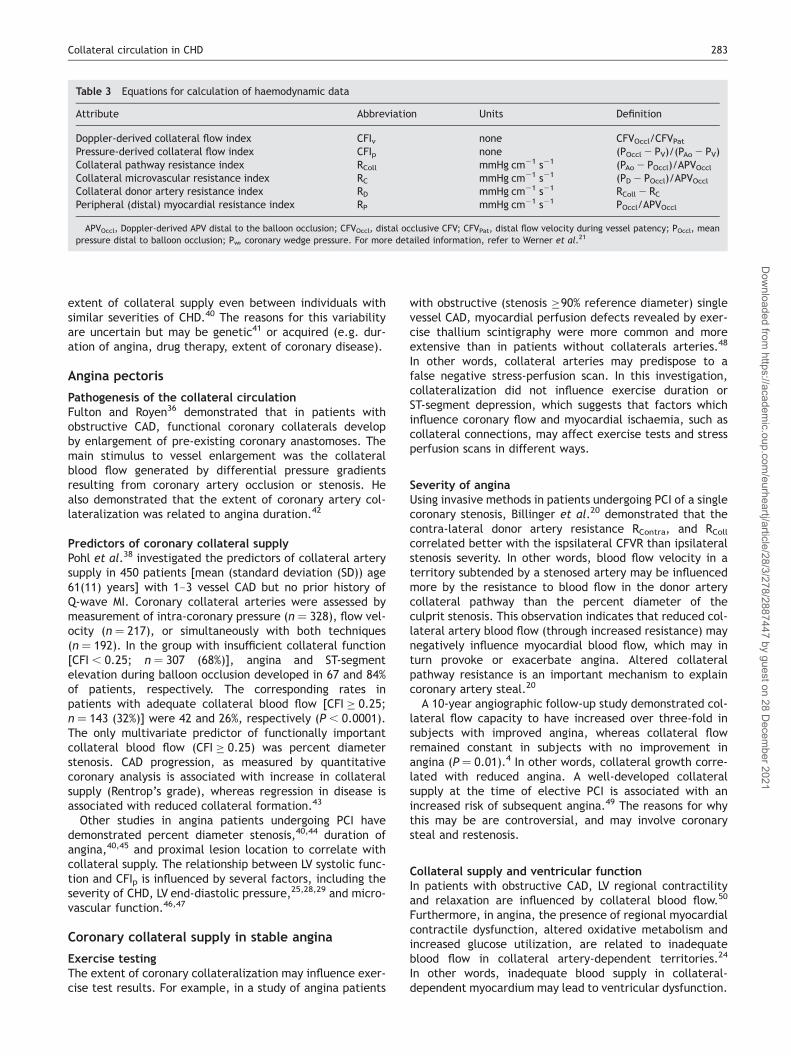
Table 3 Equations for calculation of haemodynamic data
Attribute Abbreviation Units Definition
Doppler-derived collateral flow index CFIv none CFVOccl/CFVPatPressure-derived collateral flow index CFIp none (POccl2 PV)/(PAo2 PV)Collateral pathway resistance index RColl mmHg cm21 s21 (PAo2 POccl)/APVOcclCollateral microvascular resistance index RC mmHg cm21 s21 (PD2 POccl)/APVOcclCollateral donor artery resistance index RD mmHg cm21 s21 RColl2 RCPeripheral (distal) myocardial resistance index RP mmHg cm21 s21 POccl/APVOccl
APVOccl, Doppler-derived APV distal to the balloon occlusion; CFVOccl, distal occlusive CFV; CFVPat, distal flow velocity during vessel patency; POccl, meanpressure distal to balloon occlusion; Pw, coronary wedge pressure. For more detailed information, refer to Werner et al.21
Collateral circulation in CHD 283
Dow
nloaded from https://academ
ic.oup.com/eurheartj/article/28/3/278/2887447 by guest on 28 D
ecember 2021

Risk of future myocardial infarctionIn a series of 403 patients with stable angina who underwentelective PCI and were then followed-up (mean follow-upperiod of 94 weeks) for clinical events, those with poorlydeveloped collaterals (CFIv ,0.25) experienced a higherincidence of MI (4.1%), compared with the incidence ofMI (0%; P ¼ 0.02) in patients with a well-developedcollateral supply (CFIv �0.25).49 This remarkable findingmay be explained by the contribution of collateral-derived myocardial perfusion after an acute coronarythrombosis.
SurvivalHansen51 explored the natural history of patients with CHDin relation to the presence or absence of coronary collateralarteries. Collateral arteries were evident in 104 (35%) of 300patients undergoing clinically indicated coronary angiogra-phy, with the highest proportion being in patients with anoccluded artery [95/101 (94%)]. Over 10 years, 51.1 and34.5% of patients in the collateral and non-collateralgroups survived (adjusted P ¼ 0.1). Clearly, more studiesare required to clarify the prognostic value of collateralsupply in stable CHD.
Coronary steal in stable CHD
Definition‘Coronary steal’ is a complex phenomenon in which regionalmyocardial hypoperfusion occurs through diversion of co-ronary blood flow to adjacent coronary beds. Coronarysteal may be mediated by collateral arteries. Seiler andcoworkers19,20,52 defined coronary steal as being a fall incoronary blood flow in one collateralized vascular region infavour of another during coronary arteriolar vasodilatation,i.e. a CFVR ,1 during maximal hyperaemia. Werner et al.21
defined coronary steal as a reduction in APVOccl during ade-nosine infusion, whereas an increase in APVOccl representeda positive collateral flow reserve. Thus, coronary steal mayprovoke angina, despite the presence of an apparently ade-quate collateral supply.
Mechanisms of coronary stealCoronary steal may be ‘vertical’ between different layersof the myocardium, or ‘lateral’ via branches through adja-cent vascular areas originating from a common branchbifurcation.19 Coronary steal is not necessarily collateral-dependent,21 but it is influenced by CAD severity and ventri-cular function.21
Seiler et al.19 used a Doppler wire to quantify CFVR andCFI in 100 patients undergoing coronary angioplasty. Theyassessed the occurrence of coronary steal in stenosed,collateralized coronary arteries by measurement ofmaximal mean CFV before and after intra-coronary adeno-sine injection. A CFVR ,1 during systemic adenosine-induced hyperaemia occurred in 10% of patients. Coronarysteal was associated with angiographic evidence ofenhanced coronary artery collateralization, proximal lesionlocation, and greater percent diameter stenosis. Comparedwith patients without evidence of coronary steal, fewerpatients with steal experienced angina or had intra-coronaryECG changes during balloon inflation. In other words, inpatients with coronary steal, preservation of coronary flowdistal to the balloon occlusion by collateral blood flowresulted in reduced ischaemia. Collateral flow to the vascu-lar region studied during balloon inflation decreased duringadenosine-induced hyperaemia, indicative of a steal effectvia extensive collaterals to the donor territory. These find-ings provided in vivo evidence of an association betweenthe presence and extent of steal away from the collatera-lized coronary territory and the amount of collateralstowards it.
Werner et al.21 recently dissected the contribution of indi-vidual resistance components in patients with or withoutsteal. Coronary steal occurs in �30% of chronic totallyoccluded (CTO) patients.53 Their use of CTO patients, ratherthan patients with a patent culprit artery, enabled a morecontrolled study of the coronary circulation.21 In theircohort of 56 patients undergoing CTO PCI, Werner et al.demonstrated that in patients who manifest a steal phenom-enon with adenosine infusion (46%), the donor artery FFR waslower, its resistance (RD) was higher, and DRD increasedfurther with adenosine, compared with patients withoutsteal. Collateral pathway resistance (RColl) increased withadenosine infusion in the steal group, whereas RColl and Rpfell in subjects with a positive collateral flow reserve.Overall, changes in RP were strongly correlated withchanges in RColl (R ¼ 0.8; P, 0.001). Notably, the pressure
Figure 3 Ex vivo arteriogram of a normal human heart. Radiography withthe heart submerged in saline virtually eliminated myocardial tissue shadow-ing an ensured homogenous exposure to all blood vessels. Before injection, abranch of the left CX was doubly ligated. Retrograde filling of the distalartery, in less than 20 s, demonstrated the existence of anastamotic com-munications. Stereoscopy revealed that the communicating vessels werelargely in the deep, subendocardial network. Penetration of the injectionmedium to 10–15 mm diameter blood vessels with radiological demonstrationto 20 mm. Arterio-mediastinal anastomoses are also seen above the heart(�1). Permission granted from Fulton37 and from the British Institutes ofRadiology.
284 C. Berry et al.
Dow
nloaded from https://academ
ic.oup.com/eurheartj/article/28/3/278/2887447 by guest on 28 D
ecember 2021

gradient across the collateral networks did not change duringhyperaemia. Interestingly, mean (SD) FFRD increased pro-gressively between groups [reduced collateral flow reserve(i.e. coronary steal) vs. no change vs. increased collateralflow reserve: 0.78 (0.13) vs. 0.84 (0.17) vs. 0.90 (0.09)].Therefore, they confirmed that the conditions for provokingcoronary steal involve increased collateral channelresistance, and the combination of a flow-limiting donorartery stenosis proximal to the collateral origin and/or animpaired microvascular response to vasodilator stimulus.21
In other words, coronary artery steal is unlikely to occur inlarge collateral channels (with low resistance).21,53
Clinical significance of coronary stealHolmvang et al.54 studied the relationships between coro-nary artery flow, myocardial perfusion, and LV systolic func-tion in 15 patients with symptomatic CHD. Collateral bloodflow and myocardial perfusion were assessed by angiographyand PET, respectively, and LV function was assessed by echo-cardiography. They found that the collateral-dependentcoronary flow reserve measured during adenosine-inducedvasodilatation was reduced in collateral flow-dependentsegments. The steal phenomenon was most evident in seg-ments with impaired basal contractility. Werner et al.21
also reported an association between coronary steal andimpaired ventricular function. Their hypothesis that stealcould explain regional dysfunction in CTO patients withouta history of MI merits prospective assessment.Coronary collateral steal may explain why blood flow
through collateral arteries may not always be beneficial.Diversion of blood flow through a low resistance collateralcircuit away from an ischaemic territory could be one expla-nation for the potential pro-ischaemic effects of nifedipinein angina.55 Coronary steal may also give rise to competitiveflow which could contribute to restenosis after angio-plasty.19,56 For these reasons, Seiler et al.19 questionedthe value of PCI in patients with well-developed collateralarteries and suggested medical therapy with beta-blockersas an alternative. Notably, the anti-ischaemic effects ofbeta-blockers are not influenced by coronary collateralartery supply.57
Acute myocardial infarction
Collateral artery connections may serve as a natural bypassafter an acute coronary artery occlusion. In this section,regulation of coronary collateralization and its prognosticimportance after MI are examined.
Coronary collateral artery adaptation in acuteMI: relationships with ventricular function
Most,32,58–64 but not all,65 MI studies have suggested thatmyocardial perfusion may be maintained and LV functionpreserved in patients with an adequate coronary collateralcirculation.Sabia et al.32 used MCE with intra-coronary contrast injec-
tions to study the relationships between myocardial viabilityand collateral blood flow in 43 patients who had earlierexperienced an acute MI (mean time ¼ 12 (2) days post-MI). Wall motion in the infarct region improved in 25 (78%)of the 32 patients in whom PCI had been successful(P, 0.001), and there was an inverse correlation betweencollateral flow at baseline and change in wall motion 1
month later (r ¼20.64; P, 0.001). Subjects with non-reperfused myocardium without collateral blood flow onMCE had larger infarcts, as assessed by creatine kinaseconcentrations (r ¼ 0.67; P, 0.01), and a higher preva-lence of Q-wave infarction.33
Prognostic influence of coronary collateralarteries after thrombolysis for AMI
The prognostic value of collateral arteries in MI survivorsafter thrombolysis is controversial. Anterior Q-wave MI sur-vivors with well-developed collaterals may have a worseprognosis than those without adequate collateral arteries.In one angiographic follow-up study of 803 survivors ofQ-wave anterior MI, the 8-year mortality rates of patientswith adequate collaterals compared with the rates ofthose with inadequate collaterals were 21 and 8%, respect-ively (P, 0.0034). Collateralization was positively relatedto the percent residual stenosis in the left anteriordescending (LAD) artery and negatively related to LV ejec-tion fraction. In other words, collateral supply post-MI maysimply reflect culprit stenosis severity66 and impaired ante-grade flow, which, in turn, is a major adverse prognosticfactor.66
Angiographic follow-up of Thrombolysis In Myocardial-Infarction (TIMI)-4 trial participants demonstrated that thepresence of collateral blood flow, in addition to reducedantegrade flow (TIMI grade �2 vs. grade 3 flow), lesionulceration, and greater percent diameter stenosis at90 min after thrombolytic therapy, predicted early re-occlu-sion of the infarct-related artery.67
Coronary collateral arteries and outcome afterprimary PCI for AMI
Presence of collateral arteries at the time ofprimary PCICollateral arteries are usually only evident in a minority(10–40%) of MI patients undergoing interventional manage-ment.64,68–70
Temporal evolution of collateral supply after acutecoronary artery occlusionDe novo collateral artery formation after an acute coronaryocclusion takes at least 24 h to become angiographicallyevident.9 In a series of 393 MI patients undergoing acuteintervention, 27% had evidence of a collateral circulation.71
At repeat angiography 10–14 days later, the prevalence ofangiographically detectable collateral arteries in patientswith a persistent coronary occlusion had increased from33–90%, indicating a second wave of collateral arteryrecruitment. Whereas, in patients with a complete occlusionat baseline followed by sustained reperfusion, collateralflow had reduced from 38–7%. In fact, collateral arteriesmay take up to 3 months to become usefully developed.72
The temporal evolution of collateral growth after ischaemicinjury may result in preservation of myocardium. Thus,collateral growth represents a protective host response.
Predictors of collateral supply atthe time of primary PCI
Antoniucci et al.68 evaluated the Rentrop’s collateral gradein 1164 consecutive STEMI patients undergoing primary PCI
Collateral circulation in CHD 285
Dow
nloaded from https://academ
ic.oup.com/eurheartj/article/28/3/278/2887447 by guest on 28 D
ecember 2021

in their centre and found at least grade 2 collateralizationevident in 264 (23%) patients. Recruitment of collateralswas more evident in patients in whom the infarct-relatedartery was the right coronary artery (RCA), indicatingmore extensive collateral potential from the left coronaryartery. Compared with patients without adequate collat-erals, the group of patients with evidence of adequate col-lateralization included fewer diabetic patients (11 vs. 16%;P ¼ 0.033), a higher proportion with pre-infarction angina(43 vs. 32%; P ¼ 0.001), and a lower proportion with anteriorMI (41 vs. 55%; P, 0.001) or cardiogenic shock (9 vs. 14%;P ¼ 0.029). Angiographic differences included more multi-vessel disease (59 vs. 47%; P ¼ 0.001) and chronic occlusions(20 vs. 10%; P, 0.001) in the collateral group, but a lowerfrequency of antegrade coronary flow (TIMI flow grade .1:10 vs. 21%; P , 0.001).In 1059 patients who underwent primary PCI in the
Netherlands, Rentrop’s collateral grades 0, 1, and 2/3 atfirst contrast injection were detected in 53, 37, and 10%of patients, respectively.64 Increased collateral flow grade(Rentrop grade 2/3 vs. 0/1) at first contrast injection wasassociated with non-LAD-related infarction, suggesting agreater donor potential from the LAD than the right or cir-cumflex coronary arteries.64 In a Spanish study of 238patients with an incident anterior MI due to LAD occlusion,proximal LAD occlusion was more common (55%) in patientswith some collateral filling of the LAD or its side branchescompared with subjects with no collateral filling (37%;P ¼ 0.01).In other primary PCI studies, collateral supply was pre-
dicted by duration of angina,73,74 pre-infarction angina,69
severity of the infarct-related artery stenosis,75 level ofantegrade flow collateral blood flow,75 and cholesterol-lowering therapy.74
Collateral supply and ventricular function afterprimary PCIBeygui et al.63 studied 41 patients with an acute MI andsingle vessel CAD. At the time of angiography, Rentrop’s col-lateral grades 0, 1, 2, and 3 were evident in 32, 24, 22, and24% patients, respectively. The myocardial recovery indexwas calculated as a function of the reduction in thenumber of hypokinetic segments vs. baseline as measuredby 201thallium scintigraphy 7 (1) days after primary PCI. Ina multivariate analysis, pre-reperfusion Rentrop collateralflow grade (r ¼ 0.55; P ¼ 0.0005) and TIMI flow (r ¼ 0.41;P ¼ 0.01) were determinants of myocardial contractilerecovery after MI.In the Dutch study,64 compared with patients with
Rentrop’s grade 0/1, patients with Rentrop’s collateralgrade 2/3 had a lower incidence of heart failure post-MI(Killip class �2), lesser need for intra-aortic balloon pumpcounterpulsation, improved microvascular perfusion [asassessed by myocardial blush grade (MBG) post-PCI], andsmaller infarct size. These benefits were most apparent inLAD infarcts.64
Acute MI patients with an occluded coronary artery andfunctional collateral networks are more likely to sub-sequently achieve optimal (TIMI 3) flow after PCI.76 Inrescue PCI, CFIp in the infarct-related artery is inverselyrelated to LVEDP, indicating CFIp may actually reflect micro-vascular obstruction in acute MI.29,77
Collateral circulation and adverse outcome afterprimary PCIThe absence of a collateral circulation predicts mechanicalcomplications, such as ventricular septal rupture, afterprimary PCI.70 In the Spanish study, cardiogenic shock wasa complicating factor in 75% of in-hospital deaths. Of the31 (17%) patients who died, the majority (84%) had no col-lateral supply, and lack of collaterals was a multivariate pre-dictor of cardiogenic shock (odds ratio (95% CI) 5.6 (1.9–17);P ¼ 0.002). This observation implicates inadequate collat-eral supply in the aetiology of cardiogenic shock and mor-tality after anterior MI.69
In Antoniucci et al.’s68 study, patients with collaterals hada lower 6-month mortality rate (4%) than those withoutcollaterals (9%) (P ¼ 0.011). In the Dutch study, the 1-yearsurvival rates post-primary PCI for grades 0, 1, and 2/3were 95 vs. 96.2 vs. 97.2%, respectively (P ¼ 0.66). Reasonsfor the lack of prognostic importance of collateral arteriesin this setting may reflect the effectiveness of early revascu-larization (,6 h after the onset of symptoms) and the lowmortality rate during follow-up.
Coronary collateral arteries and non-primary PCI
The presence of collaterals, and their anatomical distri-bution and functional adequacy, are important factorswhen considering PCI, as successful PCI has a fairly highchance of resulting in loss of collateral artery flow.11
Although patients with a well-developed coronary collat-eral circulation may have an increased risk of angina andrestenosis after PCI,49,78 this is controversial. The study byWahl et al.78 lacked long-term angiographic follow-up in136 (68%) patients therefore selection bias may have influ-enced their results. Werner et al.79 prospectively studied111 consecutive CTO patients and 106 (95%) of thesepatients had repeat angiography at follow-up [mean (SD)duration of follow-up 5 (1.4)]. The pre-PCI CFIP in patientswho subsequently were found to have a patent artery(n ¼ 50), restenosis (n ¼ 38), or re-occlusion (n ¼ 18) atfollow-up were 0.39 (0.13), 0.41 (0.11), and 0.41 (0.10),respectively (P ¼ 0.62). Thus, invasive (and angiographic)measures of collateral supply pre-PCI were unrelated torestenosis or re-occlusion.
A recent prospective study of 58 patients undergoingelective PCI for single vessel CAD found that CFIP at baselinedid not correlate with either neo-intimal volume, asmeasured by IVUS, or percent diameter stenosis, at 6months follow-up.44 Taken together with Werner’s79 study,we conclude that collateral supply does not predict resteno-sis after PCI. As stenosis severity is correlated with thedegree of collateralization,38,40,44,45 and as stenosis severity(or stent length) also predicts future restenosis,44,79 thismay explain the historical association between collateralarteries and restenosis.80
Coronary collateral circulation in patientswith a chronically occluded artery
Collateral flow to the territory of a CTO artery is influencedby the extent and the anatomical distribution of the donorartery,81 microvascular function,18,46 the duration of vesselocclusion,72,82 and LV function.11,72,82 Collateral flow inmyocardium subtended by an occluded artery may be bidir-ectional with systolic and diastolic components.11
286 C. Berry et al.
Dow
nloaded from https://academ
ic.oup.com/eurheartj/article/28/3/278/2887447 by guest on 28 D
ecember 2021

Utility of collateral artery opacification duringPCI in a chronically occluded arteryDuring CTO PCI, retrograde contrast opacification of thedistal artery through ipsi- or contralateral collateral arteriescan greatly facilitate the intervention (Figure 4). Advancesin CTO PCI include use of collateral connections for retro-grade guidewire access to the occlusion, and the availabilityof equipment, such as specialist coronary guidewires,designed for CTO PCI.
Fate of collateral arteries after successful PCICollateral blood flow reduces after successful PCI, whereasin patients with restenosis78 or re-occlusion,23 collateralsupply may return to pre-PCI levels. Acute loss of recruitablecollateral flow after CTO PCI is due to augmented resistancein the collateral artery, the distal vascular bed, or both.11
Collateral flow regression associated with an increase inRColl after CTO PCI may persist in the long-term, andin some patients, collaterals may not be recruitable inresponse to acute ischaemia (e.g. upon intra-coronaryballoon inflation).23 In the case of acute culprit arteryre-occlusion this could represent loss of collateral protec-tion leading to MI. In reality, however, the incidence of MIis lower than that of re-occlusion,23 and this discrepancymay be explained by the presence of persistent collaterals(i.e. spontaneous visible rather than recruitable) orgradual re-occlusion of the culprit artery post-PCI. Themore extensive the collateral network at baseline(pre-PCI) the lower the RColl at in the longer-term.Collateral recruitment after PCI, as assessed by Doppler orpressure methods, is directly related to baseline collateralsupply pre-PCI.23 In other words, large collateral networksare more likely to remain present and have recruitable func-tion after PCI than small collateral networks.
Ventricular function and CTO PCIThe relationship between collateral supply and recovery ofimpaired regional LV function after PCI is complex. A func-tional myocardial microcirculation associated with viablemyocardium is a prerequisite for improvement in LV functionafter CTO PCI.46,47
Therapeutic improvement ofcollateral artery supply
ExerciseSenti et al.83 selected 79 patients with CAD but without ahistory of MI who were referred for elective PCI. Physicalexercise, retrospectively quantified by structured interview,was related to CFIv obtained during PCI. The investigatorsfound that long-term physical activity during leisure time,in addition to conventional predictors of collateralization,such as coronary stenosis severity, predicted CFI.83
In a randomized trial of exercise therapy in 113 patients,CAD severity was reduced at 1 year (81% follow-up), whereascollateral supply measured with the Rentrop’s collateral gradewas unchanged.43 Collateral supply might have been expectedto increase with exercise. However, exercise-inducedregression in CAD severity may explain lack of change incollateral supply with exercise, as collateralization is stronglyinfluenced by the presence of flow-limiting stenoses.44
Furthermore, physiological measures of collateral supply, suchas those employed by Senti et al.,83 may be amore appropriatemethod for collateral artery assessment, rather than an ordinalscale rating in studies with a limited sample size.
Pharmacological therapies
Although experimental data suggest statins4 and ACE-inhibitors promote blood vessel growth, human data aregenerally lacking. Beta-blockers reduce collateral blood flow,probably by inducing an increase in coronary collateral arteryresistance and a reduction in myocardial oxygen demand.84
Biological therapies for CHD
Biological therapies designed to improve blood vesselgrowth and myocardial perfusion are a major therapeuticgoal. Despite recent concerns about the effectiveness of
Figure 4 Opacification of contralateral collateral arteries to assist therecannalization of a chronically occluded LAD coronary artery. (A) RCA injec-tion in a frontal projection with cranial angulation, illustrating contralateralcollateral connections (CC) from an RV branch and inferior septal branches tothe distal LAD, which is occluded proximally. (B) RCA injection in an lateral(LAO 908) projection, illustrating contralateral collateral supply (CC) fromthe same RV branch of the RCA to the distal LAD. (C) An 0.01400 ACS coronaryguidewire (Guidant Corp., Santa Clara, CA, USA) and co-axial over-the-wireballoon catheter (Guidant Corp.) are seen in the LAD proximal to its site ofocclusion. The distal LAD is opacified by contralateral collateral flow fromthe RCA (lateral projection). (D) The coronary guidewire has been advancedinto the LAD lesion. The distal LAD is opacified by contrast at the end of injec-tion into the RCA (RA0 108 Cranial 408). (E) Opacification of the distal LAD byinjection of contrast through the Voyager catheter (RA0 108 Cranial 408 pro-jection). (F) Final result post-PCI. Opacification of the left coronary artery(LAD 108 Cranial 408 projection) after deployment of a Cypher 2.5 � 33 mm(Cordis Inc., Miami, FL, USA) drug-eluting stent in the mid-LAD and a moreproximal, overlapping Cypher 3 � 18 mm stent.
Collateral circulation in CHD 287
Dow
nloaded from https://academ
ic.oup.com/eurheartj/article/28/3/278/2887447 by guest on 28 D
ecember 2021

emerging therapies,85 recent trials have reported encoura-ging results.The Reinfusion of Enriched Progenitor Cells and Infarct
Remodelling in Acute Myocardial Infarction (REPAIR-AMI)trial was a randomized, double-blind, multicentre trial ofautologous intra-coronary infusion of either bone marrow-derived cells (BMCs) or placebo medium in acute MI patientswith impaired LV systolic function (EF , 45%).86 The primaryendpoint was absolute change in global LVEF at 4 months, asmeasured by LVangiography. The combination of death, myo-cardial infarction, or coronary revascularization at 12 monthswas a pre-specified secondary endpoint. A total of 217patients provided informed consent and 204 were includedin the follow-up analysis. The median time from reperfusiontherapy to study therapy was 4 days in each group, and theaverage number [mean (SD)] of CD34+CD45+ cells adminis-tered in the BMCs group was 3.6+ 3.6 � 1026. The invasivetherapywas generally well tolerated, problemswere encoun-tered, including air embolism and difficulties with coronaryguidewire advancement. Regarding the primary endpoint,mean (SD) LVEF improved by 5.5+ 7.3% in the BMC groupand by 3.0+ 6.5% in the placebo group (P ¼ 0.01). TheBMC-related improvement in LVEF was negatively related tobaseline EF (R ¼20.21, P ¼ 0.04). Improvements in regionalLV contractility were greater for BMC-treated patients than inplacebo-treated patients, and end-systolic volumesincreased in placebo-treated patients but not in BMC-treatedpatients. The BMC group had fewer major adverse cardiacevents at 12 months (secondary endpoint ¼ 41%) than in theplacebo group (24%; P ¼ 0.009), and in an adjusted Coxanalysis, BMC treatment was a negative predictor of this end-point [hazard ratio (95% CI): 0.53 (0.30–0.91); P ¼ 0.022].The improvements in myocardial function in BMC patientswere probably due to enhanced microvascular perfusion,87
which may have been due to BMC-mediated microvascularrepair, new blood vessel growth, or both.Of the other intra-coronary BMC therapy MI trials, one
reported positive effects on regional LV function andinfarct size,88 whereas the other, the ASTAMI trial,89 didnot. Compared with REPAIR-AMI, this trial involved fewerpatients (n ¼ 100), lacked a blinded placebo group (noaspiration or sham procedure was performed), and involvedadministration of smaller doses of progenitor cells [median(interquartile range) number of CD34þ cells (0.7 � 106
(0.4–1.6 � 106)]. These factors may have contributed tothis trial’s neutral result.Granulocyte colony stimulating factor (GCSF)-based inter-
ventions for CAD have had less convincing results. Seiler andcoworkers90,91 first demonstrated that short-term subcu-taneous administration of GCSF could lead to enhanced CFIand reduced myocardial ischaemia during coronary arteryballoon occlusion. However, in their most recent study,91
two patients experienced an acute coronary syndrome,and plaque rupture could plausibly have been promoted byGCSF therapy.92 Another GCSF study in patients with stablechronic CAD has also raised safety concerns.93 Some con-trolled clinical trials of GCSF therapy in patients witheither acute94,95 or recent96 MI suggest the potential forpreservation of LV function, whereas another acute MItrial found no effect of GCSF on LV function.97 AlthoughGCSF-based therapy may lead to improvements in coronaryflow reserve (or microvascular function) in patients withestablished MI,96 safety issues remain a concern.
Presently, there are several trials recruiting patients totest growth factor- and cell-based interventions in CAD(www.clinicaltrials.gov: search terms ‘angiogenesis’ and‘coronary’). Some of these trials are designed to testwhether therapeutic angiogenesis can lead to improvedcardiac function in patients with advanced CHD unsuitablefor revascularization.
Therapeutic improvement in cardiac perfusion and func-tion may be facilitated through careful selection of patients,such as those who have had large infarctions but also havesome preservation of microvascular function. Therapeuticefficacy may also be achieved through greater retention ofviable BMCs in the infarct zone. Technical advances shouldinclude improvements in the type, dose, and timing of bio-logical therapies post-MI, and refinements of the stem cell‘niche’ environment used to support cell therapies forCHD.98 Larger clinical trials are required to determinewhether bone marrow harvesting followed by autologoustransfusion may safely lead to meaningful improvements inoutcome in acute and chronic CHD.
Conclusion
Coronary collateral blood flow is an important protectiveresponse to acute and chronic ischaemia. Catheter labora-tory investigations have led to important advances in ourunderstanding of collateral function in CHD. Non-invasiveimaging of coronary collaterals and their therapeutic modi-fication by novel biological therapies hold promise for thefuture.
Acknowledgements
C.B. and K.G.O. receive research funding from the British HeartFoundation.
Conflict of interest: none declared.
References
1. Seiler C. The human coronary collateral circulation. Heart 2003;89:1352–1357.
2. Levin DC. Pathways and functional significance of the coronarycollateral circulation. Circulation 1974;50:831–837.
3. Gensini GG, DaCosta BCB. The coronary collateral circulation in livingman. J Am Coll Cardiol 1969;24:393–400.
4. Rockstroh J, Brown BG. Coronary collateral size, flow capacity, andgrowth. Estimates from the angiogram in patients with obstructivecoronary disease. Circulation 2002;105:168–173.
5. Piek JJ, Van Liebergen RAM, Koch KT, de Winter RJ, Peters RJG, DavidGK. Pharmacological modulation of the human collateral vascular resist-ance in acute and chronic coronary occlusion assessed by intracoronaryblood flow velocity analysis in an angioplasty model. Circulation 1997;96:106–115.
6. Rentrop KP, Cohen M, Blanke H, Phillips RA. Changes in collateralchannel filling immediately after controlled coronary artery occlusionby an angioplasty balloon in human subjects. J Am Coll Cardiol 1985;5:587–592.
7. Cohen M, Sherman W, Rentrop KP, Gorlin R. Determinants of collateralfilling observed during sudden controlled coronary-artery occlusion inhuman-subjects. J Am Coll Cardiol 1989;13:297–303.
8. Seiler C, Billinger M, Fleisch M, Meier B. Washout collaterometry: a newmethod of assessing collaterals using angiographic contrast clearanceduring coronary occlusion. Heart 2001;86:540–546.
9. Rentrop KP, Thornton JC, Feit F, Vanbuskirk M. Determinants andprotective potential of coronary arterial collaterals as assessed by anangioplasty model. J Am Coll Cardiol 1988;61:677–684.
288 C. Berry et al.
Dow
nloaded from https://academ
ic.oup.com/eurheartj/article/28/3/278/2887447 by guest on 28 D
ecember 2021

10. Yamada T, Okamato M, Sueda T, Hashimoto M, Kajiyama G. Relationbetween collateral flow assessed by doppler guide-wire and angio-graphic collateral grades. Am Heart J 1995;130:32–37.
11. Werner GS, Richartz BM, Gastmann O, Ferrari M, Figulla HR. Immediatechanges of collateral function after successful recanalization of chronictotal coronary occlusions. Circulation 2000;102:2959–2965.
12. Meier B, Luethy P, Finci L, Steffenino GD, Rutishauser W. Coronarywedge pressure in relation to spontaneously visible and recruitablecollaterals. Circulation 1987;75:906–913.
13. Doucette JW, Corl PD, Payne HM, Flynn AE, Goto M, Nassi M, Segal J.Validation of a doppler guide wire for intravascular measurement ofcoronary-artery flow velocity. Circulation 1992;85:1899–1911.
14. Pijls NHJ, VanSon JAM, Kirkeeide RL, De Bruyne B, Gould KLG.Experimental basis of determining maximum coronary, myocardial,and collateral blood flow by pressure measurements for assessingfunctional stenosis severity before and after percutaneous transluminalcoronary angioplasty. Circulation 1993;86:1354–1367.
15. Pijls NHJ, Bech GJW, Elgamal MIH, Bonnier HJRM, De Bruyne B, VanGelder B, Michels HR, Koolen JJ. Quantification of recruitable coronarycollateral blood-flow in conscious humans and its potential to predictfuture ischaemic events. J Am Coll Cardiol 1995;25: 1522–1528.
16. Seiler C, Fleisch M, Garachemani A, Meier B. Coronary collateralquantitation in patients with coronary artery disease using intravascularflow velocity or pressure measurements. J Am Coll Cardiol 1998;32:1272–1279.
17. Mohri M, Egashira K, Kuga T, Shimokawa H, Takeshita A. Correlationsbetween recruitable coronary collateral flow velocities, distal occlusionpressure, and electrocardiographic changes in patients undergoingangioplasty. Japanese Circulation J 1997;61:971–978.
18. Werner GS, Emig U, Bahrmann P, Ferrari M, Figulla HR. Recovery ofimpaired microvascular function in collateral dependent myocardiumafter recanalisation of a chronic total coronary occlusion. Heart2004;90:1303–1309.
19. Seiler C, Fleisch M, Meier B. Direct intracoronary evidence of collateralsteal in humans. Circulation 1997;96:4261–4267.
20. Billinger M, Fleisch M, Eberli FR, Meier B, Seiler C. Collateral andcollateral-adjacent hyperemic vascular resistance changes and theipsilateral coronary flow reserve: documentation of a mechanismcausing coronary steal in patients with coronary artery disease.Cardiovasc Res 2001;49:600–608.
21. Werner GS, Fritzenwanger M, Prochnau D, Schwarz G, Ferrari M,Aarnoudse W, Pijls NHJ, Figulla HR. Determinants of coronary steal inchronic total coronary occlusions: donor artery, collateral, and micro-vascular resistance. J Am Coll Cardiol 2006;48:51–58.
22. Werner GS, Jandt E, Krack A, Schwarz G, Mutschke O, Kuethe F, FerrariM, Figulla HR. Growth factors in the collateral circulation of chronictotal coronary occlusions. Relation to duration of occlusion and collat-eral function. Circulation 2004;110:1940–1945.
23. Werner GS, Emig U, Mutschke O, Schwarz G, Bahrmann P, Figulla HR.Regression of collateral function after recanalization of chronic totalcoronary occlusions. A serial assessment by intracoronary pressure andDoppler recordings. Circulation 2003;108:2877–2882.
24. Vanoverschelde JLJ, Wijns W, Depre C, Essamri B, Heyndrickx GR,Borgers M, Bol A, Melin JA. Mechanisms of chronic regional postischemicdysfunction in humans: new insights from the study of noninfarctedcollateral-dependent myocardium. Circulation 1993;87:1513–1523.
25. Vogel R, Zbinden R, Indermuhle A, Windecker S, Meier B, Seiler C.Collateral-flow measurements in humans by myocardial contrast echo-cardiography: validation of coronary pressure-derived collateral-flowassessment. Eur Heart J 2006;27:157–165.
26. Pijls NHJ. Assessment of the collateral circulation of the heart. EurHeart J 2006;27:123–124.
27. De Bruyne B, Pijls NHJ, Barbato E, Bartunek J, Bech JW, Wijns W,Heyndrickx GR. Intracoronary and intravenous adenosine50-triphosphate, adenosine, papaverine, and contrast medium to assessfractional flow reserve in humans. Circulation 2003;107:1877–1883.
28. de Marchi SF, Oswald P, Windecker S, Meier B, Seiler C. Reciprocalrelationship between left ventricular filling pressure and the recruitablehuman coronary collateral circulation. Eur Heart J 2005;26:558–566.
29. Balachandran KP, Berry C, Norrie J, Vallance BD, Malekianpour M,Gilbert TJ, Pell ACH, Oldroyd KG. Relation between coronary pressure-derived collateral flow, myocardial perfusion grade, and outcome in leftventricular function after rescue percutaneous coronary intervention.Heart 2004;90:1450–1454.
30. Seiler C, Fleisch M, Billinger M, Meier B. Simultaneous intracoronaryvelocity- and pressure-derived assessment of adenosine-induced
collateral hemodynamics in patients with one- to two-vessel coronaryartery disease. J Am Coll Cardiol 1999;34:1985–1994.
31. Matsuo H, Watanabe S, Kadosaki T, Yamaki T, Tanaka S, Miyata S,Segawa T, Matsuno Y, Tomita M, Fujiwara H. Validation of collateralfractional flow reserve by myocardial perfusion imaging. Circulation2002;105:1060–1065.
32. Sabia PJ, Powers ER, Ragosta M, Sarembock IJ, Burwell LR, Kaul S. Anassociation between collateral blood-flow and myocardial viability inpatients with recent myocardial-infarction. New Engl J Med 1992;327:1825–1831.
33. Sabia PJ, Powers ER, Jayaweera AR, Ragosta M, Kaul S. Functional-significance of collateral blood-flow in patients with recent acutemyocardial-infarction: a study using myocardial contrast echocardiogra-phy. Circulation 1992;85:2080–2089.
34. Reiner L, Molnar J, Jimenez AF, Freudenthal RR. Interarterial coronaryanastomoses in neonates. Arch Pathol 1961;71:103–112.
35. Baroldi G, Mantero O, Scomazzoni G. The collaterals of the coronaryarteries in normal and pathologic hearts. Circ Res 1956;4:223–229.
36. Fulton WFM, van Royen N. The coronary collateral circulation in man. In:Schaper W, Schaper J (eds). Arteriogenesis. Dordrecht: KluwerAcademic; 2004.
37. Fulton WFM. Immersion radiography of injected specimens. Brit J Radiol1963;36:685–688.
38. Pohl T, Seiler C, Billinger N, Herren E,Wustmann K, Mehta H,Windecker S,Eberli FR, Meier B. Frequency distribution of collateral flow and factorsinfluencing collateral channel development. Functional collateralchannel measurement in 450 patients with coronary artery disease.J Am Coll Cardiol 2001;38:1872–1878.
39. Wustmann K, Zbinden S, Windecker S, Meier B, Seiler C. Is there func-tional collateral flow during vascular occlusion in angiographicallynormal coronary arteries? Circulation 2003;107:2213–2220.
40. Piek JJ, Van Liebergen RAM, Koch KT, Peters RJG, David GK. Clinical,angiographic and hemodynamic predictors of recruitable collateralflow assessed during balloon angioplasty coronary occlusion. J Am CollCardiol 1997;29:275–282.
41. Hochberg I, Roguin A, Nikolsky E, Chanderashekhar PV, Cohen S, Levy AP.Haptoglobin phenotype and coronary artery collaterals in diabeticpatients. Atherosclerosis 2002;161:441–446.
42. Fulton WFM. The time factor in the enlargement of anastomoses inarteries in normal and pathologic hearts. Scott Med J 1964;9:18–23.
43. Niebauer J, Hambrecht R, Marburger C, Hauer K, Velich T, VonhodenbergE, Schlierf G, Kubler W, Schuler G. Impact of intensive physical exerciseand low-fat diet on collateral vessel formation in stable angina-pectorisand angiographically confirmed coronary-artery disease. J Am CollCardiol 1995;76:771–775.
44. Perera D, Postema P, Rashid R, Patel S, Blows L, Marber M, Redwood SR.Does a well developed collateral circulation predispose to restenosisafter percutaneous coronary intervention? An intravascular ultrasoundstudy. Heart 2006;92:763–767.
45. Piek JJ, Koolen JJ, Hoedemaker G, David GK, Visser CA, Dunning AJ.Severity of single-vessel coronary arterial-stenosis and duration ofangina as determinants of recruitable collateral vessels during balloonangioplasty occlusion. J Am Coll Cardiol 1991;67:13–17.
46. Werner GS, Surber R, Kuethe F, Emig U, Schwarz G, Bahrmann P,Figulla HR. Collaterals and the recovery of left ventricular functionafter recanalization of a chronic total coronary occlusion. Am Heart J2005;149:129–137.
47. Petronio AS, Baglini R, Limbruno U, Mengozzi G, Amoroso G,Cantarelli A, Vaghetti M, Distante A, Balbarini A, Mariani M. Coronarycollateral circulation behaviour and myocardial viability in chronictotal occlusion treated with coronary angioplasty. Eur Heart J 1998;19: 1681–1687.
48. Tubau JF, Chaitman BR, Bourassa MG, Lesperance J, Dupras G. Import-ance of coronary collateral circulation when interpreting exercise testresults. J Am Coll Cardiol 1981;47:27–32.
49. Billinger M, Kloos P, Eberli FR, Windecker S, Meier B, Seiler C. Physiologi-cally assessed coronary collateral flow and adverse cardiac ischaemicevents: a follow-up study in 403 patients with coronary arterydisease. J Am Coll Cardiol 2002;40:1545–1550.
50. Seiler C, Pohl T, Lipp E, Hutter D, Meier B. Regional left ventricularfunction during transient coronary occlusion: relation with coronarycollateral flow. Heart 2002;88:35–42.
51. Hansen JF. Coronary collateral circulation: clinical significance andinfluence on survival in patients with coronary occlusion. Am Heart J1989;117:290–295.
Collateral circulation in CHD 289
Dow
nloaded from https://academ
ic.oup.com/eurheartj/article/28/3/278/2887447 by guest on 28 D
ecember 2021

52. Seiler C, Kaufmann U, Meier B. Intracoronary demonstration ofadenosine-induced coronary collateral steal. Heart 1997;77:78–81.
53. Werner GS, Figulla HR. Direct assessment of coronary steal and associ-ated changes of collateral hemodynamics in chronic total coronaryocclusions. Circulation 2002;106:435–440.
54. Holmvang G, Fry S, Skopicki HA, Abraham SA, Alpert NM, Fischman AJ,Picard MH, Gewirtz H. Relation between coronary ‘steel’ and contractilefunction at rest in collateral-dependent myocardium of humans withischaemic heart disease. Circulation 1999;99:2510–2516.
55. Waters D. Proischemic complications of dihydropyridine calcium-channel blockers. Circulation 1991;84:2598–2600.
56. Urban P, Meier B, Finci L, De Bruyne B, Steffenino G, Rutishauser W.Coronary wedge pressure: a predictor of restenosis after coronaryballoon angioplasty. J Am Coll Cardiol 1987;10:504–509.
57. Egstrup K, Andersen PE. Transient myocardial-ischemia during nifedi-pine therapy in stable angina-pectoris, and its relation to coronarycollateral flow and comparison with metoprolol. J Am Coll Cardiol1993;71:177–183.
58. Miwa K, Nakagawa K, Hirai T, Inoue H. Exercise-induced U-wave altera-tions as a marker of well-developed and well-functioning collateralvessels in patients with effort angina. J Am Coll Cardiol 2000;35:757–763.
59. Habib GB, Heibig J, Forman SA, Brown GB, Roberts R, Terrin ML, Bolli R,TIMI Investigators. Influence of coronary collateral vessels on myocardialinfarct size in humans. Circulation 1991;83: 739–746.
60. Rentrop KP, Feit F, Sherman W, Stecy P, Hosat S, Cohen M, Rey M,Ambrose J, Nachamie M, Schwartz W, Cole W, Perdoncin R, ThorntonJC. Late thrombolytic therapy preserves left-ventricular function inpatients with collateralized total coronary-occlusion: primary end-pointfindings of the 2nd Mount Sinai-New York University Reperfusion Trial. JAm Coll Cardiol 1989;14:58–64.
61. Clements IP, Christian TF, Higano ST, Gibbons RJ, Gersh BJ. Residual flowto the infarct zone as a determinant of infarct size after direct angio-plasty. Circulation 1993;88:1527–1533.
62. Christian TF, Schwartz RS, Gibbons RJ. Determinants of infarct size inreperfusion therapy for acute myocardial-infarction. Circulation1992;86:81–90.
63. Beygui F, Le Feuvre C, Helft G, Maunoury C, Metzger JP. Myocardial via-bility, coronary flow reserve, and in-hospital predictors of late recoveryof cotractility following successful primary stenting myocardial infarc-tion. Heart 2003;89:179–183.
64. Elsman P, van’t Hof AWJ, de Boer MJ, Hoorntje JCA, Suryapranata H,Dambrink JHE, Zijlstra F. Role of collateral circulation in the acutephase of ST-segment-elevation myocardial infarction treated withprimary coronary intervention. Eur Heart J 2004;25:854–858.
65. Boehrer JD, Lange RA, Willard JE, Hillis LD. Influence of collateral fillingof the occluded infarct-related coronary-artery on prognosis after acutemyocardial-infarction. J Am Coll Cardiol 1992;69:10–12.
66. Gohlke H, Heim E, Roskamm H. Prognostic importance of collateral flowand residual coronary stenosis of the myocardial infarct artery afteranterior wall Q-wave acute myocardial infarction. J Am Coll Cardiol1991;67:1165–1169.
67. Gibson CM, Cannon CP, Piana RN, Breall JA, Sharaf B, Flatley M,Alexander B, Diver DJ, McCabe CH, Flaker GC, Baim DS, Braunwald E.Angiographic predictors of reocclusion after thrombolysis: results fromthe thrombolysis in myocardial-infarction (TIMI)-4 trial. J Am CollCardiol 1995;25:582–589.
68. Antoniucci D, Valenti R, Moschi G, Migliorini A, Trapani M, Santoro GM,Bolognese L, Cerisano G, Buonamici P, Dovellini EV. Relation betweenpreintervention angiographic evidence of coronary collateral circulationand clinical and angiographic outcomes after primary angioplasty orstenting for acute myocardial infarction. J Am Coll Cardiol2002;89:121–125.
69. Perez-Castellano N, Garcia EJ, Abeytua M, Soriano J, Serrano JA, ElizagaJ, Botas J, Lopez-Sendon JL, Delcan JL. Influence of collateral circula-tion on in-hospital death from anterior acute myocardial infarction. JAm Coll Cardiol 1998;31:512–518.
70. Nakatani D, Sato H, Kinjo K, Mizuno H, Hishida E, Hirayama A, Mishima M,Ito H, Matsumura Y, Hori M. Effect of successful late reperfusion byprimary coronary angioplasty on mechanical complications of acutemyocardial infarction. J Am Coll Cardiol 2003;92:785–788.
71. Rentrop KP, Feit F, Sherman W, Thornton J. Serial angiographic assess-ment of coronary artery obstruction and collateral flow in acute myocar-dial infarction. Report from the second Mount Sinai-New York UniversityReperfusion Trial. Circulation 1989;80:1166–1175.
72. Werner GS, Ferrari M, Betge S, Gastmann O, Richartz BM, Figulla HR.Collateral function in chronic total coronary occlusions is related toregional myocardial function and duration of occlusion. Circulation2001;104:2784–2790.
73. Fujita M, Nakae I, Kihara Y, Hasegawa K, Nohara R, Ueda K, Tamaki SI,Otsuka KI, Sasayama S. Determinants of collateral development in patientswith acute myocardial infarction. Clin Cardiol 1999;22: 595–599.
74. Patan S. Vasculogenesis and angiogenesis as mechanisms of vascularnetwork formation, growth and remodeling. J Neuro-Oncol 2000;50: 1–15.
75. Mason MJ, Walker SK, Patel DJ, Paul VE, Ilsley CDJ. Influence of clinicaland angiographic factors on development of collateral channels. CoronArtery Dis 2000;11:573–578.
76. Lee CW, Hong MK, Kim HS, Rhee KS, Kim JJ, Park SW, Park SJ. Determi-nants of coronary blood flow following primary angioplasty for acutemyocardial infarction. Catheter Cardiovasc Interv 2000;51:402–406.
77. Sezer M, Nisanci Y, Umman B, Umman S, Okcular I, Olcay A, Bilge A,Ozcan M, Meric M. Pressure-derived collateral flow index: a strong pre-dictor of late left ventricular remodeling after thrombolysis for acutemyocardial infarction. Coron Artery Dis 2006;17:139–144.
78. Wahl A, Billinger M, Fleisch M, Meier B, Seiler C. Quantitatively assessedcoronary collateral circulation and restenosis following percutaneousrevascularization. Eur Heart J 2000;21:1776–1784.
79. Werner GS, Bahrmann P, Mutschke O, Emig U, Betge S, Ferrari M,Figulla HR. Determinants of target vessel failure in chronic total coronaryocclusions after stent implantation. The influence of collateral functionand coronary hemodynamics. J Am Coll Cardiol 2003;42: 219–225.
80. Kurotobi T, Sato H, Kinjo K, Nakatani D, Mizuno H, Shimizu M, Imai K,Hirayama A, Kodama K, Hori M. Reduced collateral circulation tothe infarct-related artery in elderly patients with acute myocardialinfarction. J Am Coll Cardiol 2004;44:28–34.
81. Gatzov P, Manginas A, Voudris V, Pavlides G, Genchev GD, Cokkinos DV.Blood flow velocity in donor coronary artery depends on the degreeand pattern of collateral vessel development. A study using thromboly-sis in myocardial infarction frame count method. Catheter CardiovascInterv 2003;60:462–468.
82. Werner GS, Ferrari M, Heinke S, Kuethe F, Surber R, Richartz BM,Figulla HR. Angiographic assessment of collateral connections incomparison with invasively determined collateral function in chroniccoronary occlusions. Circulation 2003;107: 1972–1977.
83. Senti S, Fleisch M, Billinger M, Meier B, Seiler C. Long-term physicalexercise and quantitatively assessed human coronary collateral circula-tion. J Am Coll Cardiol 1998;32:49–56.
84. Billinger M, Raeber L, Seiler C, Windecker S, Meier B, Hess OM. Coronarycollateral perfusion in patients with coronary artery disease: effect ofmetoprolol. Eur Heart J 2004;25:565–570.
85. Lekas M, Lekas P, Latter DA, Kutryk MB, Stewart DJ. Growth factor-induced therapeutic neovascularization for ischaemic vasculardisease: time for a re-evaluation? Curr Opin Cardiol 2006;21:376–384.
86. Schachinger V, Erbs S, Elsasser A, Haberbosch W, Hambrecht R,Holschermann H, Yu JT, Corti R, Mathey DG, Hamm CW, Suselbeck T,Assmus B, Tonn T, Dimmeler S, Zeiher AM. Intracoronary bone marrow-derived progenitor cells in acute myocardial infarction. New Engl JMed 2006;355:1210–1221.
87. Schachinger V, Assmus B, Honold J, Lehmann R, Hofmann WK, Martin H,Dimmeler S, Zeiher AM. Normalization of coronary blood flow in theinfarct-related artery after intracoronary progenitor cell therapy: intra-coronary Doppler substudy of the TOPICARE-AMI trial. Clin Res Cardiol2006;95:13–22.
88. Janssens S, Dubois C, Boyaert J, Theunissen K, Deroose C, Desmet W,Kolantzi M, Herbots L, Sinnaeve P, Dens J, Maertens J, Rademakers F,Dymarkowski S, Gheysens O, Van Cleemput J, Bormans G, Nuyts J,Belmans A, Mortelmans L, Boogaerts M, Van de Werf F. Autologousbone marrow-derived stem-cell transfer in patients with ST-segmentelevation myocardial infarction: double-blind, randomised controlledtrial. Lancet 2006;367:113–121.
89. Lunde K, Solheim S, Aakhus S, Arnesen H, Abdelnoor M, Egeland T,Endresen K, Ilebekk A, Mangschau A, Fjeld JG, Smith HJ, Taraldsrud E,Grogaard HK, Bjornerheim R, Brekke M, Muller C, Hopp E, RagnarssonA, Brinchmann JE, Forfang K. Intracoronary injection of mononuclearbone marrow cells in acute myocardial infarction. J Am Coll Cardiol2006;355:1199–1209.
90. Seiler C, Pohl T, Wustmann K, Hutter D, Nicolet PA, Windecker S, EberliFR, Meier B. Promotion of collateral growth by granulocyte-macrophagecolony-stimulating factor in patients with coronary artery disease. Arandomized, double-blind, placebo-controlled study. Circulation2001;104:2012–2017.
290 C. Berry et al.
Dow
nloaded from https://academ
ic.oup.com/eurheartj/article/28/3/278/2887447 by guest on 28 D
ecember 2021

91. Zbinden S, Zbinden R, Meier P, Windecker S, Seiler C. Safety and efficacyof subcutaneous-only granulocyte-macrophage colony-stimulatingfactor for collateral growth promotion in patients with coroanryartery disease. J Am Coll Cardiol 2006;46:1636–1642.
92. van Royen N, Hoefer I, Bottinger M, Hua J, Grundmann S, Voskuil M,Bode C, Schaper W, Buschmann I, Piek J. Local monocyte chemoattrac-tant protein-1 therapy increases collateral artery formation in apolipo-protein E-deficient mice but induces systemic monocytic CD11bexpression, neointimal formation, and plaque progression. Circ Res2003;92:218–225.
93. Hill JM, Syed MA, Arai AE, Powell TM, Paul JD, Zalos G, Read EJ,Khuu HM, Leitman SF, Horne M, Csako G, Dunbar CE, Waclawiw MA,Cannon RO. Outcomes and risks of granulocyte colony-stimulatingfactor in patients with coronary artery disease. J Am Coll Cardiol2005;46:1643–1648.
94. Ince H, Petzsch M, Kleine HD, Schmidt H, Rehders T, Korber T,Schumichen C, Freund M, Nienaber CA. Preservation from leftventricular remodeling by front-integrated revascularization and stemcell liberation in evolving acute myocardial infarction by use of granulo-cyte-colony-stimulating factor (FIRSTLINE-AMI). Circulation 2005;112:3097–3106.
95. Valgimigli M, Rigolin GM, Cittanti C, Malagutti P, Curello S, Percoco G,Bugli AM, Della Porta M, Bragotti LZ, Ansani L, Mauro E, Lanfranchi A,Giganti M, Feggi L, Castoldi G, Ferrari R. Use of granulocyte-colony sti-mulating factor during acute myocardial infarction to enhance bonemarrow stem cell mobilization in humans: clinical and angiographicsafety profile. Eur Heart J 2005;26:1838–1845.
96. Kang HJ, Lee HY, Na SH, Chang SA, Park KW, Kim HK, Kim SY, Chang HJ,Lee W, Kang WJ, Koo BK, Kim YJ, Lee DS, Sohn DW, Han KS, Oh BH,Park YB, Kim HS. Differential effect of intracoronary infusion ofmobilized peripheral blood stem cells by granulocyte colony-stimulatingfactor on left ventricular function and remodeling in patients with acutemyocardial infarction versus old myocardial infarction. The MAGICcell-3-DES randomized, controlled trial. Circulation 2006;114:I145–I151.
97. Zohlnhofer D, Ott I, Mehilli J, Schoming K, Michalk F, Ibrahim T,Meisetschlager G, von Wedel J, Bollwein H, Seyfarth M, Dirschinger J,Schmitt C, Schwaiger M, Kastrati A, Schomig A. Stem cell mobilizationby granulocyte colony-stimulating factor in patients with acutemyocardial infarction. A randomized controlled trial. JAMA 2006;295:1003–1010.
98. Scadden DT. The stem-cell niche as an entity of action. Nature2006;441:1075–1079.
99. Ejiri M, Fujita M, Miwa K, Nozawa T, Asanoi H, Sasayama S. Importanceof non-stenosed donor coronary-arteries for collateral flow reserve. IntJ Cardiol 1993;39:5–11.
100. Piek JJ, Koolen JJ, Vanrijn ACM, Bot H, Hoedemaker G, David GK,Dunning AJ, Spaan JAE, Visser CA. Spectral-analysis of flow velocity inthe contralateral artery during coronary angioplasty: a new methodfor assessing collateral flow. J Am Coll Cardiol 1993;21:1574–1582.
101. Malik JA, Rubal BJ, Clarke GD, Dick EJ, Ward JA, Harris RA. Useand limitations of magnetic resonance phase-contrast assessment ofcoronary flow reserve in a model of collateral dependence. Comp Med2005;55:317–325.
Collateral circulation in CHD 291
Dow
nloaded from https://academ
ic.oup.com/eurheartj/article/28/3/278/2887447 by guest on 28 D
ecember 2021