Implantation,conception, development of placenta and establishment of fetomaternal circulation.
40
Implantation,conception, development of placenta and establishment of fetomaternal circulation
-
Upload
gertrude-crawford -
Category
Documents
-
view
225 -
download
0
Transcript of Implantation,conception, development of placenta and establishment of fetomaternal circulation.
Implantation,conception, development of placenta and establishment of fetomaternal
circulation
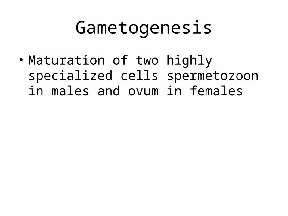
Gametogenesis
• Maturation of two highly specialized cells spermetozoon in males and ovum in females
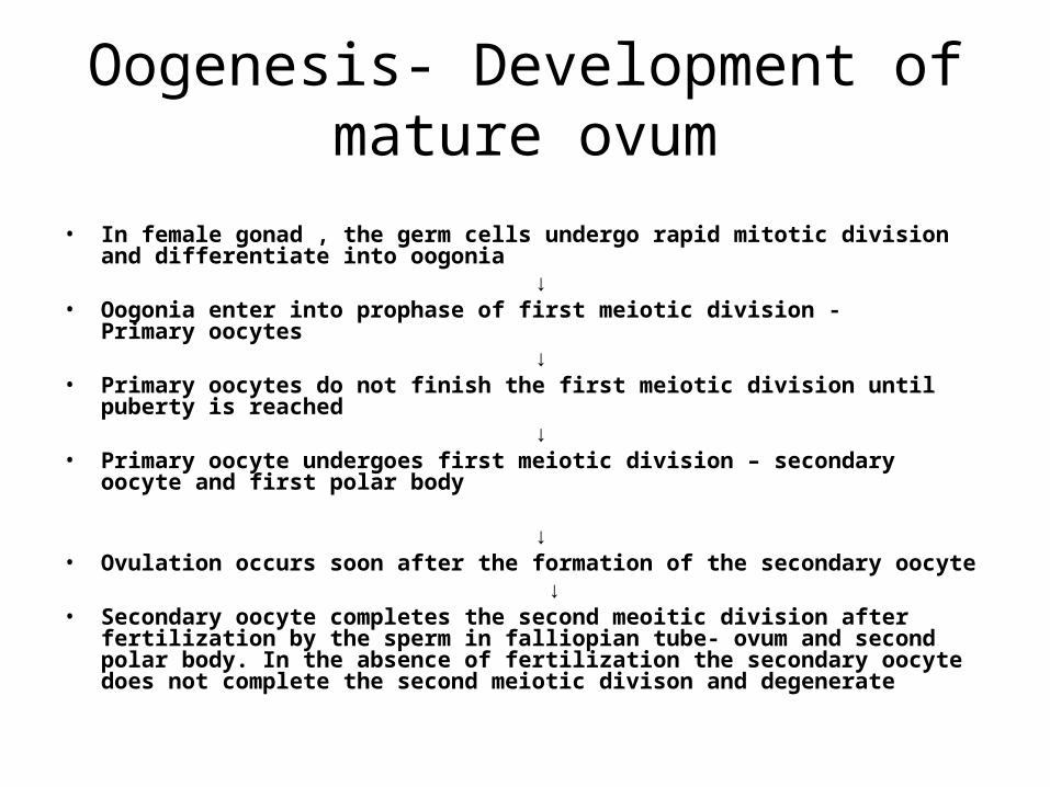
Oogenesis- Development of mature ovum
• In female gonad , the germ cells undergo rapid mitotic division and differentiate into oogonia
↓• Oogonia enter into prophase of first meiotic division - Primary oocytes ↓• Primary oocytes do not finish the first meiotic division until puberty is reached ↓• Primary oocyte undergoes first meiotic division – secondary oocyte and first
polar body ↓• Ovulation occurs soon after the formation of the secondary oocyte ↓• Secondary oocyte completes the second meoitic division after fertilization by
the sperm in falliopian tube- ovum and second polar body. In the absence of fertilization the secondary oocyte does not complete the second meiotic divison and degenerate
Ovulation
• Is the process whereby secondary oocyte is released from the ovary following rupture of mature graaafian follicle and become available for conception
• Only one secondary oocyte is likely to rupture in each ovarian cycle which starts at puberty and ends in menopause
• In relation to the menstrual period event occurs about 14 days prior to the expected period
Changes in the follicle before ovulation
• Graafian follicle becomes enlarged ( 20mm )• Follicular wall near the ovarian surface becomes
thinner• Stigma develops as conical projection which
penetrates the outer surface of the ovary and persist as thin layer
• Cumulus escapes out of the follicle as a slow oozing, process taking about 1-2 min
• Stigma is closed by a plug of plasma
Changes in oocyte before ovulation
• Increase in cytoplasmic volume
• Changes in the number and distribution of mitochondria and in the Golgi apparatus
• Completion of arrested first meiotic division occurs with formation of secondary oocyte and first polar body each containing haploid number of chromosomes(23X )
Cause of ovulationCombined FSH/LH midcycle surge is responsible for the final stage of
maturation, rupture of the follicle and expulsion of the oocyte
LH surge- • Sustained peak levels of oestrogen for 24-36 hours in the late
follicular phase cause LH surge from anterior pitutary.
• Ovulation occurs apprx 16-24 hours after LHsurge
• LH stimulates completion of reduction division of the oocyte,initiates leutinisation of the granulosa cells, synthesis of progestrone and prostaglandins
FSH rise• Preovulatory rise of progestrone facilitates the
positive feed back action of estrogen to induce FSH surge
• FSH surge causes increase in plasminogen activator which converts plasminogen into plasmin , which in turn causes lysis of the wall of the follicle
Structure of a mature ovum• Largest cell in the body
• Consists of cytoplasm and a nucleus with its nucleolus in eccentric position
• Contains 23 chromosomes (23 X )
• Sorrounded by a cell membrane called a vitelline membrane
• Outer transparent mucoprotein envelope is called zona pellucida
• Tiny channels in zona pellucida are for the transport of the materials from the granulosa cells to the oocyte
• Space between the vitelline membrane and zona pellucida is called perivitelline space which accomodates the polar bodies
• Oocyte after its escape from the follicle, retains a covering of granulosa cells known as corona radiata derived from the cumulus oophorus
Spermatogenesis
• Spermatogenesis-Development of spermatids from the primodial male germ cells and their differenciation into spermatozoa
• In man time required for a spermatogonium to develop into a mature spermatozoon is about 61 days
Spermatogenesis• Primodial germ cells undergo mitosis in seminiferous tubules to
develop into spermatogonia• ↓• Spermatogonia differenciates into primary spermatocytes (46 XY)
which remain in the stage of prophase of the first meiotic division• ↓• With completion of first meiotic division – two secondary
spermatocytes are formed( 23 X or 23 Y )• ↓• Immediately follows the second meiotic division- 4 spermatids are
formed, containing haploid number of chromosomes• ↓• Spermatids undergo extensive morphological changes to convert
them into spermatozoa
Structure of a mature spermatozoon
• It has head and a tail
• Head consist of nucleus and a acrosomal cap,rich in enzymes
• The tail is divided into four zones- neck,middle piece,the principal piece and the end piece.
Sperm capacitation and acrosome reaction
• Capacitation is the physiochemical change in the sperm by which sperm become hypermotile and is able to bind and fertilize a secondary oocyte
• Activation of acrosomal enzyme causes release of hyaluronidase, hydrolytic enzymes, proacrosin,acrosin that help the sperm to digest the zona pellucida and to enter the oocyte
Fertilization
• Is the process of fusion of spermatzoon with the mature ovum
• Fertilization occurs in the ampullary part of the uterine tube
• Ovum , following ovulation is picked by tubal fimbriae and is transported to the ampullary part
• Out of the hundreds of millions sperm deposited in the vagina at single ejaculation only thousand capacitated spermatozoa enter the tube while only 300-500 reach the ovum
• Fertilisable life span of oocyte ranges from 12-24 hours where as that of sperm is 48-72 hours
• Complete disolution of the cells of the corona radiata occurs by the chemical action of the hyaluronidase liberated from the acrosomal cap of the sperms
• Penetration of the zona pellucida is facilitated by the release of hyaluronidase from the acrosomal cap
• After the one spermatozoon has entered the ovum , others are prevented from entering by zona reaction
• Completion of the second meiotic division of the oocyte immediately follows resulting in the female pronucleus (23 X) and 2nd polar body
• At the same time the head of the spermetazoa separates from middle piece and tail and transforms into male pronucleus (23Xor 23Y)
• Male and female pronucleus unite at the center resulting in formation of zygote (46 XX or 46XY)
• Sex of the child will depend on the pattern of sex chromosome supplied by the sperm
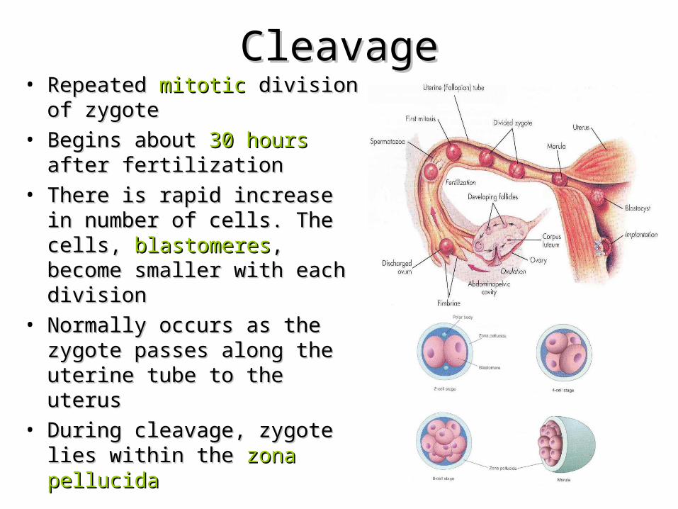
CleavageCleavage• Repeated Repeated mitoticmitotic division of division of
zygotezygote• Begins about Begins about 30 hours30 hours after after
fertilizationfertilization• There is rapid increase in There is rapid increase in
number of cells. The cells, number of cells. The cells, blastomeresblastomeres, become smaller , become smaller with each divisionwith each division
• Normally occurs as the zygote Normally occurs as the zygote passes along the uterine tube passes along the uterine tube to the uterusto the uterus
• During cleavage, zygote lies During cleavage, zygote lies within the within the zona pellucidazona pellucida
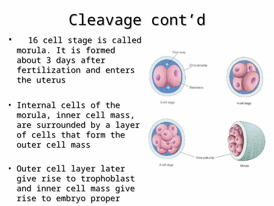
Cleavage cont’dCleavage cont’d• 16 cell stage is called morula. 16 cell stage is called morula.
It is formed about 3 days after It is formed about 3 days after fertilization and enters the fertilization and enters the uterusuterus
• Internal cells of the morula, Internal cells of the morula, inner cell mass, are inner cell mass, are surrounded by a layer of cells surrounded by a layer of cells that form the outer cell massthat form the outer cell mass
• Outer cell layer later give rise Outer cell layer later give rise to trophoblast and inner cell to trophoblast and inner cell mass give rise to embryo mass give rise to embryo properproper
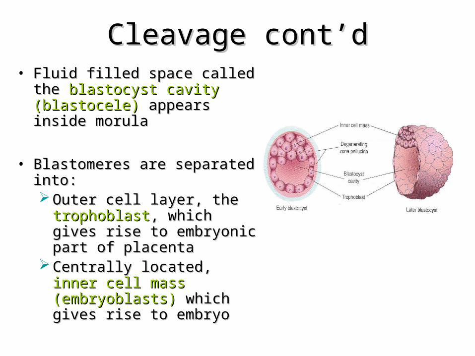
Cleavage cont’dCleavage cont’d• Fluid filled space called the Fluid filled space called the
blastocyst cavity (blastocele)blastocyst cavity (blastocele) appears inside morulaappears inside morula
• Blastomeres are separated into:Blastomeres are separated into: Outer cell layer, the Outer cell layer, the
trophoblasttrophoblast, which gives rise , which gives rise to embryonic part of placentato embryonic part of placenta
Centrally located, Centrally located, inner cell inner cell mass (embryoblasts)mass (embryoblasts) which which gives rise to embryogives rise to embryo
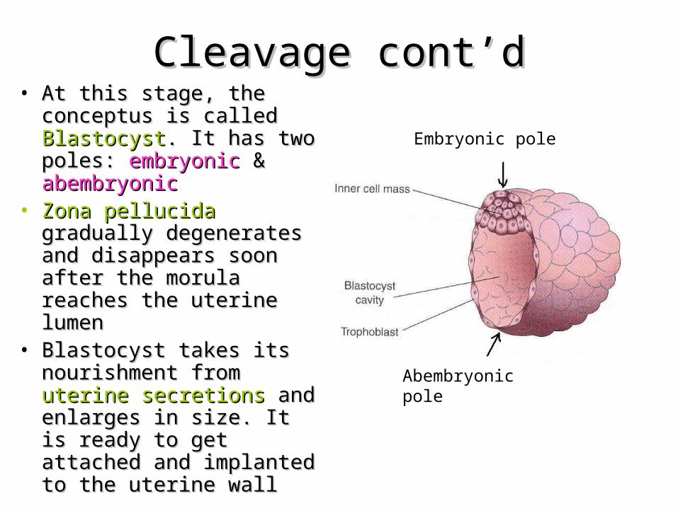
Cleavage cont’dCleavage cont’d• At this stage, the At this stage, the
conceptus is called conceptus is called BlastocystBlastocyst. It has two . It has two poles: poles: embryonicembryonic & & abembryonicabembryonic
• Zona pellucidaZona pellucida gradually gradually degenerates and degenerates and disappears soon after the disappears soon after the morula reaches the uterine morula reaches the uterine lumenlumen
• Blastocyst takes its Blastocyst takes its nourishment from nourishment from uterine uterine secretionssecretions and enlarges in and enlarges in size. It is ready to get size. It is ready to get attached and implanted to attached and implanted to the uterine wallthe uterine wall
Abembryonic pole
Embryonic pole
Formation of germ layers , chorion and amnion
• Some cells of the inner cell mass become flattened and come to lie on its free surface and constitute the endoderm
• Remaining cells of inner cell mass become columnar and constitute the ectoderm
• A space appears between the ectoderm and the trophoblast . This is the amniotic cavity filled by amniotic fluid
• The roof of the cavity is formed by amniogenic cells derived from the trophoblast, while its floor is formed by the ectoderm
• Flattened cells arising from the endoderm spread and line the inside of the blastocystic cavity
• In this way , a cavity lined on all sides by cells of endodermal origin is formed. This cavity is called primary yolk sac
• Cells of the trophoblast give origin to a mass of cells called extra-embryonic mesoderm or primary mesoderm
• These cells come to lie between the trophoblast and the flattened endodermal cells lining the yolk sac
The process by which the The process by which the developing mass gets developing mass gets
embedded within the uterine embedded within the uterine wallwall
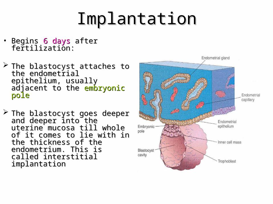
ImplantationImplantation• Begins Begins 6 days 6 days after after
fertilization:fertilization:
The blastocyst attaches to The blastocyst attaches to the endometrial epithelium, the endometrial epithelium, usually adjacent to the usually adjacent to the embryonic poleembryonic pole
The blastocyst goes deeper The blastocyst goes deeper and deeper into the uterine and deeper into the uterine mucosa till whole of it comes mucosa till whole of it comes to lie with in the thickness of to lie with in the thickness of the endometrium. This is the endometrium. This is called interstitial implantationcalled interstitial implantation
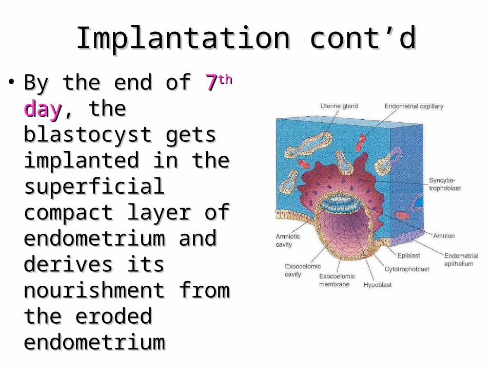
Implantation cont’dImplantation cont’d• By the end of By the end of 77thth day day, ,
the blastocyst gets the blastocyst gets implanted in the implanted in the superficial compact superficial compact layer of endometrium layer of endometrium and derives its and derives its nourishment from the nourishment from the eroded endometriumeroded endometrium
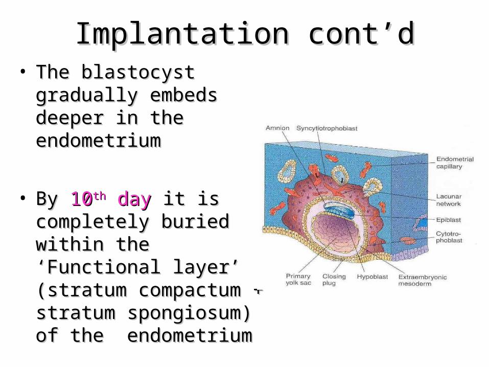
Implantation cont’dImplantation cont’d• The blastocyst gradually The blastocyst gradually
embeds deeper in the embeds deeper in the endometriumendometrium
• By By 1010thth day day it is completely it is completely buried within the buried within the ‘Functional layer’ (stratum ‘Functional layer’ (stratum compactum + stratum compactum + stratum spongiosum) of the spongiosum) of the endometriumendometrium
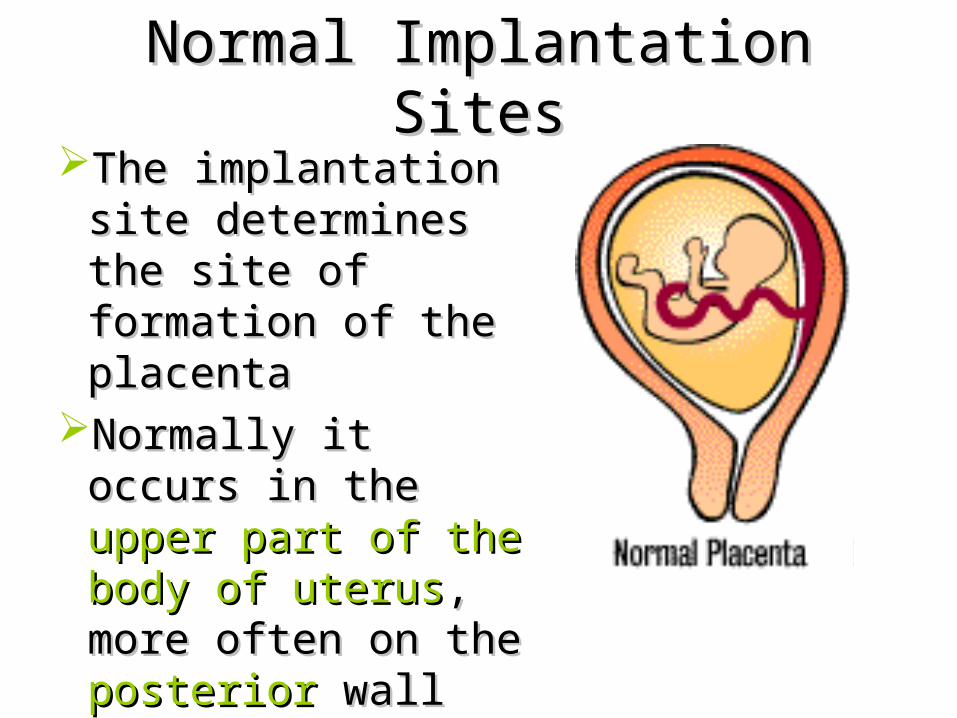
Normal Implantation SitesNormal Implantation SitesThe implantation site The implantation site
determines the site of determines the site of formation of the formation of the placentaplacenta
Normally it occurs in Normally it occurs in the the upper part of the upper part of the body of uterusbody of uterus, more , more often on theoften on the posterior posterior wallwall
• After the implantation of the embryo, the uterine endometrium is called the decidua
• When the morula reaches the endometrium , it is in the secretory phase
• After implantation , features of the endometrium in secretory phase are intensified- stromal cells enlarged , become vacuolated and store glycogen and lipids. This change in stromal cells is called decidual reaction
• The portion of the decidua where the placenta is to be formed ( deep to the developing blastocyst ) is called decidua basalis
• Part of the decidua that separates the embryo from the uterine lumen is called decidua capsularis
• Part of the decidua lining the rest of the uterine cavity is called decidua parietalis
Formation of the chorionic villi
• The essential functional elements of the placenta are very small finger like processes or villi
• These villi are surrounded by maternal blood
• In the subustance of the villi, there are capillaries through which fetal blood circulates
• Exchanges between maternal and fetal circulations take place through the tissues forming the walls of the villi
• The villi are formed as offshoots from the surface of the trophoblast
• As the trophoblast along with the underlying extra-embryonic mesoderm constitutes chorion, the villi are known as chorionic villi
• Chorionic villi are first formed all over the trophoblast and grow into the surrounding decidua
• Those related to decidua capsularis are transitory and degenrate and this part of the chorion becomes smooth and is called chorion laevae
• The villi that grow into the decidua basalis undergo considerable development
• Along with the tissues of the decidua basalis these villi form a disc shaped mass which is called the placenta
• The part of the chorion that helps to form the placenta is called the chorion frondosum
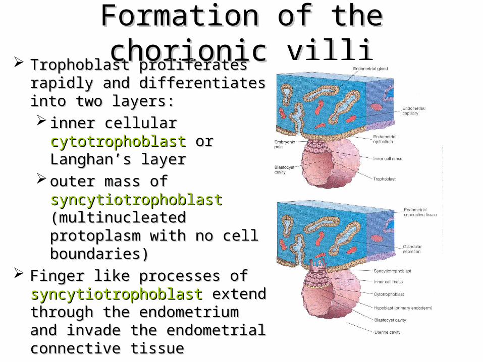
Formation of the chorionic villiFormation of the chorionic villi Trophoblast proliferates rapidly Trophoblast proliferates rapidly
and differentiates into two layers:and differentiates into two layers: inner cellular inner cellular cytotrophoblastcytotrophoblast
or Langhan’s layeror Langhan’s layer outer mass of outer mass of
syncytiotrophoblastsyncytiotrophoblast (multinucleated protoplasm (multinucleated protoplasm with no cell boundaries)with no cell boundaries)
Finger like processes of Finger like processes of syncytiotrophoblastsyncytiotrophoblast extend extend through the endometrium and through the endometrium and invade the endometrial invade the endometrial connective tissue connective tissue
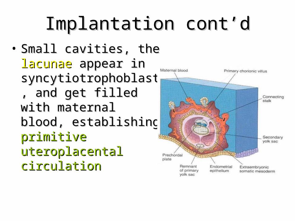
Implantation cont’dImplantation cont’d• Small cavities, the Small cavities, the
lacunaelacunae appear in appear in syncytiotrophoblast, syncytiotrophoblast, and get filled with and get filled with maternal blood, maternal blood, establishing establishing primitive primitive uteroplacental uteroplacental circulationcirculation
• The syncitotrophoblast grows into the endometrium
• As the endometrium is eroded , some of its blood vessels are opened up and blood from them fills the lacunar space
• Each trabeculus is , initially, made up entirely of sycytiotrophoblast
• Later on, cells of cytotrophoblast grow into the trabeculus , followed by extra embryonic mesoderm and blood vessels giving rise to primary villus, secondary villus and tertiary villus respectively
• Blood vessels of the villus establish connections with the circulatory system of the embryo
• Fetal blood now circulates through the villi, while the maternal blood circulates through the intervillous space
• Intially cytotrophoblast that that grows into the trabeculus does not penetrate the entire thickness of syncytium
• At a later stage ,cells of the cytotrophoblast emerge through the syncytium and spread out to form a layer that completely cuts off the syncytium from the decidua and is called cytotrophoblastic cells
• The villi that are first formed are attached on the fetal side to the embryonic mesoderm and on the maternal side to the cytotrophoblastic shell and are called anchoring villi
• Each anchoring villus consists of a stem (truncus chorii); this divides into a number of branches ( rami chorii ) which in turn divide into finer branches (ramuli chorii )
• Anchoring villi give off numerous branches which grow into the intervillous space as free villi
• As a result , the surface area available for exchanges between maternal and fetal circulation becomes enormous
Circulation of blood through placenta
• Maternal blood in the intervillous space is constantly in circulation
• Both arteries and the vein open into the roof of the cotyledon and that the pressure of the blood in the artery is sufficient to drive blood to the fetal end of the intervillious space