Impaired Motor Coordination and Learning in Mice Lacking ... · ORIGINAL PAPER Impaired Motor...
9
ORIGINAL PAPER Impaired Motor Coordination and Learning in Mice Lacking Anoctamin 2 Calcium-Gated Chloride Channels Franziska Neureither 1 & Katharina Ziegler 1 & Claudia Pitzer 2 & Stephan Frings 1 & Frank Möhrlen 1 # The Author(s) 2017. This article is an open access publication Abstract Neurons communicate through excitatory and in- hibitory synapses. Both lines of communication are adjustable and allow the fine tuning of signal exchange required for learning processes in neural networks. Several distinct modes of plasticity modulate glutamatergic and GABAergic synap- ses in Purkinje cells of the cerebellar cortex to promote motor control and learning. In the present paper, we present evidence for a role of short-term ionic plasticity in the cerebellar circuit activity. This type of plasticity results from altered chloride driving forces at the synapses that molecular layer interneu- rons form on Purkinje cell dendrites. Previous studies have provided evidence for transiently diminished chloride gradi- ents at these GABAergic synapses following climbing fiber activity. Electrical stimulation of climbing fibers in acute slices caused a decline of inhibitory postsynaptic currents re- corded from Purkinje cells. Dendritic calcium-gated chloride channels of the type anoctamin 2 (ANO2) were proposed to mediate this short-term modulation of inhibition, but the sig- nificance of this process for motor control has not been established yet. Here, we report results of behavioral studies obtained from Ano2 -/- mice, a mouse line that was previously shown to lack this particular mode of ionic plasticity. The animals display motor coordination deficits that constitute a condition of mild ataxia. Moreover, motor learning is severely impaired in Ano2 -/- mice, suggesting cerebellar dysfunction. This reduced motor performance of Ano2 -/- mice highlights the significance of inhibitory control for cerebellar function and introduces calcium-dependent short-term ionic plasticity as an efficient control mechanism for neural inhibition. Keywords Purkinje cells . Inhibition . Plasticity . Calcium . Motor performance Introduction The cerebellum controls body movements and mediates the acquisition of procedural memory [1]. The output neurons of the cerebellar cortex, the Purkinje cells, receive inhibitory GABAergic synapses from a network of molecular layer in- terneurons (MLIs), which provide fast feed-forward inhibition and co-determine the activity of the Purkinje cells [2–6]. MLIs limit the time window for synaptic integration in Purkinje cells to 1–2 ms [2], a control function that shapes the output signal of the cerebellar cortex. The MLI-Purkinje cell synapses are also subject to modulatory mechanisms, which probably con- tribute to motor learning. In particular, Purkinje cell depolar- ization that results from the activation of climbing fibers causes a slow but persistent increase of GABAergic synaptic strength termed rebound potentiation [7–9]. In genetic exper- iments, ablation of the γ2 subunit of GABA A receptors at the MLI-Purkinje cell synapse impaired inter-limb coordination and motor memory consolidation [10, 11]. Moreover, phar- macologically boosted inhibition at this synapse severely dis- turbed motor coordination [12]. Thus, the inhibitory regula- tion of Purkinje cells by MLIs is clearly crucial for motor performance. Recent studies have indicated that the inhibition efficacy at the MLI-Purkinje cell synapse can be attenuated by a postsynaptic process which is triggered by activation of climbing fibers [13, 14]. Ca 2+ signals that accompany * Stephan Frings [email protected] 1 Department of Animal Molecular Physiology, Centre for Organismal Studies, Heidelberg University, Im Neuenheimer Feld 504, 69120 Heidelberg, Germany 2 Interdisciplinary Neurobehavioral Core (INBC), Heidelberg University, Im Neuenheimer Feld 515, 69120 Heidelberg, Germany Cerebellum DOI 10.1007/s12311-017-0867-4
Transcript of Impaired Motor Coordination and Learning in Mice Lacking ... · ORIGINAL PAPER Impaired Motor...

ORIGINAL PAPER
Impaired Motor Coordination and Learning in Mice LackingAnoctamin 2 Calcium-Gated Chloride Channels
Franziska Neureither1 & Katharina Ziegler1 & Claudia Pitzer2 & Stephan Frings1 &
Frank Möhrlen1
# The Author(s) 2017. This article is an open access publication
Abstract Neurons communicate through excitatory and in-hibitory synapses. Both lines of communication are adjustableand allow the fine tuning of signal exchange required forlearning processes in neural networks. Several distinct modesof plasticity modulate glutamatergic and GABAergic synap-ses in Purkinje cells of the cerebellar cortex to promote motorcontrol and learning. In the present paper, we present evidencefor a role of short-term ionic plasticity in the cerebellar circuitactivity. This type of plasticity results from altered chloridedriving forces at the synapses that molecular layer interneu-rons form on Purkinje cell dendrites. Previous studies haveprovided evidence for transiently diminished chloride gradi-ents at these GABAergic synapses following climbing fiberactivity. Electrical stimulation of climbing fibers in acuteslices caused a decline of inhibitory postsynaptic currents re-corded from Purkinje cells. Dendritic calcium-gated chloridechannels of the type anoctamin 2 (ANO2) were proposed tomediate this short-term modulation of inhibition, but the sig-nificance of this process for motor control has not beenestablished yet. Here, we report results of behavioral studiesobtained from Ano2−/−mice, a mouse line that was previouslyshown to lack this particular mode of ionic plasticity. Theanimals display motor coordination deficits that constitute acondition of mild ataxia. Moreover, motor learning is severelyimpaired in Ano2−/− mice, suggesting cerebellar dysfunction.
This reduced motor performance of Ano2−/− mice highlightsthe significance of inhibitory control for cerebellar functionand introduces calcium-dependent short-term ionic plasticityas an efficient control mechanism for neural inhibition.
Keywords Purkinje cells . Inhibition . Plasticity . Calcium .
Motor performance
Introduction
The cerebellum controls body movements and mediates theacquisition of procedural memory [1]. The output neurons ofthe cerebellar cortex, the Purkinje cells, receive inhibitoryGABAergic synapses from a network of molecular layer in-terneurons (MLIs), which provide fast feed-forward inhibitionand co-determine the activity of the Purkinje cells [2–6].MLIslimit the timewindow for synaptic integration in Purkinje cellsto 1–2 ms [2], a control function that shapes the output signalof the cerebellar cortex. The MLI-Purkinje cell synapses arealso subject to modulatory mechanisms, which probably con-tribute to motor learning. In particular, Purkinje cell depolar-ization that results from the activation of climbing fiberscauses a slow but persistent increase of GABAergic synapticstrength termed rebound potentiation [7–9]. In genetic exper-iments, ablation of the γ2 subunit of GABAA receptors at theMLI-Purkinje cell synapse impaired inter-limb coordinationand motor memory consolidation [10, 11]. Moreover, phar-macologically boosted inhibition at this synapse severely dis-turbed motor coordination [12]. Thus, the inhibitory regula-tion of Purkinje cells by MLIs is clearly crucial for motorperformance. Recent studies have indicated that the inhibitionefficacy at the MLI-Purkinje cell synapse can be attenuated bya postsynaptic process which is triggered by activation ofclimbing fibers [13, 14]. Ca2+ signals that accompany
* Stephan [email protected]
1 Department of Animal Molecular Physiology, Centre for OrganismalStudies, Heidelberg University, Im Neuenheimer Feld 504,69120 Heidelberg, Germany
2 Interdisciplinary Neurobehavioral Core (INBC), HeidelbergUniversity, Im Neuenheimer Feld 515, 69120 Heidelberg, Germany
CerebellumDOI 10.1007/s12311-017-0867-4

climbing fiber-induced depolarization cause the opening ofchloride channels of the type anoctamin 2 (ANO2, aliasTMEM16B) [15]. It was proposed that these channels, incombination with increased Cl− uptake through the co-transporter NKCC1, cause the local intra-dendritic Cl− con-centration to increase. Such postsynaptic Cl− accumulationwould reduce the inward driving force for chloride ions atMLI-Purkinje cell synapses and, hence, induce a decline ofinhibitory postsynaptic current (IPSC) amplitudes. This mech-anism was termed depolarization-induced depression ofinhibition (DDI) and was shown to attenuate the inhibitoryinput for several minutes after climbing fiber activation [13].This mode of modulation was detected by recording fromPurkinje cells in tissue slices of rats [13] and mice [14].Climbing fiber activity triggered a rapid, transient decline ofIPSCs at the MLI-Purkinje cell synapse, and no such effectwas observed in Ano2−/−mice. This finding left the questionunanswered whether the lack of DDI would compromise mo-tor coordination and learning in Ano2−/−mice. Thus, in thepresent study, we looked for the relevance of DDI for motorcontrol. We compared the motor performance of wild-typeand Ano2−/− mice in a variety of behavioral tasks designedto specifically reveal cerebellar dysfunction. We found thatAno2−/− mice display deficiency in motor coordination andmotor learning. Our results illustrate the behavioral signifi-cance of calcium-dependent modulation of inhibitory networkactivity through short-term ionic plasticity, a novel pathwayfor controlling network function in the brain.
Materials and Methods
Animals: Housing, General Health, and Behavior
C57BL/6N mice (Charles River Laboratories, Germany) andAno2−/− mice [16] (kindly provided by Thomas Jentsch,Leipniz-Institute for Molecular Pharmacology, Berlin) werekept in groups of 2–3 with ad libitum access to food and water.A standard 12-h light/dark cycle was provided (light on: 7 amto 7 pm) and temperature was maintained at ±22 °C at arelative humidity of 40–50%. All experiments were approvedby the Regierungspräsidium Karlsruhe and were in agreementwith national and international guidelines. For general healthscreening, a modified version of the SHIRPA test [17] wasused for both genotypes. Spontaneous activity, anxiety, bodystrength, and muscle tone as well as several reflexes weretested according to the provided scoring system. General be-havior was monitored using the LABORAS animal behaviorobservation system (Metris B.V., Netherlands) individuallyfor six mice of each genotype for 72 h. Recorded activitiesincluded locomotion, climbing, rearing, grooming, eating, anddrinking.
Experimental Design
Following a habitation time of 1 week, animals were handledfor three consecutive days before the behavioral tests werestarted. Behavioral tests were performed by one female exper-imenter only in order to avoid unnecessary stress for the mice.Tests were run, if not indicated otherwise, from 1 to 4 pm.Non-automatic rated experiments (apart from the SHIRPAtest) were analyzed in a blind fashion by two different persons.Onlymale, adult micewere usedwith a starting age of 8 weeks(cohort 3) and 10–12 weeks (cohorts 1 and 2) and tests wereconducted for up to 10 weeks (cohorts 1 and 2) or 4 days(cohort 3). Numbers of animals tested were (wt/Ano2−/−) gen-eral behavior 6/6 (cohort 1); rotarod 9/8 (cohort 1); open field,grip strength, parallel rod floor, gait analysis 9/10 (cohort 2);horizontal ladder 9/9 (cohort 2); and voluntary wheel running4/5 (cohort 3).
Immunohistochemistry
To prepare cerebellar cryosections, animals were anesthetizedby isoflurane (Baxter, Germany) inhalation and killed by anoverdose of isoflurane. The cerebellum was removed and thefresh, unfixed tissue was cut on ice in phosphate-bufferedsaline (PBS; 130 mM NaCl, 8.1 mM Na2HPO4, 1.9 mMNaH2PO4, pH 7.4).) using a vibratome (VT1000S, LeicaBiosystems, Wetzlar, Germany). Sections of 180 µm thick-ness were fixed with 2% paraformaldehyde (PFA) for30 min and washed 4× 10 min with PBS (for ANO2 immu-nostaining) or with PBST (PBS containing 0.5% Tween 20(Carl Roth, Germany, 9127.1)) for calbindin immunostaining.For immunofluorescence staining, sections were incubated inblocking solution (goat serum, Sigma-Aldrich, Germany,G9023) for 2 h and then the primary antiserum (diluted inblocking solution) was applied overnight at room temperature.After washing 4× 10 min with PBS or PBST, the secondaryantisera, conjugated with fluorescence tags, were incubatedfor 2 h and washed 4× 10 min with PBST. To visualize cellnuclei, the slices were incubated in 0.3 μM DAPI (Sigma-Aldrich, 32670) for 15 min, washed 4× 5 min in PBS, andthen mounted on glass slides with Aqua-Poly/Mount(Polyscience, Germany, 18606). The sections were kept on ashaker for the whole staining process where they were gentlyshaken to improve the uptake of the antisera. The primaryantibody against ANO2 (used at 1:200) was raised in guineapig in our lab and was extensively characterized in varioustissues including cerebellum [14, 18–20]. The primary anti-body against calbindin (dilution 1:500) was raised in rabbit(Swant, Marly, Switzerland, CB-38a) and binds specifically tocalbindin D-28k. The secondary antibodies were donkey anti-rabbit conjugated with Alexa Fluor 568 (Invitrogen, A10042,dilution 1:1000) and goat anti-guinea pig conjugated withAlexa Fluor 488 (Invitrogen, A11073).
Cerebellum

Grip Strength Analysis
Forelimb strength of both mouse lines was tested using a gripstrength meter (Ugo Basile, Gemonio, Italy, model 47106),which automatically measured the force needed for the mouseto release its grip. A mouse had to grasp a trapeze wire andwould cling to it until the pulling force of the experimenterexceeded its own pulling strength. After an initial trainingsession, the mice were tested in one session only, in whicheach mouse was tested in three consecutive runs. The runswere then averaged for each animal.
Open Field Test
Mice were individually placed in a wooden box (40 × 40 cm)for a period of 5 min and their movements were cameratracked. Only one run per mouse was conducted and the dis-tance they walked was analyzed to evaluate their spontaneousactivity.
Parallel Rod Floor Test
The open field setup was adjusted with an additional rod floorwith parallel bars (3 mm in diameter, 6 mm space in between)to attain the mice’s locomotor activity at slightly aggravatedconditions. On three consecutive days, mice were placed onthe rod and their movement was video-tracked over a periodof 5 min. One session per day was conducted per mouse. Themaximal distance walked by all mice of a genotype waspooled for each session.
Horizontal Ladder Test
A horizontal ladder (40 cm in length, 43 rung positions) with arandomized and irregular rung pattern was used to assessskilled motor coordination of the animals. The metal rungswere 3mm in diameter with a minimal space of 6 mmbetweentwo rods. Rods were placed in an irregular randomized patternand their pattern was not altered during the testing phase. Aninitial training session was conducted to let the mice get ac-quired to the setup. While testing, mice were put at the begin-ning of the ladder individually and had to cross the laddervoluntarily. As a reward input, their home cage with the re-maining housing companions was placed at the end of theladder. Every walk was video-taped and the video wasstopped as soon as the mice reached the end of the ladder.Each mouse did four trails so that each body side was tapedtwice. The videos were then analyzed by two different evalu-ators in a blinded manner. The time the mice needed to tra-verse the ladder was measured and the number of steps count-ed. Final results were obtained by averaging four runs of eachmouse as well as the individual results of the two evaluators.
Gait Analysis
The animals’ gait patterns were analyzed using the CatWalkXT analysis apparatus (Noldus, Netherlands, version 10.6).The 1.3 m black corridor led over a glass plate that was illu-minated by a green LED light. As soon as the mouse’s pawtouched the glass slide, this green light was reflected and theresulting illuminated green footprint could then be captured bya high speed camera underneath the glass. As the mouse tra-versed the glass plate, its gait pattern was recorded and theresulting data automatically were analyzed by the CatWalkXTsoftware according pre-set paradigms [21]. Evaluation pa-rameters used for our analysis are specified in the legend toTable 1.
Voluntary Wheel Running
To monitor voluntary wheel running activity, mice wereplaced individually in a home cage with a running wheel (di-ameter 9.2 cm, width 5.1 cm) to which they had free access.Water and food was provided, and the experiment was con-ducted at a 12 h light/dark cycle during four successive daysand nights. Wheel revolutions per 15 min intervals were re-corded by a sensor connected to a computer interface(Columbus Instruments, Columbus, OH, USA). Only micethat did run were taken into account.
Rotarod
Accelerated rotarod tests (4–40 rpm) were performed up tomaximal 8 min to examine the animals’ motor coordinationand motor learning skills. The rotarod apparatus (Ugo Basile,Gemonio, Italy, model 47600) had a rod of 3 cm in diameterand five lanes with a width of 5 cm each. The test is designedtomeasure the timemice are able to run on the accelerated rod.As soon as a mouse falls off the rod, the time is stoppedautomatically. After an initial training session, mice were test-ed for three consecutive weeks every 2–3 days. In each ses-sion, mice were tested in three trials with a recovery phase of10 min in between. For analysis, the three trials were averagedfor eachmouse. After a test break of 3 weeks, mice were againtested for 3 weeks in the same pattern.
Results
Ano2−/− mice that lacked DDI in acute tissue slices [14] wereused for these experiments. The animals displayed normal cer-ebellar anatomy without conspicuous structural differences inthe granule cell layer, the Purkinje cell layer, the molecular celllayer, and the deep cerebellar nuclei (wt: Fig. 1a–c; Ano2−/−:Fig. 1f–h). In accordance with previous data [14], ANO2 ex-pression was detected in Purkinje cells (Fig. 1d, i) but not in the
Cerebellum
deep cerebellar nuclei (Fig. 1e, j). During home cage observa-tion, Ano2−/− mice showed normal basal voluntary activities,including similar locomotion, climbing, rearing, grooming, eat-ing, and drinking. Mean body weight increased during 6 weeksof experimentation from 25.9 ± 0.3 to 30.4 ± 0.6 g in wild-typemice and from 28.3 ± 0.7 to 31.1 ± 0.4 g in Ano2−/−animals. Inopen field tests, they displayed similar explorative behavior and
covered similar distances during the 5-min test period (wt2.04 ± 0.15 m; Ano2−/−1.79 ± 0.18 m, t test, p = 0.31).Furthermore, the grip strength test yielded similar results formuscle strength in the front limbs (wt 523 ± 32 mN; Ano2−/−
574 ± 20 mN; n = 9, t test, p = 0.19), indicating normal muscledevelopment. Ano2−/− mice scored normally in SHIRPA testsfor spontaneous activity, anxiety, body strength, muscle tone,
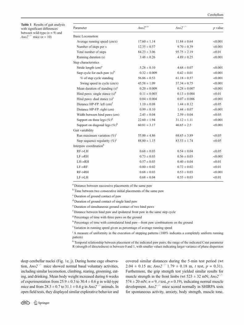
Table 1 Results of gait analysiswith significant differencesbetween wild-type (n = 9) andAno2−/− mice (n = 10)
Parameter Ano2+/+ Ano2−/− p value
Basic Locomotion
Average running speed (cm/s) 17.60 ± 1.14 11.84 ± 0.64 <0.001
Number of steps per s 12.35 ± 0.57 9.70 ± 0.39 <0.001
Total number of steps 84.23 ± 3.06 95.75 ± 2.19 <0.01
Running duration (s) 3.48 ± 0.26 4.89 ± 0.25 <0.001
Step characteristics
Stride length (cm)a 5.28 ± 0.10 4.68 ± 0.07 <0.001
Step cycle for each paw (s)b 0.32 ± 0.009 0.42 ± 0.01 <0.001
% of step cycle standing 56.86 ± 0.53 61.18 ± 0.57 <0.001
Swing speed in cycle (cm/s) 45.58 ± 1.09 37.34 ± 0.75 <0.001
Mean duration of standing (s)c 0.20 ± 0.009 0.28 ± 0.007 <0.001
Hind paws: single stance (s)d 0.11 ± 0.003 0.13 ± 0.004 <0.01
Hind paws: dual stance (s)e 0.04 ± 0.004 0.07 ± 0.006 <0.001
Distance HP-FP: left (cm)f 1.10 ± 0.08 1.44 ± 0.12 <0.05
Distance HP-FP: right (cm) 0.99 ± 0.10 1.44 ± 0.07 <0.001
Width between hind paws (cm) 2.45 ± 0.04 2.59 ± 0.04 <0.05
Support on three legs (%)g 22.60 ± 1.94 31.12 ± 1.11 <0.001
Support on diagonal legs (%)h 60.81 ± 3.17 46.65 ± 2.5 <0.001
Gait variability
Run maximum variation (%)i 55.00 ± 4.86 68.65 ± 3.89 <0.05
Step sequence regularity (%)j 88.80 ± 1.15 83.53 ± 1.74 <0.05
Interpaw coordinationk
RF->LH 0.68 ± 0.03 0.54 ± 0.04 <0.05
LF->RH 0.73 ± 0.03 0.56 ± 0.03 <0.001
LH->RH 0.57 ± 0.03 0.40 ± 0.04 <0.01
LF->RF 0.80 ± 0.02 0.72 ± 0.02 <0.01
RF->RH 0.68 ± 0.03 0.53 ± 0.03 <0.001
LF->LH 0.68 ± 0.04 0.55 ± 0.03 <0.01
a Distance between successive placements of the same pawb Time between two consecutive initial placements of the same pawcDuration of ground contact of pawdDuration of ground contact of single hind paweDuration of simultaneous ground contact of two hind pawsf Distance between hind paw and ipsilateral front paw in the same step cycleg Percentage of time with three paws on the groundh Percentage of time with contralateral hind paw—front paw combinations on the groundi Variation in running speed given as percentage of average running speedj A measure of uniformity in the execution of stepping patterns (100% indicates a completely uniform runningpattern)k Temporal relationship between placement of the indicated paw pairs; the range of the indicated Cstat parameterR (strength of directedness) is between 0 and 1, with smaller values indicating larger variance of phase dispersion
Cerebellum

and various reflexes, with the exception of the negative geotax-is test where they displayed a reduced tendency to climb up-ward on a vertical grid. Thus, Ano2−/−mice appeared basicallyhealthy and active.
To specifically examine motor coordination abilities, we firstused the parallel rod floor test [22]. Mice walked spontaneouslyand voluntarily on a grid of steel bars while being video-taped.The distance that the animals covered in three 5-min sessionswas significantly shorter in Ano2−/− mice (Fig. 2a). In the re-lated horizontal ladder test, mice had to walk over a series of 27rungs, with 16 randomly positioned gaps. Ano2−/− mice re-quired significantly more time (Fig. 2b) and more steps(Fig. 2c) to perform this task. Although Ano2−/−mice appearedinsecure and cautious on the horizontal ladder, their forelimb
and hindlimb placement was not significantly impaired. Thenumber of slips (Fig. 2d) and corrective paw movements(Fig. 2e) was similar to wild-type animals. To closer inspectwalking parameters for differences between wild-type andAno2−/− mice, we used the CatWalk XT gait analysis system[21]. In this voluntary walking assay, the position of each pawprint was recorded and was analyzed with respect to walkingpatterns and leg coordination. Walking patterns of wild-typemice were typically regular and repetitive (Fig. 3a), whileAno2−/− mice displayed a more irregular gait when walkingalong the test lane (Fig. 3b). Table 1 summarizes those param-eters of the quantitative gait analysis that differed significantlybetween wild-type and Ano2−/− mice. Briefly, the data set indi-cates that locomotion was slower in Ano2−/− mice (basic
Fig. 1 Anatomical integrity of cerebellar cortex and deep cerebellarnuclei in Ano2−/− mice. a Overview of the cerebellar cortex of a wild-type mouse with calbindin immunostaining (red) and DAPI nuclear stain(blue), depicting the molecular layer (ML), the granule cell layer (GCL),and, in between, a row of Purkinje cell somata. b Structural details ofcalbindin-expressing Purkinje cells with their dendrites in the molecularlayer. Nuclei of molecular layer interneurons are visible by DAPI stain. c
Calbindin-staining of a deep cerebellar nucleus in a wild-type mouse. dANO2-immunosignals in Purkinje cell somata and dendrites. e Lack ofspecific immunostain in a deep cerebellar nucleus of a wild-type mouse.f–j Immunostains against calbindin (red) and ANO2 (green) as in theupper row of images, but with cryosections from Ano2−/− mice.Calibration bars are 10 μm in b, g, d, and i and 100 μm in all others
Fig. 2 Ano2−/− mice display gait coordination problems when walkingon grids. a Ano2−/−mice (red) covered shorter distances during voluntarywalking on a parallel rod floor (wt 1.38 ± 0.05m; Ano2−/− 1.08 ± 0.04m;two-way ANOVA, p < 0.001, Bonferroni correction). bOn the horizontalladder with several missing rungs, Ano2−/− mice (red) needed more timeto cross the 40-cm-long ladder (wt 5.3 ± 0.7 s; Ano2−/− 8.9 ± 1.1 s;Student’s t test, p = 0.016). c Ano2−/− mice (red) also needed more stepsto walk over the horizontal ladder, with significant differences in bothfront paws (wt 8.9 ± 0.2 steps; Ano2−/− 10.2 ± 0.13 steps; Student’s t test,
p < 0.001) and hind paws (wt 8.6 ± 0.2 steps; Ano2−/− 9.7 ± 0.16 steps;Student’s t test, p = 0.001). d, e Analysis of paw placement on thehorizontal ladder revealed no significant differences between wild-typeand Ano2−/− mice (n = 9; all t test p values > 0.05). Scoring correspondsto a standard foot fault scoring system [23]. Scores 0–2 represent variouspatterns of slipping from a rung, while scores 3–4 depict correctivemovements following a faulty step. Mean values from nine wild-typeand ten Ano2−/− mice are displayed ± SEM; *p < 0.05, ***p < 0.001
Cerebellum

locomotion in Table 1), that stepping patterns and steppingspeed were different (step characteristics in Table 1), thatAno2−/− mice displayed reduced gait regularity (gaitvariability in Table 1), and that the coordination of paw posi-tioning was compromised (interpaw coordination in Table 1).Several of these differences point to cerebellar problems inAno2−/− mice. For example, the increased width betweenhindpaws is indicative of ataxia that does not depend on walk-ing speed [24]. The increased gait variability is consistent withan earlier report on a cerebellar small-lesion model [25]. Takentogether, the gait analysis produced indications of mild ataxiaduring spontaneous, voluntary walking in Ano2−/−mice, a con-dition that appeared to enforce a slow, hesitant walking pattern.
To examine whether motor learning was also affected in theAno2−/−mice, each mouse was provided with a running wheelover a test period of 4 days and nights. Both wild-type and
Ano2−/− mice used the wheels during the night over a totalperiod of 10–12 h with only short intermissions (Fig. 4a).Both mouse lines spent similar time on wheel running,displaying equally strong motivation for this activity(Fig. 4b). Ano2−/− mice, however, reached 70% less rotationswithin the testing period (Fig. 4c). Moreover, while wild-typemice almost doubled their motor performance over four nights,Ano2−/− mice were unable to increase the number of rotations(Fig. 4d). These results indicate impaired motor coordinationand learning in Ano2−/− mice in a voluntary locomotion task.
To find out whether the animals were able to learn in anenforced locomotion task, we challenged them with the accel-erating rotarod test. Both wild-type and Ano2−/− mice per-formed similarly well on the first day of testing (Fig. 5a).Ano2−/− mice, however, did not significantly improve theirmotor skills over 3 weeks of rotarod training, while wild-typemice showed a steady learning curve and almost doubled theirtime on the rotarod (Fig. 5a). To test whether this learned motorskill represented consolidated procedural memory, we repeatedthe entire experiment after a 20-day interval. Wild-type miceregained their full rotarod performance already on the secondday of rotarod training, while Ano2−/− mice again did not im-prove significantly (Fig. 5b). These results demonstrate thatAno2−/−mice have a reduced ability for motor learning in boththe initial (Fig. 5c) and the repeated (Fig. 5d) period of theexperiment, corroborating the evidence for cerebellar dysfunc-tion in this mouse line.
Discussion
Cl− concentrations in dendrites are not uniform but can changelocally and transiently [26–33]. The results presented here areconsistent with the idea that ANO2 channels modulate Purkinjecell inhibition by MLIs and that this modulation plays a role in
Fig. 3 Walking patterns ofAno2−/−mice differ fromwild types. aRegularwalking pattern of a wild-type mouse as recorded by the Catwalk XT gaitanalysis system. The mouse walked the 36-cm test lane within 20 s. Pawpositions are indicated as dots for the right forepaw (RF), right hindpaw(RH), left forepaw (LF), and left hindpaw (LH). b Representative walkingpattern of an Ano2−/− mouse that walked along the same test lane in 27 s.The results of a detailed gait analysis are given in Table 1
Fig. 4 Impaired voluntary motor performance in Ano2−/− mice. a Bothwild-type (black) and Ano2−/− mice (red) used running wheels fornocturnal activity. The shaded boxes depict the dark periods from 7 pmto 7 am during the 84-h experiment. Plotted are the rounds accumulatedover successive 2-h intervals. b Both mouse lines used the wheel forsimilar durations within the active period, proving comparablemotivation for the task. c Over the four nights of activity, Ano2−/− mice
ran on average fewer rounds on the wheel than wild-type animals (wt2599 ± 336 rotations/2 h; Ano2−/− 939 ± 95.6 rotations/2 h; two-wayANOVA, p < 0.001, Bonferroni correction). d The motor performanceon the running wheel increased in wild-type but not in Ano2−/− mice.Two-way ANOVA: genotypes, p < 0.001; time dependence, p = 0.11;interaction, p = 0.2. Mean values from four wild-type and five Ano2−/−
mice are displayed ± SEM; ***p < 0.001
Cerebellum

motor coordination and motor learning. The behavioral proto-cols described here are suitable for the detection of cerebellardysfunctions. Several mouse models, mostly animals with de-fect Purkinje cells, display a motor phenotype in these tasks[11, 34, 35]. We thus interpret the motor problems of Ano2−/−
mice as indicative of a cerebellar problem. This view is sup-ported by previous results from mice with genetically disabledMLI-Purkinje cell synapses. L7-Δγ2 mice do not expressfunctional synaptic GABAA receptors in Purkinje cells [10].These animals tend to use smaller steps on the ErasmusLadder, a sophisticated version of the horizontal-grid task[11], and generally display only subtle changes in gait, evenless conspicuous than the walking pattern observed in Ano2−/−
mice. In comparing these two mouse lines, one has to take intoaccount that the effective removal of fast-forward inhibition inL7-Δγ2 mice constitutes a severe interference with cerebellarcircuitry, which probably triggers developmental compensa-tions to extenuate any phenotype [12]. In contrast, the removalof the modulatory pathway in Ano2−/− mice appears to be lesssevere, and its effects on motor performance are not obliteratedby developmental compensation.
Taken together, the lack of DDI reported previously [14] andthe Ano2−/− phenotype described here match the notion ofdysregulation on the level of MLIs. ANO2 is not expressedin skeletal muscle, motoneurons, or other spinal cord neurons.Muscular dysfunction can, therefore, be ruled out as a cause forthe Ano2−/− phenotype, a conclusion supported by the normalgrip strength of Ano2−/− mice. Apart from the cerebellum,ANO2 channels are also expressed in the cilia of olfactorysensory neurons [16, 36, 37], in rod photoreceptor terminals[19, 38], in dendrites of hippocampal pyramidal neurons [39],and in thalamocortical neurons [40]. Contribution to the Ano2−/− phenotype from these structures still has to be examined. In
olfaction, ANO2 ablation leads to a sensory phenotype thatshows no altered sensitivity in operant conditioning experi-ments [16] but displays problems with the detection of novelodors [41]. Mice in our experiments were habituated to theirolfactory surroundings and should, consequently, not be com-promised in olfactory performance. No Ano2−/− phenotypewas reported so far for the visual system. ANO2 channels seemto be involved in the regulation of glutamate release from rodterminals under scotopic conditions [19], but the contributionof ANO2 to visual performance is not yet understood. Whetherthe loss of ANO2 in hippocampal neurons may contribute tothe motor problems of the Ano2−/− mice cannot be assessed atthis stage. Hippocampal lesions tend to cause hyperactivity anddo not reduce rotarod performance [42], whereas the Ano2−/−
mice present the opposite phenotype. Thalamocortical neuronstransmit sensory information from the thalamus to the cortex.They show a distinct, ANO2-mediated type of self-inhibitionthat reduces their spike frequency during sensory stimulation[40]. Ano2−/−mice respond more strongly to visceral—but notto acute—pain than wild-type mice, and it is conceivable thatincreased sensory perception may also affect motor perfor-mance. However, a possible link between thalamocorticalself-inhibition and cerebellar function still needs to be investi-gated. A possible contribution to the Ano2−/− phenotype maycome from the striatum, as striatal low-threshold spiking, NPY-positive interneurons express ANO2 and appear to require thechannels for maintaining membrane potential oscillations of 3–7 Hz [43]. The inhibitory interneuron circuits of the striatumserve to filter incoming signals, and their dysfunction maycause problems in motor coordination [44]. A striatal aspectof the Ano2−/− phenotype must be examined in future whenconditional ANO2 knockouts become available [45]. For thepresent, the global Ano2−/− mouse provides a consistent set of
Fig. 5 Impaired enforced motor performance and learning in Ano2−/−
mice. aMotor performance on the accelerating rotarod increased in wild-typemice (black) over 6 days of testing (Student’s t test, p < 0.001), whileAno2−/− mice (red) did not improve significantly (p = 0.12). Two-wayANOVA: genotypes, p < 0.01; time dependence, p < 0.01; interaction,p = 0.38. bWild-type mice rapidly regained motor performance levels onthe rotarod after a 20-day intermission, indicating consolidated procedur-al memory. Ano2−/− mice displayed no significant motor learning ability.Two-way ANOVA: genotypes, p < 0.001; time dependence, p = 0.15;
interaction, p = 0.93. c Statistical analysis of the latency to fall fromrotarod (data from trials 1–6) illustrates a significantly reduced motorperformance in Ano2−/− mice (wt 321.6 ± 13.3 s; Ano2−/− 270 ± 15 s;two-way ANOVA, p < 0.01, Bonferroni correction). d In the repeatedrotarod session (data from trials 7–12), a persistent difference in motorperformance was seen (wt 360 ± 12 s; Ano2−/− 261 ± 11 s; two-wayANOVA, p < 0.001, Bonferroni correction). Mean values from ninewild-type and eight Ano2−/− mice are displayed ± SEM; *p < 0.05,**p < 0.01, ***p < 0.001
Cerebellum

results linking mild ataxia and reduced motor learning to thelack of DDI in cerebellar Purkinje cells.
The attenuation of MLI input through DDI limitsGABAergic inhibition of Purkinje cells after climbing fiberstimulation, an effect that may last for several minutes [13].Our results indicate that the loss of this process in Ano2−/−
mice causes problems in motor coordination as brought out bymildly ataxic behavior on horizontal grids, running wheels,and rotarods. However, motor memory consolidation also ap-peared compromised, as Ano2−/−mice seemed to be unable toimprove motor performance significantly and could not main-tain the small progress they achieved in rotarod training over 2weeks. Such impaired procedural memory may be related tothe ablation of ANO2 inasmuch as the reduced disinhibitionof Purkinje cells in Ano2−/− mice attenuates the output signalthat the cerebellar cortex conveys to the deep cerebellar nuclei.While this concept has not yet been tested experimentally, it isconsistent with the notion that signal flow from Purkinje cellsto cerebellar nuclei is necessary for memory consolidation[46]. As ANO2 appears not to be expressed in neurons ofthe cerebellar nuclei, the reduced motor learning ability ofAno2−/− mice is more likely caused by impaired communica-tion between cerebellar cortex and nuclei than by functionalproblems in the nuclei themselves. In any case, ANO2 chan-nels contribute to motor coordination and motor learning inthe cerebellum.
Conclusion
Behavioral experiments revealed mild ataxia and reduced motorlearning in Ano2−/− mice, which lack the calcium-dependentmode of ionic plasticity termed depolarization-induced depres-sion of inhibition at theMLI-Purkinje cell synapses. This findingdemonstrates that modulation of inhibitory input to Purkinje cellsis an important component of signal processing in the cerebellarcortex. Modulation appears to protect the cells from excessiveinhibition during and shortly after an excitatory signal is deliv-ered by a climbing fiber. It precedes the long-lasting (>1 h) in-crease of inhibitory strength termed rebound potentiation. Thus,a biphasic modulation of synaptic strength appears to shape theGABAergic inhibition of cerebellar Purkinje cells.
Acknowledgements This work was funded by the DeutscheForschungsgemeinschaft (FR 937/17). We are grateful to Dr. ThomasJentsch for providing the Ano2-/- mouse line. We are grateful for thecompetent assistance of Barbara Kurpiers.
Compliance with Ethical Standards All experiments were approvedby the Regierungspräsidium Karlsruhe and were in agreement with na-tional and international guidelines.
Conflict of Interest The authors declare that they have no conflictinterest.
Open Access This article is distributed under the terms of the CreativeCommons At t r ibut ion 4 .0 In te rna t ional License (h t tp : / /creativecommons.org/licenses/by/4.0/), which permits unrestricted use,distribution, and reproduction in any medium, provided you give appro-priate credit to the original author(s) and the source, provide a link to theCreative Commons license, and indicate if changes were made.
References
1. Ito M. The cerebellum. Upper Saddle River: FT Press; 2012.2. Mittmann W, Koch U, Hausser M. Feed-forward inhibition shapes
the spike output of cerebellar Purkinje cells. J Physiol. 2005;563:369–78.
3. Dizon MJ, Khodakhah K. The role of interneurons in shapingPurkinje cell responses in the cerebellar cortex. J Neurosci.2011;31:10463–73.
4. Park SM, Tara E, Khodakhah K. Efficient generation of reciprocalsignals by inhibition. J Neurophysiol. 2012;107:2453–62.
5. Hausser M, Clark BA. Tonic synaptic inhibition modulates neuro-nal output pattern and spatiotemporal synaptic integration. Neuron.1997;19:665–78.
6. Blot A, de Solages C, Ostojic S, Szapiro G, Hakim V, Lena C.Time-invariant feed-forward inhibition of Purkinje cells in the cer-ebellar cortex in vivo. J Physiol. 2016;594:2729–49.
7. Kano M, Fukunaga K, Konnerth A. Ca(2+)-induced rebound po-tentiation of gamma-aminobutyric acid-mediated currents requiresactivation of Ca2+/calmodulin-dependent kinase II. Proc. Nat.Acad. Sci. USA. 1996;93:13351–6.
8. Kano M, Rexhausen U, Dreessen J, Konnerth A. Synaptic excita-tion produces a long-lasting rebound potentiation of inhibitory syn-aptic signals in cerebellar Purkinje cells. Nature. 1992;356:601–4.
9. Tanaka S, Kawaguchi SY, Shioi G, Hirano T. Long-term potentia-tion of inhibitory synaptic transmission onto cerebellar Purkinjeneurons contributes to adaptation of vestibulo-ocular reflex. JNeurosci. 2013;33:17209–20.
10. Wulff P, Schonewille M, Renzi M, Viltono L, Sassoe-Pognetto M,Badura A, Gao Z, Hoebeek FE, van Dorp S, Wisden W, et al.Synaptic inhibition of Purkinje cells mediates consolidation ofvestibulo-cerebellar motor learning. Nature Neurosci. 2009;12:1042–9.
11. Vinueza Veloz MF, Zhou K, Bosman LW, Potters JW, Negrello M,Seepers RM, Strydis C, Koekkoek SK, De Zeeuw CI. Cerebellarcontrol of gait and interlimb coordination. Brain Struct Funct.2015;220:3513–36.
12. Wisden W, Murray AJ, McClure C, Wulff P. Studying cerebellarcircuits by remote control of selected neuronal types withGABA(A) receptors. Front Mol Neurosci. 2009;2:29.
13. Satoh H,Qu L, Suzuki H, Saitow F. Depolarization-induced depres-sion of inhibitory transmission in cerebellar Purkinje cells. PhysiolRep. 2013;1:e00061.
14. Zhang W, Schmelzeisen S, Parthier D, Frings S, Mohrlen F.Anoctamin calcium-activated chloride channels may modulate in-hibitory transmission in the cerebellar cortex. PLoS One. 2015;10:e0142160.
15. Schroeder BC, Cheng T, Jan YN, Jan LY. Expression cloning ofTMEM16A as a calcium-activated chloride channel subunit. Cell.2008;134:1019–29.
16. Billig GM, Pal B, Fidzinski P, Jentsch TJ. Ca2+-activatedCl- currents are dispensable for olfaction. Nature Neurosci.2011;14:763–9.
17. Rogers DC, Fisher EM, Brown SD, Peters J, Hunter AJ, Martin JE.Behavioral and functional analysis of mouse phenotype: SHIRPA, a
Cerebellum

proposed protocol for comprehensive phenotype assessment.Mammal Genome. 1997;8:711–3.
18. Dauner K, Lissmann J, Jeridi S, Frings S, Mohrlen F. Expressionpatterns of anoctamin 1 and anoctamin 2 chloride channels in themammalian nose. Cell Tiss Res. 2012;347:327–41.
19. Dauner K, Mobus C, Frings S, Mohrlen F. Targeted expression ofanoctamin calcium-activated chloride channels in rod photorecep-tor terminals of the rodent retina. Invest Ophthal Vis Sci. 2013;54:3126–36.
20. Hengl T, Kaneko H, Dauner K, Vocke K, Frings S, Mohrlen F.Molecular components of signal amplification in olfactory sensorycilia. Proc. Nat. Acad. Sci. USA. 2010;107:6052–7.
21. Hamers FP, Koopmans GC, Joosten EA. CatWalk-assisted gaitanalysis in the assessment of spinal cord injury. J Neurotrauma.2006;23:537–48.
22. Kamens HM, Crabbe JC. The parallel rod floor test: a measure ofataxia in mice. Nature Prot. 2007;2:277–81.
23. Metz GA, Whishaw IQ. The ladder rung walking task: a scoringsystem and its practical application. J Visual Exp. 2009.
24. Batka RJ, Brown TJ, McMillan KP, Meadows RM, Jones KJ,HaulcombMM. The need for speed in rodent locomotion analyses.Anat Rec. 2014;297:1839–64.
25. Stroobants S, Gantois I, Pooters T, D'Hooge R. Increased gait var-iability in mice with small cerebellar cortex lesions and normalrotarod performance. Behav Brain Res. 2013;241:32–7.
26. Mohapatra N, Tonnesen J, Vlachos A, Kuner T, Deller T, NagerlUV, Santamaria F, Jedlicka P. Spines slow down dendritic chloridediffusion and affect short-term ionic plasticity of GABAergic inhi-bition. Sci Rep. 2016;6:23196.
27. Kaila K, Price TJ, Payne JA, Puskarjov M, Voipio J. Cation-chloride cotransporters in neuronal development, plasticity and dis-ease. Nat Rev Neurosci. 2014;15:637–54.
28. Glykys J, Dzhala V, Egawa K, Balena T, Saponjian Y, KuchibhotlaKV, Bacskai BJ, Kahle KT, Zeuthen T, Staley KJ. Localimpermeant anions establish the neuronal chloride concentration.Science. 2014;343:670–5.
29. Delpire E, Staley KJ. Novel determinants of the neuronal Cl(-)concentration. J Physiol. 2014;592:4099–114.
30. Alger BE, Nicoll RA. GABA-mediated biphasic inhibitory re-sponses in hippocampus. Nature. 1979;281:315–7.
31. Staley KJ, Proctor WR. Modulation of mammalian dendriticGABA(A) receptor function by the kinetics of Cl- and HCO3-transport. J Physiol. 1999;519:693–712.
32. Doyon N, Vinay L, Prescott SA, De Koninck Y. Chloride regula-tion: a dynamic equilibrium crucial for synaptic inhibition. Neuron.2016;89:1157–72.
33. Jedlicka P, Deller T, Gutkin BS, Backus KH. Activity-dependentintracellular chloride accumulation and diffusion controls
GABA(A) receptor-mediated synaptic transmission. Hippocampus.2011;21:885–98.
34. van der Vaart T, van Woerden GM, Elgersma Y, de Zeeuw CI,Schonewille M. Motor deficits in neurofibromatosis type 1 mice:the role of the cerebellum. Genes Brain Behav. 2011;10:404–9.
35. Cendelin J. From mice to men: lessons from mutant ataxic mice.Cereb Atax. 2014;1:4.
36. Stephan AB, Shum EY, Hirsh S, Cygnar KD, Reisert J, Zhao H.ANO2 is the cilial calcium-activated chloride channel that maymediate olfactory amplification. Proc. Nat. Acad. Sci. USA.2009;106:11776–81.
37. Li RC, Ben-Chaim Y, Yau KW, Lin CC. Cyclic-nucleotide-gatedcation current and Ca2+−activated cl current elicited by odorant invertebrate olfactory receptor neurons. Proc Nat Acad Sci USA.2016;113:11078–87.
38. Stohr H, Heisig JB, Benz PM, Schoberl S, Milenkovic VM, StraussO, AartsenWM,Wijnholds J,Weber BHF, Schulz HL. TMEM16B,a novel protein with calcium-dependent chloride channel activity,associates with a presynaptic protein complex in photoreceptor ter-minals. J Neurosci. 2009;29:6809–18.
39. Huang WC, Xiao S, Huang F, Harfe BD, Jan YN, Jan LY.Calcium-activated chloride channels (CaCCs) regulate action po-tential and synaptic response in hippocampal neurons. Neuron.2012;74:179–92.
40. Ha GE, Lee J, Kwak H, Song K, Kwon J, Jung SY, Hong J, ChangGE, Hwang EM, Shin HS, et al. The Ca2+-activated chloride chan-nel anoctamin-2 mediates spike-frequency adaptation and regulatessensory transmission in thalamocortical neurons. Nature Comm.2016;7:13791.
41. Pietra G, Dibattista M, Menini A, Reisert J, Boccaccio A. TheCa2+-activated Cl- channel TMEM16B regulates action potentialfiring and axonal targeting in olfactory sensory neurons. J GenPhysiol. 2016;148:293–311.
42. Goddyn H, Leo S, Meert T, D'Hooge R. Differences in behaviouraltest battery performance between mice with hippocampal and cer-ebellar lesions. Behav Brain Res. 2006;173:138–47.
43. Song SC, Beatty JA, Wilson CJ. The ionic mechanism of mem-brane potential oscillations and membrane resonance in striatal LTSinterneurons. J Neurophysiol. 2016;116:1752–64.
44. Gittis AH, Kreitzer AC. Striatal microcircuitry and movement dis-orders. Trends Neurosci. 2012;35:557–64.
45. Slugocka A, Wiaderkiewicz J, Barski JJ. Genetic targeting incerebellar Purkinje cells: an update. Cerebellum. 2017;16:191–202.
46. D'Angelo E,Mapelli L, Casellato C, Garrido JA, Luque N,MonacoJ, Prestori F, Pedrocchi A, Ros E. Distributed circuit plasticity: newclues for the cerebellar mechanisms of learning. Cerebellum.2016;15:139–51.
Cerebellum