Impacted Mandibular Canine-J Contemp Dent Pract
9
The Journal of Contemporary Dental Practice, Volume 8, No. 7, November 1, 2007 Impacted Mandibular Canines Aims: The aim of the present study was to investigate t he incidence of impacted mandibular canines, the associated pathology of these teeth, and to classify them. Methods and Materials: This is a retrospective cohort study of 5022 panorami c radiographs taken of pati ents who presented to the Oral and Maxillofacial Surgery Service of the Faculty of Dentistry at Ataturk University in Erzurum, Tur key etween Januar y, 1998 an Marc , 2006. T e panor amic ra iogr ap s an clinical ata were reviewed. Observations were made on the status of missing permanent mandibular canines; retained deciduous canines; side and number of mandibular canines; sex and age of patients; and any other associated pathology or symptoms as well as treatment methods employed. Results: The incidence of mandibular canine impaction is 1. 29% in the 5022 individuals of this T urkish subpopulation. A total of 65 patients had impacted mandibular canines with 33 being females and 32 males. In t is stu y 41 impact e man i ul ar cani nes we re ex tr acte . Twenty- t ree cani nes we re at ta c e to on e buttons for orthodontic eruption purposes. After surgical exposure, one impacted canine was transplanted and the others were left in place for observation. Abstract
-
Upload
oprisan-alex -
Category
Documents
-
view
222 -
download
0
Transcript of Impacted Mandibular Canine-J Contemp Dent Pract

8/12/2019 Impacted Mandibular Canine-J Contemp Dent Pract
http://slidepdf.com/reader/full/impacted-mandibular-canine-j-contemp-dent-pract 1/9
The Journal of Contemporary Dental Practice, Volume 8, No. 7, November 1, 2007
Impacted Mandibular Canines
Aims: The aim of the present study was to investigate the incidence of impacted mandibular canines, the
associated pathology of these teeth, and to classify them.
Methods and Materials: This is a retrospective cohort study of 5022 panoramic radiographs taken of patients
who presented to the Oral and Maxillofacial Surgery Service of the Faculty of Dentistry at Ataturk University
in Erzurum, Turkey etween January, 1998 an Marc , 2006. T e panoramic ra iograp s an clinical ata
were reviewed. Observations were made on the status of missing permanent mandibular canines; retained
deciduous canines; side and number of mandibular canines; sex and age of patients; and any other associated
pathology or symptoms as well as treatment methods employed.
Results: The incidence of mandibular canine impaction is 1.29% in the 5022 individuals of this Turkish
subpopulation. A total of 65 patients had impacted mandibular canines with 33 being females and 32 males.
In t is stu y 41 impacte man i ular canines were extracte . Twenty-t ree canines were attac e to on e
buttons for orthodontic eruption purposes. After surgical exposure, one impacted canine was transplanted and
the others were left in place for observation.
Abstract
8/12/2019 Impacted Mandibular Canine-J Contemp Dent Pract
http://slidepdf.com/reader/full/impacted-mandibular-canine-j-contemp-dent-pract 2/9
The Journal of Contemporary Dental Practice, Volume 8, No. 7, November 1, 2007
Introduction
Impacted teeth are those with a delayed eruptiontime or that are not expected to erupt completely
ase on clinical an ra iograp ic assessment.1
Failure of tooth eruption may be the consequence
of local factors. These factors may includemechanical obstruction (by a supernumerary
tooth, cyst, or tumor); insufficient space inthe dental arch due to skeletal incongruities
(micrognathia); or to the premature loss ofdeciduous teeth or a tooth arch size discrepancy.Systemic factors such as genetic disorders,
endocrine de iciencies, and previous irradiation othe jaws are also associated with a failure of tooth
eruption. In systemic conditions multiple teethare usually impacte . In most cases, owever,the specific cause of failure of eruption remains
unknown.2
ll teet can e impacte , owever, t ir molars,maxillary canines, maxillary and mandibular
premolars, and maxillary central incisors are theteeth most requently involved.
3T e prevalence
of impacted maxillary canines is 0.9–2.2%,
but mandibular canine impaction occurs lessrequently.
4,5,6
A large number of completely impacted teeth
may e retaine w en asymptomatic.7 However,
Bishara et al. suggested the following sequelae ofcanine impaction:
8,9
• Labial or lingual malpositioning of the
impacted tooth
• Migration of the neighboring teeth and loss ofarc lengt
• External root resorption of the impacted toothas well as the neighboring teeth
• In ection particularly with partial eruptionresulting in pain and trismus
• Referred pain
The aim of the present study was to investigatethe incidence o impacted mandibular canines,the associated pathology of these teeth, and to
classify them.
Methods and Materials
This is a retrospective cohort study of 5022
panoramic radiographs taken of patientswho presented to the Oral and MaxillofacialSurgery Service of the Faculty of Dentistry at
Ataturk University in Erzurum, Turkey betweenanuary, 1998 an Marc , 2006. T e status
of missing permanent mandibular canines,retained deciduous canines, side and number
o mandibular canines, sex and age o patients,any other associated pathology or symptoms, aswell as treatment methods were evaluated with
ra iograp ic an clinical ata. Ra iograp ic anclinical data from this study are presented.
In the present study impacted mandibular canineswere classified based on angulations and depthsof the involved teeth. In terms of angulationimpacted mandibular canines can be classified as
mesioangular, istoangular, vertical, or orizontal.Depth of the impactions were classified as Level
A, Level B, and Level C as follows:
Conclusions: Maxillary canine impaction is more frequent than mandibular canine impaction. Mandibular
canine impaction incidence in this study was ound higher than in the published literature to date. This result
may be evidence of an actual increase of the number of impacted mandibular canine teeth among patients.
eywords: Impacte canine, man i ular canine impaction, cuspi , inci ence
Citation: Yavuz MS, Aras MH, Büyükkurt MC, Tozoglu S. Impacted Mandibular Canines. J Contemp Dent Pract
2007 November; 8 7:078-085.
8/12/2019 Impacted Mandibular Canine-J Contemp Dent Pract
http://slidepdf.com/reader/full/impacted-mandibular-canine-j-contemp-dent-pract 3/9
The Journal of Contemporary Dental Practice, Volume 8, No. 7, November 1, 2007
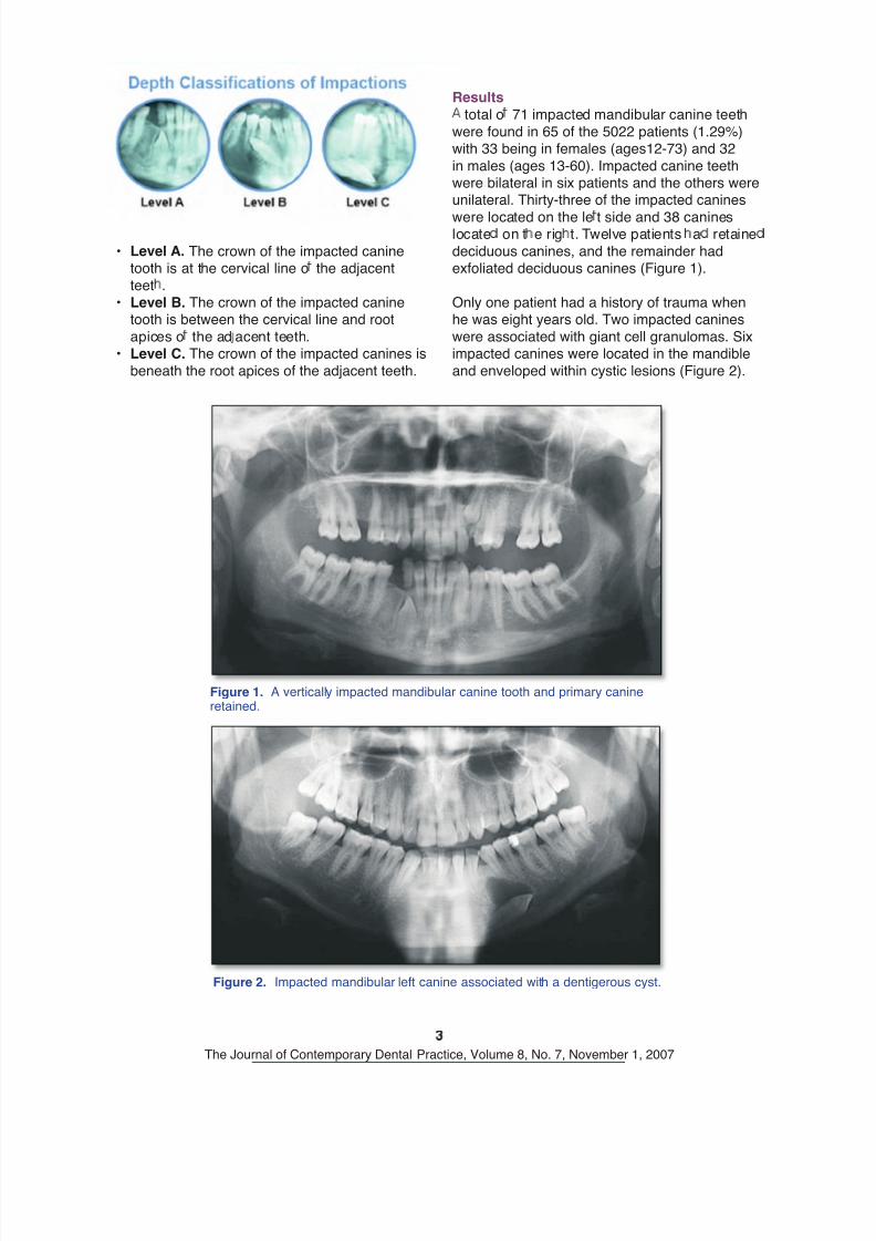
• Level A. The crown of the impacted caninetooth is at the cervical line o the adjacent
teet .• Level B. The crown of the impacted canine
tooth is between the cervical line and root
apices o the ad acent teeth.• Level C. The crown of the impacted canines is
beneath the root apices of the adjacent teeth.
Results
total o 71 impacted mandibular canine teeth
were found in 65 of the 5022 patients (1.29%)with 33 being in females (ages12-73) and 32
in males (ages 13-60). Impacted canine teethwere bilateral in six patients and the others wereunilateral. Thirty-three of the impacted canines
were located on the le t side and 38 canineslocate on t e rig t. Twelve patients a retaine
deciduous canines, and the remainder hadexfoliated deciduous canines (Figure 1).
Only one patient had a history of trauma whenhe was eight years old. Two impacted canines
were associated with giant cell granulomas. Siximpacted canines were located in the mandible
and enveloped within cystic lesions (Figure 2).
Figure 1. A vertically impacted mandibular canine tooth and primary canineretained.
Figure 2. Impacted mandibular left canine associated with a dentigerous cyst.
8/12/2019 Impacted Mandibular Canine-J Contemp Dent Pract
http://slidepdf.com/reader/full/impacted-mandibular-canine-j-contemp-dent-pract 4/9
The Journal of Contemporary Dental Practice, Volume 8, No. 7, November 1, 2007
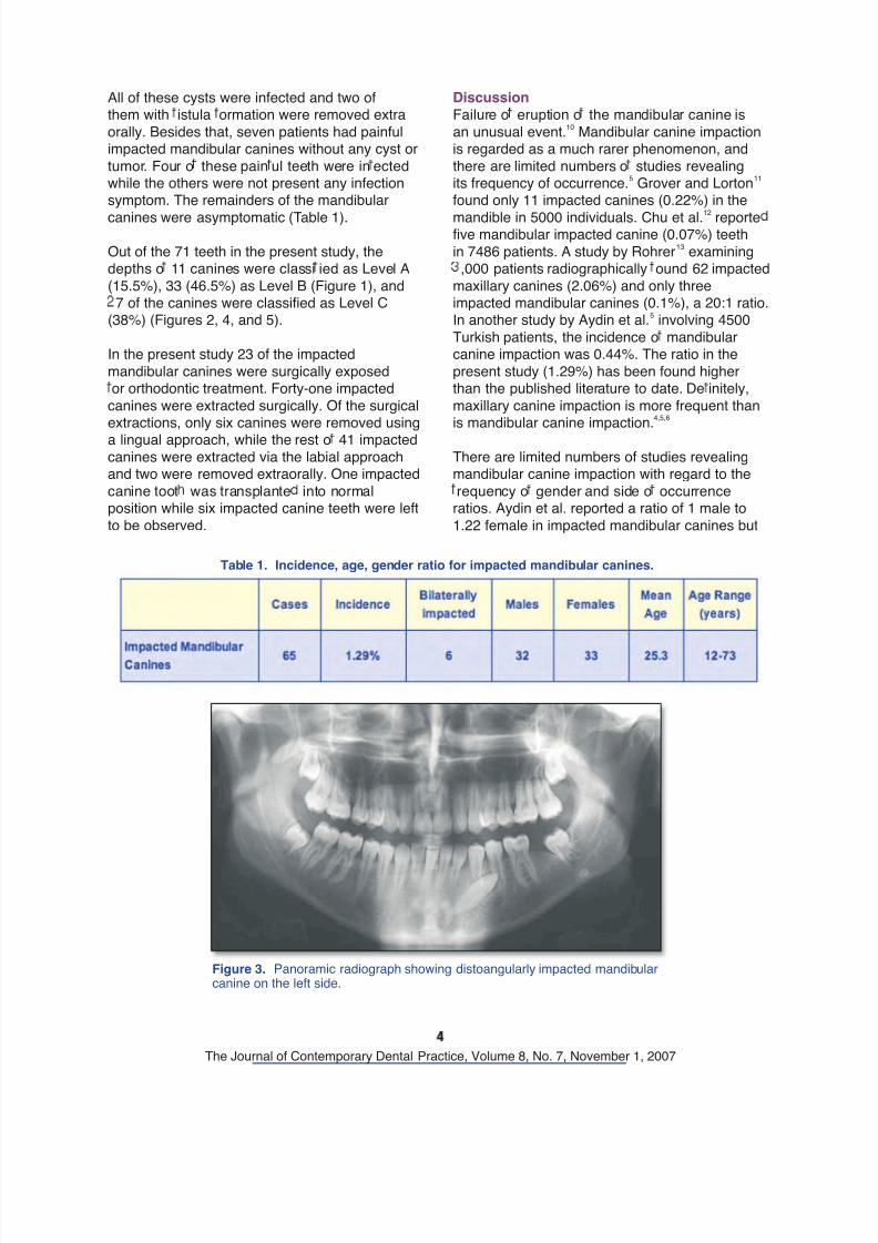
All of these cysts were infected and two ofthem with istula ormation were removed extra
orally. Besides that, seven patients had painfulimpacted mandibular canines without any cyst or
tumor. Four o these pain ul teeth were in ectedwhile the others were not present any infectionsymptom. The remainders of the mandibular
canines were asymptomatic (Table 1).
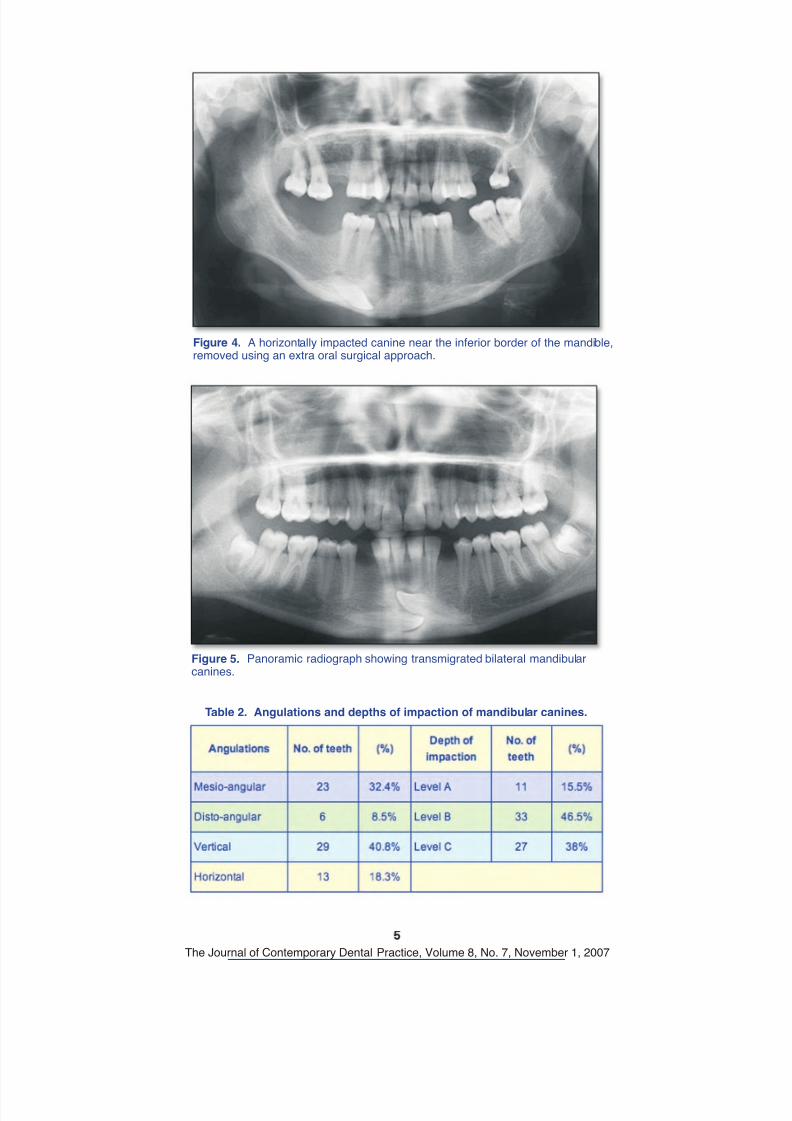
Out of the 71 teeth in the present study, thedepths o 11 canines were classi ied as Level A
(15.5%), 33 (46.5%) as Level B (Figure 1), and7 of the canines were classified as Level C
(38%) (Figures 2, 4, and 5).
In the present study 23 of the impacted
mandibular canines were surgically exposedor orthodontic treatment. Forty-one impacted
canines were extracted surgically. Of the surgicalextractions, only six canines were removed using
a lingual approach, while the rest o 41 impactedcanines were extracted via the labial approachand two were removed extraorally. One impacted
canine toot was transplante into normalposition while six impacted canine teeth were left
to be observed.
Discussion
Failure o eruption o the mandibular canine is
an unusual event.10
Mandibular canine impactionis regarded as a much rarer phenomenon, and
there are limited numbers o studies revealingits frequency of occurrence.
5 Grover and Lorton
11
found only 11 impacted canines (0.22%) in the
mandible in 5000 individuals. Chu et al.12
reportefive mandibular impacted canine (0.07%) teeth
in 7486 patients. A study by Rohrer13 examining
,000 patients radiographically ound 62 impacted
maxillary canines (2.06%) and only threeimpacted mandibular canines (0.1%), a 20:1 ratio.In another study by Aydin et al.
5 involving 4500
Turkish patients, the incidence o mandibularcanine impaction was 0.44%. The ratio in the
present study (1.29%) has been found higherthan the published literature to date. De initely,
maxillary canine impaction is more frequent thanis mandibular canine impaction.
4,5,6
There are limited numbers of studies revealingmandibular canine impaction with regard to the
requency o gender and side o occurrenceratios. Aydin et al. reported a ratio of 1 male to
1.22 female in impacted mandibular canines but
Table 1. Incidence, age, gender ratio for impacted mandibular canines.
Figure 3. Panoramic radiograph showing distoangularly impacted mandibularcanine on the left side.
8/12/2019 Impacted Mandibular Canine-J Contemp Dent Pract
http://slidepdf.com/reader/full/impacted-mandibular-canine-j-contemp-dent-pract 5/9
The Journal of Contemporary Dental Practice, Volume 8, No. 7, November 1, 2007
Figure 4. A horizontally impacted canine near the inferior border of the mandible,removed using an extra oral surgical approach.
Figure 5. Panoramic radiograph showing transmigrated bilateral mandibularcanines.
Table 2. Angulations and depths of impaction of mandibular canines.

8/12/2019 Impacted Mandibular Canine-J Contemp Dent Pract
http://slidepdf.com/reader/full/impacted-mandibular-canine-j-contemp-dent-pract 6/9
The Journal of Contemporary Dental Practice, Volume 8, No. 7, November 1, 2007
did not report a ratio of between right and left sideoccurrences in impacte man i ular canines.
5
A total of 65 patients (33 females ages 12 to 73years and 32 males ages 13 to 60 years) had
impacte man i ular canines in t e present stu y.Sex predilection was not noticeably differentfrom the Aydin et al.
5 study. Most of the impacted
man i ular canines are unilateral, owever, sixpatients a ilaterally impacte man i ular
canines.
There are many reasons why canines fail toerupt.
14 Most surgeons agree the reasons may
include a suspected pathological condition,
in ection, inter erence with prosthetic devices,disturbance of the existing dentition, pain,
and ectopic eruption.15 Many authors have
also speculated about the cause o impacted
mandibular canines.15
These causes includeinadequate space, supernumerary teeth,
premature loss o the deciduous dentition,retention of the deciduous canine, excessivecrown length, hereditary factors, functional
disturbances o the endocrine glands, tumors,cysts, and trauma.
10,14-18Mitchell
17 reported trauma
also has an effect on the impaction of a toothas an etiologic actor. In the patient group o thepresent study only one patient had a history of
trauma when he was eight years old. While 12primary canines were not exfoliated or extracted
in t is case, t e aut ors o not t ink t at traumacan be an etiologic factor for impaction of teeth.
Impacte man i ular canines are also more likelyto be located on the labial aspect of the dental
arch than are maxillary canines, and the removalo impacted teeth routinely involves an intraoral
surgical approach. But Plumpton19 suggested that
some extractions of the impacted mandibular
canine teet may one via an extraoral surgicalapproach. In the present study six canineswere removed using a lingual approach, while
the remainder o the removed canines wereextracte via a la ial approac an two impacte
mandibular canine teeth removed extraorally. In
the present study the most common angulation ofimpaction of the canine teeth was vertical (40.8%),followed by mesioanguler (32.4%), horizontal(18.3%), and then distoanguler (8.5%).
The mandibular canines are affected by pathology
in a lower ratio than the third molars andpremo ars,
10,12However, some aut ors reporte a
few cases such as dentigerous cyst, squamouso ontogenic tumors, an amelo lastoma were
associated with impacted mandibular canineteeth.
20,21,22 Dentigerous cysts caused by impacted
mandibular canine teeth were ound in six cases,and two impacted canines were associated withreparative giant cell granulomas in the present
study. All o these cysts were in ected and pain ulwith two of them having fistulas developing
extraorally below the mandible.
emoval of the entire cyst along with the impactedtooth is the principle treatment to preventrecurrence of the cyst.
3The reparative giant
cell granulomas ound were asymptomatic. Theimpacted mandibular canines associated with
dentigerous cysts and giant cell granulomas in thisstu y were remove surgically.
Most impacted teeth are asymptomatic, butchronic in ection with istula ormation and some
symptoms such as pain and swelling have beenreported in the literature.
10,12 In the present study
seven patients a pain ut t ese patients ano tumor or cyst formation. Only four impactedcanines of these seven patients were infected.
The remainder o the impacted mandibularcan nes were asymptomat c.
There are several treatment options proposed forimpacte man i ular canines inclu ing surgicalremoval, exposure and orthodontic alignment,transplantation, and observation.
24
If adequate space for alignment of an impacted
mandibular canine exists and it is mechanicallypossi le to reposition an impacte man i ular
8/12/2019 Impacted Mandibular Canine-J Contemp Dent Pract
http://slidepdf.com/reader/full/impacted-mandibular-canine-j-contemp-dent-pract 7/9
The Journal of Contemporary Dental Practice, Volume 8, No. 7, November 1, 2007
• If the deciduous canine has a good root lengthan it is est etically accepta le, o servation
of an asymptomatic mandibular canine can berecommended.
24
Surgical extraction is necessary in the followingsituations:
• The existence o in ection, cyst, or tumorrelate to t e impacte canine.
• The impacted tooth causes the periodontaldisturbances o the adjacent teeth.
• The presence of neuralgic symptoms.6
• Crowding of the mandibular arch requiringtherapeutic extractions to correct crowded
incisor teet .10
• The impacted canine is ankylosed and cannot
be transplanted.• There is evidence o root resorption a ecting
the adjacent teeth.• The root of impacted canine is severely
ilacerate .• Severe impaction of canine tooth.• Patient rejection of orthodontic treatment or
transp antat on.9
In this study 41 impacted mandibular canineswere extracted, 23 were attached to buttons ororthodontic treatment following surgical exposure,
one was transplanted, and the others were left inplace for observation.
Conclusion
Although maxillary canine impaction is morerequent than mandibular canine impaction, the
impaction ratio of the mandibular canine teeth
has been found to be higher than the publishediterature to date. The reason or this higher
incidence may be related to the patient selectionfrom the surgery clinic. However, this result may
also be evidence o the increase o the number oimpacted mandibular canine teeth.
canine into proper position, then orthodontictreatment is in icate .
24,25Following surgical
exposure, the impacted tooth may be allowed toerupt passively, especially if it has a favorable
angulation. Alternatively, orced eruption maybe carried out in conjunction with orthodonticalignment.
9,25As a third alternative, if an impacted
canine cannot be positioned avorably but thereis space for its full eruption, then orthodontic
treatment may help align the adjacent teeth intheir migrated order ollowed by crowning or
recontouring of some teeth to improve esthetics.26
It should be noted orthodontic treatment ofimpacted canines started after the conclusion
o the pubertal growth spurt are likely to beprotracted.
27
I the mandibular incisors are in a normal position,
space for the impacted canine is sufficient, andthe patient is symptom free, then transplantation
may e a reasona le treatment c oice.16,19
Autogenous transplantation of teeth with completeroot formation may be considered as a viable
treatment option to conventional prost etic animplant rehabilitation for both therapeutic and
economic reasons.28
This procedure is relativelyquick ut as an uncertain long-term prognosis.
Some authors believe asymptomatic impactedteeth can be left in place, but in these patients a
series of successive radiographs should be takenperiodically.
19 Observation of impacted mandibular
canines may be indicated in the followingcircumstances:
• A systemic contraindication to a surgery
exists.• T ere is a eeply impacte asymptomatic
mandibular canine with no associatedpathology, particularly in an older patient.
• Whenever the patient has a satis actorydental appearance and does not want surgicalintervention.
9,25

8/12/2019 Impacted Mandibular Canine-J Contemp Dent Pract
http://slidepdf.com/reader/full/impacted-mandibular-canine-j-contemp-dent-pract 8/9
The Journal of Contemporary Dental Practice, Volume 8, No. 7, November 1, 2007
References
1. Richardson G, Russel l KA. A Review of Impacted Permanent Maxillary Cuspids: Diagnosis and
Prevention. J Can Dent Assoc 2000: 66:497-501.2. Fonseca JR. Oral and Maxillofacial Surgery. Philadelphia: W. B. Saunders, 2000: Vol. 1:342-371.
3. Rajic S, Muretic Z, Percac S. Impacted canine in a prehistoric skull. Angle Orthod 1996: 66:477-80.4. D’Amico RM, Bjerklin K, Kurol J, Falahat B. Long-term Results of Orthodontic Treatment of Impacted
Maxillary Canines. Angle Orthod 2003: 73:231–238.
. Aydin U, Yilmaz HH, Yildirim D. Incidence o canine impaction and transmigration in a patientpopulation. Dentomaxillofac Radiol 2004: 33:164-9.
6. Alaejos-Algarra C, Berini-Aytes L, Gay-Escoda C. Transmigration of mandibular canines: Report ofsix cases and review of the literature. Quintessence Int. 1998: 29:395-398.
7. Yamaoka M, Furusawa K, FujimotoK, Uematsu T. Completely impacted teeth in dentate andedentulous jaws. Aust Dent J 1996: 41:169-72.
8. Bishara SE, Kommer DD, McNeil MH, Montagano LN, Oesterle LJ, Youngquist W. Management of
impacted canines. Am J Orthod 1976: 69:371-87.9. Bishara SE. Impacted maxillary canines. Am J Orthod Dentofac Orthop 1992: 101:159-71.
10. Camilleri S, Scerri E. Transmigration of mandibular canines-A review of the literature and a report offive cases. Angle Orthod 2003: 73:753-762.
11. Grover PS, Lorton L. The incidence of unerupted permanent teeth and related clinical cases. OralSurg Oral Med Oral Pathol 1985: 59:420-425.
12. Chu FCS, Li TKL, Lui VKB, Newsome PRH, Chow RLK, Cheung LK. Prevalence of impacted teethand associated pathologies - a radiographic study of the Hong Kong Chinese population. Hong KongMed J 2003: 9:158-163.
3. Rohrer A. Displaced and impacted canines. Int J rthod ral urg 1929: 15:1003.14. Wright DM. A case report: Forced eruption of an impacted lower canine in a 48-year-old man. J Am
Dent Assoc 1995: 126:1025-1027.5. Milano M, Barrett L, Marshall E. Extraction of a Horizontally Impacted Mandibular anine Through a
genioplasty Approach: Report of a case. J Oral Maxillofacial Surg 1996: 54:1240-1242.
16. Joshi MR. Transmigrant Mandibular Canines: A record of 28 Cases and a Retrospective Review ofthe Literature. Angle Orthod 2001: 71:12-22.
17. Mitchell L. Displacement of a mandibular canine following fracture of the mandible. Br Dent J 1993:74:417-41 .
18. Brezniak N, Ben-Yehuda A, Shapira Y. Unusual mandibular canine transposition: A case report. Am JOrthod Dentofacial Orthop. 1993: 104:91-94.
19. Plumpton S. The extraction of mandibular teeth via an extra-oral approach. Br J Oral Surg 1966:
4:127-131.20. Monteil RA, Terestri P. Squamous odontogenic tumor related to an unerupted lower canine. J Oral
Maxillofac Surg 1985: 43:888-895.21. Liposky RB. Decortication and bone replacement technique for the treatment of a large mandibular
cyst. J Oral Surg 1980: 38:42-45.22. El-Hakim IE, El-Khashab MM. Peripheral and Mural Ameloblastoma in the mandibular canine region
of a 13-year-old boy. J Oral Maxillofac Surg 2000: 58:1150-1154.
23. Hyomoto M, Kawakami M, Inoue M, Kirita T. Clinical conditions for eruption of maxillary canines andmandibular premolars associated with dentigerous cysts. Am J Orthod Dentofacial Orthop 2003:
24:515-520.
24. McDonald F, Yap WL. The surgical exposure and application of direct traction of unerupted teeth.Am J Orthod. 1986: 89:331-40.
25. Ferguson JW. Management of the unerupted maxillary canine. Br Dent J. 1990: 169:113-114.26. Shanmuhasuntharam P, Boon LC. Transmigration of permanent mandibular canines. Case report.
ust Dent J. 1991: 36:209-213.27. Orton HS, Garvey MT, Pearson MH. Extrusion of the ectopic maxillary canine using a lower
removable appliance. Am J Orthod Dentofacial Orthop. 1995: 107:349-59.28. Teixeira CS, Pasternak B Jr, Vansan LP, Sousa-Neto MD. Autogenous transplantation of teeth with
complete root formation: two case reports. Int Endod J. 2006: 39:977-985.
8/12/2019 Impacted Mandibular Canine-J Contemp Dent Pract
http://slidepdf.com/reader/full/impacted-mandibular-canine-j-contemp-dent-pract 9/9
The Journal of Contemporary Dental Practice, Volume 8, No. 7, November 1, 2007
About the Authors