Impact of the upper gut on body fluid regulation and blood ...
52
Impact of the upper gut on body fluid regulation and blood pressure – potential involvement of a locally expressed renin‐angiotensin system Doctoral thesis Peter Hallersund M.D. Department o d Education f Gastrosurgical Research an Institute of Clinical Sciences The Sahlgrenska Academy at University of Gothenburg Gothenburg, Sweden 2011
Transcript of Impact of the upper gut on body fluid regulation and blood ...
Impactoftheuppergutonbodyfluidregulationandbloodpressure
–potentialinvolvementofalocallyexpressedrenin‐angiotensinsystem
Doctoral thesis
Peter Hallersund
M.D.
Department o d Education f Gastrosurgical Research an
Institute of Clinical Sciences
The Sahlgrenska Academy at University of Gothenburg
Gothenburg, Sweden
2011

Correspondence:
Peter Hallersund
search and Education Department of Gastrosurgical Re
y Hospital Sahlgrenska Universit
Gothenburg SE41345
Sweden
2 68 59 Telephone: +46 31 34
Fax: +46 31 41 18 82
mail: [email protected] E
7/24855 http://hdl.handle.net/207
ISBN 978‐91‐628‐8287‐7
© Peter Hallersund, 2011
Printed by Intellecta Infolog AB, Gothenburg, Sweden

Tomychildren


ABSTRACT
This thesis explores the role of the upper gut in the regulation of diuresis and blood pressure control in relation to the novel finding of a mucosa‐located renin‐angiotensin system (RAS). RAS is a regulatory super‐system vital for body fluid homeostasis and blood pressure control. Recent research demonstrates that RAS is not only an endocrine (blood orne) system, but also in all respects locally expressed influencing tissue growth and ifferentiabd tion as well as inflammatory responses. A first aim of the present thesis‐project was to explore if RAS was expressed in the mucosa of the stomach and duodenum. Indeed, by use of western blot and immunohistochemistry most components of RAS were found in several compartments of the gastric mucosa of the Mongolian gerbil (model for human Helicobacter pylori infection) and also in the human mucosa. It was also observed that a subset of gastric mucosal endocrine cells expressed AT1 eceptors suggesting that activity in a local RAS can influence enteroendocrine signalling. AS componenrR ts were found also in the mucosa of the human duodenum. The second aim of the thesis was to examine the potential functionality of the local mucosal RAS described above. The project was focussed on a previously described sodium/volume sensor postulated to be situated in upper gut. Such a sensor is activated by food ingestion/drinking and increases renal diuresis already in the pre‐absorptive state. The upper‐gut location of this regulatory principle was demonstrated in healthy volunteers by intragastric instillation of 750 ml saline that almost promptly was followed by an increased diuresis, whereas intrajejunal instillation had an additional 60 min lag‐time until response. In a second set of experiments, the volunteer were first exposed to gastric instillation of saline (with sham‐intubation as time control) and after 30 to 40 min a gastroduodenoscopy with sampling of mucosal biopsies was performed. The tissue specimens were examined for RAS components and the principal finding was that the concentration of the pro‐hormone angiotensinogen decreased in the duodenal mucosa, but not in the stomach. The results confirm that a volume sensor is located to the upper gut in man. Furthermore, local mucosal AS, particularly in the duodenum, may be involved in mediating the diuresis occurring in he pre‐absorRt ptive state after drinking and eating. The third aim of the project was related to the physiological and clinical relevance of the sodium/volume monitor described above. Patients participating in the Swedish Obese Subjects (SOS) study were investigated. Gastric bypass (GBP), meaning that food and drinks are led directly into the jejunum thus bypassing the major part of the stomach and duodenum, was compared to gastric band constructions. The latter type of weight reducing surgery restricts the food intake capacity with the alimentary route intact. Interestingly, after adjustments for weight loss the GBP patients exhibited a larger 24h diuresis and a markedly more reduced systolic and diastolic pressure than the gastric band patients. These changes were prominent also 10 years after surgical intervention and were not related to the reduced body weight. Furthermore, the GBP patients consumed, despite a lowered blood pressure, approximately 1 g dietary salt more per day than patients operated with the restrictive banding techniques. This picture is compatible with that the sodium/volume sensor induces diuresis in an anticipatory fashion in relation to ingestive load and also inhibits salt appetite. Upon removal of this pre‐absorptive regulatory mechanisms (as following GBP), more rough post‐absorptive regulatory principles dominate that very probably results in an overshooting diuretic effect with depressor action and an increased salt intake.
1

LISTOFPAPERS
his thesis is based on the following papers, which will be referred to in the text by their TRoman numerals:
ction in I. Hallersund P, Helander HF, Casselbrant A, Edebo A, Fändriks L, Elfvin A. ngiotensin II receptor expression and relation to Helicobacter pylori‐infehe stomach of the Mongolian gerbil. BMC Gastroenterol. 2010 Jan 14;10:3 At II. Hallersund P, Elfvin A, Helander Hhe expression of renin‐angiotensin stric mucosa.
F, Fändriks L. system components in the human ga
ReninAngiotensinAldosteroneSyst.2011 Mar 12;54‐64. Epub 2010 Aug T J III. Hallersund, P, Edebo A, Casshe sodium/volume sensor in thocal renin‐angiotensin system. Inmanuscript
elbrant A, Spak E, Fändriks L. e upper gut in man – potential involvement of a T
l IV. Hallersund P, Sjöström L, Olbers T, Lönroth H, Jacobson P, Wallenius V, Näslund I,
L. Long‐term effects on blood pressure and dietary salt intake rgery – an analysis of 10‐year follow‐up data from the Swedish
Carlsson LM, Fändriksby weight reducing suObese Subjects study. Inmanuscript
2
TABLEOFCONTENTS
L
ISTOFABBREVIATIONS_____________________________________________________________________________________ 4
BACKGROUND
INTRODUCTION _________________________________________________________________________________________________________ 5
THE RENIN‐ANGIOTENSIN SYSTEM (RAS) ___________________________________________________________________________ 6
BODY FLUID HOMEOSTASIS AND THE GUT SODIUM/VOLUME SENSOR __________________________________________9
HEMOSENSING IN THE GUT MUCOSA AND ENTEROENDOCRINE CELLS ______________________________________ 12 C
HYPOTHESESANDAIMSOFTHETHESIS ______________________________________________________________ 15
REVIEWOFRESULTS
PRESENCE AND LOCATION OF RAS COMPONENTS IN THE GASTRIC MUCOSA ________________________________ 16
RAS EXPRESSION IN RELATION TO HELICOBACTER PYLORI INFECTION _____________________________________ 18
RAS COMPONENTS IN THE DUODENAL MUCOSA ________________________________________________________________ 20
DIURETIC RESPONSE AND PLASMA HORMONES AFTER A GASTRIC OR JEJUNAL SALINE LOAD ___________ 21
AGT AND ANGII IN THE GASTRO‐DUODENAL MUCOSA SUBSEQUENT TO A GASTRIC SALINE LOAD ________ 24
IURNAL URINE OUTPUT, SALT INTAKE AND BLOOD PRESSURE AFTER GASTRIC BYPASS SURGERY _____ 27 D
CONCLUSIONS___________________________________________________________________________________________________31
GENERALDISCUSSION
SOME METHODOLOGICAL CONSIDERATIONS ___________________________________________________________________ 32
RAS IN THE UPPER GUT MUCOSA __________________________________________________________________________________ 33
IS THE GASTRODUODENAL RAS INVOLVED IN SENSING OF LUMINAL CONTENT? ___________________________ 35
THE GUT SODIUM/VOLUME SENSOR? ______________________________________________________________________________ 36
ROLE OF THE GUT IN BODY FLUID HOMEOSTASIS AND ARTERIAL PRESSURE CONTROL ____________________ 37
THE RELATION BETWEEN “PRE‐ABSORPTIVE” AND “POST‐ABSORPTIVE” MECHANISMS ____________________ 38
PHYSIOLOGICAL AND CLINICAL RELEVANCE _____________________________________________________________________ 39
ACKNOWLEDGEMENTS_______________________________________________________________________________________ 40
REFERENCES____________________________________________________________________________________________________ 41
3
LISTOFABBREVIATIONS
erting enzyme ACE angiotensin‐conv
AGT angiotensinogen
) AngII angiotensin II (1‐8
‐7) Ang‐(1 angiotensin (1‐7)
AT1R AngII type 1 receptor
AT2R AngII type 2 receptor
ANP atrial natriuretic peptide
c peptide BNP B‐type natriureti
CgA chromogranin A
id ECF extracellular flu
P GB gastric bypass
testinal GI gastroin
um Na sodi
NaCl salt
NEP neprilysin
RAS renin‐angiotensin system
tudy SOS study Swedish Obese Subjects s
ut Upper g stomach and duodenum
VBG/B gastric banding procedures
4
BACKGROUND
INTRODUCTION
This thesis project explores the role of the upper gut in the regulation of diuresis and blood
pressure control in relation to the novel finding of a mucosa‐located renin‐angiotensin
system (RAS) in the stomach and duodenum. The project emanated from an exploration of
mechanisms by which the human pathogen Helicobacterpylori “manipulated” host defence‐
dependent cytotoxic radical formation in the human gastric mucosa1. The findings were
further explored in Mongolian gerbils being regarded as a good model for the human H.
pyloriinfection and its related pathology2. The rationale for investigating RAS in this animal
model can perhaps not be conceived as intuitive and therefore deserves some explanation.
Our research team had previously linked H.pylori to inhibition of duodenal mucosal
bicarbonate secretion. This secretion provides a neutralising zone close to the surface
epithelium protecting the mucosa from intraluminal acid disposed by the stomach. Hence,
the ulcerogenic property of H.pyloricould to some degree be explained as due to inhibited
mucosal bicarbonate secretion3. In a parallel project in our laboratory, RAS was found to
regulate such duodenal mucosal bicarbonate transport4, 5. In addition, data in the literature
show that RAS is involved in inflammation, tissue growth and differentiation, as well as
carcinogenesis, all being of great clinical interest for GI pathology. Based on this background
we occasionally checked for the presence of angiotensin II (AngII) receptors in the H.pylori
infected and inflamed gastric mucosa of the abovementioned Mongolian gerbils. The
intriguing finding of a widespread presence of AngII receptors in gastric mucosae, also in
those devoid of infection/inflammation, became the starting point for this thesis project.
The project has since then evolved from mucosal expression of RAS to the role of the upper
gut as part of fluid homeostasis and arterial pressure control. Below are the today’s
paradigms regarding RAS, body fluid homeostasis and gut chemosensing briefly reviewed.
Novel findings are then presented and discussed.
5

THERENIN‐ANGIOTENSINSYSTEM(RAS)
TheclassicalRAS
Textbooks in physiology still describe RAS as an endocrine system for hemodynamic
regulation and body fluid homeostasis (Figure 1). This classical picture relates to a system
that is activated when blood circulation is challenged, for example due to hemorrhage or
uncompensated profuse sweating. The reduced blood volume will be manifested as a
lowered arterial pressure or a sodium deficiency that will initiate the release of the enzyme
renin from the juxtaglomerular apparatus of the kidneys. Renin cleaves off the decapeptide
angiotensin I (AngI) from the precursor protein angiotensinogen (AGT; 452 amino acids
long) released by the liver. AngI is then degraded to the signal mediator octapeptide
angiotensin II (AngII) by angiotensin‐converting enzyme (ACE) expressed by endothelial
cells mainly in pulmonary vessels. Circulating AngII acts vasoconstrictive and induces renal
sodium and fluid retention to maintain arterial pressure and to compensate for the reduced
blood volume. AngII also mediates the thirst sensation and salt appetite driving the
individual to a final fluid compensation by increased oral intake of water and sodium6, 7
(Figure 1). AngII regulates cardiovascular and body fluid homeostasis both directly on the
vascular system, kidney and brain, as well as indirectly via other regulatory factors, for
example by liberation of aldosterone from the adrenals, or by facilitation of vasoconstrictive
sympathetic nervous activity8, 9.
Figure 1.
Theclassicalendocrine
renin‐angiotensinsystem(RAS)
6
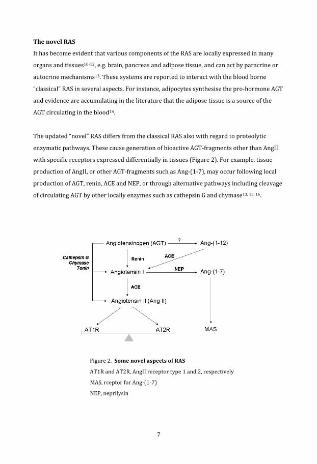
ThenovelRAS
It has become evident that various components of the RAS are locally expressed in many
organs and tissues10‐12, e.g. brain, pancreas and adipose tissue, and can act by paracrine or
autocrine mechanisms13. These systems are reported to interact with the blood borne
“classical” RAS in several aspects. For instance, adipocytes synthesise the pro‐hormone AGT
and evidence are accumulating in the literature that the adipose tissue is a source of the
GT circulating in the blood14. A
The updated “novel” RAS differs from the classical RAS also with regard to proteolytic
enzymatic pathways. These cause generation of bioactive AGT‐fragments other than AngII
with specific receptors expressed differentially in tissues (Figure 2). For example, tissue
production of AngII, or other AGT‐fragments such as Ang‐(1‐7), may occur following local
production of AGT, renin, ACE and NEP, or through alternative pathways includingcleavage
f circulating AGT by other locally enzymes such as cathepsin G and chymase13, 15, 16. o
Figure 2.SomenovelaspectsofRAS
1 and 2, respectively AT1R and AT2R, AngII receptor type
r Ang‐(1‐7)
MAS, rceptor fo
NEP, neprilysin
7

AngiotensinII–theprincipalmediatorofRAS
AngII works principally through two receptors (Figure 2) designated AngII type 1 receptor
(AT1R) and AngII type 2 receptor (AT2R)17. Classical effects of AngII, such as
vasoconstriction and aldosterone release, are mediated via AT1R. Less is known about the
actions of AT2R. Several studies indicate that activation of the AT2R generally has effects
that oppose those mediated by the AT1R, thus modulating the responses to stimulation
with AngII18. These two receptor subtypes belong to the seven‐transmembrane G‐protein‐
coupled receptor superfamily, and binding of AngII to AT1R and AT2T can activate several
intracellular second‐messenger systems19‐21, resulting in e.g. hormonal release (e.g. AGT,
aldosterone and vasopressin) or activation of transcription factors inducing gene
expression, such as Activator Protein 1 (AP‐1), Signal Transducer and Activator of
ranscription (STATs), and Nuclear Factor‐kB (NF‐kB). T
RASinthegastrointestinal(GI)mucosa
The presence of RAS in the GI mucosa is sparsely reported in the literature, and particularly
so with regard to the situation in man22. Nevertheless, AngII receptors of both subtypes
(AT1R and AT2R) have been reported to be expressed in the esophageal23, small intestinal24
and in the colonic mucosa25, and data suggest that in these parts of the GI tract, RAS is
involved in epithelial fluid/electrolyte and glucose transport, as well as in mucosal
nflammation12, 26‐29. i
d. Potential roles for the RAS in the gastric and duodenal mucosa are very little explore
Effects on duodenal bicarbonate secretion and gastric blood perfusion in relation to
circulatory stress (and reperfusion) have been reported in animal studies5, 30, 31, and also
involvement in the postulated sodium monitor suggested to be situated in the upper part of
the gut32‐34. This latter mechanism is of particular interest for the present thesis project and
will be described in detail below. Briefly, it represents the sensor of an entero‐renal
signalling mechanism demonstrated by the phenomenon that dietary sodium induces a
more prompt natriuresis than does the similar amount sodium given intravenously35, 36.
Filling the gap regarding information on RAS location in the human gastric and duodenal
mucosa is one important aim of the present thesis project (see Aims of the thesis).
8

BODYFLUIDHOMEOSTASISANDTHEGUTSODIUM/VOLUMESENSOR
Body fluid homeostasis is a core element in physiology and detailed descriptions are given
in most textbooks and many comprehensive reviews37‐39. A brief summary is given below
with some extra attention given to systematic mediators of importance for the experimental
roject presented later in this thesis. p
Of the total water content in the body, the intracellular fluid compartment constitutes 2/3.
The remaining 1/3 is extracellular fluid (ECF). 3/4 of the ECF volume surrounds the cells
(interstitial fluid) and the rest (1/4) circulates in blood as plasma. Because of its abundance,
sodium (Na) is the major determinant of the osmolarity of the ECF. Therefore, the sodium
concentration of the ECF constitutes the major osmotic force that moves water in or out of
cells. It follows that body fluid homeostasis requires mechanisms that strive to maintain an
optimal distribution within the intracellular and extracellular fluid compartments; as well
as mechanisms that maintain a precise balance between the intake and excretion of sodium
nd water of the body. a
The input of sodium and water to the ECF is determined by the net absorptive capacity
(mucosal absorption minus secretion) of the intestinal mucosa and by the ingested
amounts. The latter is in turn dependent on central regulation of ingestive behavior in
relation to the sensations of thirst and salt (NaCl) appetite. Sodium and water output is
during resting conditions determined mainly by the kidneys, which can control the rates of
excretion of water and sodium independently of each other40. During exercise one also has
to count losses by transpiration and respiration. Stool water contents can vary considerably
ut is during physiological conditions not regarded of importance for volume output. b
Aberrations from normal body fluid conditions are counteracted by regulatory mechanisms
on all functional levels of the organism. Local ion concentrations influence directly the state
of membrane transporters to protect functions on the cellular level. On the tissue and organ
level, specialized sensor structures activate humoral factors and neural activity that forces
distant organs to compensatory actions. One such principle is the sensing of blood pressure
at specific sites within the cardiovascular system. Blood pressure is by definition dependent
9
on blood volume which in turn is associated to ECF and its sodium concentration. Pressure
sensing takes place in the heart and pulmonary vessels (low‐pressure sensing) and in the
carotid sinus, aortic arch, and juxtaglumerular apparatus of the kidneys (high‐pressure
sensing). In addition to integrating pressure information, the organism also senses sodium
concentration persein e.g. the juxtaglumerular cells and at certain brain areas. Regulatory
signals are mediated via the sympathetic nervous system (partly by renin release), via RAS
(AngII and indirectly via the production of aldosterone), and via cardiac natriuretic
eptides41 (ANP and BNP), as well as via vasopressin from the pituitary. p
Peripheralmarkersofbodyfluidcontrol
From a research perspective, sympathetic neural activity is a difficult variable to assess
whereas the humoral mediators (e.g. AngII, aldosterone, BNP, vasopressin) are easy
accessible by blood sampling and therefore often are used as good markers on actions
related to body fluid control. As mentioned, the circulating blood volume is part of the body
fluids and consequently hemodynamic regulation and body fluid homeostasis are
integrated. Therefore, mechanisms that regulate blood circulation are also the major
determinants of sodium and water balance. It follows that ECF volume partly determines
venous and arterial pressure. Blood pressure recordings (particularly in the low pressure
parts) can briefly reflect the state of the ECF.
10

Thegutsodium/volumesensor
The above described sensors in the cardiovascular system, brain and kidneys detect
changes in plasma volume or sodium concentration. Additionally, experiments have
indicated that there also exists a “pre‐absorptive” sodium/volume sensor in the GI tract33,
42‐44. This sensor is activated by salt ingestion and drinking and signals to the kidneys to
increase natriuresis before any detectable changes in plasma sodium concentration are
observed. A similar mechanism inhibits salt appetite and thirst in an anticipatory fashion
Figure 3). (
Figure 3. Theproposedsodium/volumesensorintheuppergut
For example, gastric salt loading inhibits salt appetite in sodium depleted rats before
plasma sodium concentration is enhanced by absorption of the salt43. Likewise, water
intake causes satiety in thirsty humans and animals (initially given hypertonic saline
ntravenously to induce thirst) before plasma sodium concentration is corrected44. i
The cellular and molecular mechanisms underlying pre‐absorptive body fluid regulation are
unknown, as well as the exact location of the sensor. Suggested mediators linking the
sodium/volume sensor to the central nervous system and the kidneys include vagal
. afferents45, enteroendocrine “taste” cells33 and several humoral factors including AngII32, 46
For example, experiments in rats given ACE‐inhibitors (decreasing AngII generation) have
indicated that an intact renin‐angiotensin system is necessary for the interplay between the
gastrointestinal tract and kidney47.
11

CHEMOSENSINGINTHEGUTMUCOSAANDENTEROENDOCRINECELLS
The physicochemical properties of the luminal bulk influence markedly the secretion of
gastric acid and proteolytic enzymes, the gastric emptying rate and the type of intestinal
motility. Sensing of the luminal contents by the GI mucosa is necessary for these adaptive
responses that optimize the digestive and absorptive conditions. In addition, the detection
of constituents within the GI tract is important also for extra‐GI organs and the organism as
a whole. Many important physiological processes are initiated or modulated from the GI
tract, e.g. immune responses, glycemic control (demonstrated for example by the fact that
oral ingestion of glucose triggers more insulin release than glucose delivered intravenously)
nd food intake48‐51. a
Gut chemosensing is usually regarded as a neuro‐endocrine process involving hormone
releasing cells in the gut mucosa; the enteroendocrinecells. When activated, these cells exert
endocrine actions (the hormone reach distant targets via the blood stream), or paracrine
activation (local release and actions) of, for example, local enteric nerves and/or afferent
ibers of the vagal nerve mediating the signal to the central nervous system (Figure 4). f
Figure 4.Endocrineandparacrinesignallingbyenteroendocrinecells
12

The enteroendocrine cells are confined to the epithelial layer of the mucosa and have two
principal morphological shapes, the “open type” having contact with the GI lumen, and the
“closed type” not reaching the luminal contents. Despite being a numerically small
proportion of the total epithelial cells these cells are regarded as the largest endocrine
“organ” of the body, both in terms of number of cells and variety of hormones produced.
Vagal mucosal fibers do not reach the epithelial surface, but are closely associated with the
nteroendocrine cells (Figure 4) and express specific receptors for GI hormones. e
A common feature for enteroendocrine cells is the presence of chromogranins52 which are
vesicle storage proteins, reflecting the secretory granules present in endocrine cells.
Chromogranin A (CgA) is often visualized in the initial immunohistochemical identification
of enteroendocrine cells53. The release of hormones from enteroendocrine cells (Figure 4)
is partly regulated by agents in the GI lumen, such as nutrients54, 55 (lipids, proteins and
carbohydrates), acidity, and gas tensions56, 57 (e.g. C02, NO). Some enteroendocrine cells are
also acting secondary to other signalling principles, e.g. neural impulses, blood borne signal
substances and nutrients, and gastrointestinal mechanical properties (for example wall
tension reflecting the degree of distension due to presence of food and/or muscular
activity). On the other hand, chemosensitivive enteroendocrine cells can elicit muscular
activity that in turn activate mechanosensors belonging to the extrinsic vagal and spinal
afferents that in turn mediates signals to the central nervous system eliciting reflex
eedback and/or perceptions. f
One example of polymodal enteroendocrine signalling is the mediator glucagon‐like peptide
1 (GLP‐1)58. This peptide is liberated when nutrients reach the enteroendocrine L‐cells in
the distal small intestine and colon. GLP‐1 has multiple effects based on its endocrine mode
of action (for example stimulates insulin secretion from the pancreas) but does also activate
agal afferents in turn resulting in rapid reflex effects. v
Recently, much interest has been focused on the role of “taste cells” in the GI mucosa. These
cells express modality‐specialized sensing molecules originally described in the taste
receptor cells of the tongue59, 60. Interestingly, recent findings suggest that the molecular
pathways similar to those mediating oral taste perceptions also operate in the gut mucosa55.
13

Taste molecules have been found in enteroendocrine cells and in other morphologically
similar cells, called “brush cells”. However, no secretory granules of the type that
characterize endocrine cells can be demonstrated in brush cells61. Studies indicate that
brush cells can release nitric oxide (NO) that may be an important signalling molecule62,
ctivating vagal afferent nerve fibers or influencing adjacent mucosal cells. a
In general, nutrient sensing mechanisms in the gut are not well understood but this is an
area of increasing scientific interest, given its importance in the regulation of glucose
homeostasis and food intake. It is difficult to study enteroendocrine cells directly within the
gut mucosa and particularly their paracrine actions because plasma levels may not be
helpful in assessing local roles of a particular hormone or determing the mechanism of its
release. Consequently, much of what we know of direct chemosensing by enteroendocrine
cells comes from experiments on cell lines50.
14

HYPOTHESES
It is well established that the endocrine renin‐angiotensin system (RAS) is a
powerful signalling system involved in the electrolyte and fluid homeostasis and
blood pressure control. It was hypothesised that RAS components expressed locally
in the mucosa of the upper gut exert such regulatory impact already in relation to the
ingestion of electrolytes and fluid. Based on this hypothesis it was assumed that
intervention with the gastrointestinal continuity should affect blood pressure
control.
AIMSOFTHETHESIS
The general aim of this thesis was to investigate the presence of the renin‐
angiotensin system (RAS) in the mucosa of the human stomach and duodenum and
o position the findings in a physiological and clinically relevant context. t
The specific aims of the project were related to the following questions:
1. s RAS present in the gastric and duodenal mucosa? I
2. Is the upper‐gut mucosal RAS involved in gut‐renal diuretic responses?
3. Does permanent exclusion of the upper‐gut sodium/volume sensor influence
diuresis, salt appetite and blood pressure?
15

REVIEWOFRESULTS
1.IsRASpresentinthegastricandduodenalmucosa?
The presence and location of representative RAS components in the gastric and duodenal
mucosa was investigated by use of Western blot and immunohistochemistry (I, II and III).
Gastric mucosal infection with Helicobacterpylori is extremely common in the population.
Although severe morbidity, e.g. peptic ulcers and gastric carcinomas, can be associated to
this infection most individuals remain asymptomatic63. Because of its high prevalence it was
considered of importance to rule out if and how an H.pylori infection influenced the
expression of RAS. The mapping of RAS components in the gastric mucosa (I, II), therefore,
was related to if H.pylori was present or not.
Studysetting
A systematic mapping of immunoreactivity to AngII receptors (AT1R and AT2R) was first
performed in the stomach of the Mongolian gerbil (commonly used as a model for human H.
pyloriassociated gastritis) in presence or absence of experimentally induced H.pylori
infection (I). These results were subsequently confirmed in endoscopic biopsies from the
human mucosa of H.pylori‐negativeandH.pylori‐positivevolunteers, where also
immunoreactivity to angiotensin generating enzymes (renin, ACE and NEP) and the
prohormone AGT were assessed (II). Mapping of RAS components in the human duodenal
ucosa was performed on endoscopic biopsies from healthy volunteers (III). m
PresenceandlocationofRAScomponentsinthegastricmucosa
The proteins of the examined RAS components were all identified by Western blotting in
samples from the gerbil and human stomach, and immunoreactivity to AT1R and AT2R was
found in a variety of cells in the gastric mucosa (I, II). A summary of the
immunohistochemical results from various mucosal compartments of the human stomach is
given in Table 1.
16

Table 1.LocationofRASproteinsinthehumangastricmucosa(fromII)
Interesti f ngly, strong immunoreactivity to the AT1R protein was found (independent o
H.pylori infection) in some epithelial cells in the antral mucosa of both the gerbil and
human stomach. These cells had the typical appearance of enteroendocrine cells, e.g. in
some cases a narrow string of cytoplasm was observed. Co‐expression of AT1R and CgA (a
marker for endocrine cells53) by a subpopulation of gastric enteroendocrine cells was
confirmed using double immunostaining (Figure 5). Hence, these results suggest that
activity in a local RAS can influence enteroendocrine signalling.
17

Figure 5. EnteroendocrinecellsingastricmucosastainingpositiveforAT1R
A marker for endocrine cells (Cga) was used for confirmation. Stainings from the
gerbil (upper sections) and human (lower sections) gastric mucosa are displayed.
RASexpressioninrelationtoHelicobacterpyloriinfection
In the human gastric mucosa, immunoreactivity to the proteins of AGT, renin, ACE, NEP did
not differ quantitatively between H.pylori‐positive and H.pylori‐negative subjects.
However, AT1R protein expression was significantly more pronounced in the gerbil and
human H.pylori‐positive mucosa compared to H.pylori‐negative mucosa.
Immunohistochemistry also showed an abundance of inflammatory cells (lymphocytes and
neutrophils) in the mucosa with immunoreactivity to AT1R (I, II). By quantifying
lymphocytes and neutrophils present in the mucosa, we found that the AT1R protein
expression correlated with the number of neutrophils, but not with the number of
lymphocytes (Figure 6). Thus, these results indicate that H.pylori induced gastritis is
associated with higher prevalence of AT1R, most probably due to presence of infiltrating
neutrophils carrying this receptor.
18
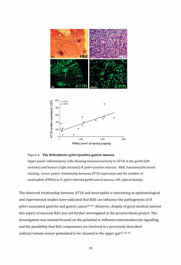
Figure 6.TheHelicobacterpyloripositivegastricmucosa
Upper panel: inflammatory cells showing immunoreactivity to AT1R in the gerbil (left
sections) and human (right sections) H.pylori positive mucosa. H&E, haematoxylin/eosin
staining. Lower panel: relationship between AT1R expression and the number of
neutrophils (PMNs) in H.pylori infected gerbil antral mucosa. OD, optical density.
The observed relationship between AT1R and neutrophils is interesting as epidemiological
and experimental studies have indicated that RAS can influence the pathogenesis of H.
pylori associated gastritis and gastric cancer64‐67. However, despite of great medical interest
this aspect of mucosal RAS was not further investigated in the present thesis project. The
investigation was instead focused on the potential to influence enteroendocrine signalling
and the possibility that RAS components are involved in a previously described
sodium/volume sensor postulated to be situated in the upper gut32, 34, 43.
19

RAScomponentsintheduodenalmucosa
Ingested liquid meals are rapidly disposed by the stomach into the duodenum. The gastric
emptying rate differs depending on the physicochemical properties of the stomach
contents, e.g. energy density, osmolality etc. Water and non‐caloric isotonic solutions are
almost instantly delivered into the duodenal lumen68, 69. Thus, drinks do not only expose the
gastric mucosa, but also the duodenal one. The expression of RAS in the human duodenum
during basal conditions had not been previously investigated so this was done as part of
Paper III. Indeed, the proteins of AT1R, AT2R, renin, ACE, NEP and AGT were all identified
by Western blotting in samples of duodenal mucosa fromhealthy volunteers (III).
Immunohistochemistry showed staining for AT1R and AT2R in the basal parts of most
epithelial cells. Interestingly, immunoreactivity to AGT was found in the basal parts of
solitary epithelial cells in the duodenal mucosa (Figure 7).
Figure 7. ImmunoreactivitytoAT1R,AT2R,andAGTinthehumanduodenalmucosa
Left: Immunostainings for AT1R and AT2R. Right: AGT was found in the basal parts of solitary
epithelial cells in villi and crypts (arrows) and in blood vessels (not indicated). Original
magnification of images: x40
1stconclusion
Promine . nt components of RAS are present in the human gastric and duodenal mucosa
H.pylori induced gastritis is associated with higher prevalence of AT1 receptors, most
probably due to presence of infiltrating neutrophils carrying this receptor.
Immunoreactivity to AT1R and AGT in solitary epithelial cells suggest that local RAS activity
can influence gastro‐duodenal enteroendocrine signalling.
20

2.Istheupper‐gutmucosalRASinvolvedingut‐renaldiureticresponses?
The project was then directed towards the potential functionality of RAS in the gastric and
duodenal mucosa. The aim was to investigate if the mucosa‐located RAS is involved in the GI
sodium/volume sensor that upon drinking and eating induces diuresis in an anticipatory
fashion. The presence and location of the gastrointestinal sodium/volume sensor was first
investigated. Acute signs of mucosal RAS reactions to an intraluminal saline load were then
xplored. e
Studysetting
To confirm presence and location of pre‐absorptive regulation, 750 ml isotonic NaCl was
installed intralluminally via a nasogastro (‐jejunal) tube either in the stomach or in the
jejunum of healthy male volunteers. The time course of the diuretic response was
characterized. Blood borne factors of importance for body fluid homeostasis were also
analyzed using radioimmunoassay (RIA) or enzyme immunoassay (EIA). Potential changes
in the gastroduodenal mucosal RAS to an intragastric luminal saline load were assessed by
Western blot and EIA targeting AGT and AngII levels in the mucosa, respectively. In these
experiments, the volunteers were first exposed to instillation of saline via a nasogastric
tube (with sham‐intubation as time control) and after 30‐40 min a gastroduodenoscopy
with sampling of mucosal biopsies (usually 45 min after the exposure procedure) was
performed. All subjects were instructed to avoid high salt intake 4 days before examinations
and each subject participated at two separate study days to be able to serve as its own
ontrol. c
Diureticresponseandplasmahormonesafteragastricorjejunalsalineload
The latency of onset to a diuretic response was markedly shorter after gastric loading than
after jejunal loading of 750 ml isotonic saline (Figure 8). Thus, these results confirm that a
diuresis regulating mechanism is activated in the upper gut at a time point where blood
volume expansion following absorption is unlikely to have occurred.
21
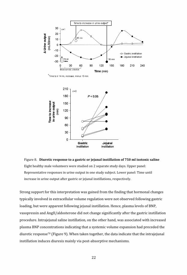
Figure 8.Diureticresponsetoagastricorjejunalinstillationof750mlisotonicsaline
Eight healthy male volunteers were studied on 2 separate study days. Upper panel:
el: Time until
Representative responses in urine output in one study subject. Lower pan
increase in urine output after gastric or jejunal instillations, respectively.
Strong support for this interpretation was gained from the finding that hormonal changes
typically involved in extracellular volume regulation were not observed following gastric
loading, but were apparent following jejunal instillation. Hence, plasma levels of BNP,
vasopressin and AngII/aldosterone did not change significantly after the gastric instillation
procedure. Intrajejunal saline instillation, on the other hand, was associated with increased
plasma BNP concentrations indicating that a systemic volume expansion had preceded the
diuretic response70 (Figure 9). When taken together, the data indicate that the intrajejunal
instillation induces diuresis mainly via post‐absorptive mechanisms.
22
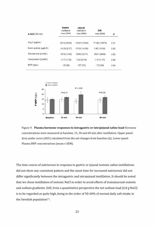
Figure 9.Plasmahormoneresponsestointragastricorintrajejunalsalineload Hormone
concentrations were measured at baseline, 15, 30 and 60 min after instillation. Upper panel:
Area under curve (AUC) calculated from the net changes from baseline (Δ). Lower panel:
lasma BNP concentrations (mean ± SEM). P
The time course of natriuresis in response to gastric or jejunal isotonic saline instillations
did not show any consistent pattern and the onset time for increased natriuresis did not
differ significantly between the intragastric and intrajejunal instillation. It should be noted
that we chose instillation of isotonic NaCl in order to avoid effects of transmucosal osmotic
and sodium gradients. Still, from a quantitative perspective the net sodium load (6.8 g NaCl)
is to be regarded as quite high, being in the order of 50‐60% of normal daily salt intake in
the Swedish population71.
23

AGTandAngIIinthegastro‐duodenalmucosasubsequenttoagastricsalineload
In the next set of experiments, instillation of isotonic saline in the stomach was used to
provoke the gastroduodenal mucosa and potentially the local RAS. The volume saline
installed was, compared to the previous experimentation, reduced from 750 to 500 ml (4.5
g NaCl) to minimize the risk for aspiration in association to the subsequently performed
endoscopy. The endoscopic mucosal biopsy takings were performed approximately 45 min
after instillation. According to the previous results, this time point corresponded to onset of
the diuretic response following gastric instillation (Figure 9) and, hypothetically, the
mucosal reaction inducing gastroduodeno‐renal signalling. For practical reasons the tissue‐
analyses were limited to AngII and the prohormone AGT representing two important
mediator factors. Vasopressin, that in addition of being a pituitary hormone also is
xpressed in the GI mucosa72, was measured as reference. e
Interestingly, the content of AGT in the duodenal mucosa decreased significantly
subsequent to the gastric saline instillation and this was not the case in antral specimens
(Figure 10). The saline load did not significantly influence AngII or vasopressin, neither in
antral nor in duodenal mucosae. These observations suggest that the duodenum might be
the primary site for this type of luminal sensing and that the local duodenal RAS reacts upon
a luminal saline load with a mobilization of stored AGT.
2ndconclusion
The temporal relationship between increased diuresis induced by an intragastric saline
load and the reduced quantity of AGT in duodenum suggest a role for RAS in the duodenal
mucosa in the pre‐absorptive induction of diuresis occurring after drinking and eating.
24

Figure 10.Tissuelevelsofangiotensinogen(AGT),AngIIandvasopressininantraland
duodenalmucosasubsequenttoagastricsalineload.The assessments were performed 45
min after gastric instillation of isotonic saline (500 ml) or a gastric sham instillation procedure.
ADU, arbitrary densitometric units. Gray circles (AngII in antral mucosa) denote levels under
the limit of detection or absorbance levels to high to be quantified.
25
3. Does permanent exclusion of the upper‐gut sodium/volume sensor influence
iuresis,saltappetiteandbloodpressure?d
To further investigate the upper‐gut sodium/volume sensor, as well as its potential
physiological and clinical relevance, the next study focused on body fluid regulation
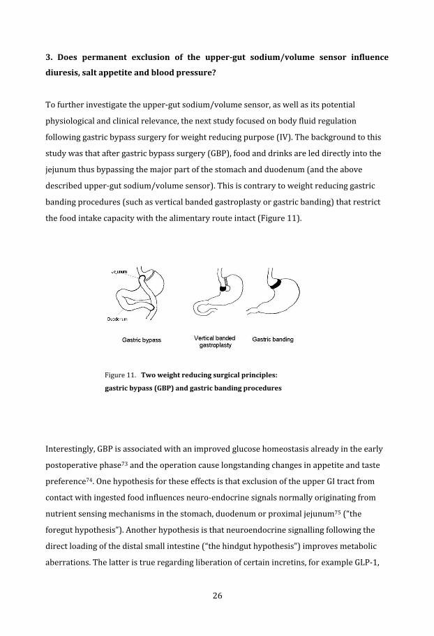
following gastric bypass surgery for weight reducing purpose (IV). The background to this
study was that after gastric bypass surgery (GBP), food and drinks are led directly into the
jejunum thus bypassing the major part of the stomach and duodenum (and the above
described upper‐gut sodium/volume sensor). This is contrary to weight reducing gastric
banding procedures (such as vertical banded gastroplasty or gastric banding) that restrict
he food intake capacity with the alimentary route intact (Figure 11). t
Figure 11.Twoweightreducingsurgicalprinciples:
gastricbypass(GBP)andgastricbandingprocedures
Interestingly, GBP is associated with an improved glucose homeostasis already in the early
postoperative phase73 and the operation cause longstanding changes in appetite and taste
preference74. One hypothesis for these effects is that exclusion of the upper GI tract from
contact with ingested food influences neuro‐endocrine signals normally originating from
nutrient sensing mechanisms in the stomach, duodenum or proximal jejunum75 (“the
foregut hypothesis”). Another hypothesis is that neuroendocrine signalling following the
direct loading of the distal small intestine (“the hindgut hypothesis”) improves metabolic
aberrations. The latter is true regarding liberation of certain incretins, for example GLP‐1,
26

that stimulate insulin release from the pancreas. However, neither the foregut, nor the
hindgut hypothesis can fully explain the early effect on glucose homeostasis by GBP,
implicating that unknown mechanisms are operating as well. It has been reported that GBP
also reduces blood pressure before significant weight loss has occurred76. It was therefore
hypothesised that the exclusion of the gastroduodenum and the previously mentioned gut
sodium/volume monitor could be a mechanism of action. If so, the GBP‐patients should
exhibit a diuretic pattern and/or salt ingestive behaviour that differ from patients operated
ith banding procedures and with their GI continuity intact. w
Studysetting
Subjects participating in the Swedish Obese Subjects (SOS) study77, 78 were examined. The
prospective large scale SOS study compares obese patients undergoing weight‐reducing
surgery, with contemporaneously matched, non‐operated obese control patients. The
subjects who underwent weight‐reducing surgery were for the purpose of the present
analysis divided into two groups: gastric bypass (GBP) and vertical banded gastroplasty or
gastric banding (VBG/B) (Figure 11). Diurnal urine collections and blood pressure levels
were investigated at baseline and at 2y and 10y after study‐inclusion. Dietary salt intake
was assessed by measurement of 24h urinary excretion of sodium, which is considered the
old standard for assessing salt intake79. g
Diurnalurineoutput,saltintakeandbloodpressureaftergastricbypasssurgery
After adjustments for weight loss, the GBP patients exhibited a larger 24h urine output and
a larger 24h natriuresis than the gastric band or control patients. The GBP operated
individuals also displayed a markedly more reduced systolic and diastolic pressure (Figure
12). These changes were prominent also 10 years after surgical intervention (median
follow‐up time) and were not related to the reduced body weight. Furthermore, regression
analyses demonstrated that changes in diuresis were linearly associated with blood
pressure changes only in the GBP cohort, indicating that blood pressure reduction following
GBP can be attributed to its diuretic action (Figure 13).
27

Figure 12. Changesindiurnalurinaryoutput(U‐Volume)andexcretionofsodium(U‐Na+)inrelation
tobodyweight(upperpanels),andchangesinbloodpressure(lowerpanels)aftergastricbypass
surgery(GBP),afterpurerestrictivebariatricsurgery(VBG/B)andinnon‐operatedobesecontrols.
Changes from baseline (∆) at the 2y and 10y follow‐up visits are displayed. Data are mean values adjusted for
sex, age, baseline BMI and the baseline level of the respective variables. The bars represent the 95%
confidence intervals. Differences between groups are given as mean (95% confidence intervals). *P<0.05,
**P<0.01 and ***P<0.001
28
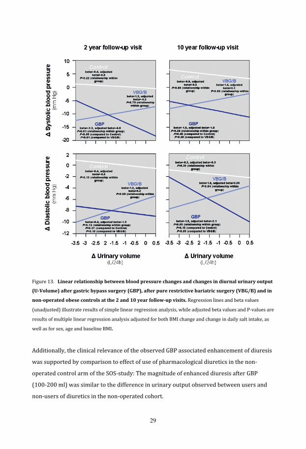
Figure 13.Linearrelationshipbetweenbloodpressurechangesandchangesindiurnalurinaryoutput
(U‐Volume)aftergastricbypasssurgery(GBP),afterpurerestrictivebariatricsurgery(VBG/B)andin
non‐operatedobesecontrolsatthe2and10yearfollow‐upvisits.Regression lines and beta values
(unadjusted) illustrate results of simple linear regression analysis, while adjusted beta values and P‐values are
results of multiple linear regression analysis adjusted for both BMI change and change in daily salt intake, as
ell as for sex, age and baseline BMI. w
Additionally, the clinical relevance of the observed GBP associated enhancement of diuresis
was supported by comparison to effect of use of pharmacological diuretics in the non‐
operated control arm of the SOS‐study: The magnitude of enhanced diuresis after GBP
(100‐200 ml) was similar to the difference in urinary output observed between users and
non‐users of diuretics in the non‐operated cohort.
29

Interpretation
If GBP silences out the diuresis promoting monitor of the upper gut, why did these patients
excrete more urine than did the weight matched subjects that had their gastrointestinal
continuity intact? In order to explain gastrointestino‐renal diuretic regulation one has to
consider the short‐time course of the gastroduodeno‐signalling (related to ingestive
behaviour) and that after GBP there is probably an additional effect of direct volume‐
loading into the rapidly absorbing jejunum. Over 24h it may be that diuresis‐promoting
post‐absorptive mechanisms become more pronounced when gastroduodenal short‐term
coordination is bypassed, resulting in an “overshoot” of fluid excretion by the kidneys. The
situation after GBP is actually mimicked by the jejunal infusion in Paper III where plasma
BNP increased already within 1h, strongly indicating the induction of post‐absorptive
iuresis‐promoting mechanisms. d
However, a primary natriuretic mechanism seems unlikely as the GBP patients were found
to have a slightly increased serum sodium concentration. Contributing to this intriguing
picture was the finding that GBP patients consumed approximately 1 g dietary salt more per
day than the group operated with the restrictive banding techniques. The picture can be
compatible with that the upper‐gut sodium/volume sensor, in addition to short‐term (i.e.
<2h) diuretic regulation, normally inhibits salt appetite and that salt intake increases
following GBP. Alternative explanations and need for future research will be discussed
below in General Discussion.
3dconclusion
Permanent exclusion of the upper GI tract increases salt intake and diuresis suggesting that
the upper gut sodium/volume sensor participates in the regulation of salt appetite. Blood
pressure reduction following gastric bypass surgery can be attributed to a diuretic action.
30

CONCLUSIONS
1. Prominent components of RAS are present in the human gastric and duodenal
mucosa. H.pylori induced gastritis is associated with higher prevalence of AT1
receptors, most probably due to presence of infiltrating neutrophils carrying this
receptor. Immunoreactivity to AT1R and AGT in solitary epithelial cells suggest that
ocal RAS activity can influence gastroduodenal enteroendocrine signalling. l
2. The temporal relationship between increased diuresis induced by an intragastric
saline load and the reduced quantity of AGT in duodenum suggest a role for RAS in
the duodenal mucosa in the pre‐absorptive induction of diuresis occurring after
drinking and eating.
3. Permanent exclusion of the upper GI tract increases salt intake and diuresis
suggesting that the upper gut sodium/volume sensor participates in the
regulation of salt appetite. Blood pressure reduction following gastric bypass
surgery is attributed to a diuretic action.
31

GENERALDISCUSSION
Somemethodologicalconsiderations
This thesis project includes several methodological principles regarding both laboratory
analyses and to the design of studies. Papers I, II and III are exploratory studies on
carefully standardized small samples of populations, whereas IV is an adhoc analysis of
data from a prospective interventional trial involving in total more than 4000 included
atients. p
The antibody dependent assessments (Western blotting and immunohistochemistry)
were performed in accordance to validated standard procedures and included internal
controls. The quality of the commercially acquired antibodies can differ markedly and all
new batches were therefore tested for specificity. The immunoassay (EIA) used for
plasma‐AngII analysis was used carefully according to the manufacturer’s instructions.
Immunoassays used for other plasma peptide analyses, and the analyses of electrolyte
concentrations in serum and urine, were outsourced to accredited clinical chemistry
laboratories. In general, all the analytical procedures were confirmed as specific and
sensitive. In Paper III, however, tissue concentration of AngII was assessed using an EIA
not validated for its use in solid tissues. The biological halftime of AngII in tissue is short
and it cannot be excluded that, despite addition of enzyme inhibitor during work up, the
amount of the octapeptide in the mucosal samples had already been reduced due to
ndogenous enzymatic activity. e
Another potential source of error in the present project is the 24h sampling of urine in
the SOS study (IV). As the urine was collected over 24h by the study subjects them
selves, sampling errors related to over‐ or under‐collections must be considered.
However, there is little reason to believe that any of the surgery groups would be more
likely to provide over‐ or under‐collections. Furthermore, the differences in diurnal
32
urine output and sodium excretion between GBP‐ and gastric banding subjects remained
ignificant after adjusting these analyses for U‐creatinine79, 80. s
RASintheuppergutmucosa
The presence of AngII receptors is a good indication of the potential impact by RAS on
the functional state of a tissue. A few previous studies have indicated expression of AngII
receptors in the upper GI tract. Autoradiography of the rat stomach indicated that AT1R
and AT2R are present in all layers of the stomach81. Matsuo etal. used
immunohistochemistry to locate AT1R in human antrum and found it in vascular
smooth muscle cells, mesenchymal cells and smooth muscle cells in the muscular layers
of the mucosa and muscularis propria82. Further, Bregonzio etal. located the AT1R
protein by means of immunohistochemistry in vascular endothelial cells in the rat
stomach81. Johansson etal localised both AT1 and AT2 receptors in the lamina propria
in duodenal mucosal villi in the rat5. The present study confirms presence of AngII
receptors in the human gastroduodenal mucosa and provides novel data on the
presence of several other RAS components (I, II, III). Of particular interest is the finding
of storage/depletion of the prohormone AGT in the duodenal mucosa (III). These novel
spects of RAS will be further discussed later. a
The presence of AngII receptors at specific locations suggests functional importance. Not
surprisingly, vascular endothelium expressed both the AT1R and AT2R subtypes in the
present study (I, II). The former mediates the classical vasoconstrictive effect of AngII,
whereas AT2R has been reported to mediate vasodilatation12. However, the superficial
vessels of the mucosa studied in the present study were very thin‐walled (i.e. without
blood flow regulating vascular smooth muscle cells) and consisted mainly of capillaries
and venules. AngII receptors in this tissue compartment are probably related to
microvascular permeability as has been reported for rat mesenteric venules83, 84. To
what extent AngII influences mucosal microvascular permeability in the human upper
gut remains to be tested. Interestingly, AT1R and AT2R were localised also to cells in the
lamina propria and the epithelium suggesting functional impact on epithelial functions.
33

In the duodenal mucosa, distinct immunoreactivity for AT1R and AT2R was seen
primarily in the basal part of most epithelial cells. This differs from the picture in rat
duodenum where immunoreactivity was confined to cells in the lamina propria5.
Speculatively, sodium/water26 and glucose transport28, 29 as well as bicarbonate
secretion5 or regulation of cell growth12 can be targeted by activation of these juxta‐
epithelial and epithelial receptors, but more research is needed to elucidate
hysiological significances in man. p
The present study also indicated that AT1R is highly expressed by a subpopulation of
antral endocrine cells in the stomach. It has been reported that AngII through AT1R can
influence gastric acid secretion in rats85. In the present study, however, no AT1R was
found on the acid producing parietal cells so if AT1R influences gastric acid secretion in
man, it is possible that such an effect first involves antral endocrine cells. In the human
duodenal mucosa, distinct immunoreactivity for AT1R and AT2R was found principally
in all epithelial cells (which will also include the endocrine cells), as mentioned above.
Therefore, it’s reasonable to assume that also some duodenal enteroendocrine cells
express the AT1R protein. The localisation of AT1R to endocrine cells suggests that
activity in a local RAS can influence enteroendocrine signalling and represent an
nteresting field for future research. i
A perquisite for AT1R and AT2R activation is the presence of the ligand AngII. This
peptide is the result of enzymatic degradation of AGT by renin and ACE and both these
enzymes were found in the human gastric and duodenal mucosa. The present study also
identified significant levels of AngII in these tissues (III), supporting that the main
effector peptide of RAS is also generated in the mucosa of the upper gut. In addition to
AngII receptors, angiotensin generating enzymes and AngII, human gastric and duodenal
mucosa also showed immunoreactivity to AGT, the prohormone of RAS. AGT was
located to solitary epithelial cells in the duodenal mucosa. This observation indicates
that AGT is produced and stored at certain cellular locations in the upper gut mucosa.
This is a novel finding that certainly demand further investigation. For example, by use
of laser capture microscopy it will be possible to assess mRNA expression of the AGT‐
34

immunoreactive epithelial cells without any interference of other celltypes (such as for
xample the vascular endothelial cells). e
Furthermore, data in Paper III suggest that luminal stimulation (in this case isotonic
NaCl) influence AGT liberation in the duodenal mucosa that, depending on presence and
type of degradation enzymes, may result in formation of bioactive angiotensins with
potential to exert endocrine or paracrine effects. Furthermore, mRNA expression of AGT
by intestinal “taste cells” in mice has been reported by others86, supporting that this
protein has a relation to mucosal sensing mechanisms. Future double immunostaining
studies, using specific markers to taste cells, endocrine cells and brush cells, are needed
to further characterise the AGT‐positive solitary epithelial cells found in the human
duodenal mucosa. Still, this observation supports the possibility that paracrine RAS
ctivity might influence also duodenal enteroendocrine signalling. a
IsthegastroduodenalRASinvolvedinsensingofluminalcontents?
We considered that the previously postulated salt/volume monitor in the upper gut32, 34
could to be of particular interest as a physiological context for the mucosal RAS in the
stomach and duodenum. Therefore, levels of AGT and AngII in the human gastro‐
duodenal mucosa subsequent to a gastric saline load were measured in‐situ, at a time
point corresponding to onset of the diuretic response following gastric instillation of
saline (determined in our previous experiments). Indeed, the content of AGT in the
duodenal mucosa decreased significantly subsequent to the gastric saline instillation
and that was not the case in antral specimens. This observation thus suggests that the
duodenum rather than the stomach is the primary site for luminal sensing of a volume
oad. l
Enzymatic degradation of the pro‐hormone AGT can result in several bioactive
“angiotensins” and the one of particular interest is of course the octapeptide AngII, being
the principal mediator in RAS. However, the present investigation did not show
35
increased AngII concentration in the duodenal mucosa upon intragastric saline loading,
suggesting that this mediator is of less importance for the diuretic response. Yet, the
absence of changes in local mucosal AngII levels does not completely rule out a role for
this mediator. The paracrine mode of action of AngII on, for example, enteroendocrine
cells in turn eliciting distant hormonal or autonomic neural effects, may demand local
o the y. liberation of rather small amounts that are subthreshold t presently used assa
Paracrine arrangements are difficult to study in human’s in‐situ. A feasible way for
further investigation of the role of RAS in duodenal sensing of luminal content may be to
isolate the mucosa in an Ussing chamber and use different luminal stimuli (e.g.
hypertonic saline and nutrients) with pharmacological inhibitors to for example renin,
ACE etc. Also other bioactive AGT‐fragments with diuretic effects have been described87,
such as Ang‐(1‐7). These peptides can be generated locally in the GI‐mucosa, depending
on type of proteolytic enzymes present in the tissues, and remain to be investigated in
he future. t
Thegutsodium/volumesensor?
The pre‐absorptive sodium/volume sensor in the GI tract is postulated to be situated in
the upper gut but the exact location of this sensor has not been determined. In the
present study, we approached this mechanism by studying the diuretic response to
instillation of saline at two sites along the gut lumen (III). These results indeed support
that a pre‐absorptive diuretic signal is elicited when the gastroduodenal region is
subjected to a sodium‐water load. The latency of onset to increased diuresis was
markedly shorter after gastric loading than after jejunal loading. Furthermore, the
diuretic response was not associated with hormonal changes that typically are involved
in relation to extracellular volume regulation. The diuretic response to intrajejunal
saline instillation, on the other hand, had a much slower onset and was associated with
increased plasma BNP concentrations suggesting that a systemic volume expansion had
preceded the diuretic response70. Hence, the data indicate that the intrajejunal
instillation induces diuresis mainly via post‐absorptive mechanisms. The interpretation
of these experiments is that a pre‐absorptive fluid sensor is situated in the upper gut.
36

Because the rate of gastric emptying of non‐energetic, isotonic liquids is high with
halftimes in the order of 8‐10 min68, 69, the duodenum almost instantly will be targeted
by liquid disposed in the stomach. Consequently, it seems reasonable to assume a more
prec m. ise location of the pre‐absorptive sensor being the stomach and/or the duodenu
The natriuretic response in the present study did not show any consistent temporal
patterns in relation to infusion site. Still, total sodium outputs were similar over the
observation time. Therefore, these results do not support that the upper gut senses
sodium specifically. However, we chose instillation of isotonic NaCl (6.8 g NaCl) in order
to avoid effects of osmotic gradients. It follows that isotonic saline mainly is a volume
stimulus. A transmucosal sodium gradient might be needed to stimulate the sodium
sensor mechanism. Such experiments, using higher luminal salt concentrations, are in
rogress. p
Roleofthegutinbodyfluidhomeostasisandarterialpressurecontrol
To further investigate the upper‐gut sodium/volume sensor, as well as its potential
impact on arterial blood pressure control, we analysed measurements of salt intake,
diuresis and arterial blood pressure following two weight reducing surgical principles:
gastric banding and gastric bypass (GBP). The GBP operated individuals consumed
approximately 1 g dietary salt more per day and had a slightly increased serum sodium
concentration, compared to the weight matched subjects that had undergone a
restrictive procedure with the gastrointestinal continuity intact. The increased salt
consumption together with a small hypernatremia is consistent with an increased
preference for salty foods88. Actually, Tichansky etal. reported that GBP patients
experience an increased salt appetite89. However, a diet‐induced hypernatremia is
controversial in relation to the view that increased salt intake should contribute to
increased blood pressure90‐92, not the opposite as was observed in the present study.
Furthermore, the GBP patients exhibited a larger 24h diuresis being linearly associated
with blood pressure reduction.
37

Apparently, the GBP procedure functions as a diuretic agent, very probably explaining
the long term lowered blood pressure. The increased diuresis, however, cannot simply
be explained as a compensation for increased salt intake as hypernatremia normally is
associated with reduced diuresis. Low vasopressin concentrations can be associated
with hypernatremia, but does not explain the increased salt intake. It is likely that the
GBP procedure elicits a mixed action, for example increased salt appetite/intake and
increased natriuresis due to increased BNP‐levels. The nature of the increased diurnal
diuresis following GBP should be reasonably easy to sort out by assessing plasma and
rine osmolality as well as the humoral mediators of interest for fluid homeostasis. u
Therelationbetween“pre‐absorptive”and“post‐absorptive”mecha
In Paper III, a diuresis promoting pre‐absorptive mechanism sensitive to
gastroduodenal filling was described. Permanent exclusion of this mechanism due to
GBP was followed by an even more pronounced diuretic action (IV). How to put this
together? Well, it is a very common phenomenon in physiology that anticipatory
mechanisms override the more basal adaptation to an existing aberration. In this case,
volume and sodium loading of the upper gut (III) probably activates anticipatory
mechanisms via the circulation or the central nervous system that counteract the
oncoming absorptive phase and plasma volume expansion by changing appetite,
ingestive behaviour and urinary output. This “pre‐absorptive” (or foregut) fluid
regulation normally avoids activation of “post‐absorptive” mechanisms. For example,
the liberation of BNP is not needed because the organism has already taken the relevant
measures so that an expansion of ECF has been prevented. In the case of GBP, the
anticipatory foregut regulatory mechanisms are removed and the organism has to count
on its post‐absorptive regulatory capacity (or perhaps so far unknown hindgut factors).
nisms
38

Physiologicalandclinicalrelevance
From the perspective depicted above, one cannot say that GBP normalise blood pressure
control. The procedure rather manipulates the physiology so that post‐absorptive
“overshooting” makes the blood pressure decrease. In that sense, the foregut
sodium/volume sensor is of physiological significance by being one principal
mechanism behind why GBP surgery has beneficial effect on obesity‐associated blood
pressure elevation. Furthermore, Paper IV also shows that GBP reduced blood pressure
with a magnitude of clinical relevance93 over long time (median follow‐up time: 10
years). This contrasted to gastric banding techniques that exerted a short‐lasting and
weight‐related depressor effect.
The clinical relevance of excluding the foregut and the volume/sodium monitor is
obvious. Several findings suggest that a local mucosal RAS is involved in the pre‐
absorptive volume and sodium sensor of the upper gut, but conclusive data are still
lacking and represents an area for urgent future research.
39

ACKNOWLEDGEMENTS
wish to express my sincere gratitude and appreciation to all the people who have in one to this thesis.
Iway or another contributed In particular, I would like to thank: y supervisor – Professor LarsFändriks, for sharing your knowledge in physiology and
you for your guidMscience with me. Thank ance and support throughout this process. My co‐supervisor – Dr AnnaCasselbrant, for your encouragement, advice and generous laboratory support.
erbertHelan edge in the field of histology. H der for your enthusiasm, support and knowl
ndersElfvin dship. A for your support, positivism and frien
atory assistance. ChristinaEkfor your skilful labor y colleagues and ex‐colleagues: MalinWerling, EmmaSpak,ErikElias, MajHedtjärnndTomasSjöbeMa
rg for friendship, inspiring discussions and fun times.
elp with te SörenLundberg,for your invaluable h chnical matters and data management.Eva‐LottaEen, MyEngström, NiclasJohanssonandDianaLustgarten at the “Gastlab‐eam” for advice and skilful assistance. GunillaPervik, foryourexcellent secretarial tservice. My co‐authors:AndersEdebo for skilful endoscopy; VilleWalleniusfor methodological dvice and good ideas; HansLönroth, LenaCarlsson,LarsSjöström, IngemarNäslund,a
PeterJacobsonandGerdBergmarkfor fruitful collaboration. The Centre for Cellular Imaging (CCI) and the EpiStat facility at the Sahlgrenska Academy for the support from the staff. y family for supporting me, especially my daughter Denise for helping me at home during e finishin
Mth
g of this thesis.
njaPaust for being a caring friend and a wonderful mother to our son. A This study was supported financially by the Swedish Medical Research Council (VR medicin), Swedish Federal Government under the LUA/ALF agreement and Gothenburg Medical Association.
40

REFERENCES
1. von Bothmer C, Edebo A, Lonroth H, Olbe L, Pettersson A, Fandriks L. Helicobacter pylori infection inhibits antral mucosal nitric oxide production in humans. Scand J Gastroenterol 2002;37:404-8.
2. Elfvin A, Bolin I, Von Bothmer C, et al. Helicobacter pylori induces gastritis and
intestinal metaplasia but no gastric adenocarcinoma in Mongolian gerbils. Scand J Gastroenterol 2005;40:1313-20.
3. Fandriks L, von Bothmer C, Johansson B, Holm M, Bolin I, Pettersson A. Water extract
of Helicobacter pylori inhibits duodenal mucosal alkaline secretion in anesthetized rats. Gastroenterology 1997;113:1570-5.
4. Johansson B, Holm M, Chen L, Pettersson A, Jonson C, Fandriks L. ANG II prolongs
splanchnic nerve-mediated inhibition of duodenal mucosal alkaline secretion in the rat. Am J Physiol 1997;273:R942-6.
5. Johansson B, Holm M, Ewert S, Casselbrant A, Pettersson A, Fandriks L. Angiotensin II
type 2 receptor-mediated duodenal mucosal alkaline secretion in the rat. Am J Physiol Gastrointest Liver Physiol 2001;280:G1254-60.
6. Fitzsimons JT. Angiotensin, thirst, and sodium appetite. Physiological reviews
1998;78:583-686. 7. Daniels D, Yee DK, Fluharty SJ. Angiotensin II receptor signalling. Experimental
Physiology 2007;92:523-7. 8. Harrison-Bernard LM. The renal renin-angiotensin system. Advances in Physiology
Education 2009;33:270-4. 9. Singh M, Mensah GA, Bakris G. Pathogenesis and clinical physiology of hypertension.
Cardiol Clin 2010;28:545-59. 10. Bader M. Tissue renin-angiotensin-aldosterone systems: Targets for pharmacological
therapy. Annu Rev Pharmacol Toxicol 2010;50:439-65. 11. Fyhrquist F, Saijonmaa O. Renin-angiotensin system revisited. J Intern Med
2008;264:224-36. 12. Paul M, Poyan Mehr A, Kreutz R. Physiology of local renin-angiotensin systems.
Physiological reviews 2006;86:747-803. 13. Lavoie JL, Sigmund CD. Minireview: overview of the renin-angiotensin system--an
endocrine and paracrine system. Endocrinology 2003;144:2179-83. 14. Thatcher S, Yiannikouris F, Gupte M, Cassis L. The adipose renin-angiotensin system:
role in cardiovascular disease. Mol Cell Endocrinol 2009;302:111-7.
41

15. Nagata S, Kato J, Kuwasako K, Kitamura K. Plasma and tissue levels of proangiotensin-12 and components of the renin-angiotensin system (RAS) following low- or high-salt feeding in rats. Peptides 2010;31:889-92.
16. Olszanecki R, Madej J, Suski M, Gebska A, Bujak-Gizycka B, Korbut R. Angiotensin
metabolism in rat stomach wall: prevalence of angiotensin-(1-7) formation. J Physiol Pharmacol 2009;60:191-6.
17. de Gasparo M, Catt KJ, Inagami T, Wright JW, Unger T. International union of
pharmacology. XXIII. The angiotensin II receptors. Pharmacol Rev 2000;52:415-72. 18. Fandriks L. The angiotensin II type 2 receptor and the gastrointestinal tract. J Renin
Angiotensin Aldosterone Syst 2010;11:43-8. 19. Balla T, Baukal AJ, Eng S, Catt KJ. Angiotensin II receptor subtypes and biological
responses in the adrenal cortex and medulla. Mol Pharmacol 1991;40:401-6. 20. Brasier AR, Jamaluddin M, Han Y, Patterson C, Runge MS. Angiotensin II induces gene
transcription through cell-type-dependent effects on the nuclear factor-kappaB (NF-kappaB) transcription factor. Mol Cell Biochem 2000;212:155-69.
21. Matsukawa T, Miyamoto T. Angiotensin II-stimulated secretion of arginine vasopressin is
inhibited by atrial natriuretic peptide in humans. Am J Physiol Regul Integr Comp Physiol 2011;300:R624-9.
22. Fandriks L. The renin-angiotensin system and the gastrointestinal mucosa. Acta Physiol
(Oxf) 2011;201:157-67. 23. Casselbrant A, Edebo A, Hallersund P, et al. Angiotensin II receptors are expressed and
functional in human esophageal mucosa. Am J Physiol Gastrointest Liver Physiol 2009;297:G1019-27.
24. Spak E, Bjorklund P, Helander HF, et al. Changes in the mucosa of the Roux-limb after
gastric bypass surgery. Histopathology 2010;57:680-8. 25. Hirasawa K, Sato Y, Hosoda Y, Yamamoto T, Hanai H. Immunohistochemical
localization of angiotensin II receptor and local renin-angiotensin system in human colonic mucosa. J Histochem Cytochem 2002;50:275-82.
26. Jin XH, Wang ZQ, Siragy HM, Guerrant RL, Carey RM. Regulation of jejunal sodium
and water absorption by angiotensin subtype receptors. Am J Physiol 1998;275:R515-23. 27. Suzuki Y, Ruiz-Ortega M, Lorenzo O, Ruperez M, Esteban V, Egido J. Inflammation and
angiotensin II. Int J Biochem Cell Biol 2003;35:881-900. 28. Wong TP, Debnam ES, Leung PS. Involvement of an enterocyte renin-angiotensin system
in the local control of SGLT1-dependent glucose uptake across the rat small intestinal brush border membrane. J Physiol 2007;584:613-23.
42
29. Wong TP, Debnam ES, Leung PS. Diabetes mellitus and expression of the enterocyte renin-angiotensin system: implications for control of glucose transport across the brush border membrane. Am J Physiol Cell Physiol 2009;297:C601-10.
30. Heinemann A, Sattler V, Jocic M, Wienen W, Holzer P. Effect of angiotensin II and
telmisartan, an angiotensin1 receptor antagonist, on rat gastric mucosal blood flow. Aliment Pharmacol Ther 1999;13:347-55.
31. Nakagiri A, Sunamoto M, Murakami M. Angiotensin AT1 receptor blockers suppress
ischemia/reperfusion-induced gastric injury in rats. Inflammopharmacology 2007;15:171. 32. Duggan KA, Ye VZ, Jones DM, Macdonald GJ. Angiotensin II: a humoral mediator for
the gastric sodium monitor. Am J Physiol 1996;270:F406-10. 33. Michell AR, Debnam ES, Unwin RJ. Regulation of renal function by the gastrointestinal
tract: potential role of gut-derived peptides and hormones. Annual review of physiology 2008;70:379-403.
34. Mu JY, Hansson GC, Lundgren O. The small intestine, salt intake and arterial
hypertension. Blood pressure 1995;4:77-9. 35. Lennane RJ, Carey RM, Goodwin TJ, Peart WS. A comparison of natriuresis after oral
and intravenous sodium loading in sodium-depleted man: evidence for a gastrointestinal or portal monitor of sodium intake. Clin Sci Mol Med 1975;49:437-40.
36. Lennane RJ, Peart WS, Carey RM, Shaw J. A comparison on natriuresis after oral and
intravenous sodium loading in sodium-depleted rabbits: evidence for a gastrointestinal or portal monitor of sodium intake. Clin Sci Mol Med 1975;49:433-6.
37. Patel S. Sodium balance-an integrated physiological model and novel approach. Saudi J
Kidney Dis Transpl 2009;20:560-9. 38. Skorecki KL, Brenner BM. Body fluid homeostasis in man. A contemporary overview.
Am J Med 1981;70:77-88. 39. Skott O. Body sodium and volume homeostasis. Am J Physiol Regul Integr Comp Physiol
2003;285:R14-8. 40. Bie P, Wamberg S, Kjolby M. Volume natriuresis vs. pressure natriuresis. Acta Physiol
Scand 2004;181:495-503. 41. Rubattu S, Sciarretta S, Valenti V, Stanzione R, Volpe M. Natriuretic peptides: an update
on bioactivity, potential therapeutic use, and implication in cardiovascular diseases. Am J Hypertens 2008;21:733-41.
42. Tordoff MG, McCaughey SA. Influence of oral and gastric NaCl preloads on NaCl intake
and gastric emptying of sodium-deficient rats. Am J Physiol Regul Integr Comp Physiol 2001;281:R1152-60.
43. Stricker EM, Hoffmann ML. Presystemic signals in the control of thirst, salt appetite, and
vasopressin secretion. Physiol Behav 2007;91:404-12.
43

44. Bourque CW. Central mechanisms of osmosensation and systemic osmoregulation. Nat Rev Neurosci 2008;9:519-31.
45. Geerling JC, Loewy AD. Central regulation of sodium appetite. Experimental Physiology
2008;93:177-209. 46. Qian X, Moss NG, Fellner RC, Goy MF. Circulating prouroguanylin is processed to its
active natriuretic form exclusively within the renal tubules. Endocrinology 2008;149:4499-509.
47. Mu JY, Johansson M, Lundgren O. The effects of enalapril on the natriuretic response
evoked by an oral sodium load in sodium deprived normotensive and hypertensive rats. Acta Physiol Scand 1997;160:157-64.
48. Berthoud HR. The vagus nerve, food intake and obesity. Regul Pept 2008;149:15-25. 49. Maljaars J, Peters HP, Masclee AM. Review article: The gastrointestinal tract:
neuroendocrine regulation of satiety and food intake. Aliment Pharmacol Ther 2007;26 Suppl 2:241-50.
50. Raybould HE. Gut chemosensing: interactions between gut endocrine cells and visceral
afferents. Auton Neurosci 2010;153:41-6. 51. Roges OA, Baron M, Philis-Tsimikas A. The incretin effect and its potentiation by
glucagon-like peptide 1-based therapies: a revolution in diabetes management. Expert Opin Investig Drugs 2005;14:705-27.
52. Taupenot L, Harper KL, O'Connor DT. The chromogranin-secretogranin family. N Engl J
Med 2003;348:1134-49. 53. Lee CS, Perreault N, Brestelli JE, Kaestner KH. Neurogenin 3 is essential for the proper
specification of gastric enteroendocrine cells and the maintenance of gastric epithelial cell identity. Genes & development 2002;16:1488-97.
54. Alpers DH. Nutrient sensing in the gastrointestinal tract. Curr Opin Gastroenterol
2010;26:134-9. 55. Steinert RE, Beglinger C. Nutrient sensing in the gut: Interactions between chemosensory
cells, visceral afferents and the secretion of satiation peptides. Physiol Behav 2011; Mar 2 [Epub ahead of print].
56. Akiba Y, Kaunitz JD. Luminal chemosensing in the duodenal mucosa. Acta Physiol (Oxf)
2011;201:77-84. 57. Holm M, Olbe L, Fandriks L. Intragastric CO2 and nitric oxide participate in the
regulation of peptone-induced gastrin release in humans. Scand J Gastroenterol 2000;35:1260-5.
58. Hellstrom PM. GLP-1 playing the role of a gut regulatory compound. Acta Physiol (Oxf)
2011;201:151-6.
44

59. Rozengurt E, Sternini C. Taste receptor signaling in the mammalian gut. Curr Opin Pharmacol 2007;7:557-62.
60. Sternini C, Anselmi L, Rozengurt E. Enteroendocrine cells: a site of 'taste' in
gastrointestinal chemosensing. Curr Opin Endocrinol Diabetes Obes 2008;15:73-8. 61. Sbarbati A, Osculati F. A new fate for old cells: brush cells and related elements. J Anat
2005;206:349-58. 62. Lundgren O. Interface between the intestinal environment and the nervous system. Gut
2004;53 Suppl 2:ii16-8. 63. Olbe L, Fandriks L, Hamlet A, Svennerholm AM, Thoreson AC. Mechanisms involved in
Helicobacter pylori induced duodenal ulcer disease:an overview. World J Gastroenterol 2000;6:619-23.
64. Carl-McGrath S, Grantzdorffer I, Lendeckel U, Ebert MP, Rocken C. Angiotensin II-
generating enzymes, angiotensin-converting enzyme (ACE) and mast cell chymase (CMA1), in gastric inflammation may be regulated by H. pylori and associated cytokines. Pathology 2009;41:419-27.
65. Huang W, Yu LF, Zhong J, et al. Stat3 is involved in angiotensin II-induced expression of
MMP2 in gastric cancer cells. Digestive diseases and sciences 2009;54:2056-62. 66. Ramos-Nino ME, Maclean CD, Littenberg B. Association of angiotensin-converting
enzyme inhibitor therapy and comorbidity in diabetes: results from the Vermont diabetes information system. BMC endocrine disorders 2008;8:17.
67. Sugimoto M, Furuta T, Shirai N, et al. Role of angiotensinogen gene polymorphism on
Helicobacter pylori infection-related gastric cancer risk in Japanese. Carcinogenesis 2007;28:2036-40.
68. Brener W, Hendrix TR, McHugh PR. Regulation of the gastric emptying of glucose.
Gastroenterology 1983;85:76-82. 69. Collins PJ, Houghton LA, Read NW, et al. Role of the proximal and distal stomach in
mixed solid and liquid meal emptying. Gut 1991;32:615-9. 70. Lobo DN, Stanga Z, Aloysius MM, et al. Effect of volume loading with 1 liter
intravenous infusions of 0.9% saline, 4% succinylated gelatine (Gelofusine) and 6% hydroxyethyl starch (Voluven) on blood volume and endocrine responses: a randomized, three-way crossover study in healthy volunteers. Crit Care Med 2010;38:464-70.
71. Hulthen L, Aurell M, Klingberg S, Hallenberg E, Lorentzon M, Ohlsson C. Salt intake in
young Swedish men. Public Health Nutr 2009;13:601-5. 72. Friedmann AS, Memoli VA, North WG. Evidence for vasopressin production in the
human gastrointestinal system. Peptides 1991;12:1051-6. 73. Rubino F, Schauer PR, Kaplan LM, Cummings DE. Metabolic surgery to treat type 2
diabetes: clinical outcomes and mechanisms of action. Annu Rev Med 2010;61:393-411.
45

74. Miras AD, le Roux CW. Bariatric surgery and taste: novel mechanisms of weight loss. Curr Opin Gastroenterol 2010;26:140-5.
75. Rubino F, R'Bibo S L, del Genio F, Mazumdar M, McGraw TE. Metabolic surgery: the
role of the gastrointestinal tract in diabetes mellitus. Nat Rev Endocrinol 2010;6:102-9. 76. Ahmed AR, Rickards G, Coniglio D, et al. Laparoscopic Roux-en-Y gastric bypass and its
early effect on blood pressure. Obes Surg 2009;19:845-9. 77. Sjostrom L, Lindroos AK, Peltonen M, et al. Lifestyle, diabetes, and cardiovascular risk
factors 10 years after bariatric surgery. N Engl J Med 2004;351:2683-93. 78. Sjostrom L, Narbro K, Sjostrom CD, et al. Effects of bariatric surgery on mortality in
Swedish obese subjects. N Engl J Med 2007;357:741-52. 79. Liu K, Stamler J. Assessment of sodium intake in epidemiological studies on blood
pressure. Ann Clin Res 1984;16 Suppl 43:49-54. 80. Taylor EN, Curhan GC. Differences in 24-hour urine composition between black and
white women. J Am Soc Nephrol 2007;18:654-9. 81. Bregonzio C, Armando I, Ando H, Jezova M, Baiardi G, Saavedra JM. Anti-inflammatory
effects of angiotensin II AT1 receptor antagonism prevent stress-induced gastric injury. Am J Physiol Gastrointest Liver Physiol 2003;285:G414-23.
82. Matsuo T, Ikura Y, Ohsawa M, et al. Mast cell chymase expression in Helicobacter
pylori-associated gastritis. Histopathology 2003;43:538-49. 83. Newton CR, Curran B, Victorino GP. Angiotensin II type 1 receptor activation increases
microvascular hydraulic permeability. Surgery 2004;136:1054-60. 84. Newton CR, Curran B, Victorino GP. Angiotensin II type 2 receptor effect on
microvascular hydraulic permeability. J Surg Res 2004;120:83-8. 85. Chow L, Zakrzewska K, De Gasparo M, Cumin F, Levens N. Gastric acid secretion after
blockade of angiotensin AT1 receptors in the Na(+)-depleted rat. Eur J Pharmacol 1995;294:309-17.
86. Bezencon C, Furholz A, Raymond F, et al. Murine intestinal cells expressing Trpm5 are
mostly brush cells and express markers of neuronal and inflammatory cells. J Comp Neurol 2008;509:514-25.
87. Joyner J, Neves LA, Stovall K, Ferrario CM, Brosnihan KB. Angiotensin-(1-7) serves as
an aquaretic by increasing water intake and diuresis in association with downregulation of aquaporin-1 during pregnancy in rats. Am J Physiol Regul Integr Comp Physiol 2008;294:R1073-80.
88. He FJ, Markandu ND, Sagnella GA, de Wardener HE, MacGregor GA. Plasma sodium:
ignored and underestimated. Hypertension 2005;45:98-102.
46

89. Tichansky DS, Boughter JD, Jr., Madan AK. Taste change after laparoscopic Roux-en-Y gastric bypass and laparoscopic adjustable gastric banding. Surg Obes Relat Dis 2006;2:440-4.
90. Bibbins-Domingo K, Chertow GM, Coxson PG, et al. Projected effect of dietary salt
reductions on future cardiovascular disease. N Engl J Med 2010;362:590-9. 91. Sacks FM, Campos H. Dietary therapy in hypertension. N Engl J Med 2010;362:2102-12. 92. Strazzullo P, D'Elia L, Kandala NB, Cappuccio FP. Salt intake, stroke, and cardiovascular
disease: meta-analysis of prospective studies. BMJ 2009;339:b4567. 93. Law M, Wald N, Morris J. Lowering blood pressure to prevent myocardial infarction and
stroke: a new preventive strategy. Health Technol Assess 2003;7:1-94.
47

48