Impact of hookworms and their secreted proteins on the ...
126
ResearchOnline@JCU This file is part of the following work: Agha, Zainab Khudhair (2020) Impact of hookworms and their secreted proteins on the microbiota and subsequent development of type 2 diabetes in mice. PhD Thesis, James Cook University. Access to this file is available from: https://doi.org/10.25903/2ngn%2Dwv51 © 2020 Zainab Khudhair Agha. The author has certified to JCU that they have made a reasonable effort to gain permission and acknowledge the owners of any third party copyright material included in this document. If you believe that this is not the case, please email [email protected]
Transcript of Impact of hookworms and their secreted proteins on the ...

ResearchOnline@JCU
This file is part of the following work:
Agha, Zainab Khudhair (2020) Impact of hookworms and their secreted proteins
on the microbiota and subsequent development of type 2 diabetes in mice. PhD
Thesis, James Cook University.
Access to this file is available from:
https://doi.org/10.25903/2ngn%2Dwv51
© 2020 Zainab Khudhair Agha.
The author has certified to JCU that they have made a reasonable effort to gain
permission and acknowledge the owners of any third party copyright material
included in this document. If you believe that this is not the case, please email

i
Impact of hookworms and their secreted
proteins on the microbiota and subsequent
development of type 2 diabetes in mice
By Zainab Khudhair Agha
M.Sc. Biology/Genetics
For the degree of Doctor of Philosophy in Medical and Molecular Sciences
College of Public Health, Medical and Veterinary Sciences
James Cook University, Cairns, Australia April 2020

ii
Acknowledgments First, I would like to express my profound appreciation to my mentor supervisor Distinguished Prof.
Alex Loukas for offering me the opportunity to join his lab to obtain my PhD and be in a group of
scientists that I’m proud to be a part of. I was honoured for him to be my supervisor and I could not
have asked for a better supervisor for my PhD. I appreciate your continuous support, patience,
immense knowledge and invaluable practical advice during my research. This was a big support to
encourage me to complete my thesis and developing my skills for the future.
My appreciation also extends to my secondary advisor Dr. Javier Sotillo for his kind help and co-
operation, availability and quickly responding to revise manuscripts and help whenever needed. The
advice he has provided throughout my time as his student has been valuable. Your encouragement,
patient guidance and persistence help supported me in completing the project.
I’d like to recognise the Australian Institute of Tropical Health and Medicine (AITHM) and James
Cook University for providing curriculum, financial support and opportunities to connect with my
fellow students.
Thanks to past and present lab members for their support during my graduate career with special
emphasis on Biotech project manager Ms. Mel Campbell for her persistent help and support
throughout my PhD. I really appreciate all your help and support from the very first day of completing
the application for my PhD; your fingerprint is on every piece of paper from this work! Mrs. Trilby
Butcher and Ms. Julie Woodward also significantly contributed to my overall achievements in this
thesis. I would like to acknowledge Dr. Matt Field, Dr. Ramon Eichenberger, Dr. Andreas Kupz,
Dr. Paul Giacomin, Dr. Saparna Pai, Dr. Bemnet Tedla, Ms. Geraldine Buitrago, Dr. Jayden
Logan, Dr. Stephanie Ryan, Dr. Claudia Cobos, Dr. Mohadeseh Dastpeyman, Ms. Linda Jones,
Ms. Sally Troy, Mr. Bjoernar Hauge, Dr. Atik Susianto and Mr. Luke Becker; you are all
successful scientists and maintain the atmosphere that makes the AITHM lab unique. I would also
like to thank Ms. Paula Broughton and Ms. Kristy Grant for their kind words and moral support.
I’d like to thank our research collaborator, Dr. Lutz Krause, for his support and guidance with the
Calypso (software) and microbiota analysis.
I appreciate the Australian Centre for Ecogenomics team, for performing the 16S rRNA sequencing
for my microbiota data. Asha Kakkanat and Isabelle Krippner, thank you for the organisation,
quick response and the prompt submission of the results. Special thanks to Dr Nicola Angel for your
patience in answering all my questions and concerns with the samples, the primers, the 16S rRNA
sequencing process and the valuable information that you provide me with.
Thank you very much for the help, support and grants from the College of Public Health, Medical
and Veterinary Sciences team. A special thank you to: Mrs. Tina Cornell, A/Prof Kerrianne Watt,
Mrs. Kerry Knight, Mr. Shane Walker and Prof. Alan Baxter.

iii
Thank you to my thesis committee for the valuable feedback on my research projects.
I would like also to express my sincere appreciation to Prof. Norelle Daly and Dr. David Wilson
for their patience and guidance and for providing me with necessary practical NMR techniques and
MetaboAnalyst skills during my research.
My special thanks to Assoc. Prof. Liz Tynan, co-ordinator of the professional development program
at the JCU Graduate Research School for her hard work and helpful advice in PELA and SKIP
programs.
I am grateful to all my friends, Hala Hijleh and all Arabic ladies’ group in Cairns, thank you for your
moral support and endurance despite my rejection of many of your invitations due to my PhD study
responsibilities.
Last but not least, my sincere appreciation to my guiding lights, symbols of sacrifice and tender hearts
my mother Sahra Mohammed and father Khudhair Agha, my sister Zahraa Agha, my brother in
heaven and my aunts Maida and Dr. Najlaa for their endurance, unconditional love, care,
encouragement and support. This degree is my humble gift to you. My family in law for their moral
support. My other half, my husband Dr. Rafid Al-hallaf, for the teamwork and moral and persistent
efforts to complete this work. My real treasure, my wonderful kids Ali and Mohammed for their
endurance throughout this journey, you just want me to get done. You are my motivation for success.

iv
Statement of contributions
This thesis contains my original research, and contains no material previously published or written
by another person except where due reference has been made in the text.
I have clearly stated the contribution of others throughout the thesis, including statistical assistance,
survey design, data analysis, significant technical procedures, professional editorial advice and any
other original research work I used or reported in my thesis.
The content of my thesis is the results I have obtained for my research higher degree candidature and
has not been submitted to qualify for the award or any other degree in any university or institution. I
have clearly stated which parts of my thesis, if any, have been submitted to qualify for another award.
I acknowledge that an electronic copy of my thesis will be lodged with the University Library and,
subject to the policy and procedures of James Cook University, the thesis be made available for
research and study in accordance with the Copyright Act 1968 unless a period of embargo has been
approved by the Dean of the Graduate School.
I acknowledge that copyright of all material contained in my thesis resides with the copyright
holder(s) of that material. Where appropriate I have obtained copyright permission from the copyright
holder to reproduce material in this thesis.

v
Abstract
In the past few decades there has been a significant increase in the number of people with type 2
diabetes (T2D) worldwide. Socioeconomic developments have led to changes in diet, lifestyle, and
environmental factors, which have been associated with complex changes in disease patterns, evident
from the increasing prevalence of non-communicable diseases, including T2D. Cumulative evidence
suggests that long term activation of pro-inflammatory immune responses contribute to the
development of T2D. This phenomenon has piqued the interest of researchers in the potential of
targeting inflammation to prevent and/or treat T2D. Despite the strong evidence that T2D is
associated with an increased risk of infectious diseases, recent evidence suggests that helminths and
their secreted proteins can modulate the host’s immune system by activating regulatory pathways
which can control inflammatory immune responses associated with T2D. Moreover, the gut
microbiota is another substantial player in host metabolism, physiology, nutrition, and immune
function, and impacts on T2D. On the other hand, helminths have been shown to alter the composition
of the gut microbiota by promoting species that correlate with good gut health. These helminth-driven
changes to immunity and microbiota have a major impact on inflammatory metabolic disorders such
as diabetes.
This thesis aimed to examine the potential of Nippostrongylus brasiliensis infections and their
excretory/secretory (ES) proteins as therapeutics in mouse models of T2D, via providing an anti-
inflammatory milieu by normalising gut microbiota composition and restoring/maintaining metabolic
homeostasis. Firstly, I showed that N. brasiliensis infection is associated with changes in local and
systemic immune cell populations, and induction of Th2 immune responses measured by IL-4, Rentla
and Chil3, and might be responsible for the improved glucose metabolism and reduced weight gain
observed. I next assessed whether administration of N. brasiliensis ES products induces a similar
phenotype in mice by regulating glucose metabolism. Similar to the results observed with N.
brasiliensis infection, treatment with adult ES products (AES) or infective third-stage larvae ES
products (L3ES) from N. brasiliensis improved glucose tolerance and reduced body weight gain in
mice fed a high glycaemic index (HGI) diet as a model of T2D. In addition, I found for the first time
that treatment of mice with N. brasiliensis ES was associated with type 2 immune responses measured
by increased numbers of eosinophils and M2 macrophages and IL-5 in peripheral tissues, and a
corresponding decrease in the levels of IL-6 in adipose tissue. Thus, helminth infections and their ES
products are important for suppressing immune responses that drive metabolic dysfunction and T2D.
Lastly, I investigated for the first time the role of N. brasiliensis infection and their ES products in
modulation the gut microbiota composition in normal control (NC) mice and in mice fed both high-
fat (HF) and HGI diets to induce T2D. I found modest shifts in species diversity in the intestinal

vi
microbial communities of mice with N. brasiliensis infection and mice treated with ES products.
Infection with N. brasiliensis or treatment with their ES products resulted in non-significant changes
in a-diversity.However, infection with N. brasiliensis or treatment with L3ES or AES resulted in
significant compositional changes in the gut microbiota that occurred at both the phylum and order
levels. Abundance of the phyla Actinobacteria, Verrucomicrobia, Proteobacteria and TM7, and orders
Clostridiales, Desulfovibrionales, Burkholderiales, Verrucomicrobiales and Coriobacteriales were
the most affected by infection or treatment with ES products. Whether these microbial changes are
essential components in maintenance of glucose metabolism and T2D is yet to be confirmed.
This project sheds light on the mechanisms by which parasitic helminths and their ES products can
manipulate their host’s immune and metabolic networks and reveals novel mechanisms by which
gastrointestinal nematodes and their ES proteins can be harnessed as a source of next generation
therapeutics for inflammatory and metabolic diseases.

vii
Table of Contents
Chapter 1- Introduction.............................................................................................................................21.1. Diabetes Mellitus.............................................................................................................................................2
1.1.1. Definition and Prevalence ........................................................................................ 2
1.1.2. Aetiology .................................................................................................................. 4
1.1.2.1. Genetics ................................................................................................................. 4
1.1.2.2. Environmental factors ........................................................................................... 5
1.1.2.2.1. Lifestyle factors .................................................................................................. 5
1.1.2.3. Inflammation ......................................................................................................... 5
1.1.3. Diagnosis .................................................................................................................. 8
1.1.4. Treatment ................................................................................................................. 81.2. The Hygiene hypothesis.................................................................................................................................9
1.2.1. Studies supporting the hygiene hypothesis ............................................................ 101.3. Helminth infection.......................................................................................................................................11
1.3.1. Helminths as a therapeutic modality for immune-mediated inflammation ............ 12
1.3.2. Helminths as a therapy for diabetes mellitus ......................................................... 141.4. Helminth-derived products......................................................................................................................15
1.4.1. Helminth-derived products as therapies for immune-mediated inflammation ...... 17
1.4.2. Helminth-derived products as therapeutics for diabetes mellitus .......................... 181.5. The microbiota, mucous and inflammation...........................................................................................18
1.5.1. Role of microbiota in immune mediated diseases ................................................. 22
1.5.2. Role of microbiota in diabetes mellitus ................................................................. 231.6. Helminth-microbiota interactions............................................................................................................241.7. Hypothesis underpinning this thesis........................................................................................................26
2. Gastrointestinal helminth infection improves insulin sensitivity and decreases systemic inflammation in a mouse model of type 2 diabetes...........................................................................29
2.1. Abstract..........................................................................................................................................................302.2. Introduction..................................................................................................................................................312.3. Materials and Methods...............................................................................................................................32
2.3.1. Ethics statement ..................................................................................................... 32
2.3.2. Animals and diet .................................................................................................... 32
2.3.3. Helminth infection ................................................................................................. 32
2.3.4. Fasting blood glucose (FBG) and oral glucose tolerance test (OGTT) ................. 33
2.3.5. Blood collection for cytokine analyses .................................................................. 33
2.3.6. Isolation of MLNs, AT and liver ........................................................................... 33
2.3.7. Flow cytometry ...................................................................................................... 33
2.3.8. Quantitative real-time PCR .................................................................................... 34
2.3.9. Data analysis .......................................................................................................... 342.4. Results............................................................................................................................................................34
viii
2.4.1. Infection with N. brasiliensis maintained glucose homeostasis ............................ 34
2.4.2. N. brasiliensis infection slowed weight gain in HGI and HF diet models of T2D 37
2.4.3. N. brasiliensis infection induces local eosinophilia and Th2 immune responses .. 38
2.4.4. Increased expression of genes involved in Th2 responses in AT, liver and small intestine
of infected groups ............................................................................................................. 392.5. Discussion......................................................................................................................................................40
3. Immune modulation by hookworm excretory/secretory proteins improves insulin sensitivity in a mouse model of type 2 diabetes.....................................................................................................45
3.1. Abstract..........................................................................................................................................................463.2. Introduction..................................................................................................................................................473.3. Materials and Methods...............................................................................................................................49
3.3.1. Ethics statement ..................................................................................................... 49
3.3.2. Animals and diet .................................................................................................... 49
3.3.3. ES preparation and administration ......................................................................... 49
3.3.4. Fasting Blood Glucose (FBG) and Oral Glucose Tolerance Test (OGTT) ........... 50
3.3.5. Tissue collection and cytokine analysis ................................................................. 50
3.3.6. Isolation of the mesenteric lymph nodes, adipose tissue and liver for flow cytometry
analysis ............................................................................................................................. 51
3.3.7. Data analysis .......................................................................................................... 513.4. Results............................................................................................................................................................52
3.4.1. Treatment with either AES or L3ES from N. brasiliensis improves glucose tolerance and
attenuates body weight gain ............................................................................................. 52
3.4.2. Treatment with N. brasiliensis ES induces tissues eosinophilia and Th2 immune responses
.......................................................................................................................................... 543.5. Discussion......................................................................................................................................................56
4. Role of helminths and their excretory/secretory products on the composition of the gut microbiota in type 2 diabetic mice........................................................................................................60
4.1 Abstract...........................................................................................................................................................614.2. Introduction..................................................................................................................................................624.3. Materials and Methods:.............................................................................................................................64
4.3.1 Ethics statement: ..................................................................................................... 64
4.3.2 Animals and Diet: ................................................................................................... 64
4.3.3. Whole worm infection ........................................................................................... 64
4.3.4. Administration of AES and L3ES products ........................................................... 64
4.3.5. DNA extraction and bacterial 16S rRNA Illumina sequencing ....................... 66
4.3.6. Bioinformatics and statistical analysis ................................................................... 664.4. Results............................................................................................................................................................67
4.4.1. Infection with N. brasiliensis resulted in altered microbial profile in normal control and
T2D mice .......................................................................................................................... 67
ix
.......................................................................................................................................... 68
4.4.2. Infection with N. brasiliensis alters the abundance of bacterial taxa in different diets 68
4.4.3. Treatment with N. brasiliensis L3ES and AES resulted in alternation in the microbial
profile of both normal control diet and high glycaemic index diet groups ...................... 72
4.4.5. Treatment with L3ES or AES of N. brasiliensis alters the abundance of bacterial taxa 734.5Discussion.....................................................................................................................................................76
5. General Discussion...............................................................................................................................835.1 Overview..........................................................................................................................................................835.1.1 N. brasiliensis infection and ES products improve glucose metabolism and regulate the immune responses associated with T2D...........................................................................................................................845.1.2 Mechanism(s) by which N. brasiliensis and their ES regulate immune responses to T2D...........855.1.3 Mechanism(s) by which N. brasiliensis and host microbiota regulate immune responses and T2D..................................................................................................................................................................................865.2Conclusion....................................................................................................................................................885.3Futureperspective.....................................................................................................................................89
6. References..............................................................................................................................................90
x
List of Figures
Figure 1.1. Number of people with all forms of diabetes as determined by the International Diabetes Federation, 2017 (3)...................................................................................................................3According to the IDF 2017, 425 million people have diabetes mellitus. The majority of cases can be found in the Pacific region............................................................................................................3Figure 1.2 Changes in the environment of resident immune cells in adipose tissue as a result of obesity (31)....................................................................................................................................................7Resident immune cells of lean individuals provide an anti-inflammatory milieu to maintain glucose homeostasis, however adiposity induces pro-inflammatory immune responses that are an important trigger of insulin resistance..............................................................................................7Figure 1.3. The gut microbiota and host immune responses in the steady state (modified from (199))............................................................................................................................................................20Fig 2.4.1. Infection with N. brasiliensis maintained glucose homeostasis.....................................36Fig 2.4.2. N. brasiliensis infection slowed weight gain in HGI and HF diet models of T2D.....37Fig 2.4.3. N. brasiliensis infection induces local and systemic eosinophilia.................................38Fig 2.4.4. Increased expression of IL-4, Rentla and Chil3 in AT and liver of infected groups.......................................................................................................................................................................39Fig 2.4.5. Increased expression of IL-4, Rentla and Chil3 in gut of infected groups.................40Fig 3.4.1. Treatment with either AES or L3ES from N. brasiliensis improves glucose tolerance and attenuates body weight gain...........................................................................................................53Fig 3.4.2. Treatment with N. brasiliensis ES induces tissues eosinophilia and Th2 immune responses.....................................................................................................................................................55Fig 4.4.1. Infection with N. brasiliensis resulted in altered microbial profile in normal control and T2D mice.............................................................................................................................................68Fig 4.4.2. Infection with N. brasiliensis alters the abundance of bacterial phyla in different diets..............................................................................................................................................................70Fig 4.4.3. Infection with N. brasiliensis alters the abundance of bacterial order in different diets..............................................................................................................................................................71Fig 4.4.4. Treatment with L3ES or AES of N. brasiliensis resulted in alternation in the microbial profile of both normal control diet and high glycaemic index diet groups...............73Fig 4.4.5. Treatment with L3ES or AES of N. brasiliensis alters the abundance of bacterial phyla............................................................................................................................................................74Fig 4.4.6. Treatment with L3ES or AES of N. brasiliensis alters the abundance of bacterial order............................................................................................................................................................75Supplementary Fig 4.4.1: Infection with N. brasiliensis resulted in altered alpha diversity and microbial richness in normal control and T2D mice........................................................................80Supplementary Fig 4.4.3: Treatment with N. brasiliensis L3ES and AES resulted in alternation in the alpha diversity and species richness of both normal control diet and high glycaemic index diet groups...................................................................................................................81

xi
List of Abbreviations
A
AAMs = Alternatively activated macrophages
AcES = ES of adult hookworm Ancylostoma caninum
ADA = American diabetes association
ALDEx = Anova-like differential expression analysis
AT = Adipose tissue
APC = Antigen presenting cells
Arg1 = Arginase-1
C
CAMs = Classically activated macrophages
CD = Crohn’s disease
CDKAL1 = CDK5 Regulatory Subunit Associated Protein 1-Like 1
CDKN2A/B = Cyclin-dependent kinase inhibitor 2A
CTLA-4 = Cytotoxic T-lymphocyte-associated protein 4
CeD = Celiac disease
CIA = Collagen-induced arthritis
D
DCs = dendritic cells
DM = Diabetes mellitus
DSS = Dextran Sodium Sulfate
E
IEC = Intestinal epithelial cell
ES = Excretory/secretory
ES-62 = Secreted phosphorylcholine-containing glycoprotein
F
FBG = Fasting Blood Glucose
FDR = False discovery rate
FFAs = Free fatty acids
FHES = Fasciola hepatica ES products
Foxp3+ = Forkhead box P3
xii
G
GAD = Glutamic Acid Decarboxylase
GF = Germ free
H
HbA1c = Glycosylated Haemoglobin
HDL = High density lipoprotein
HES = Heligmosomoides polygyrus ES products
HF = High fat diet
HFN = High fat diet naïve group
HFNb = High fat diet N. brasiliensis infected group
HGI = High glycemic index
HGIAES = High glycaemic index diet group treated with adult excretory/secretory
HGIL3ES = High glycaemic index diet group treated with larvae 3 excretory/secretory
HGIN = High glycaemic index naïve group
HGINb = High glycaemic diet N. brasiliensis infected group
HLA = Human leukocyte antigen
HOMA-IR = Homeostatic model assessment for insulin resistance
I
IA-2 = Islet antibody
IAA = Insulin autoantibody
IBD = Inflammatory bowel disease
IDDM = Insulin dependent diabetes mellitus
IDF = International Diabetes Federation
IGF1 = Insulin-like growth factor 1
IGT = Impaired glucose tolerance
IFN-γ = Interferon-gamma
IL-1β = Interleukin-1 beta
IL-2 = Interleukin-2
IL2RA = Interleukin 2 receptor alpha
IL-4 = Interleukin-4
IL4Ra = Interleukin-4 receptor alpha
IL-6 = Interleukin-6
IL-10 = Interleukin-10

xiii
IL-13 = Interleukin-13
IL-23 = Interleukin-23
IR = Insulin resistance
IRS1 = Insulin Receptor Substrate 1
K
KCNJ11 = Potassium Channel, Inwardly Rectifying Subfamily J, Member 11
L
LDL = Low density lipoprotein
LMWM-ESP = Low molecular weight metabolites derived from somatic extract ES products
LMWM-SE = Low molecular weight metabolites derived from ES products
M
MACs = Macrophages
M1 = Classically activated macrophages
M2 = Alternatively activated macrophages
MCP-1 = Monocyte Chemoattractant Protein-1
MS = Multiple sclerosis
MUC = Mucin
N
NC = Normal control diet
NCAES = Normal control diet group treated with adult excretory/secretory
NCL3ES = Normal control diet group treated with larvae 3 excretory/secretory
NCN = Normal control diet naïve group
NCNb = Normal control diet N. brasiliensis infected group
NIDDM = Non-insulin dependent diabetes mellitus
NOD = Non-obese diabetic
O
OGTT = Oral Glucose Tolerance Test
P
PPAR𝛾= Peroxisome proliferator-activated receptors
PTPN22 = Protein tyrosine phosphatase non-receptor type 22

xiv
R
RA = Rheumatoid arthritis
RDA = Multivariate redundancy analysis
RegIII-γ = Regenerating islet-derived protein III-gamma
rNB-Cys = Recombinant protein cysteine protease inhibitor Nippocystatin
S
SEA = Schistosoma mansoni-soluble egg antigens
SPF = Specific pathogen free
T
T1D = Type 1 diabetes
T2D = Type 2 diabetes
TCF7L2 = Transcription Factor 7-like
TG = Triglycerides
Th = T helper 1
Th2 = T helper2
Th17 = T helper 17
Th22 = T helper 22
TLR= Toll like receptor
TNBS = 2,4,6-trinitrobenzene sulfonic acid
TNF-α = Tumor necrosis factor alpha
Treg = Regulatory T cell
TsES = Trichinella spiralis ES products
TSLP = Thymic stromal lymphopoietin
U
UC = Ulcerative colitis
V
VAT = Visceral adipose tissue
W
WAT = White adipose tissue

1
CHAPTER 1
INTRODUCTION

2
Chapter 1- Introduction
1.1. Diabetes Mellitus
1.1.1. Definition and Prevalence
Diabetes mellitus (DM) is a metabolic disorder characterised by chronic hyperglycemia with
disturbances of carbohydrate, fat, and protein metabolism due to an absence of, or deficiency in,
insulin secretion, insulin action or both. This condition is associated with long-term dysfunction and
failure of many organs such as the eyes, kidneys, nerves, heart, and blood vessels (1). DM is divided
into two main classes. Type 1 diabetes (T1D), also called insulin-dependent diabetes mellitus
(IDDM), represents around 10% of all cases of diabetes, and is the result of autoimmune destruction
of beta cells in the pancreas that leads to an absolute deficiency of insulin secretion. Type 2 diabetes
(T2D) or non-insulin-dependent diabetes mellitus (NIDDM) represents around 90% of all diabetes,
and is the result of abnormalities in carbohydrate, fat and protein metabolism that results in resistance
to insulin action on target tissues, leading to a relative deficiency in insulin secretion (1).
The prevalence of DM is increasing worldwide in both genders, and in all ethnic groups (2). The
trend of developing diabetes is increasing towards younger people (3). According to International
Diabetes Federation (IDF), DM is the fourth leading cause of non-communicable disease deaths, and
is responsible for about 4 million deaths, and accounted for USD 727 billion of the world’s health
expenditure in 2017 (3).
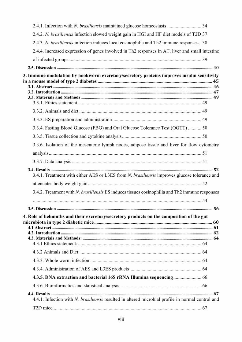
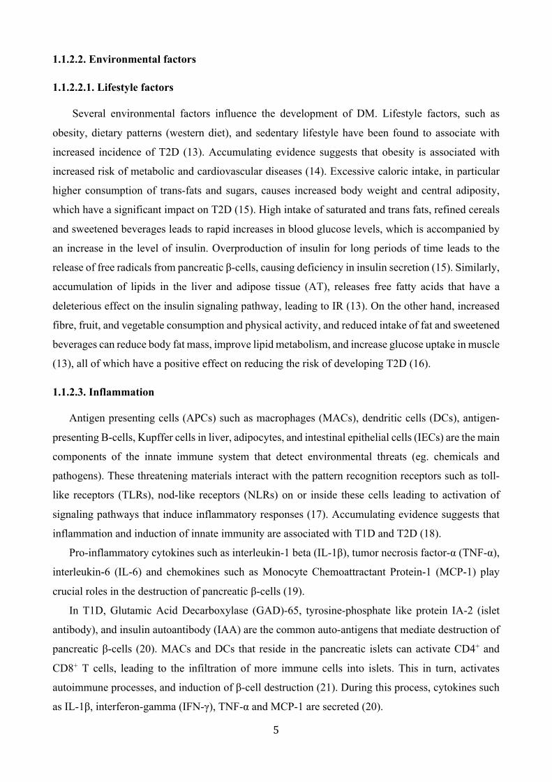
IDF estimated that the number of people with all forms of diabetes worldwide will increase from
424.9 million people in 2017 to 628.6 million people in 2045 (3). There are more people with all
forms of diabetes living in urban (279.2 million), than in rural (145.7 million) areas, and this gap is
predicted to widen to 472.6 million people living in urban areas and 156 million people in rural areas
by 2045 (3). There are however wide variations in prevalence worldwide. The Western Pacific region
has the highest number of people with all forms of diabetes, with 159 million people, followed by
South-East Asia (82 million people), Europe (58 million people) and North America (46 million
people), respectively (Fig.1). The incidence of T1D is increasing, particularly in the age groups under
20 years, with 1,106,500 cases recorded in 2017 worldwide and approximately 132,600 newly
diagnosed T1D cases annually (3). T2D occurs in youth, with the highest rates observed in people
aged 15–19 (4). Moreover, it has been estimated that 352.1 million people have impaired glucose
tolerance (IGT) worldwide in 2017 (almost half of them under the age of 50), and nearly 212 million
people remain undiagnosed (3).

3
Figure 1.1. Number of people with all forms of diabetes as determined by the International Diabetes Federation, 2017 (3).
According to the IDF 2017, 425 million people have diabetes mellitus. The majority of cases can be found in the Pacific region.
9
Number of people with diabetes worldwide and per region in 2017 and 2045 (20-79 years)

4
1.1.2. Aetiology
DM is a multifactorial disease where genetic, environmental, and immunological factors interplay
in the development of the disease.
1.1.2.1. Genetics
DM is a multigenic disease. A strong association between family history, and the risk of
development of diabetes has been documented (5). In addition, family history has been shown to
affect the phenotype of patients with DM (6). Individuals with first degree relatives who are diabetic
are at risk of developing diabetes (1). Indeed, the risk of developing T2D increases by 40% if
individuals have one parent with T2D, and 70% if both parents are affected. Moreover, in
monozygotic twins the concordance of T2D increases by 70% compared with 20-30% in dizygotic
twins (5, 7). Similarly, the risk of developing T1D increases by 6% if individuals have one parent
with T1D and by >30% if both parents are affected. In monozygotic twins the concordance of T1D
increases by 50% compared with 8% in dizygotic twins (8). Many studies have focused on the
analysis of the genetic factors associated with DM, and over 60 genes have been shown to be
associated with this disease. The human leukocyte antigen HLA class II region on chromosome 6p21
is the primary region that contributes to approximately 40–50% of the heritable risk for T1D (9). In
addition, the insulin gene on chromosome 11p15, the cytotoxic T-lymphocyte-associated protein 4
(CTLA-4) gene on chromosome 2q33, a protein tyrosine phosphatase non-receptor type 22 (PTPN22),
the gene on chromosome 1p13 and the interleukin 2 receptor alpha (IL2RA) gene on chromosome
10p15 have also been found to be associated with T1D (8, 9). Genome-wide association studies have
identified a number of genetic loci on chromosomes 3q, 4q, 7q, 9q, 10q, 11q 13q, 15q and 17q that
contribute to the susceptibility to T2D. In European and Asian populations, 45 and 29 loci were
identified respectively (10). The Transcription Factor 7-like (TCF7L2) gene is the most susceptible
gene found to be associated with T2D (11). Moreover, genes like Potassium Channel, Inwardly
Rectifying Subfamily J, Member 11 (KCNJ11) and peroxisome proliferator-activated receptors
(PPAR𝛾) which are targets for anti-diabetic medications have been shown to affect insulin sensitivity
(5). Insulin Receptor Substrate 1 (IRS1) and Insulin-like growth factor 1 (IGF1) genes are associated
with fasting insulin and homeostatic model assessment for insulin resistance (HOMA-IR), and
deletion of these genes results in insulin resistance (IR) (11). Furthermore, proteins like CDK5
Regulatory Subunit Associated Protein 1-Like 1 (CDKAL1), cyclin-dependent kinase inhibitor 2A
(CDKN2A/B) and Insulin-Like Growth Factor 2 may have an effect on β -cell function (10, 12).
5
1.1.2.2. Environmental factors
1.1.2.2.1. Lifestyle factors
Several environmental factors influence the development of DM. Lifestyle factors, such as
obesity, dietary patterns (western diet), and sedentary lifestyle have been found to associate with
increased incidence of T2D (13). Accumulating evidence suggests that obesity is associated with
increased risk of metabolic and cardiovascular diseases (14). Excessive caloric intake, in particular
higher consumption of trans-fats and sugars, causes increased body weight and central adiposity,
which have a significant impact on T2D (15). High intake of saturated and trans fats, refined cereals
and sweetened beverages leads to rapid increases in blood glucose levels, which is accompanied by
an increase in the level of insulin. Overproduction of insulin for long periods of time leads to the
release of free radicals from pancreatic β-cells, causing deficiency in insulin secretion (15). Similarly,
accumulation of lipids in the liver and adipose tissue (AT), releases free fatty acids that have a
deleterious effect on the insulin signaling pathway, leading to IR (13). On the other hand, increased
fibre, fruit, and vegetable consumption and physical activity, and reduced intake of fat and sweetened
beverages can reduce body fat mass, improve lipid metabolism, and increase glucose uptake in muscle
(13), all of which have a positive effect on reducing the risk of developing T2D (16).
1.1.2.3. Inflammation
Antigen presenting cells (APCs) such as macrophages (MACs), dendritic cells (DCs), antigen-
presenting B-cells, Kupffer cells in liver, adipocytes, and intestinal epithelial cells (IECs) are the main
components of the innate immune system that detect environmental threats (eg. chemicals and
pathogens). These threatening materials interact with the pattern recognition receptors such as toll-
like receptors (TLRs), nod-like receptors (NLRs) on or inside these cells leading to activation of
signaling pathways that induce inflammatory responses (17). Accumulating evidence suggests that
inflammation and induction of innate immunity are associated with T1D and T2D (18).
Pro-inflammatory cytokines such as interleukin-1 beta (IL-1β), tumor necrosis factor-α (TNF-α),
interleukin-6 (IL-6) and chemokines such as Monocyte Chemoattractant Protein-1 (MCP-1) play
crucial roles in the destruction of pancreatic β-cells (19).
In T1D, Glutamic Acid Decarboxylase (GAD)-65, tyrosine-phosphate like protein IA-2 (islet
antibody), and insulin autoantibody (IAA) are the common auto-antigens that mediate destruction of
pancreatic β-cells (20). MACs and DCs that reside in the pancreatic islets can activate CD4+ and
CD8+ T cells, leading to the infiltration of more immune cells into islets. This in turn, activates
autoimmune processes, and induction of β-cell destruction (21). During this process, cytokines such
as IL-1β, interferon-gamma (IFN-γ), TNF-α and MCP-1 are secreted (20).
6
MAC polarization has been shown to have a key role in systemic insulin resistance, glucose
tolerance and the development of metabolic disorders, and T2D (22, 23). MACs can differentiate into
two major effector cells: M1 (pro-inflammatory), and M2 (anti-inflammatory) (24). Activation of M1
MACs, or classically activated macrophages, requires two signals: firstly, IFN-γ through the IFN-γ
receptor, and secondly, TNF-α associated with production of IL-1 β, IL-6 and additional TNF-α (25,
26). On the other hand, activation of M2 MACs, or alternatively activated macrophages requires
interaction with Th2 cytokines such as interleukin-4 (IL-4), interleukin-13 (IL-13), interleukin-10
(IL-10), transforming growth factor-beta (TGF-β) and galectin-3 (26).
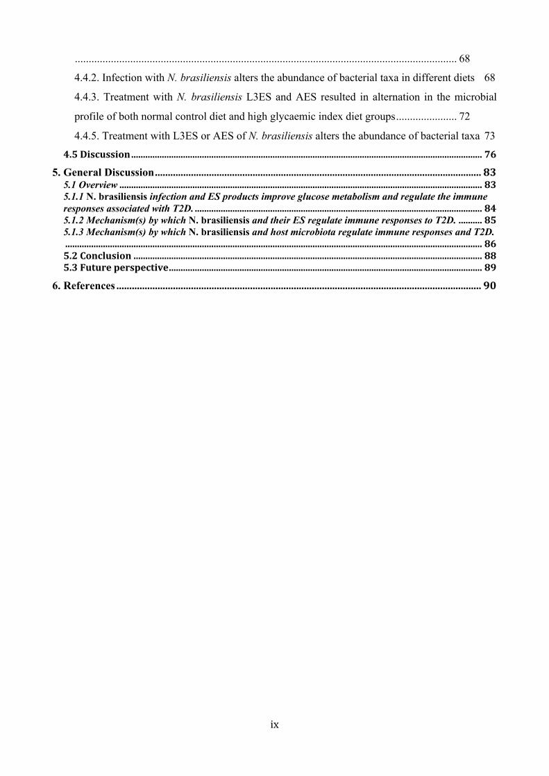
Obesity is the main cause of insulin resistance and plays a key role in T2D (23). Liver, AT, and
muscle are the major players in maintaining glucose homeostasis. Liver maintains glucose
homeostasis between meals via two processes: glycogenolysis and gluconeogenesis. However, AT
may regulate glucose homeostasis after a meal indirectly by regulating lipid homeostasis (27).
Accumulation of lipid in AT, releases free fatty acids (FFAs) that induce the production of adipokines
such as leptin and the activation of pro-inflammatory cytokines such as IL-1 β, IL-6 and TNF-α. M1
MACs are also recruited into AT, inducing the secretion of pro-inflammatory cytokines (28). On the
other hand, resident M2 MACs are responsible for maintaining tissue homeostasis in metabolic
organs (AT, pancreas, liver, and skeletal muscles) (29). Obesity induces local inflammation and
recruitment of M1 MACs, leading to an imbalance between M1/M2 MACs and causing impaired
glucose tolerance in adipocytes, hepatocytes, and myocytes (23, 30). M1 MAC numbers were found
to be elevated in the islets of humans with T2D, and in a mouse model of T2D. However, in normal
conditions the islets exhibited large numbers of resident M2 MACs (25, 31). Moreover, the M2 MACs
comprise 10% of total cells in lean white adipose tissue (WAT) and are associated with the
maintenance of insulin sensitivity (32).
Eosinophils play important roles in the induction of Th2 immunity via production of many Th2
cytokines such as, IL-4, IL-10, IL-13, and TGF-β that participate in anti-inflammatory immune
responses (27). In a human study, it was found that elevated eosinophil numbers were associated with
a decreased risk of T2D (33). Interestingly, it has been demonstrated that eosinophils might play an
important role in the polarisation of MACs towards the M2 phenotype, which in turn allows
maintenance of metabolic homeostasis, and glucose tolerance (34, 35). In particular, Wu and
colleagues have highlighted the role of residential eosinophils in AT as a major IL-4-expressing cell
type that might sustain production of M2 MACs (36, 37). Eosinophil numbers correlated inversely
with body mass in mice on a high fat diet (HFD). Obesity was associated with decreased AT
eosinophil numbers, and increased eosinophil expression in IL-5 transgenic mice improved obesity-
induced insulin resistance (37). Interestingly, distinct groups of innate lymphoid cells (ILCs), a

7
recently discovered innate immune cell type, have been shown to be involved in regulating obesity
(38). Group 2 innate lymphoid cells (ILC2s) are considered as anti-obese immune regulators, and
they are involved in the browning of AT that induce the lean phenotype. These cells participate in the
anti-inflammatory immune response deriving the secretion of Th2 cytokines IL-4, IL-5, and IL-13,
the accumulation of eosinophils in AT, and the polarisation of MACs into the M2 phenotype (38, 39).
On the other hand, group 1 innate lymphoid cells (ILC1s) have been found to participate in tissue
inflammation via induction of IFN-γ, and TNF-α, which drives the polarisation of MACs into the M1
phenotype, in the obese state (38). Administration of the alarmin IL-33 was shown to reduce adiposity
and fasting blood glucose (FBG), and to improve glucose and insulin tolerance (40). Signaling via
IL-33 was required for ILC2-induced IL-5 production for induction of visceral adipose tissue (VAT)
eosinophils, and polarization of VAT MACs towards an M2 phenotype (39-41).
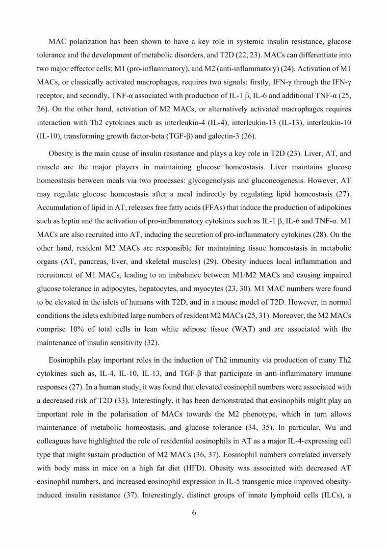
Figure 1.2 Changes in the environment of resident immune cells in adipose tissue as a result of obesity (31). Resident immune cells of lean individuals provide an anti-inflammatory milieu to maintain glucose homeostasis, however adiposity induces pro-inflammatory immune responses that are an important trigger of insulin resistance.
Recently, new subpopulations of CD4+ T cells such as T helper 17 (Th17) and T helper 22 (Th22)
cells have been associated with the pathogenesis of T1D. Interleukin-23 (IL-23), which is mainly
secreted by MACs, is responsible for the expansion of Th17 cells and is considered to be one of the
most important cell populations involved in the development of T1D (28). CD4+ T cells skewed
toward a Th17/Th22 phenotype in AT of obese subjects with insulin resistance and had a greater
percentage of AT cells producing IL-17 and IL-22 (42). However, IL-22 was found to protect
endothelial cells from glucose, and lysophosphatidylcholine-induced injury. Blocking the IL-22

8
receptor-1 (IL-22R1) diminished the protective role of IL-22, suggesting a double function of IL-22
in T2D (43). Moreover, mice deficient in IL-22R1 were prone to develop metabolic disorders when
fed a HFD. Administration of IL-22 to genetically obese leptin receptor deficient mice (db/db) or HF
mice had beneficial effects in improving insulin sensitivity, and regulating lipid metabolism in liver
and AT (44). Many studies have revealed that CD4+ CD25+ Foxp3+ regulatory T cells (Tregs)
producing IL-10, IL-4 and IL-13 are involved in the suppression of pro-inflammatory Th1/17
responses (45). In addition, the prospective role of Treg cells in the pathogenesis and treatment of
T1D and T2D has been highlighted. In a human study, Tregs have been shown to induce M2 MACs,
maintaining tissue homeostasis (45). Furthermore, in non-obese diabetic (NOD) mice, T1D
progression is associated with a progressive loss of Treg cells in the inflamed islets and treatment of
mice with interleukin-2 (IL-2), which mediates the induction of CD25, promoted Treg cell survival
and subsequently prevented the onset of diabetes (46). Moreover, depletion of Treg cells accelerates
the development of T1D (47). A study by Feuerer and colleagues demonstrated higher numbers of
CD4+ Foxp3+ Treg cells in the AT of mice fed a normal diet in comparison with those fed a HFD
(48). Moreover, transferring Tregs from healthy donor mice to db/db mice improved insulin
sensitivity (22).
1.1.3. Diagnosis
Diabetes is diagnosed by a blood glucose test from venous blood. The biochemical tests that are
considered for diagnosis of diabetes are: FBG, two hours Oral Glucose Tolerance Test (OGTT) and
Glycosylated Haemoglobin (HbA1c). The FBG test measures the level of glucose in the blood at a
single time point, usually after overnight fasting for at least eight hours. The diagnostic level of blood
glucose for diabetes in this test is ≥ 126 mg/dL (7.0 mmol/L). The OGTT test measures the level of
glucose in the blood at two time points. This test is also performed after overnight fasting. The patient
is given glucose solution containing the equivalent of 75 g glucose, and the blood glucose is measured
at baseline (before the glucose load) and two hours after the glucose load. The diagnostic value in
this test is blood glucose ≥ 200 mg/dL (11.1 mmol/L). The HbA1c test is commonly used to diagnose
diabetes in individuals with risk factors and measures the average level of blood glucose over a period
of three months. The diagnostic cut-off point of diabetes in this test is ≥ 6.5% (1).
1.1.4. Treatment
Diabetes is a complex, chronic illness, and thus, long-term medical care and treatment are
needed to help control blood sugar levels and prevent disease complications. Nutritional therapy
includes managing the amount and type of food taken, weight loss and regular exercise (1). Indeed,

9
150 min/week of moderate exercise or 75 min/week of vigorous aerobic physical activity have been
shown to improve blood glucose control, reduce cardiovascular risk factors by an increase in high
density lipoprotein (HDL) cholesterol, decrease in triglycerides (TG), and decrease in blood pressure
(1). Furthermore, regular exercise may prevent the risk of developing T2D in high risk individuals
(1). However, for many patients, these methods alone are insufficient in managing the disease. Thus,
pharmacological therapy is also used to manage insulin secretion and blood glucose levels. Patients
with T1D require lifelong insulin therapy, usually twice or more daily, with doses adjusted on the
basis of self-monitoring of blood glucose levels (49). Metformin, sulphonylureas, thiazolidinediones,
and other drugs are the first line therapy for T2D patients (50), however treatment with insulin is
sometimes needed. Despite the wide use of these medications in the treatment of T2D, and their
proven efficacy, these medications can have side effects including hypoglycaemia, weight gain, and
increased low density lipoprotein (LDL) and TG levels (51).
1.2. The Hygiene hypothesis
The transition of nations from primarily agricultural to industrial societies has been associated
with a rapid rise in the incidence of many immune diseases, including inflammatory bowel disease
(IBD), multiple sclerosis (MS), rheumatoid arthritis (RA), and T1D (52). The hygiene hypothesis,
formulated by David Strachan in 1989 (53), proposed that changes in human environment such as
changed dietary habits, a cleaner environment with improved sanitation, vaccination programmes and
excessive antibiotic use have reduced our exposure to many infectious agents (53), and symbiotic
microorganisms (including helminths and gut microbiota) that had an evolutionary relationship with
humans (54). The coevolution of humans and infectious agents established an immunological
interaction that ensured the development of regulatory pathways to keep inflammation in check,
thereby reducing inappropriate immune responses, which are considered the key mediators in many
immune disorders (54).
At first, the imbalance between Th1 (pro-inflammatory)/Th2 (anti-inflammatory) responses was
suggested to explain the epidemiology underlying the hygiene hypothesis, whereby reduced exposure
to microbial products that stimulate Th1 responses resulted in excessive Th2 responses, culminating
in an increase in the incidence of Th2-mediated allergic diseases (55). However, it soon became
apparent that this theory of opposing Th1 and Th2 responses was insufficient to explain the increased
incidence of Th1/Th17 mediated autoimmune diseases in addition to Th2-mediated allergic
conditions in developed countries (56).
As the important role of Treg cells in controlling inappropriate inflammation was revealed, the
importance of pathogen exposure on the subsequent development of a functional regulatory immune
response became apparent (57). Indeed, the failure of immunoregulatory mechanisms (primarily Treg

10
responses) can lead to increased immunopathology (58). In DM, mutation in Foxp3, a transcription
factor that plays a crucial role in the development and function of Treg cells is associated with T1D
(59). Moreover, the depletion of Treg cells leads to the development of various autoimmune diseases,
such as gastritis, T1D, and IBD (60). Hence, the balance between Tregs, Th1, Th2, and Th17
responses is essential to prime immunoregulation and provide protection against diseases that result
from a dysfunctional immune system.
1.2.1. Studies supporting the hygiene hypothesis
The biota that inhabits the mammalian body is a major driving force in shaping the mammalian
immune system (61). The mode of child delivery (Cesarian section) and feeding (formula vs. breast
feeding), as well as antibiotic therapy, have a strong effect on the early distribution of gut microbiota.
These, in turn, affect the susceptibility of individuals to many inflammatory diseases and allergic
disorders (61-63). Helminth parasites are another important element of the human biome, and they
have co-evolved over millennia, resulting in “pathogen tolerance” through the induction of regulatory
immune responses (64). Many epidemiological studies have demonstrated the importance of an early
exposure to different pathogens on the development of the immune system. Absence of early
exposure to pathogens is thought to increase susceptibility to allergic and autoimmune diseases like,
asthma, IBD, and T1D (65). This phenomenon is also supported by many studies showing a higher
incidence of allergic conditions such as asthma (66), atopy (67), and IBD (68) in urban areas
compared to rural areas. Similarly, other studies have found an increase in the prevalence of allergy
and MS in the developed world compared to helminth-endemic areas or regions with poor sanitation
(69, 70). Moreover, studies have found an increase in allergen skin reactivity after anthelmintic
treatment (71). Birth mode, breast feeding, and antibiotic usage have an effect on the composition of
gut microbiota and have been associated with the development of DM and other metabolic diseases.
For instance, babies born via cesarean section harbored bacterial communities dominated by
Staphylococcus, Corynebacterium, and Propionibacterium spp. However, vaginally delivered babies
harbored bacterial communities dominated by Lactobacillus, Prevotella, or Sneathia spp. (72).
Cesarean delivery and short term of breast feeding positively associated with atopic dermatitis (73).
Moreover, a meta-analysis found a 20% increase in the risk of asthma and T1D in children delivered
by cesarean section compared to vaginal delivery (74, 75). In a cohort study of 1,650 German school
children, cesarean delivery resulted in a more than two-fold increase in childhood T1D risk compared
to vaginal delivery (76). The use of antibiotics early in life has been associated with paediatric IBD
(77), and increases the risk of asthma in early childhood (78). Moreover, antibiotic exposure during
the first 6 months of age has been associated with an increase in body mass (63).

11
1.3. Helminth infection
Nematodes (also known as roundworms), trematodes (also known as flatworms), and cestodes
(also known as tapeworms) are the three main groups of parasitic helminths (79). The major
gastrointestinal (GI) nematodes of humans, often referred to as soil-transmitted helminths (STH), are
the cause of one of the most important neglected tropical diseases, and include the ascarids (eg.
Ascaris lumbricoides), hookworms (eg. Necator americanus, Ancylostoma sp.), threadworms (eg.
Strongyloides stercoralis) and whipworms (eg. Trichuris trichiura) (80). Successful parasites employ
different strategies to maintain a harmonious relationship with their host in order to survive (64). It
is well established that helminth infections induce a profound Th2 response but without the hallmark
features of serious allergy such as urticaria or anaphylaxis, and this phenomenon is attributed to the
ability of helminths to promote regulatory networks that prevent excessive inflammation (81).
Induction of a Th2 immune response is associated with an increase in the levels of IL-4, IL-5, IL-9,
IL-13 and IL-21 as well as expansion of specific effector cells, such as eosinophils, MACs, mast cells,
neutrophils, basophils and ILCs (82). M2 MACs have been shown to play an important role in host
protective immunity against helminth infections (83). IL-4 and IL-13 induce M2 MACs by signalling
through the IL-4 receptor alpha/ signal transducer, and activator of transcription 6 (IL-4Rα /STAT6).
These cells are multi-faceted and orchestrate diverse processes such as immune regulation, tissue
repair, and worm expulsion and resistance (64, 83). The protective effect of M2 MACs depends on
the expression of arginase-1 (Arg1), chitinase-like 3 (Ym1) and Resistin-like molecules (RELMα and
β) (83). Eosinophil numbers are increased in the blood following helminth infection and they are
rapidly recruited to the site of infection where they participate in worm killing through the secretion
of toxic mediators such as major basic protein, eosinophil peroxidase and eosinophil neurotoxin, in
addition to the production of various chemokines, and cytokines such as, IL-4, IL-5, TGF-β (82, 84).
Moreover, a rapid rise in Foxp3+ Tregs mediated by IL-10 and TGF-β are reported after helminth
infections. This response plays an instrumental role in regulating inflammation in response to
helminth infection. Depletion of these cells has an influence on both pathology and resistance to
infection (reviewed in ref (85)). Generally, the infective stage of STHs enter their host either orally
or via skin penetration, and mature to become adult worms which inhabit different niches within the
intestine (86). N. brasiliensis is a GI hookworm-like nematode of rats but has been studied extensively
in the mouse (87). The lifecycle of this parasite involves two phases; a free-living phase in the external
environment, and parasitic phase that takes place inside the definitive rodent host. This latter phase
commences when infective third-stage larvae (L3) penetrate the skin and enter the circulation to reach
the lung vasculature. In the lungs they moult to (L4), break through the alveoli and are then coughed
up and swallowed as they enter the digestive system to reach the small intestine. In the small intestine,

12
they moult to L5 then become dioecious adults which mate. The female worm starts releasing eggs
in the host faeces, which then hatch into L1 then L2 and finally L3 in the soil, before the L3 penetrates
the tissues of a new host (88). Following GI nematode infections, APCs such as DCs, MACs,
basophils and ILC2s play different roles in response to infection through influencing the induction of
both Th2 and Treg immune responses (reviewed in ref (86)). Eosinophils (89), neutrophils, MACs
(90, 91), and ILCs (92, 93) are essential in maintaining protection against N. brasiliensis infection in
mice through orchestrating Th2 immune responses (94, 95). Both eosinophils and M2 MACs
accumulate at sites of N. brasiliensis infection where they participate in worm killing and expulsion,
and tissue remodelling. Activation of STAT6 by IL-4 and IL-13 play an important role in this process.
Resistance to N. brasiliensis infection was impaired in eosinophil-deficient mice, and mice lacking
IL-5 (89, 96, 97), STAT6, IL-4Rα and IL-13 (96). Eosinophils in these mice were recruited (skin,
lung, small intestine) in very small numbers, more larvae migrated to the lungs, and adult worms
displayed prolonged production of eggs compared to control mice (89). Rapid upregulation of YM1,
RELM-α, and Arg1 were found in the lungs of mice infected with N. brasiliensis with increased IL-
4 and IL-13, indicating the role of M2 alveolar MACs in the induction of host immune responses to
the infection (91, 98). By day 8 post-infection, these cells suppressed inflammation caused by larval
migration through the pulmonary environment (99).
1.3.1. Helminths as a therapeutic modality for immune-mediated inflammation
1.3.1.1. Evidence from human studies
Epidemiological studies from helminth-endemic areas show an inverse relationship between
helminth infection and inflammatory diseases. For instance, there is an inverse relationship between
the frequency of S. stercoralis infection and the incidence of autoimmune liver disease (100).
Different clinical trials have highlighted the therapeutic roles of helminths in immune mediated
diseases. To assess the immunosuppressive properties of N. americanus in coeliac disease (CeD), two
clinical trials have been conducted where CeD patients were infected with low numbers of live N.
americanus L3. Hookworm infection induced strong systemic and mucosal Th2 (IL-4, IL-5, IL-9 and
IL-13) and regulatory (IL-10 and TGF- β) cytokines, supressed the production of IFN-γ and IL-17A
and increased the numbers of CD4+ CD25+ Foxp3+ cells in duodenal biopsy cultures (105). Moreover,
suppression of mucosal IL-23 and upregulation of IL-22 (which promotes mucous production) of
hookworm-infected participants with CeD after gluten challenge was observed. In these studies,
treatment with N. americanus appears to be safe and hookworm-infected mucosa retained a healthy
appearance (101-105). Most importantly, moderate gluten challenge of hookworm-infected patients
did not induce any immunopathology in the gut (villous height to crypt depth ratio) and anti-tissue
transglutaminase levels remained unchanged (105).

13
In two different studies, infection with live N. americanus was safe for crohn’s disease (CD)
patients, and resulted in a reduced CD activity index score (106). In the second study on seven IBD
patients (four with active CD and three with UC), administration of eggs of the porcine GI whipworm
Trichuris suis was shown to be safe and patients showed improvement in the common clinical indices
of the disease (107). Another study on 29 patients with CD who received T. suis eggs revealed a
significant reduction in symptoms (108). Furthermore, a randomized, double blind, placebo-
controlled trial including 54 patients with active colitis showed improvement in 43.3% of the patients
that received T. suis compared with 16.7% that received placebo treatment (109).
Moreover, in a cohort study, 12 patients with MS infected with different GI nematodes showed
significantly lower relapse frequency than uninfected patients, accompanied by an increase in IL-10-
and TGF-β-secreting cells, and a decrease in IL-12- and IFN-γ-producing cells compared with non-
infected patients (110). When these infected patients received anthelmintic treatment, a significant
decrease in IL-10- and TGF-β-secreting cells, and a significant increase in IFN-γ- and IL-12-
producing cells was observed (110). Also, a clinical trial of 4 MS patients using experimental T. suis
therapy revealed a downregulation of Th1 responses (particularly IL-2 and IFN-γ) and an increase in
Th2 associated IL-4 (111). A survey of self-treatment with helminths (T. suis ova, T. trichiura, N.
americanus and tapeworm Hymenolepis diminuta) revealed that helminth therapy was effective for
many people in reducing a variety of inflammatory diseases including IBD, allergies, and
autoimmune diseases (112). All of these trials have revealed the potential use of helminths as
therapeutic agents in a wide range of inflammatory mediated diseases. You are mostly reporting the
positive outcomes. There are many papers that show tht it doesn’t work very well too. You might cite
a few of them to keep the balance. For example, phase 2 trials of TSO in CD and UC failed to meet
clinical endpoints. See attached review paper of Stephanie’s that is in press at PLoS Path. If I forget
to send it to you, remind me.
1.3.1.2. Evidence from animal models
A variety of helminths and their products have been tested in different mouse strains to assess
their roles in the prevention of immune mediated diseases. For instance, in a mouse model of asthma,
infection with N. brasiliensis suppresses the development of allergen-induced airway eosinophilia
via the production of IL-10. Interestingly, N. brasiliensis infection alone (in the absence of allergen
challenge) induced airway and blood eosinophilia (113). Restimulating the MLNs and spleen cells of
infected mice with anti-CD3 and IL-2 led to increased amounts of IL-4, IL-5, IL-10 and IL-13 in
comparison to uninfected mice. However, when N. brasiliensis infected mice were challenged with
OVA allergen, a significant decrease in airway eosinophilia was observed, accompanied by reduced
levels of eotaxin in the broncho-alveolar lavage fluid of these mice in comparison to uninfected mice

14
challenged with OVA. This effect was possibly dependent on IL-10, as the suppressive effect was
not observed in mice deficient in IL-10 (113). Infection with the GI nematode of mice
Heligmosomoides polygyrus showed reduction of airway eosinophilia, and neutrophilia after OVA
challenge. In addition, elevation in Tregs and regulatory B cells (Bregs), and increased expression of
TGF-β and IL-10 were also detected in MLNs after challenge (114, 115).
In two different model of arthritis, N. brasiliensis infection suppressed inflammatory arthritis
through induction of Th2 immune response. The anti-arthritis effect was dependent on the activation
of the STAT6 pathway by IL-4/IL-13. Eosinophil numbers increased in the joints of infected mice
and neutrophil numbers decreased. Expression markers of M2 MACs were increased while
expression markers of M1 MACs decreased in the joints of infected mice (116). In another model of
arthritis, mice infected with either N. brasiliensis or H. polygyrus also showed reduced arthritis
severity that was associated with increased levels of IL-4 (117).
In a mouse model of dinitrobenzene sulfonic acid (DNBS)-induced colitis, infection with the GI
nematode Trichinella spiralis reduced the severity of colitis. This was accompanied by a significant
reduction in IL-12 levels and elevated production of IL-4 and IL-13 (118). In addition, H. polygyrus
infection of Rag IL-10-/- mice was protective in the T cell transfer model of colitis, and was
accompanied by alterations in mucosal DC function and reduction in the capacity of intestinal T cells
to produce IFN-γ and IL-17 (119).
In a mouse model of MS, rats infected with T. spiralis showed reduction in the clinical score of
the disease, which was associated with increased levels of IL-4 and IL-10 and decreased levels of
IFN-γ and IL-17 (120). Likewise, mice infected with Trichinella pseudospiralis showed amelioration
in the clinical score of the disease with reductions in the levels of IL-17, IL-6, IL-1β, IFN-γ and TNF-
α (121). Infection with H. polygyrus also showed beneficial effects on disease severity, which is
associated with increased levels of IL-10, TGF-β, and IL-6 in the cerebrospinal fluid and in the serum,
and decreased IL-17A and IL-2 levels in the serum (122).
1.3.2. Helminths as a therapy for diabetes mellitus
Recent epidemiological studies in indigenous communities of north-west Australia, Indonesia,
rural China and India revealed an inverse correlation between helminth infection and T2D (123-126).
Additionally, infection with S. stercoralis in Australian Aboriginal communities seems to protect
against the onset of T2D (127). Moreover, there is an inverse relationship between the prevalence of
lymphatic filariasis and T1D in southern India (128). NOD mice are widely used as a model to study
human T1D. In this model of DM, Th1 responses drive the development of inflammation. Many
studies have investigated the protective role of nematodes (T. spiralis, H. polygyrus, and
Litomosoides sigmodontis (filarial nematode)) in the treatment of T1D in NOD mice. The data show

15
increased levels of IL-4, IL-5, IL-10, Foxp3, IgG1 and IgE; and decreased levels of IFN-γ and IL-12
as well as a reduction in CD8+ lymphocyte infiltrate in the pancreas (129-132). However, in IL-4
deficient mice the protection against diabetes does not depend on the Th2 shift, and requires TGF-β
and IL-10, which suggests that other regulatory mechanisms might be involved in diabetes prevention
(129, 130). These findings provide evidence that GI nematodes in particular can induce a mixed
Th2/Treg response that holds potential for treating T1D (133).
In the context of T2D, diabetic mice infected with H. polygyrus had improved glucose tolerance,
decreased HOMA-IR and body weight gain with decreased fat accumulation and fatty acid synthase
gene expression in the liver compared to uninfected diabetic mice (134, 135). The infection also
increased the expression of uncoupling protein 1 (UCP1), the M2 MAC markers, RELMα, Arg1, and
Ym1 and resulted in increased levels of IL-4, IL-13, and IL-10 in the small intestine and MLNs (134,
135). This shift towards a Th2 environment was accompanied by increased expression of GATA3,
and Foxp3+ and decreased IFN-γ and IL-17 in the MLNs (134). Similarly, in a mouse model of
obesity, infection with N. brasiliensis resulted in reduced body weight gain, decreased adipose tissue,
and liver masses with decreased levels of hepatic triglycerides. This was accompanied by improved
glucose homeostasis and expression of M2 MAC markers Arg1 and Ym1 and Th2 cytokines IL-4 and
IL-5 in AT and liver (136). Two additional studies showed that N. brasiliensis infection improved
insulin sensitivity through induction of IL-33, which mediates activation of resident VAT, ILC2
producing IL-5- and IL-13-dependent accumulation of VAT, and eosinophilia (37, 41). Additionally,
mice infected with the filarial nematode L. sigmodontis also showed improved glucose tolerance
which was associated with increased numbers of eosinophils, M2 MACs, and CD4+ T cells in the AT
(137). C57BL/6 mice fed a HFD and infected with the blood fluke trematode Schistosoma mansoni
showed an increase in WAT eosinophils, and M2 MACs. This was associated with metabolic
homeostasis, and improved insulin sensitivity (138). Moreover, in a mouse model of atherosclerosis,
treatment with Schistosoma eggs reduced total serum cholesterol and triacylglycerol and cholesteryl
ester lipids in the liver (139).
1.4. Helminth-derived products
Helminths release soluble mediators that interact with host immune cells. These helminth-
derived molecules, referred to as excretory/secretory (ES) products, play important roles in host
immune modulation and represent the molecular interface between host and parasite. Several
helminth-derived molecules that interfere with host immune processes have been described (140).
These molecules include proteases, protease inhibitors, cytokine homologues, anti-oxidants, various
esterases, proteins of unknown function, and glycans and lipids (140). Numerous schistosome soluble
egg antigens (SEA) direct DCs to drive Th2 responses in vitro and in vivo (141). For instance, Lacto-

16
N-fucopentaose III (LNFPIII) was capable of inducing Th2 immune responses, with decreased levels
of IFN-γ, and increased levels of IL-4, IL-5 and IL-10 (142). Additionally, recombinant alpha-1
induces basophils to produce IL-4 (143), and omega-1 induces Th2 responses by driving production
of IL-4 (144). Likewise, recombinant thioredoxin peroxidase from the liver fluke Fasciola hepatica
has been shown to induce the recruitment of M2 MACs, which is associated with the induction of
Arg1, and high levels of IL-10 (145). Parasitic nematode ES products also drive regulatory immune
responses. One of the best known examples is ES-62, a phosphorylcholine-bearing glycoprotein from
Acanthocheilonema viteae that promotes the differentiation of DCs and MACs towards an anti-
inflammatory phenotype (146). ES products of T. spiralis have been shown to modulate the function
of MACs via inhibiting the production of TNF-α and IL-6 (147) and inducing type 2 cytokine
responses via increased production of IL-4 and IL-10 (148). H. polygyrus ES (HES) products have
been reported to inhibit T cell proliferation, and HES-exposed DCs can induce differentiation of IL-
10-producing CD4+ Tregs (149). Moreover, hookworms express molecules with immunosuppressive
properties, including neutrophil inhibitory factor, anticoagulant peptides and protease inhibitors
(150). ES products of adult N. americanus bind selectively to NK cells and induce IFN-γ production
in the presence of both IL-2 and IL-12 (151). Proteases secreted by N. americanus have been shown
to induce type 2 cytokine production by basophils, and this was associated with increased levels of
IL-4, IL-5 and IL-13 but not IFN-γ (152). Importantly from the perspective of my PhD project, ES
products of N. brasiliensis (NES) have been shown to stimulate maturation of DCs towards a Th2
phenotype (153-156).
Furthermore, helminths were recently described to secrete particles known as extracellular
vesicles (EVs). These EVs might have a role in parasite-host interaction and modulation of the host
immune response (157). Researchers have described EVs from F. hepatica (158), Schistosoma spp.
(159-161), and N. brasiliensis (162, 163), among others (164, 165), and highlighted their roles in
parasite-host interactions (161), including both suppression of inflammation and enhanced
inflammatory responses that predispose to cancer (158). For instance, EVs from S. japonicum
increase numbers of host peripheral monocytes in vivo (166) and skew MACs towards the M1
phenotype accompanied by increased levels of TNF-α and IL-12 in vitro (167). Internalization of H.
polygyrus EVs by MACs caused downregulation of Th1 and Th2 responses which was associated
with suppressed expression of IL-6, TNF, Ym1, RELMα, and expression of the IL-33 receptor subunit
ST2 (168).

17
1.4.1. Helminth-derived products as therapies for immune-mediated inflammation
Many studies have indicated the therapeutic potential of helminth ES products for treating a
diverse array of inflammatory conditions. ES products of the hookworm A. caninum (AcES)
suppressed intestinal pathology in a dextran sulphate sodium (DSS) mouse model of colitis, and was
associated with the upregulation of Th2/anti-inflammatory cytokines IL-4, IL-5 and IL-10, and
downregulation of the pro-inflammatory cytokines IL-6, IL-17 and IFN-γ (169, 170). NES of N.
brasiliensis suppressed asthma by inducing a Th2 immune response that was associated with
increased IL-4 and IL-5-producing CD4+ T cells that simultaneously inhibited the development of
OVA-specific allergic responses (171, 172). ES products of adult T. spiralis attenuated the severity
of DSS colitis in mice, characterised by a significant reduction in IL-17 levels in the colon and MLNs
(173). Additionally, rats administered with crude T. spiralis larval (L1) ES products exhibited
improved clinical scores in experimental autoimmune encephalomyelitis (EAE) as a model of human
MS. This was associated with increased levels of IL-4, IL-10 and TGF-β, and decreased levels of
IFN-γ and IL-17 with increases in the proportion of CD4+ CD25+ Foxp3+ T cells at the systemic level,
and in target organs (spleen and spinal cord) (174). In another study, soluble products of both T. suis
and T. spiralis significantly reduced the clinical signs of experimental EAE in mice (175).
Administration of EVs from H. polygyrus, and L. sigmodontis reduced eosinophil numbers and IL-5,
IL-13 and IL-33 levels in bronchoalveolar lavage, expression induced by allergen Alternaria (176).
In a more recent study, EVs from N. brasiliensis protected against chemically-induced colitis
inflammation in mice, which was associated with the suppression of pro-inflammatory cytokines such
as IL-6, IL-1β, IFNγ and IL-17a and increase of the anti-inflammatory cytokine IL-10 in EV-treated
groups compared to control groups (163). H. diminuta antigen-pulsed DCs were found to be
protective in di-nitrobenzene sulphonic acid (DNBS)-induced colitis, and required IL-4Rα and the
capacity to secrete IL-10 (177, 178). However, ES from helminth is very complex and difficult to
produce in large scale, hence recombinant Es proteins would be better to produce as new therapeutic
modality. S. mansoni recombinant protein, 28-kDa glutathione S-transferase (P28GST) prevented
inflammation in the TNBS model of colitis in both mice and rats, characterised by decreased
expression of pro-inflammatory cytokines and increased expression of Th2 and anti-inflammatory
cytokines (179). Recently, one of the most abundant AcES proteins, AIP-2, was produced in
recombinant form and shown to suppress airway inflammation in a mouse model of asthma via
induction of tolerogenic DCs, and Foxp3+ Tregs (180). Recombinant filarial cystatin was also found
to suppress allergic airway inflammation via induction of IL-10-producing MACs that inhibited
eosinophil recruitment, reduced levels of total IgE and downregulated IL-4 production (181).
Recombinant T. spiralis Ts-specific 53-kDa glycoprotein (TsP53) also protected mice from colitis,
typified by a reduced mucosal damage and a reduced inflammatory cell infiltration in the colonic

18
mucosa and submucosa (182). A systematic review reported that treatment of mice with multiple
helminth recombinant proteins including A. viteae ES-62 - and its small molecule analogue SMA-
12b, 11a and PC- as well as recombinant SJMHE1 from S. japonicum resulted in significant reduction
in clinical arthritis score. In all of these studies the treatment with these helminth recombinant proteins
was associated with a significant decrease in the pro-inflammatory cytokines, such as TNF-α, IL-6,
IL-1β, IFNγ and IL-17, and with a significant increase in IL-10 (183).
1.4.2. Helminth-derived products as therapeutics for diabetes mellitus
T1D being an autoimmune inflammatory condition could potentially be treated with helminth
ES products. ES of F. hepatica liver flukes prevented T1D in NOD mice through induction of Bregs
and Tregs, and was associated with induction of M2 MACs and increased IL-10 and TGF-β levels
(184). Schistosome crude SEA also prevented T1D in the same mouse model, inducing Th2 and Treg
responses which involved induction of DCs and M2 MACs and increased levels of IL-4, IL-10 and
TGF-β (185). SEA was also protective in an atherosclerosis model of mice. In this model low dose
exposure to SEA promoted Th2 responses and reduced the levels of total cholesterol and LDL (186).
Likewise, mice treated with SEA had lower plasma cholesterol levels, mainly LDL and VLDL, which
had an effect on reducing the size of atherosclerotic plaques. SEA treatment resulted in reduced
circulating neutrophils and M1 MACs and TNF-α expression, and increases in M2 MACs producing
IL-10 (187). The LNFPIII glycan found in schistosome SEA (also found in human breast milk) has
anti-inflammatory properties via induction of IL-10 from DCs and Arg1+ M2 MACs. HFD mice
treated with LNFPIII had improved insulin sensitivity and reduced liver TG (188). In two different
studies the researchers found that ES products from L. sigmosoides and S. mansoni increased the
number of eosinophils and M2 MACs in AT and were able to improve metabolic homeostasis, and
insulin sensitivity in murine obesity models (137, 138).
1.5. The microbiota, mucous and inflammation
The mammalian body is inhabited by a microbial community that is essential for the formation
and maintenance of a balanced immune system with functioning effector and regulatory capacity
(60). Mode of birth and feeding, antibiotic use, dietary habits, and helminth infection can all have an
impact on the composition and diversity of the intestinal microbiota (189-191). The gut microbiota
plays an important role in host metabolism, physiology, nutrition, and immune function. In order to
maintain a homeostatic relationship with these microbes, the intestinal immune system has to reduce
direct contact with resident bacteria, and limit their exposure to immune system components (60).
The intestine is protected by mucous secreted mostly by goblet cells. Mucous consists of mucins,
which are glycoproteins that have more than 50% of their mass as O-glycans. The mucin (Muc) family
includes members Muc1 to Muc12 (192). Defects in the mucous layer allow resident gut bacteria to

19
be in direct contact with the epithelium, leading to induction of inflammatory responses as
demonstrated by increased permeability and bacterial adherence to the epithelial cell surface in Muc2-
deficient mice (193), and their progression to develop colitis (194). RELMβ upregulates Muc2
secretion in human goblet-like cells in culture as well as in mouse goblets cells in vivo where it
reduces the colonic damage in a mouse model of inducible colitis (195). Moreover, germ-free (GF)
mice showed a dramatic reduction in RELMβ, however, enhanced expression and robust secretion of
RELMβ were found following colonisation by commensal bacteria (196). Th2 immune-mediated
responses can activate RELMβ gene transcription in a STAT6-dependent manner (196). Interestingly,
induction of Th2 cytokines by GI nematode infections induces RELMβ expression by IECs that is
essential for helminth expulsion (197). IL-4/IL-13–dependent goblet cell hyperplasia drives N.
brasiliensis expulsion through production of mucous-containing RELMβ that can encapsulate worms
and inhibit their ability to sense their source of nutrition (198). In this context, IEC may have an
immunoregulatory function in addition to their role in maintenance of mucosal barrier integrity.
Paneth cells are also specialised cells in the intestinal epithelium that secrete antimicrobial molecules
such as regenerating islet-derived protein III (RegIII-β, RegIII-γ), which is capable of killing Gram
positive bacteria, limiting the microbial penetration of the epithelial surface and preventing them from
damaging the epithelium. This homeostatic relationship with gut microbes stimulates a highly
regulated innate and adaptive immune response (199).

20
Figure 1.3. The gut microbiota and host immune responses in the steady state (modified from (199)). Microbiota and their associated molecular patterns such as short chain fatty acids (SCFAs) and LPS induce immune cells such as MACs, DCs and Tregs to provide an anti-inflammatory milieu that maintains immune homeostasis. The resident M2 MACs and DCs in the intestinal lamina propria have developed mechanisms to
downregulate pro-inflammatory responses (200). Interaction of intestinal DCs with B and T cells
results in IgA production, preventing gut microbes from penetrating systemic secondary lymphoid
tissues (60). Intestinal MACs through MyD88 signaling downregulate pro-inflammatory cytokines,
resulting in wound healing and tolerance to these commensal bacteria (200). Moreover, stimulation
of resident ILCs in the lamina propria results in production of IL-22, which plays an important role
in preventing the spread of bacteria to systemic sites (60). IL-22 is required for protection during
bacterial infection through direct induction of antimicrobial proteins, RegIII-β, and RegIII-γ (201).
commensal microbiota, SCFAs, LPS
Inflammatoryresponse
IL-18

21
IL-22 enhanced activation of STAT3-mediated enhancement of mucous-associated proteins (Muc1,
Muc3, Muc13) within colonic epithelial cells, leading to attenuation of local intestinal inflammation
(202). Th17 cells also contribute to host defence via producing IL-22. Smaller Peyers patches, lymph
nodes, and spleens of GF mice have an impact on the maturation of the immune system. These mice
show decreased (or a complete absence) intestinal Th17 cell development, and decreased production
of mucosal IgA (203). Interestingly, infection with N. brasiliensis increases IL-22, which directly
increased goblet cell numbers and RELMβ, Muc1, Muc2 and Muc3 production in the small intestine,
and which was shown to be essential for helminth expulsion (204). It has been shown that IL-33-
indcued IgA production is important for maintaining microbial homeostasis in the intestine and
confers protection against colitis. However, IL-33 deficient mice exhibit microbial dysbiosis and are
prone to develop colitis (205). Noteworthy, in N. brasiliensis infection, IL-33 induced ILC2-
producing IL-13.
In the healthy state, a number of commensals have been found to induce CD4+Foxp3+ Tregs that
secrete IL-10 and TGF- β (206). However, there is a significant decrease in these cells with a
reduction in IL-10 levels in GF mice (207). Microbial-associated molecular patterns such as bacterial
LPS and SCFAs such as, acetate, propionate and butyrate, which are the main end-products of the
bacterial fermentation of non-digestible dietary fibres, also participate in modulation of host immune
responses (208). In the homeostatic state, sensing of LPS molecules occurs directly by host protein
lipid A-binding protein (LBP). LBP binds to LPS forming LBP/LPS complexes which then bind to
CD14. The latter LPS/LBP/CD14 complex signals through TLR4, leading to downstream
inflammatory responses (209). SCFAs, apart from their role as a source of energy for IECs, also play
an important role in the homeostasis of the intestinal epithelium. These bacterial metabolites induce
a tolerogenic and anti-inflammatory response, and under some conditions they may also induce Th1
and Th17 responses through activation of the host G-protein- coupled receptors Gpr-41 and -43 (210).
The immune mechanisms by which they do this involves an increase in the expression of
antimicrobial peptides and IL-18 (a key cytokine for the repair, and maintenance of epithelium
homeostasis) by IECs, and regulation of the differentiation and activation of immune cells such as
neutrophils, DCs, MACs and T lymphocytes which is most likely associated with activation of
FoxP3+ Tregs cells, and increased production of IL-10 (208). Moreover, depending on the cytokine
milieu, these metabolites can promote the differentiation of naïve T cells into T cells producing IL-
17 and IFN-γ (208, 210, 211). Thus, the shift in the composition or density of gut microbiota leads to
changes in the inflammatory state associated with imbalance in intestinal and systemic immune
homeostasis (199).

22
1.5.1. Role of microbiota in immune mediated diseases
There is substantial evidence highlighting the beneficial role of gut microbiota in shaping the
mammalian immune response. Disturbance of the intestinal microbial community leads to altered
immune responses that can result in various human inflammatory disorders (212). Firmicutes (Gram
positive) and Bacteroidetes (Gram negative) represent the largest phyla in the human and mouse
microbiota, and a shift in the ratio of these phyla has been associated with many disease conditions
(213). Studies using GF mice have revealed the crucial role of the microbiota in the pathogenesis of
many immune-mediated diseases.
A recent study demonstrated that OVA-sensitized GF mice subjected to an OVA aerosol
challenge showed increased allergic airway inflammation. Recolonisation of these mice with a
complex specific pathogen free (SPF) microbiota for 3 to 4 weeks protected mice from increased
allergic airway inflammation. This was associated with a decrease in regulatory DCs and alveolar M2
MACs and an increase in proallergic basophils in the lung tissue and airways of GF mice compared
with SPF mice (214). Administration of SCFAs to mice attenuated the severity of allergic airway
inflammation. This was associated with increased production of DC and MAC precursors in the bone
marrow with high phagocytic activity, and reduced expression of MHCII that were less effective at
inducing Th2 effector cells in the lung. Alteration in the ratio of Bacteroidetes to Firmicutes was
detected in the gut and the lungs of SCFA-treated mice (215). Administration of DSS to GF mice
caused severe colitis and led to death of mice on day three (216). Mono-colonisation of GF mice with
Bacteroides fragilis (BF) significantly ameliorated DSS-induced colitis and increased animal survival
by 40%. BF-DSS mice exhibited decreased production of the pro-inflammatory cytokine TNF-α and
increased production of the anti-inflammatory cytokine IL-10 (217). Moreover, administration of
SCFAs (butyrate) ameliorated the development of colitis-mediated induction of functional Tregs in
the colonic mucosa (218).
In human studies, differences in the gut flora of healthy infants in comparison with allergic
infants were observed. Increases in Clostridia and decreases in Bifidobacteria were shown to
predispose infants to allergy, and early colonisation with B. fragilis at 3 weeks of age is an early
indicator of possible asthma later in life (219). Moreover, RA patients showed a reduction in the
abundance of Bacteroides with an increase in the abundance of Prevotella (220), and an increased
number and community diversity of Lactobacillus spp. compared to controls. These changes may
also represent the outcome of disease progression (221). Furthermore, IBD has been associated with
an alteration in the abundance of Firmicutes and Enterobacteriaceae (222). Different studies have
reported a reduction in Firmicutes including Clostridium, Ruminococcus and Lactobacillus with a
decrease or increase of Bacteroidetes in UC patients compared with healthy controls (223). However,
treatment with SCFAs attenuated the severity of UC (224). Moreover, several studies have

23
highlighted the beneficial effect of SCFAs on human health and their role in the attenuation of the
severity of different inflammatory diseases (225). These studies highlight the important role of the
microbiota in the aetiology and pathogenesis of inflammatory diseases.
1.5.2. Role of microbiota in diabetes mellitus
The connection between gut microbiota and regulation of host homeostasis and inflammation in
obesity, metabolic syndrome and DM has been demonstrated (226). The intestine is the primary site
of nutrient absorption in the body, thus, the immune system and commensal microbiota are sensitive
to changes in diet. The first evidence came from studies on GF mice, and revealed that GF mice
administered a Western diet (high-fat, high-carbohydrate) were resistant to obesity-associated insulin
resistance, hepatic steatosis, dyslipidemia, and elevated systemic TNF-α (227). Conventionally raised
mice have a 40% increase in body fat content compared to GF mice, and when the latter group were
colonised with microbiota from conventionally raised mice they experienced a 57% increase in total
body fat, a 2.3-fold increase in hepatic triglycerides and a dramatic increase in IR (226). Changes in
the composition and number of gut microbiota were observed in HFD mice compared to mice fed a
normal chow diet. HFD mice had more Firmicutes and a 75% decrease in Lactobacillus, 279%
increase in Oscillibacter and fewer Bacteroidetes. These changes were associated with weight gain
and increased expression of TNF-α in the colon, mesenteric fat and liver, and IL-6 in mesenteric fat
and liver (228). A reduction in Bacteroidetes and an increase in Firmicutes have also been observed
in genetically obese ob/ob mice (229). Consistent with the animal studies, a study in obese children
showed an elevation in the Firmicutes to Bacteroidetes ratio compared with lean children (230).
Notably, several human studies have reported a decrease in the abundance of Bacteroidetes in obese
individuals (231) but an increase in the abundance of Firmicutes (232). Others have found a decrease
in the abundance of Bacteroidetes and an increase in the abundance of Actinobacteria, with no
differences in Firmicutes (232). However, four studies reported reductions in Firmicutes with an
increase, decrease or no change in Bacteroidetes (233).
In the context of diabetes, different studies have associated changes in the intestinal microbiota
with T2D (234, 235). Reduction in the abundance of Firmicutes, and increase in the proportion of
Bacteroidetes was observed in diabetic patients (236). Increased levels of Lactobacillus spp., and
decreased levels of Clostridium spp. have also been observed in T2D patients (230).
Alteration in the composition of gut microbiota has also been observed in T1D patients. Four
studies with children reported an increase in Bacteroides with T1D, whereas Firmicutes levels were
higher in healthy children. Furthermore, a large increase in Bacteroides dorei was observed in
autoimmune-prone children (237). These results suggest a role of gut microbiota dysbiosis in diabetic
pathology. Different mechanisms have been proposed linking the effect of dysbiosis in the microbiota

24
on glucose metabolism, and their link to diabetes. Changes in the amount or the types of Gram
positive bacteria (which lack LPS) vs. Gram negative bacteria (contain LPS) led to increases in gut
permeability and increases in systemic LPS which is a key mediator in the induction of low grade
chronic inflammation leading to T2D. Changes in the gut microbiota also lead to changes in the
concentration of SCFAs which have an effect on the regulation of the intestinal hormones glucagon-
like peptide-1 and the anorectic hormone peptide YY, and stimulating fatty acid oxidation and
inhibiting de novo lipogenesis and lipolysis. The effect of microbial dysbiosis also extends to
influencing the concentration of bile acids in the liver and circulating branched chain amino acids
which have been found to play a role in glucose homeostasis (reviewed in (238, 239).
1.6. Helminth-microbiota interactions
Both helminths and gut microbiota play a role in the modulation of the host’s immune system
(240, 241), and helminth presence has been linked with microbiota diversity and composition (242).
One of the first positive effects of this interaction was observed in a model of T. muris infection where
the gut microflora was shown to be essential for T. muris eggs to hatch in the gut and establish an
infection (243). Chronic infection of C57BL/6 mice with T. muris demonstrated a reduction in
microbial α-diversity and led to decreased diversity and abundance of Bacteroidetes, specifically
Prevotella and Parabacteroides, and increased abundance of Firmicutes (244). This was associated
with decreases in IL-10 and Treg cells and increases in IFN-γ, T-bet+ cells and TCR β+ cells (245).
N. brasiliensis infection reduced the total bacterial load of Firmicutes and increased the total load of
Bacteroidetes and Actinobacteria in the ileum of infected mice. At a family level increases in the
abundance of family Lactobacillaceae with decreases in the abundance of Peptostreptococcaceae,
Clostridiaceae and Turicibacteraceae. This was associated with increased expression of
antimicrobial peptides, RELMβ, Muc2 and mucins and increased expression of the M2 MAC
expression markers YM1 and RELMα, and decreased IL-17 expression in the ileum (246).
Additionally, H. polygyrus infection resulted in a significant increase in the numbers of γ-
Proteobacteria/ Enterobacteriaceae and members of the Bacteroides/Prevotella in the cecum. Higher
loads of Gram-positive bacteria such as Lactobacillus and Clostridium were detected in the small
intestine. Alteration in gut microbiota composition was independent of IL-4/IL-13/STAT6 pathway
as similar increases in the bacterial load were also observed in infected IL-4 KO mice (247),
particularly an abundance of Lactobacillus species in the duodenum and ileum of infected mice, with
increases in Foxp3+ cells and IL-17 levels in the MLNs (248, 249). Infection with H. polygyrus altered
the intestinal bacterial communities, which led to attenuation of allergic airway inflammation in mice
inoculated with house dust mite (HDM), increased production of SCFAs and decreased eosinophil
numbers in the airways of the infected group. On the other hand, in antibiotic-treated mice helminth

25
infection did not attenuate the severity of inflammation. Noteworthy, when antibiotic-treated mice
were cohoused with helminth infected mice they exhibited the same protective effect (250).
Nucleotide-binding and oligomerization domain containing protein 2 (NOD2)-/- mice showed
increases in Bacteroides vulgatus, whereas infection with the GI nematodes T. muris or H. polygyrus
completely inhibited growth of B. vulgatus, and was accompanied by a significant decrease in
Prevotella and Bacteroides, and significant increases in the Lachnospiraceae family of the order
Clostridiales. The NOD2−/− infected mice showed increased IL-4 and IL-13 with decreased IFN-γ
levels. Administration of a mixture of Clostridiales strains or cohousing of the NOD2−/− uninfected
mice with NOD2−/− infected mice both resulted in reduction in B. vulgatus levels (251). H. polygyrus
was also found to have a protective effect against respiratory syncytial virus infection in the lung, and
this was dependent on the presence of the microbiota, as GF mice showed no protective effect against
viral infection when infected with the helminths. The protective effect was Th2-independent as Rag-
/- and IL-4-/- mice maintained the same effect (252). Colonisation of rats with the tapeworm H.
diminuta led to a shift in the microbial community. Most of the changes observed within the
Firmicutes phylum involved an increase in Clostridia and a decrease in Bacilli (253). Moreover,
infection with H. polygyrus suppresses obesity via increased UCP1 expression in AT and serum
norepinephrine (NE) concentration. This was associated with an increase in Firmicutes and
Proteobacteria phyla, and mainly in Bacillus and Escherichia species (254). In another model,
infection with Strongyloides venezuelensis improved insulin sensitivity in a mouse model of obesity.
This was associated with an increase in Firmicutes phylum mainly in Lactobacillus spp., and decrease
in Bacteroidetes phylum with a decrease in circulating LPS, and an increase in M2 MACs in the
adipose tissue, and circulating IL-10 (255).
Furthermore, in the collagen-induced arthritis model, administration of ES-62 protects against
arthritis-dependent modulation of the gut microbiome (256). ES-62 administration normalises the gut
microbiota communities (mainly Clostridiaceae, Lachnospiraceae and Ruminococcaceae) and
maintains barrier integrity. This was associated with an increase in splenic IL-10+ B cells and serum
IL-10 levels and a decrease in serum IL-6 levels. This effect was lost after antibiotic treatment (256).
In human studies, infection with S. stercoralis has been associated with a shift in the abundance
of faecal microbiota, in particular Leuconostocaceae, Ruminococcaceae and Paraprevotellaceae,
which were significantly increased in the infected groups while bacteria belonging to the order
Turicibacterales were significantly decreased compared to the control groups (257). A significant
increase in the intestinal microbial species richness of CeD patients was observed following infection
with N. americanus with a trends towards increased abundance of species within the Bacteroides
phylum (258-260). Moreover, higher microbial diversity was found in helminth-infected subjects
26
from indigenous communities in Malaysia compared to helminth negative subjects. Additionally, in
a comparison between Malaysian and New York residents, Firmicutes were more abundant in the
New York subjects while Cyanobacteria, Actinobacteria, Tenericutes and Proteobacteria were more
abundant in Malaysian subjects (261).
This highlights the role of helminth-induced microbiota alterations in triggering host
immunomodulatory pathways, which contributes to inhibition of inflammation and reduced disease
severity (189).
1.7. Hypothesis underpinning this thesis
Microorganisms, including parasitic worms and gut microbiota, have co-evolved with their hosts
over millennia. This coevolutionary relationship has established an immunological interaction that is
essential for the formation and maintenance of a balanced immune system that is associated with
induction of Th2/regulatory immune responses and suppression of Th1/Th17 inflammatory
responses. As mentioned previously in this chapter, data from helminth endemic areas have revealed
negative associations between helminth infection and incidence of many inflammatory and metabolic
diseases such as IBD, T1D and T2D. In addition, experimental studies from mouse models and human
trials showed protective effects of helminths and their ES products against different inflammation-
mediated diseases. On the other hand, many inflammatory diseases, and in particular T2D, were found
to associate with gut microbiota alterations. Moreover, other studies have highlighted the role of
helminth infections in altering the composition of the gut microbiota. There is therefore a real need
to uncouple this three-way relationship between helminths, the microbiome and
inflammatory/metabolic diseases. Induction of Th1 immune responses that increase production of
pro-inflammatory cytokines and M1 MACs in AT, liver and pancreas have been associated with T2D.
Helminth infections induce eosinophils, which play a key role in maintaining M2 MACs. I
hypothesise that induction of Th2 immune responses by N. brasiliensis and its L3 and adult ES
products, characterised by eosinophilia in particular, will reduce the inflammatory milieu in target
tissues associated with T2D and improve insulin sensitivity. Therefore, the major aim of this thesis
is to examine the impact of infection with N. brasiliensis or administration of their ES products on
the development of T2D. Microbial dysbiosis promotes the accumulation of pro-inflammatory Th1
and Th17 cells in the small intestine, and increases production of IL-12, IL-17, IFN-γ and TNF-α.
However, in homeostatic conditions IECs promote Tregs, and increase production of RELMβ, Muc2,
IL-10 and TGF-β that downregulate Th1/Th17 immune responses. Moreover, induction of Th2
immune responses, expansion of eosinophils, and M2 MACs in the IECs and increased production of
Th2/regulatory cytokines such as IL-4, IL-5, IL-13, IL-10, TGF-β, RELMβ and Muc2 are the main
features of the host immune response to many GI helminth infections. Targeting the gut microbiota

27
to counter obesity and metabolic-related diseases is highly topical. Given that helminth infections
promote diversity in the composition of the gut microbiota culminating in a phenotype associated
with good gut health, improved understanding of these interactions is essential to determine if
helminths and their secreted products are to be considered as a novel therapeutic platform. Thus, I
hypothesise that helminth infection provides an anti-inflammatory milieu that normalises gut
microbiota composition and restores/maintains gut homeostasis. Therefore, in the second aim of this
thesis I will investigate the effects of N. brasiliensis infection and their ES products on the diversity
of the gut microbiota. In the final aim, I will further explore the interaction between helminths and
the microbiota by determining whether N. brasiliensis and its ES products confer protection from
T2D in a manner that is dependent on the alteration of the gut microbiota. Hence, the overall
hypothesis of this project is that N. brasiliensis and its secreted products restore gut microbial
homeostasis and reduce systemic inflammation, which in turn, can be exploited as a novel therapeutic
for T2D.

28
CHAPTER 2

29
2. Gastrointestinal helminth infection improves insulin sensitivity and decreases
systemic inflammation in a mouse model of type 2 diabetes
Zainab Aghaa, Rafid Alhallafa, Ramon Eichenbergerc, Andreas Kupza, Javier Sotillob, Alex Loukasa#
aCentre for Molecular Therapeutics, Australian Institute of Tropical Health and Medicine, James Cook
University, Cairns, QLD, Australia bCentro Nacional de Microbiología, Instituto de Salud Carlos III, Majadahonda, Madrid, Spain cInstitute of parasitology, University of Zurich
#Corresponding author:
Prof. Alex Loukas. Centre for Molecular Therapeutics, James Cook University, Cairns, 4878,
Queensland, Australia. Email: [email protected]

30
2.1. Abstract
Diabetes is a major health problem and is considered one of the top 10 diseases leading to
death globally. This disease has been widely associated with systemic and local inflammatory
responses, and with alterations in the gut microbiota. Microorganisms, including parasitic worms and
gut microbiota, have co-evolved with their hosts over millennia. This co-evolutionary relationship
has established an immunological interaction that is essential for the formation and maintenance of a
balanced immune system, including suppression of excessive inflammation. Herein we have shown
that infection with the parasitic nematode N. brasiliensis significantly reduced fasting blood glucose,
oral glucose tolerance and body weight in two different mouse models of T2D. We also found that
the infection was associated with elevated type 2 immune responses including increased eosinophil
numbers in the mesenteric lymph nodes, liver and adipose tissues as well as increased expression of
IL-4 and alternatively activated macrophage marker genes Rentla, and Chil3 in adipose tissue, liver,
and gut.
Our findings show that N. brasiliensis infection is associated with changes in local and systemic
immune cell populations. These changes are associated with a reduction in systemic and local
inflammation and might be responsible for the improved insulin sensitivity observed, suggesting that
experimental hookworm infection could be a novel therapeutic approach for preventing T2D.
Keywords: Type 2 diabetes, Nippostrongylus brasiliensis, hookworm, eosinophils, OGTT oral
glucose tolerance test, HGI high glycaemic index diet, HF high fat diet

31
2.2. Introduction
Diabetes is a metabolic disease resulting from the absence of, or deficiency in, insulin
secretion, insulin action or both, leading to an abnormal metabolism of carbohydrates and elevated
levels of glucose in the blood (3). The main types of diabetes are T1D, which represents around 10%
of all diabetes cases, and T2D, which represents around 90% of all diabetes forms. Diabetes is a fast-
growing health problem worldwide. According to the International Diabetes Federation there were
424.9 million people living with diabetes with a further 352.1 million with impaired glucose tolerance
in 2017 (3). Diabetes caused 4 million deaths and accounted for 10.7% of global all-cause mortality,
and cost USD 727 billion in healthcare spending in 2017 alone (3).
Cumulative evidence suggests that T2D is associated with inflammation. Induction of Th1
immune responses, in particular activation of M1 MACs and increased production of pro-
inflammatory cytokines such as IL-1β, IFN-γ, TNF-α and IL-6 play a crucial role in the destruction
of pancreatic β-cells, and insulin resistance in AT, liver and muscle (17). In contrast, cells such as
ILC2s, eosinophils, and M2 MACs, as well as increased levels of Th2 cytokines such as IL-5, IL-4,
and IL-13 have been found to regulate adipose tissue homeostasis (262, 263), liver regeneration (264),
and gastrointestinal homeostasis (265), leading to whole body metabolic homeostasis.
Environmental changes such as changes in dietary habits, improved sanitation, vaccination and
excessive use of antibiotics has reduced our exposure to various infectious agents and symbiotic
microorganisms (including helminths and gut microbiota) that had a co-evolutionary relationship
with humans (266). This relationship has established an immunological interaction with highly
developed regulatory pathways that serve to dampen inappropriate immune responses, which are
considered the key drivers in many immune-mediated disorders including T2D (267). Helminth
infections have been found to induce Th2 immune responses by expansion of innate immune cells
such as eosinophils, M2 MACs, ILCs, and upregulation of cytokines such as IL-4, IL-5 and IL-13.
Furthermore, it has been widely shown that helminth infections promote expansion and/or
recruitment of Tregs that play an important role in regulating inflammation (268, 269). Recent
experimental evidence in animal models has highlighted the therapeutic role of helminth-mediated
induction of Th2- and Treg-mediated immune responses in many inflammatory diseases such as IBD,
MS, arthritis, asthma and T1D (270). Likewise, helminth infections also showed promising results in
patients with IBD, CeD and MS (104, 105, 271).
In the context of diabetes, epidemiological studies from helminth-endemic areas such as
Indonesia, rural China, India and Aboriginal communities from North-West Australia found an
inverse relationship between helminth infection and incidence of T2D (123, 125, 126, 272).
Additionally, it has been shown that helminth infection of mice with N. brasiliensis, H. polygyrus, L.
sigmodontis and S. mansoni are associated with significant increases in ILC2s, eosinophils, M2

32
MACs, and Th2 cytokines that result in restoration of glucose levels and improved insulin sensitivity
in mouse models of obesity (134-137, 262, 263).
Diabetes has also been found to associate with alterations in the composition of the gut
microbiota. Human studies as well as studies in animal models of obesity and T2D revealed a shift
in the abundance of the dominant gut phyla Bacteroidetes and Firmicutes (273-275). Shifts in the
abundance of these phyla has also been observed after infection with the gastrointestinal nematodes
N. brasiliensis, T. muris and H. polygyrus (244-247, 276, 277), suggesting that helminth infections
might have a positive role in maintaining gut homeostasis and preventing the development of T2D
(255).
In this study, we found that infection with N. brasiliensis maintains glucose homeostasis,
probably via induction of Th2 immune responses characterised by increases in eosinophils, M2 MAC
markers Rentla, and Chil3, and IL-4 levels in lymphoid and non-lymphoid tissues in both high-
glycaemic index (HGI), and high fat (HF) diet-induced T2D.
2.3. Materials and Methods
2.3.1. Ethics statement
All procedures were approved by the JCU Animal Ethics Committee, ethics application
number A2244. The study protocols were in accordance with the 2007 Australian Code of Practice
for the Care and Use of Animals for Scientific Purposes and the 2001 Queensland Animal Care and
Protection Act.
2.3.2. Animals and diet
Male C57BL/6 wild-type (WT) (JCU Townsville) mice were used for all experiments (10
mice per group). At the age of 5 weeks control groups were fed a normal control diet (NC), whereas
to induce T2D mice were either fed a HGI diet (SF03-30; Speciality Feeds, Western Australia) or a
HF diet (SF07-066; Speciality Feeds, Western Australia).
2.3.3. Helminth infection
N. brasiliensis life cycle was maintained in our laboratory at James Cook University (Cairns).
Briefly, faeces from N. brasiliensis-infected rats were collected from days 5-9 post-infection. Egg-
containing faeces were mixed with an equal amount of water and charcoal, distributed into Petri dish
plates and incubated at 26°C. One week after incubation, L3 were collected from the faecal/charcoal
culture plate, washed three times with PBS, then all infections with N. brasiliensis were performed
by inoculating subcutaneously 500 third-stage larvae of N. brasiliensis (NbL3) into the skin over the
interscapular region. To determine if infection with N. brasiliensis could prevent T2D, mice were
infected once every month with N. brasiliensis starting at 6 weeks of age.
33
2.3.4. Fasting blood glucose (FBG) and oral glucose tolerance test (OGTT)
The FBG was measured in 6-hour unfed mice. Blood sampling was performed by tail
bleeding. Mice were screened for blood glucose levels every 2 weeks using Accu-Check® Performa
(Roche). Mice were considered diabetic when glucose levels reached >12.0 mmol/L. For the OGTT,
after initial blood collection (time 0) in 6-hour unfed mice, mice were administered D-glucose orally
(2 g/kg body weight) by gavage. Blood sampling was performed by tail bleeding at 15, 30, 60, 90,
and 120 minutes after administration of glucose.
2.3.5. Blood collection for cytokine analyses
Blood was collected by cardiac puncture for terminal procedures, or submandibular bleeding
for non-terminal procedures. Serum was separated, collected and stored at -30°C for further analysis.
2.3.6. Isolation of MLNs, AT and liver
In brief, epididymal fat pads or liver from male mice fed with ND, HGI or HF diet were
removed and minced into small pieces. Minced tissues were then transferred to a 50 ml conical tube
containing 1 ml DPBS (0.5% BSA) (Sigma) and 3 ml collagenase type II (Life technologies), and
incubated in a rotating shaker (200 rpm) at 37°C for 35 minutes The homogenates were filtered
through a 70 μm strainer into a new tube and centrifuged at 500 g for 10 minutes at 4°C. Following
centrifugation, the supernatant was discarded and the pellet was resuspended in 1× red blood cell
lysis buffer (Sigma) followed by a washing step with 5 ml FACS buffer, and a final centrifugation at
500 g for 10 minutes at 4°C.
MLNs were collected and transferred to a 5 ml tube containing 1 ml of RPMI media (Gibco),
then filtered through a 70 μm strainer.
Cell viability was assessed by Trypan Blue and cells were blocked using FcR blocking reagent
(BD biosciences) for FACS analysis.
2.3.7. Flow cytometry
Cell surface marker analysis was performed using flow cytometry. Single-cell suspensions
prepared from MLNs, AT and liver were collected from mice at the times indicated. Cell surface
markers were stained for 30 minutes at 4°C with rat anti-mouse CD3/CD19-CF594 (Clone:145-
2C11,1D3) F4/80-APC (Clone: T45-2342), CD11c-FITC (Clone: HL3), CD301-pecy7 (Clone:
LOM-14), CD64-PerCp-Cy5.5 (Clone: X45-5/7.1), CD11b-BV650 (Clone: M1/70), Ly6G-efluor700
(Clone: 1A8) and Siglec-F-PE (Clone: E50-2440) (BD Bioscience). All antibody incubations were
performed at 4°C for 30 minutes (isotype controls were included). Data were acquired using a BD
FACS Aria and analysed using FlowJo software (Tree Star, Inc).

34
2.3.8. Quantitative real-time PCR
A small piece (<0.5 cm) of AT, liver and small intestine (SI) was collected in a 2 ml Eppendorf
tube containing 1 ml TRIzol-reagent (Sigma) and homogenised using a TissueLyzer (QIAGEN).
Tissues were homogenised and RNA was extracted using TRIzol-reagent (Sigma) following the
manufacturer’s protocol. RNA samples were reverse transcribed to cDNA as follows. After RNA
quantification, 50-70 ng of each sample was transferred to a 0.2 ml tube and 1 μl of each of oligo(dT)
(Qiagen) and 10 mM dNTPs were added, followed by incubation at 65°C for 5 minutes in a Veriti
96-well thermal cycler (Applied Biosystems) followed by incubation on ice for 2 minutes. Four (4)
μl of first strand buffer (Qiagen), 1 μl of each of 0.1 M DTT (Qiagen), RNAse out and 0.5 μl of
Superscript III were added to the sample (Qiagen). The sample was incubated for 60 minutes at 55°C,
then 15 minutes at 70°C. Finally, cDNA was quantified on a Nanodrop 2000 (Thermo scientific).
For qPCR reactions, 100 ng of RNA was mixed with 12.5 μl of SYBR Green and 2.5 μl of
each primer of the selected genes in a total volume of 25 μl per sample. A Rotor-Gene Q (QIAGEN)
was used for real time thermal cycling. All genes were normalised for levels of transcription relative
to the housekeeping gene β-actin.
2.3.9. Data analysis
Data were tested for statistical significance using GraphPad Prism software (version 6). A
Mann-Whitney U test was applied to test statistically significant differences between two unpaired
groups with non-parametric distribution. Data that were normally distributed were tested for
statistical significance using the unpaired t test for comparisons of two groups or the ANOVA test
followed by the Holm-Sidack multiple-comparison test to compare more than two groups. Values of
p < 0.05 were considered statistically significant. Results are expressed as mean with SEM or means
± SD. *p < 0.05; **p < 0.01.
2.4. Results
2.4.1. Infection with N. brasiliensis maintained glucose homeostasis
In order to address the prophylactic and therapeutic effect of infection with N. brasiliensis on
the outcome of T2D, we used two different models of diet (HGI and HF) to induce T2D in C57BL/6
mice. At week 5 of age, male C57BL/6 mice were divided into groups fed on a NC diet, HGI diet or
HF diet for up to 30 weeks to induce T2D. To ascertain the prophylactic effect of the infection on
T2D, mice were infected at week 6 and re-infected once every month until the end of the experiment.
To ascertain the therapeutic effect, infection with N. brasiliensis started at week 24, and continued
once every 3 weeks for a total of three infections.
35
As predicted, mice on either HGI or HF diet had a significant increase in the level of FBG
compared to those on NC (Fig. 2.4.1A and B). Prophylactic infection as well as therapeutic infection
with N. brasiliensis significantly decreased the FBG levels in all groups, compared to their respective
uninfected groups (Fig. 2.4.1A and B). A similar result was also observed for the OGTT test. HGI
and HF diet infected mice had significantly lower levels of blood glucose than the respective control
(uninfected) groups at all time points, both prophylactically and therapeutically (Fig. 2.4.1 C-F). Of
note, the blood glucose level of the HGI and HF diet infected groups was also comparable to those
mice on a NC diet and infected with N. brasiliensis (Fig. 2.4.1 C-F).

36
A
0
15
10
5
Bloo
d gl
ucos
e(m
mol
/L) *
* **
N Nb N NbNC HGI
N NbHF
30
25
20
15
10
5
00 15 30 60 12090
Time (min.)
Bloo
d gl
ucos
e(m
mol
/L) HGI Nb
HGI Naive
E F
*
** * * *
NC NaiveNC Nb
30
25
20
15
10
50
0 15 30 60 12090Time (min.)
Bloo
d gl
ucos
e(m
mol
/L)
NC Nb
HF Nb
NC Naive
HF Naive
*
** * * *
0
10
5Bloo
d gl
ucos
e(m
mol
/L)
*
* **15
B
C D
N Nb N NbNC HGI
N NbHF
***
**
*
30
25
20
15
10
5
00 15 30 60 12090
Bloo
d gl
ucos
e(m
mol
/L)
30
25
20
15
10
5
00 15 30 60 12090
Time (min.)
*
**
** *
Bloo
d gl
ucos
e(m
mol
/L)
Time (min.)
**
*
Fig 2.4.1. Infection with N. brasiliensis maintained glucose homeostasis.
C57/BL6 mice were fed NC or either HGI or HF diets and frequently infected with 500 Nippostrongylus brasiliensis L3 larvae. (A) Prophylactic FBG, HGI and HF diets; (B) therapeutic FBG, HGI and HF diets; (C) prophylactic OGTT, NC and HGI diets; (D) therapeutic OGTT, NC and HGI diets; (E) prophylactic OGTT, NC and HF diets; (F) therapeutic OGTT, NC and HF diets. Statistical significance was determined with Student’s t test or Tow-way analysis of variance (ANOVA). Data are expressed as mean ± SEM or as means ± SD and are representative of 2 experiments where n = 5/group. *p < 0.05, **p < 0.01.

37
2.4.2. N. brasiliensis infection slowed weight gain in HGI and HF diet models of T2D
Reduction in the rate of body weight gain was also observed as a result of infection. Mice on
either HGI or HF diets gained significantly more weight compared to mice on a NC diet (Fig. 2.4.2);
however, infection with N. brasiliensis significantly reduced the body weight gain in all infected
groups compared to their uninfected counterparts (Fig. 2.4.2).
These data indicate that in response to N. brasiliensis infection, HGI and HF diet groups
maintained low levels of FBG and displayed improved glucose metabolism compared to uninfected
controls.
A
C
55
50
45
40
35
30
25
Body
wei
ght
55
50
4540
35
30
25
2015
Body
wei
ght
31292523 2721191715131197531Age (weeks)
****
***HGI NbHGI Naive
NC NaiveNC Nb
55
50
45
40
35
30
25
Body
wei
ght
7 8654321Age (weeks)
55
50
454035
30
25
2015
Body
wei
ght
**
**
***
HF NbHF Naive
NC NaiveNC Nb
31292523 2721191715131197531Age (weeks)
7 8654321Age (weeks)
B
D
Fig 2.4.2. N. brasiliensis infection slowed weight gain in HGI and HF diet models of T2D.
C57/BL6 mice were fed NC or either HGI or HF diets then frequently infected with 500 Nippostrongylus brasiliensis L3 larvae. (A) Prophylactic body weight, NC and HGI diets. (B) Therapeutic Body weight, NC and HGI diets. (C) Prophylactic body weight, NC and HF diets. (D) Therapeutic Body weight, NC and HF diets. Statistical significance was determined with Tow-way analysis of variance (ANOVA). Data are expressed as means ± SD and are representative of 2 experiments where n = 5/group. *p < 0.05, **p < 0.01.

38
2.4.3. N. brasiliensis infection induces local eosinophilia and Th2 immune responses
To determine whether an increase in eosinophil numbers could induce a potent Th2 cytokine
response and alternative activation of MACs in MLNs, AT, liver and gut, mice fed a NC, HGI or HF
diet were infected with 500 N. brasiliensis L3 and sacrificed at different time points. At the end of
the experiment MLNs, AT, liver and SI were collected. MLNs, AT and liver tissue were analysed by
flow cytometry for eosinophils. qPCR analysis was performed on AT, liver and SI to assess M2 MAC
expression markers. In response to N. brasiliensis infection, there was a significant increase in the
total number of eosinophils in the MLNs, AT and liver in all groups compared to their uninfected
littermates (Fig. 2.4.3A and B).
Fig 2.4.3. N. brasiliensis infection induces local and systemic eosinophilia.
C57/BL6 mice were fed NC, HGI or HF diets and frequently infected with 500 Nippostrongylus brasiliensis L3 larvae. (A) Prophylactic eosinophil frequency and total numbers in mLNs, AT and liver; (B) therapeutic eosinophil frequency and total numbers in mLNs, AT and liver. Statistical significance was determined with Student’s t test. Data are expressed as mean ± SEM and are representative of two different experiments where n = 5/group. *p < 0.05, **p < 0.01.
20
15
10
5
0
**
**
AmLNEosinophils(x105)/ml
**
N Nb N NbNC HGI
N NbHF
ATEosinophils(x105)/ml80
60
40
20
0
**
**
**
N Nb N NbNC HGI
N NbHF
LiverEosinophils(x105)/ml30
20
10
0
**
**
**
N Nb N NbNC HGI
N NbHF
8
6
4
2
0
****
B
**
N Nb N NbNC HGI
N NbHF
mLNEosinophils(x105)/ml ATEosinophils(x105)/ml60
40
20
0
****
N Nb N NbNC HGI
N NbHF
**
LiverEosinophils(x105)/ml20
15
5
0
**
N Nb N NbNC HGI
N NbHF
**
**
10

39
2.4.4. Increased expression of genes involved in Th2 responses in AT, liver and small intestine
of infected groups
N. brasiliensis infection significantly upregulated the transcripts for major Th2 cytokines and
associated proteins, including IL-4, Rentla, and Chil3 in AT, liver (Fig. 2.4.4) and SI (Fig. 2.4.5) of
all the infected groups compared to their respective naïve groups.
N Nb N NbNC HGI
N NbHF
60
40
20
0
*
*
*Fold
cha
nge
AT IL-4
N Nb N NbNC HGI
N NbHF
50
40
20
10
5
0
**
*
Fold
cha
nge
AT Rentla
N Nb N NbNC HGI
N NbHF
80
60
20
0
*
*
Fold
cha
nge
*
N Nb N NbNC HGI
N NbHF
80
60
20
0
*
*
*
Fold
cha
nge
Liver IL-4
B
40
N Nb N NbNC HGI
N NbHF
80
40
0
*
**
Fold
cha
nge
Liver Rentla
60
20
N Nb N NbNC HGI
N NbHF
80
40
0
**
*
Fold
cha
nge
Liver Chil3
60
20
40
AAT Chil3
N Nb N NbNC HGI
N NbHF
15
10
5
0
*
*
*
Fold
cha
nge
N Nb N NbNC HGI
N NbHF
25
15
10
0
*
*
*
Fold
cha
nge
20
5
N Nb N NbNC HGI
N NbHF
20
10
0
*
*
*Fold
cha
nge 15
5
D
N Nb N NbNC HGI
N NbHF
10
8
6
4
2
0
**
*
Fold
cha
nge
N Nb N NbNC HGI
N NbHF
10
8
6
4
2
0
*
*
*
Fold
cha
nge
N Nb N NbNC HGI
N NbHF
60
40
20
0
**Fold
cha
nge
*
CAT IL-4 AT Rentla AT Chil3
Liver IL-4 Liver Rentla Liver Chil3
Fig 2.4.4. Increased expression of IL-4, Rentla and Chil3 in AT and liver of infected groups.
C57/BL6 mice were fed NC, HGI or HF diets and frequently infected with 500 Nippostrongylus brasiliensis L3 larvae. (A) Prophylactic IL-4, Rentla and Chil3 levels in the AT; (B) prophylactic IL-4, Rentla and Chil3 levels in the liver; (C) therapeutic IL-4, Rentla and Chil3 levels in the AT; (D) therapeutic IL-4, Rentla and Chil3 levels in the liver. Statistical significance was determined with Student’s t test. Data are expressed as mean ± SEM and are representative of two different experiments where n = 5/group. *p < 0.05, **p < 0.01.

40
2.5. Discussion
Diabetes is recognised as the world’s fastest growing chronic condition (3). The number of
people with T2D is growing all over the world and its health and socioeconomic importance is
indisputable (3). As mentioned previously, helminth infections have been associated with a lower
prevalence of T2D, due to their ability to induce type 2 immune responses (123, 125, 126, 272). T2D
is associated with Th1 and Th17 immune responses, innate immune cells such as M1 MACs and pro-
inflammatory cytokines such as IL-1β, IL-6, IFN-γ, and TNF-α (278). However, in a normal state,
resident ILCs, eosinophils and Tregs maintain metabolic homeostasis, favoring the production of Th2
cytokines such as IL-4, IL-5, IL-13, and IL-10 which also drive proliferation of M1 MACs towards
an M2 MAC-like state (278, 279).
C57/BL6 mice were fed normal control (NC) diet or either high fat (HF) diet or high glycemic index
(HGI) diet and infected once monthly with 500 L3 N. brasiliensis. (A) Prophylactic IL-4, Rentla and
Chil3 levels in the gut. (B) Therapeutic IL-4, Rentla and Chil3 levels in the AT. Data are expressed
as mean ± SEM and are representative of 2 experiments where n = 5/group. *p < 0.05, **p < 0.01.
A80
40
20
0
**
*
Fo
ld c
ha
ng
e
Gut IL-4
60
60
40
0
**
*
Fo
ld c
ha
ng
e
Gut Rentla
20
80
40
0
*
**
Fo
ld c
ha
ng
e
Gut Chil3
60
20
Fig 5: Increase gene expression of Rentla and Chil3 in AT, liver and small intestine of infected
groups.
20
10
5
0
*
*
*
Fo
ld c
ha
ng
e 15
25
15
10
0
*
*
*
Fo
ld c
ha
ng
e
20
5
20
10
0
*
*
*Fo
ld c
ha
ng
e 15
5
BGut IL-4 Gut Rentla Gut Chil3
N Nb N Nb
NC HGI
N Nb
HF
N Nb N Nb
NC HGI
N Nb
HF
N Nb N Nb
NC HGI
N Nb
HF
N Nb N Nb
NC HGI
N Nb
HF
N Nb N Nb
NC HGI
N Nb
HF
N Nb N Nb
NC HGI
N Nb
HF
Fig 2.4.5. Increased expression of IL-4, Rentla and Chil3 in gut of infected groups
C57/BL6 mice were fed NC, HGI or HF diets and frequently infected with 500 Nippostrongylus brasiliensis L3 larvae. (A) Prophylactic IL-4, Rentla and Chil3 levels in the gut; (B) therapeutic IL-4, Rentla and Chil3 levels in the gut. Statistical significance was determined with Student’s t test. Data are expressed as mean ± SEM and are representative of two different experiments where n = 5/group. *p < 0.05, **p < 0.01.
41
N. brasiliensis is a strong inducer of type 2 immunity, involving activation and expansion of
CD4+ T cells that produce cytokines such as IL‐4, IL‐5, IL‐9, IL‐10, and IL‐13. This response induces
a systemic and localised eosinophilia and activates ILC2 and M2 MACs, which play an important
role in generating protective immunity against N. brasiliensis (89, 280-285). We therefore set out to
investigate the role of helminth-induced type 2 immunity, and the mechanisms underlying protection
against the development of T2D-induced insulin resistance. C57BL/6 mice were fed a HGI or HF
diet and infected frequently with N. brasiliensis. This strain of mice is genetically susceptible to
obesity, glucose intolerance, hyperglycaemia and T2D when fed a HF or HGI diet (286, 287). Here,
we demonstrate that infection with N. brasiliensis had a beneficial effect, both prophylactically and
therapeutically against T2D in two different diabetic diets. Our findings are consistent with a role for
helminth infection in promoting type 2 immune responses by eliciting eosinophil production in
MLNs, AT and liver, with increased levels of IL-4 and markers of M2 MACs Rentla and Chil3 in
AT, liver and SI.
Loss and gain of function studies revealed the involvement of eosinophils, M2 MACs, ILCs
and Th2 cytokines in lowering blood glucose levels, improving insulin sensitivity, increasing energy
expenditure and decreasing adiposity (262, 263, 288-291). In our work we found an increase in IL-4
gene expression in the AT, liver and SI of infected groups. Of note, overexpression of IL-4 has been
shown to reduce the adipocyte layer in the skin (292). Moreover, IL-4 restored insulin sensitivity in
the 3T3-L1 cultured cell line used for adipocyte differentiation in a dose-dependent manner (293).
The phosphoinositide 3-kinase (PI3K)/Protein kinase-B (AKT) pathway is involved in insulin
signalling in AT, muscle and liver, mediating cellular functions such as glucose homeostasis, lipid
metabolism, protein synthesis and cell proliferation and survival (294). Overexpression of IL-4
induced glucose tolerance- and insulin sensitivity-associated reductions in body weight gain and fat
mass via activation of AKT in a PI3K-dependent manner. IL-4 also regulates adipokines and free
fatty acids levels, implicating its role in lipid metabolism (295). Furthermore, immune cell signalling
via IL-4/IL-13-mediated activation of STAT6 is required for N. brasiliensis expulsion (284).
Signalling via this pathway was also found to improve insulin resistance, decrease body weight gain
and adiposity, regulate liver metabolism and decrease inflammation in AT (291). Interestingly,
infection with N. brasiliensis downregulated expression of genes encoding enzymes involved in lipid
metabolism in liver and AT, leading to weight loss via an IL-4/IL-13/STAT-6-dependent manner and
activation of M2 MAC markers (136, 296).
Recently, eosinophils in particular have been implicated in glucose homeostasis and energy
expenditure. These cells play an unexpected role in metabolic homeostasis through maintenance of
adipose M2 MACs. Absence of eosinophils resulted in increased body weight gain and impaired
42
glucose tolerance in mice (262, 263, 290). Moreover, in the absence of eosinophils, mice also exhibit
a defect in lipid metabolism in the liver and SI, an increase in the expression of pro-inflammatory
IFN-γ and a decrease in the expression of IL-4 and IL-13 in AT (288).
In our work, N. brasiliensis induced MLN, AT and liver eosinophilia, and increased gene
expression of M2 MAC markers in AT, liver and SI. This was consistent with other studies which
showed that infection with N. brasiliensis induced adipose eosinophilia and M2 MACs, enhanced
glucose tolerance and lipid metabolism and ameliorated body weight gain in different mouse models
of obesity (136, 262, 263). These studies have proposed mechanisms by which these cells might
influence AT homeostasis. Eosinophils in bone marrow and their recruitment into WAT are largely
controlled by IL-5. The main source of adipose IL-5 is a newly recognised population of ILC2s (262).
These cells induce the accumulation of eosinophil-derived IL-4 which sustains M2 MACs in the
tissue (262, 263, 290). Moreover, the chemokine eotaxin-1 expressed by adipocytes attracts and
promotes survival of eosinophils in AT (288). Eosinophils are also implicated in the AT (mainly
brown and beige fat) thermogenesis process, which contributes to whole body energy expenditure.
Mechanistically, eotaxin-1 induces eosinophils to secrete IL-4/IL-13. This leads to accumulation of
M2 MACs and proliferation of adipocytes which increases energy expenditure, reduces weight gain
and improves glucose intolerance (297, 298). Loss of eotaxin-1, eosinophils or IL-4/IL-13 impairs
the process (297-299).
In line with our results, studies utilising the HF diet model of obesity showed improvements
in glucose intolerance of obese mice after infection with the filarial nematode L. sigmodontis or
administration of soluble adult worm extract. This was associated with increased numbers of CD4+
T cells, eosinophils and M2 MACs, and depletion of eosinophils resulted in impaired glucose
tolerance (137). Chronic S. mansoni infection and administration of schistosome soluble egg antigens
resulted in increased numbers of AT eosinophils, M2 MACs and Th2 cytokines, and a corresponding
decrease in body weight gain and improved insulin sensitivity in obese mice (300). Infection with the
gastrointestinal nematode H. polygyrus also resulted in decreased body weight gain and improved
glucose and lipid metabolism, and an associated increase in Th2/Treg immune responses in the
MLNs, AT and SI. Moreover, infected mice on a HF diet displayed dysregulated expression of genes
and proteins involved in energy expenditure and lipid metabolism in AT and liver (134, 135).
This study has revealed that chronic infection with N. brasiliensis protects against metabolic
disorders in mouse models of diet-induced T2D. We have established that N. brasiliensis reduces
body weight gain, promotes peripheral glucose uptake and specific glucose sensitivity. Through
analysis of immune cell composition at the cellular level, we show that N. brasiliensis infection
promotes eosinophil accumulation and Th2 responses in MLNs, AT, liver and SI. Mechanistically,

43
the increase in eosinophil numbers in mice fed a HGI or HF diet following N. brasiliensis infection,
may be the result of local and systemic increases in eosinophils and Th2 cytokines, as well as
increases in M2 MAC numbers that regulate many key events involved in the control of metabolic
homeostasis. It is not yet clear whether the eosinophil-mediated regulation of obesity-induced insulin
resistance and AT inflammation is due to the direct and primary effects of eosinophils on insulin
resistance or due to secondary effects of eosinophils on changes in body weight and adiposity. Further
studies are required to elucidate the functions of helminth-induced eosinophils in terms of their
beneficial and detrimental effects in driving metabolic reprogramming, and the therapeutic utility of
this phenomenon for treating the global epidemic of metabolic disorders.
Acknowledgments
This work was supported by a program grant from the National Health and Medical Research
Council (NHMRC) [program grant number 1132975] and The Australian Institute of Tropical Health
and Medicine (AITHM) [program grant number SRI40200003]. The funders had no role in study
design, data collection and analysis, decision to publish, or preparation of the manuscript.

44
CHAPTER 3

45
3. Immune modulation by hookworm excretory/secretory proteins improves insulin sensitivity
in a mouse model of type 2 diabetes
Zainab Aghaa, Rafid Alhallafa, Ramon Eichenbergerc, Javier Sotillob, Alex Loukasa#
aCentre for Molecular Therapeutics, Australian Institute of Tropical Health and Medicine, James Cook
University, Smithfield, QLD, 4878, Australia bCentro Nacional de Microbiología, Instituto de Salud Carlos III, Majadahonda, Madrid, Spain cInstitute of parasitology, University of Zurich
#Corresponding author:
Prof. Alex Loukas. Centre for Biodiscovery and Molecular Development of Therapeutics,
James Cook University, Cairns, 4878, Queensland, Australia. Email: [email protected]
46
3.1. Abstract
Diabetes is recognised as the world’s fastest growing chronic condition. The number of people
with T2D is growing all over the world. Helminth infections have been shown to be associated with
a lower prevalence of T2D, mainly due to their ability to induce a type 2 immune response. Therefore,
to understand the molecular mechanisms that underlie the development of T2D-induced insulin
resistance, we treated mice fed on normal or diabetes-promoting diets with Nippostrongylus
brasiliensis excretory/secretory products (ES). We demonstrated that treatment with crude adult
(AES) or infective L3 ES (L3ES) from N. brasiliensis improved glucose tolerance and attenuated
body weight gain in mice fed a HGI diet. In addition, we found for the first time that N. brasiliensis
ES treatment was associated with type 2 immune responses measured by an increase of eosinophils
and IL-5 in peripheral tissue but not IL-4 and with a decrease in the level of IL-6 in AT while increase
in its level in the liver
These data highlight a role for N. brasiliensis ES in modulating the immune response
associated with T2D, and suggest that N. brasiliensis ES contain molecules with therapeutic potential
for treating metabolic syndrome and T2D.
Keywords: Nippostrongylus brasiliensis, helminth, diabetes, excretory/secretory products, Th2,
eosinophils, M2 MAC
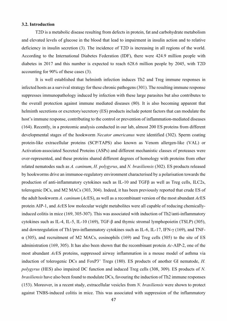
47
3.2. Introduction
T2D is a metabolic disease resulting from defects in protein, fat and carbohydrate metabolism
and elevated levels of glucose in the blood that lead to impairment in insulin action and to relative
deficiency in insulin secretion (3). The incidence of T2D is increasing in all regions of the world.
According to the International Diabetes Federation (IDF), there were 424.9 million people with
diabetes in 2017 and this number is expected to reach 628.6 million people by 2045, with T2D
accounting for 90% of these cases (3).
It is well established that helminth infection induces Th2 and Treg immune responses in
infected hosts as a survival strategy for these chronic pathogens (301). The resulting immune response
suppresses immunopathology induced by infection with these large parasites but also contributes to
the overall protection against immune mediated diseases (80). It is also becoming apparent that
helminth secretions or excretory/secretory (ES) products include potent factors that can modulate the
host’s immune response, contributing to the control or prevention of inflammation-mediated diseases
(164). Recently, in a proteomic analysis conducted in our lab, almost 200 ES proteins from different
developmental stages of the hookworm Necator americanus were identified (302). Sperm coating
protein-like extracellular proteins (SCP/TAPS) also known as Venom allergen-like (VAL) or
Activation-associated Secreted Proteins (ASPs) and different mechanistic classes of proteases were
over-represented, and these proteins shared different degrees of homology with proteins from other
related nematodes such as A. caninum, H. polygyrus, and N. brasiliensis (302). ES products released
by hookworms drive an immunoe-regulatory environment characterised by a polarisation towards the
production of anti-inflammatory cytokines such as IL-10 and TGFβ as well as Treg cells, ILC2s,
tolerogenic DCs, and M2 MACs (303, 304). Indeed, it has been previously reported that crude ES of
the adult hookworm A. caninum (AcES), as well as a recombinant version of the most abundant AcES
protein AIP-1, and AcES low molecular weight metabolites were all capable of reducing chemically-
induced colitis in mice (169, 305-307). This was associated with induction of Th2/anti-inflammatory
cytokines such as IL-4, IL-5, IL-10 (169), TGF-β and thymic stromal lymphopoietin (TSLP) (305),
and downregulation of Th1/pro-inflammatory cytokines such as IL-6, IL-17, IFN-γ (169), and TNF-
α (305), and recruitment of M2 MACs, eosinophils (169) and Treg cells (305) to the site of ES
administration (169, 305). It has also been shown that the recombinant protein Ac-AIP-2, one of the
most abundant AcES proteins, suppressed airway inflammation in a mouse model of asthma via
induction of tolerogenic DCs and FoxP3+ Tregs (180). ES products of another GI nematode, H.
polygyrus (HES) also impaired DC function and induced Treg cells (308, 309). ES products of N.
brasiliensis have also been found to modulate DCs, favouring the induction of Th2 immune responses
(153). Moreover, in a recent study, extracellular vesicles from N. brasiliensis were shown to protect
against TNBS-induced colitis in mice. This was associated with suppression of the inflammatory

48
cytokines IL-6, IL-1β, IFNγ and IL-17a and an increase in the anti-inflammatory cytokine IL-10
(163). Furthermore, ES products from adult N. brasiliensis (171), and recombinant forms of the
cysteine protease inhibitors Nippocystatin (rNB-Cys) from N. brasiliensis and (rHp-CPI) from H.
polygyrus suppressed antigen-specific antibody production (172) and IFNγ production (310), and
simultaneously induced ES-specific Th2 immune responses (171) in ovalbumin-immunised mice
(171, 172, 310). It has also been revealed that the filarial secreted protein ES-62 inhibited pro-
inflammatory Th1 cytokines (i.e. TNF-α, IL-6 and IFNγ) and suppressed severity and progression of
collagen-induced arthritis (CIA) in vivo using DBA/1 mice in a murine model of rheumatoid arthritis
(RA), and in vitro using synovial fluid and peripheral blood mononuclear cells (PBMCs) from RA
patients (311). ES-62 administered to mice also induced M2 MACs and IL-10-producing B cells, and
decreased the levels of IL-22 in the kidney that suppressed development of proteinuria and protected
against kidney damage in a mouse model of systemic lupus erythematosus (312).
In the context of diabetes, the ES products from different parasites have been shown to confer
protection against T1D and T2D. For instance, the ES products of F. hepatica, and S. mansoni SEA
prevented T1D in NOD mice via induction of Bregs, Tregs, M2 MACs and DCs, and increased IL-4,
IL-10 and TGF-β levels (184, 185). Moreover, treatment with SEA (138), LNFPIII glycan from SEA
(also found in human breast milk) (188), SEA T2 RNase recombinant protein-derived ω1 (ω1) (313)
or soluble products from Trichuris suis (TsSP) (314) improved insulin sensitivity and suppressed
liver lipogenesis in HF diet-induced obesity in mice. This was associated with increased numbers of
ILC2s, eosinophils and M2 MACs and increased levels of Th2 cytokines and the alarmin IL-33 in
AT and liver (138, 188, 313, 314). In another study, administration of somatic extract from the filarial
nematode L. sigmosoides improved metabolic homeostasis and insulin sensitivity in a murine obesity
model. This was also associated with increase in the eosinophils, M2 MACs, ILC2s, and expression
of Arg-1, Foxp3, Gata3 and IL-10 (137). In a recent study, using a mouse model of T2D, researchers
found that administration of S. japonicum SEA protect against T2D via induction of Tregs and Th2
immune responses, which was characterised by an increase in the frequency of CD25+Foxp3+ T cells,
and the expression of IL-4 and IL-5 in the spleen of the treated groups (315).
In the present study, we describe a role for ES products from the gastrointestinal nematode N.
brasiliensis in modulating the immune response associated with T2D. Intraperitoneal injection of N.
brasiliensis ES resulted in reduced glucose levels and reduced body weight gain in a mouse model of
T2D, which was mediated by activation of systemic and local eosinophils and IL-5 but not IL-4. N.
brasiliensis ES administration also decreased the levels of IL-6 in AT but increased them in the liver.
Together, these data highlight for the first time the importance of N. brasiliensis ES products in
modulating the immune response associated with T2D.

49
3.3. Materials and Methods
3.3.1. Ethics statement
All procedures were approved by the JCU Animal Ethics Committee, ethics application
number A2244.
3.3.2. Animals and diet
Male C57BL/6 wild-type (WT) (JCU Townsville) mice were used. At the age of 5 weeks mice
were separated into 2 main groups: one group was fed a normal control (NC) diet and the other group
was fed a high glycaemic index (HGI) diet (SF03-30; Speciality Feeds, Western Australia) to induce
T2D.
3.3.3. ES preparation and administration
For preparation of the crude L3ES proteins, the N. brasiliensis life cycle was maintained in
our laboratory at James Cook University (Cairns). Briefly, faeces from N. brasiliensis-infected rats
were collected from days 5-9 post-infection. Egg-containing faeces were mixed with an equal amount
of water and charcoal, distributed into Petri dish plates and incubated at 26°C. One week after
incubation, L3 were collected from the faecal/charcoal culture plate, washed three times with PBS,
then three times with PBS supplemented with 5% antibiotic-antimycotic (AA) (Gibco). L3 were then
transferred into a flat bottom 24 well-plate, cultured in serum-free RPMI media (Gibco) supplemented
with 2% AA and 1% D-glucose (Sigma) and incubated at 37°C and 5% CO2 at a density of 500
L3/well. The supernatant was then collected daily from days 1-10, ensuring motility of the worms at
all times. Supernatants were stored at -30°C before protein concentration and lipopolysaccharide
(LPS) content were determined.
For adult ES (AES) preparation, rats were infected with 3,000 L3 N. brasiliensis. At day 6
post-infection rats were euthanized and the small intestine collected and opened in a Petri dish
containing RPMI and incubated at 37°C and 5% CO2 for 2 hours. Adult worms were then collected,
washed three times with PBS followed by three washes with PBS supplemented with 5% AA and one
wash with RPMI media supplemented with 2% AA. 100 worms/well were then transferred into a flat
bottom 24 well-plate and incubated in RPMI media supplemented with 2% AA, 1% D-glucose and
1% Glutamax at 37°C and 5% CO2. After 24h the supernatant was discarded to minimise
contamination with host proteins and replaced with new media. AES was collected at day 2 and
replaced subsequently every day until day 10. Supernatants were stored at -30°C before protein
concentration and lipopolysaccharide (LPS) content were determined .
Amicon ultrafiltration 3 kDa tubes (Thermo scientific) were used for protein concentration
and buffer exchange with PBS. The Pierce BCA protein assay kit (Thermo scientific) was used for
50
quantification of the protein content following the manufacturer’s instructions. The Pierce LAL
Chromogenic Endotoxin Quantification Kit (Thermo Scientific) was used for the quantification of
LPS in AES and L3ES preps. Firstly, AES and the L3ES were incubated with 1% Triton-114 at 4°C
on a rotor (Ratek) overnight with low constant stirring. After that, ES proteins were incubated at 37°C
for 10 minutes using a dry bath incubator (Major Science), followed by centrifugation in a Microfuge
22R centrifuge (Beckman Coulter) at 14,000 g for 5 minutes. The top layer was collected and
prepared for LPS quantification. Briefly, 4 standards were prepared, ranging from 1- 0.1 EU/ml.
Then, 50 μl of the standards and ES samples were added in duplicate to a 96 well plate and incubated
at 37°C and 5% CO2 for 5 minutes Subsequently, 50 μl of Limulus Amebocyte Lysate was added and
the plate was incubated at 37°C for 10 minutes, followed by addition of 50 µl of Chromogenic
Substrate and incubation for 6 minutes at 37°C. Then, 50 μl of the stop solution was added and the
results were read on a BMG Polarstar Omega fluorescence microplate reader at 405 nm. The ES
proteins were suitable for use when the LPS content was below 5 EU/mg of protein.
The ES products were administered to mice intraperitoneally twice weekly from week 6 until
the end of the experiment at a dose of 1 mg/kg. Control groups were administered intraperitoneally
with the same volume of PBS.
3.3.4. Fasting Blood Glucose (FBG) and Oral Glucose Tolerance Test (OGTT)
The FBG was measured in 6-hour unfed mice. Blood sampling was performed by tail bleeding
using Accu-Check® Performa (Roche). Mice were considered diabetic when glucose levels reached
> 12.0 mmol/L. The OGTT was measured in 6-hour unfed mice, after initial blood collection (time
0). Mice were administered D-glucose orally (2 g/kg body weight) by gavage. Blood sampling was
performed by tail bleeding at 15, 30, 60, 90, and 120 minutes after administration of glucose.
3.3.5. Tissue collection and cytokine analysis
The AT, liver and small intestine were collected and washed with PBS; one cm of the tissue
was combined with 0.5 mL of PBS, homogenised using a TissueLyzer (QIAGEN), centrifuged and
supernatants stored at -80°C until use. The ELISA Ready-Set-Go set (eBioscience) for the cytokines
IL-4, IL-5, IL-6 was used following the manufacturer’s instructions of standard sandwich ELISA
protocols. In brief, a 96-well flat bottom plate was coated with 50 μl of capture antibody and incubated
overnight at 4°C. The plate was then washed 3 times with washing buffer. Then, 50 μl of blocking
reagent was added and the plate was incubated for 1 hour at room temperature. The washing step was
performed twice. 50 μl from the samples and from serial dilution of the standards were added to the
plate and incubated overnight at 4°C, followed by 3 more washing steps. Subsequently, 50 μl of
detection antibody was added, incubated for 30 minutes at room temperature and followed by 3 more
washes. Then, 50 μl of substrate solution was added and incubated for 15 minutes at room

51
temperature. Finally, 50 µl of stop solution (1M HCl) was added and the absorbance was read on a
BMG Polarstar Omega at 450 nm. The lower detection limit for all cytokines was ≥ 6 pg/ml.
3.3.6. Isolation of the mesenteric lymph nodes, adipose tissue and liver for flow cytometry
analysis
In brief, epididymal fat pads or liver from male mice fed a NC or HGI diet were removed and
minced into small pieces, then transferred to 50 ml conical tubes containing 1 ml DPBS (0.5% BSA)
(Sigma) and 3 ml collagenase type II (Life Technologies), and incubated on a rotational shaker (200
rpm) at 37°C for 35 minutes The AT or liver homogenates were filtered through a 70 μm strainer
into a new tube and centrifuged at 500 g for 10 minutes at 4°C. Following centrifugation, the
supernatant was discarded and the pellet was resuspended in 1× red blood cell lysis buffer (Sigma),
followed by a washing step with 5 ml FACS buffer and centrifugation at 500 g for 10 minutes at 4°C
(316, 317). MLNs were removed and filtered through a 70 μm strainer. Cell viability was assessed
by Trypan Blue and cells were blocked for FACS analysis. Cell surface marker analysis was
performed using flow cytometry. Single-cell suspensions prepared from MLNs, AT and liver were
collected from mice at the times indicated. Cell surface markers were stained for 30 minutes at 4°C
with CD3/CD19-CF594 (Clone:145-2C11,1D3) F4/80-APC (Clone: T45-2342), CD11c-FITC
(Clone: HL3), CD301-pecy7 (Clone: LOM-14), CD64-PerCp-Cy5.5 (Clone: X45-5/7.1), CD11b-
BV650 (Clone: M1/70), Ly6G-efluor700 (Clone: 1A8) and Siglec-F-PE (Clone: E50-2440) (BD
Bioscience). All antibody incubations were performed at 4°C for 30 minutes (isotype controls were
included). Data were acquired using a BD FACS Aria with BD FACS DIVA software (BD
Bioscience) and analysed using FlowJo software (Tree Star, Inc).
3.3.7. Data analysis
Data were tested for statistical significance using GraphPad Prism software (version 6). The
Mann-Whitney U test was applied to test differences between two unpaired groups with
nonparametric distribution for statistical significance. Data that were normally distributed were tested
for statistical significance using the unpaired t test for comparisons of two groups or the ANOVA test
followed by the Holm-Sidack multiple-comparison test to compare more than two groups. Values of
p < 0.05 were considered statistically significant. Results are expressed as mean with SEM or means
± SD. *p < 0.05; **p < 0.01.

52
3.4. Results
3.4.1. Treatment with either AES or L3ES from N. brasiliensis improves glucose tolerance and
attenuates body weight gain
In order to address the prophylactic effect of N. brasiliensis ES on the outcome of T2D, 5
week-old male C57BL/6 mice were used; half the mice were fed NC diet and the other half were fed
a HGI diet to induce T2D for up to 30 weeks. Mice were treated twice weekly with N. brasiliensis
AES or L3ES at a dose of 1 mg/kg starting at week 6 until the end of the experiment. Mice treated
with AES or L3ES showed a significant decrease in the level of FBG compared to the control groups
(Fig. 3.4.1A). Moreover, results from the OGTT test showed that NC and HGI diet groups treated
with AES or L3ES had significantly lower levels of blood glucose at all time points (Fig. 3.4.1B and
C). Reduction in body weight gain was also observed as a result of the treatment. Mice on either NC
or HGI diet treated with AES or L3ES gained less weight compared to control groups (Fig. 3.4.1D
and E).
53
Age (weeks)
E
A
9
0
15
12
6
3
FBG
Naive
AES
NC HGI
L3ES
B30
25
20
15
10
5
00 15 30 60 12090
Time (min.)
NC OGTT
30
25
20
15
10
5
00 15 30 60 12090
HGI OGTT
Bloo
d gl
ucos
e (m
mol
/L)
C
Time (min.)
5550454035302520
15252321191715131197531
DNC
Body
wei
ght
27
Naive
AES
L3ES
Age (weeks)
5550454035302520
15252321191715131197531
HGI
Body
wei
ght
27
Naive
AES
L3ES
*
Naive
AES
L3ES
Naive
AES
L3ES
**
**
**
*
ns
**
**
****
**ns
**
****
**
ns**
**ns
ns
**
**ns
ns**
*
*
*
Bloo
d gl
ucos
e (m
mol
/L)
Bloo
d gl
ucos
e (m
mol
/L)
Fig 3.4.1. Treatment with either AES or L3ES from N. brasiliensis improves glucose tolerance and attenuates body weight gain.
C57/BL6 mice were fed NC or HGI diet and treated frequently with L3ES or AES of N. brasiliensis. (A) FBG; (B) OGTT, ND; (C) OGTT, HGI; (D) Weight, NC; (E) Weight, HGI. Statistical significance was determined with Student’s t test or Tow-way analysis of variance (ANOVA). Data are expressed as mean ± SEM or as means ± SD and are representative of 1 experiment where n = 10/group. *p < 0.05, **p < 0.01.

54
3.4.2. Treatment with N. brasiliensis ES induces tissues eosinophilia and Th2 immune responses
Mice were fed an HGI diet and treated with AES or L3ES of N. brasiliensis as described
above. At the end of the experiment, various tissues were collected such as MLNs, AT and liver for
further characterisation of the Th2 immune response. Interestingly, in response to N. brasiliensis AES
or L3ES injection, there was a significant increase in the total number of eosinophils in the mLNs,
AT and liver of the HGI and NC diet groups compared to the control group (Fig. 3.4.2A).
To assess whether the increase in the eosinophils numbers induced a potent Th2 cytokine response
by elevating cytokine levels in AT and liver we analysed the levels of several cytokines by ELISA.
Injection of AES or L3ES significantly increased the levels of Th2 cytokines, measured by IL-5 in
AT and liver (Fig. 3.4.2B and C) compared to the naïve groups. However, our study showed no
significant differences in the levels of IL-4 between treated and un-treated groups (Fig. 3.4.2B, and
C). Moreover, groups treated with either AES or L3ES had significantly lower levels of IL-6 in the
AT (Fig. 3.4.2B) while this level was higher in the liver of mice treated with AES (Fig. 3.4.2C).

55
Fig 3.4.2. Treatment with N. brasiliensis ES induces tissues eosinophilia and Th2 immune responses.
C57/BL6 mice were fed normal control (NC) or high glycaemic index (HGI) diet and treated twice weekly with L3ES or AES of N. brasiliensis. (A) Eosinophil frequency and total numbers in mLNs, AT and Liver. (B) AT IL-4, IL-5 and IL-6 expression. (C) Liver IL-4, IL-5 and IL-6 expression. Statistical significance was determined with Student’s t test. Data are expressed as mean ± SEM and are representative of 1 experiment where n = 10/group. *p < 0.05, **p < 0.01.
Naive
L3ES
NC HGI
ATEosinophils(x104)/mlA
MLNEosinophils(x104)/ml
AES15
10
5
0
**
40
30
20
10
0NC HGI
**
*
**
**
LiverEosinophils(x104)/ml
ND HGI
4
3
2
1
0
*
**
****
**
**
B
C
ATNaiveAESL3ES
NC HGI
80
60
40
0
20
AT60
40
20
0NC HGI
AT
ND HGI
50
3020100
40
LiverNaiveAESL3ES
NC HGI
60
40
0
20
Liver60
40
20
0NC HGI
Liver60
40
20
0NC HGI
***
*
**
nsns
nsns
****
** **
nsns
nsns
****
**
***ns
*ns
IL-4
pg/
ml
IL-5
pg/
ml
IL-6
pg/
ml
IL-4
pg/
ml
IL-5
pg/
ml
IL-6
pg/
ml

56
3.5. Discussion
Incidence and prevalence of diabetes is on the rise globally, imposing a drastic socio-
economic burden on public health (3). Many researchers have reported that helminths and their
secretions can modulate the host’s immune response, which might have beneficial effects in
controlling or preventing the inflammatory responses associated with immune-mediated diseases
(164). Understanding the mechanisms by which helminth ES products can skew the immune response
away from a T2D-promoting phenotype is critical for the rational design of preventative therapies
against this disease. It has been found that N. brasiliensis and its secreted products are able to induce
a Th2 immune response. This was characterised by an increase in a systemic and localised
eosinophilia, ILC2s, M2 MACs, DCs, and increased production of cytokines such as IL‐4, IL‐5, IL‐
9, IL‐10 and IL‐13 (89, 153, 154, 280). In addition, it has been previously reported that administration
of SEA from S. mansoni or S. japonicum (138, 185, 315) or LsAg from the adult filarial nematode L.
sigmodontis (137) protect against T1D and T2D in mice (185, 315) and improved insulin sensitivity
in obese mice (137, 138).
In the present study, we showed that administration of N. brasiliensis crude ES from adult
and L3 developmental stages attenuated the clinical indicators of T2D in the HGI diet mouse model.
Our data showed that AES and L3ES administration ameliorates glucose intolerance and attenuates
body weight gain in the treated groups. Previous findings using SEA from S. japonicum significantly
reduced the level of blood sugar, however the level was still higher than those on a normal diet (315).
This was consistent with our results, as we also found that treatment with AES or L3ES of N.
brasiliensis significantly reduced the blood glucose level in the diabetic group, although they were
still higher than those mice fed a normal chow diet.
Our results showed non-significant differences in the levels of IL-4 in the liver and adipose
tissue of treated groups compared to their naïve counterparts; however we found a significant increase
in the number of eosinophils in the MLNs, AT and liver with a significant increase in the level of IL-
5 in the AT and liver. IL-5 is responsible for the release, expansion and survival of eosinophils (318-
320), and N. brasiliensis infection is characterised by high eosinophil production induced by IL-5
secreted from CD4+ T cells (285). Importantly, it has been demonstrated that AT eosinophils are
highly dependent on IL-5 (41) and eosinophils play an unexpected role in metabolic homeostasis
through maintenance of adipose M2 MACs (37). As with eosinophil deficiency, IL-5 deficiency in
mice impairs eosinophil accumulation in AT, and mice develop increased body fat, impaired glucose
tolerance and insulin resistance when fed a HF diet (41). Moreover, ILC2s have been found to play
an important role in sustaining AT eosinophils (41). Infection with N. brasiliensis induces adipose
eosinophilia and enhances glucose tolerance in mice fed with a HF diet via activation of AT ILC2s,
which induces IL-5 production that leads to accumulation of eosinophils in the AT (41). Deletion of

57
IL-5 resulted in the loss of eosinophil activation in AT (41) and impaired glucose tolerance (37). It
has been reported that N. brasiliensis infection protects against experimental autoimmune
encephalomyelitis (EAE) via increased IL-5 levels and subsequent increases in eosinophils and
CD4+CD25+ Tregs. Moreover, anti-IL-5 or anti-CD25, but not anti-IL-4, can abolish the beneficial
effect of the parasitic infection (321). Furthermore, treatment of mice with F. hepatica ES products
attenuates EAE independently of IL-4 and IL-10, but via production of IL-33 and IL-5, which
promoted accumulation of eosinophils (322).
In our study, treatment with AES or L3ES of N. brasiliensis significantly decreased the
expression of IL-6 in AT. However, in the liver IL-6 expression was comparable between treated and
untreated groups. In one study, treatment of intestinal pig epithelial cells in vitro with T. suis ES
products increased the level of IL-6 and IL-10, but not IL-4 in both differentiated and undifferentiated
cells (323). IL-6 is a multifaceted cytokine that is involved in the induction of insulin resistance and
pathogenesis of T2D (324), and secretion of IL6 by AT resulted in the induction of hepatic insulin
resistance (325). On the other hand, IL-6 is involved in liver regeneration and maintenance of liver
tissue homeostasis (326). Moreover, IL-6 is required for protection against hepatic inflammation and
insulin resistance, and lack of IL-6 reversed the effect (327, 328). Short term preincubation with IL-
6 of human PBMC stimulated with anti-CD3 and anti-CD28 antibodies led to up-regulation of IL-4
and IL-5. However, the effect on IL-4 production gradually disappeared but the effect on IL-5 became
more pronounced in the long term preincubation of these cells with IL-6 (329). Moreover, IL-6
enhanced expansion and survival of antigen-stimulated CD4+ T cells and reduced their apoptosis in
the blood, lymph nodes, spleen, liver, and lung in vivo and in vitro (330). Furthermore, in the presence
of eosinophil-derived neurotoxin (EDN), splenocytes of TLR2+/+ mice immunized with OVA
produced IL-5, IL-6, IL-10 and IL-13, whereas in the presence of LPS they produced IFN-γ (331).
One possibility for the increase in liver IL-6 we observed might be the effect of IL-6 as a survival
factor in liver tissue-mediated EDN in activation of TLR2 and expansion and survival of CD4+ T
cells in the liver.
Here we found that treatment with L3ES or AES from N. brasiliensis using a dose of 1 mg/kg
twice weekly significantly decreased the level of blood glucose in diabetic mice, although levels were
still higher than control group. We also found a significant increase in the number of eosinophils in
the MLNs, AT and liver associated with an increase in the levels of IL-5 in AT and liver but no
differences in the levels of IL-4 in the same tissues. Moreover, a decrease in the level of IL-6 was
observed in AT whereas it was increased in the liver. It must be pointed out, that N. brasiliensis ES
products induced airway eosinophilia in a dose-dependent manner (156). Moreover, we (data not
published) and others (37, 41, 136) have found a robust increase in the systemic and peripheral
eosinophil number, IL-4 level and IL-5 level after infection with N. brasiliensis. Infection with N.

58
brasiliensis requires parasite development through different larval stages and migration through
different tissues, which may induce distinct mechanisms of modulating the host immune response
that differ from the administration of its ES proteins. This might explain the differences in the ability
of L3ES and AES to suppress T2D. So, it is worth considering the route of infection and the
therapeutic/prophylactic dose of ES proteins. As previously mentioned, ES products induce a variety
of immune responses that promote innate and adaptive immune cells, and these cells play very
different roles in inducing or protecting against metabolic diseases. Understanding the precise
immunological mechanism of action that underpins the clinically beneficial effect is now paramount.
To our knowledge, this study is the first to uncover a novel effector response of helminth ES product-
mediated reduction of insulin resistance in a mouse model of T2D. Future work should focus on
defining the bioactive ES components and their recombinant or synthetic production and validation.
Acknowledgments
This work was supported by a program grant from the National Health and Medical Research
Council (NHMRC) [program grant number 1132975] and The Australian Institute of Tropical Health
and Medicine (AITHM) [program grant number SRI40200003]. The funders had no role in study
design, data collection and analysis, decision to publish, or preparation of the manuscript.

59
CHAPTER 4

60
4. Role of helminths and their excretory/secretory products on the composition of the gut
microbiota in type 2 diabetic mice
Zainab Aghaa, Rafid Alhallafa, Ramon Eichenbergerc, Matt Fielda, Lutz Kraused, Javier Sotillob, Alex
Loukasa# aAustralian Institute of Tropical Health and Medicine, James Cook University, Smithfield, QLD,
4878, Australia bCentro Nacional de Microbiología, Instituto de Salud Carlos III, Majadahonda, Madrid, Spain cInstitute of parasitology, University of Zurich d Microba, Brisbane, QLD, 4000, Australia
#Corresponding author:
Prof. Alex Loukas. Centre for Biodiscovery and Molecular Development of Therapeutics,
James Cook University, Cairns, 4878, Queensland, Australia. Email: [email protected]

61
4.1 Abstract
T2D is a highly prevalent metabolic disorder characterised by an imbalance in the blood
glucose level. Dysbiosis of the gut microbiome may reshape intestinal barrier functions, host
metabolic and signalling pathways, which are directly or indirectly related to insulin resistance in
T2D. Recently, a growing body of evidence has highlighted a role for helminth infection and/or their
ES products in modulating the composition of the gut microbiota, which in turn might maintain and
promote intestinal health. We demonstrated that infection with N. brasiliensis or treatment with L3ES
or AES were associated with significant compositional changes in the gut microbiota that occurred
at both the phylum and order level. Phyla that were particularly affected included Actinobacteria,
Verrucomicrobia, Proteobacteria and TM7. Orders that were most impacted by infection or treatment
with ES products included Clostridiales, Desulfovibrionales, Burkholderiales, Verrucomicrobiales
and Coriobacteriales. The effects on microbiota composition were also impacted by diet. However,
whether these microbiota changes are a necessary component for helminth- or -ES product- mediated
protection against T2D is unclear and has yet to be investigated. These findings indicate that
gastrointestinal nematode ES products hold promise as a novel intervention approach for the
treatment of T2D.
Keywords: Type 2 diabetes, Microbiota, Nippostrongylus brasiliensis, Excretory/Secretory,
Clostridiales
62
4.2. Introduction The gut microbiota is not only essential for host metabolism but is also important in the
maintenance of immune homeostasis (275, 332). The immune system has largely evolved as a means
to maintain a symbiotic relationship with these highly diverse and evolving microbes. When operating
optimally, the immune system-microbiota alliance permits the induction of protective responses to
pathogens, and the maintenance of regulatory pathways that govern tolerance to innocuous antigens
(241). Indeed, a decrease in our exposure to infectious agents such as helminths, and alteration of the
gut microbiota composition has resulted in an enhanced inflammatory environment and poorly
developed regulatory networks, and has been strongly associated with the increased incidence of
immune mediated diseases (333). There is cumulative evidence highlighting the beneficial role of gut
microbiota in shaping the mammalian immune system (212).
Disturbance of the intestinal microbial community leads to altered immune responses that can
result in various inflammatory disorders (212). Regulation of metabolic homeostasis and
inflammation in obesity, metabolic syndrome and DM has been increasingly connected with the gut
microbiota (226). The first evidence came from studies on GF mice, as they were resistant to obesity-
associated insulin resistance and dyslipidemia when fed a Western HF or HGI diet; however,
colonization of these mice with microbiota from conventionally raised mice led to an increase in the
total body fat, increased lipids in the liver and a dramatic increase in IR (227, 334, 335). Many studies
have demonstrated differences in the composition of gut microbiota between healthy, obese and T2D
patients. The ratio of Firmicutes to Bacteroidetes found to be changed with obesity, favoring increase
in Bacteroidetes in some reports (336) or decrease in its ratio in others (337) and increase in the
Firmicutes (338). Moreover, T2 diabetic subjects had significantly lower proportions of the phylum
Firmicutes and class Clostridia compared to control group. Also the ratio of Bacteroidetes to
Firmicutes, as well as the ratio of Bacteroides-Prevotella to Clostridium coccoides-Eubacterium
rectale were correlated positively and significantly with the plasma glucose concentration (236).
Other metagenomics association studies of gut microbiota in T2D subjects found a reduction in the
number of Clostridiales bacteria (Roseburia species and Faecalibacterium prausnitzii) compared to
diabetic patients (339, 340).
A growing body of literature has linked helminth presence with microbiota diversity and
composition (242). For instance, infection with the roundworm T. muris led to a reduction in the
microbial α-diversity and the abundance of Bacteroidetes, specifically Prevotella and
Parabacteroides (244), and an increase in the abundance of Proteobacteria (245), Firmicutes (244)
and the Lactobacillus genus belonging to Firmicutes (245). Infection with H. polygyrus increases the
members of γ-Proteobacteria/Enterobacteriaceae group and members of the Bacteroides/Prevotella
genera (247), as well as members of Lactobacillaceae family (249). N. brasiliensis infection reduces

63
the abundance of Firmicutes/Peptostreptococcaceae, Clostridiaceae and Turicibacteraceae and
increases the abundance of Lactobacillaceae, Bacteroidetes/S24-7 and Actinobacteria/
Coriobacteriaceae. (246). Human studies also highlighted the role of helminth infection in modulating
the composition of gut microbiota between helminth-infected and helminth-naive individuals (257,
258, 261). CeD subjects infected with hookworm N. americanus and challenged with gluten showed
tolerance to gluten and had increase in the intestinal microbial species richness and a trend towards
an increase in the abundance of species within the Bacteroidetes phylum in comparison with the
uninfected group (103-105, 258-260). This was also associated with increase in the Th2 and Tregs
cytokines (102, 103). Moreover, increase in the abundance of Paraprevotellaceae, Mollicutes,
Bacteroidales and α-proteobacteria was observed in subjects naturally infected with multiple
helminths (Trichuris spp., Ascaris spp. and hookworm). However, the helminth-negative group
showed increased abundance of Bifidobacterium (261). Furthermore, people infected with S.
stercoralis had a higher abundance of the families Leuconostocaceae, Ruminococcaceae,
Paraprevotellaceae and the genus Peptococcus , while the S. stercoralis-negative group showed a
significant increase in the order Pseudomonadales/Pseudomonas and an unidentified species of the
genus Bacteroides (257).
In mice studies, others have highlighted a role for helminth-microbiota interaction in the
protection against many inflammatory diseases such as allergy (250), IBD (251), arthritis (256),
Lupus (341) and obesity (254, 255). NOD2-/- mice develop spontaneous colitis, and infection of these
mice with T. muris or H. polygyrus inhibited growth of Bacteroides vulgatus and decreased the
abundance of Bacteroidales/Bacteroides and Prevotella and increased the abundance of
Clostridiales/Lachnospiraceae. Administration of a mixture of clusters IV, XIVa and XVIII
Clostridiales and Erysipelotrichales strains to NOD2-/- mice can inhibit colonization of B. vulgatus
(251). Mice infected with H. polygyrus then challenged with HDM, this altered the intestinal bacterial
communities that attenuated allergic airway inflammation (250). Suppression in obesity was also
observed when mice fed HF diet then infected with H. polygyrus (254). This was associated with an
increase in order Clostridiales (250), Bacillus and Escherichia species as well as in the Proteobacteria
phylum (254). Infection of obese mice with S. venezuelensis improved insulin sensitivity and was
associated with an increase in Firmicutes/Lactobacillus spp. and a decrease in Bacteroidetes (255).
Administration of the A. viteae immunomodulator ES-62 maintained a healthy microbiome diversity
and normalised gut microbiota communities, mainly Clostridiaceae, Lachnospiraceae and
Ruminococcaceae, that protect against arthritis (256). The anti-inflamamtory moiety of ES-62 is
phosphorylcholine, and treatment with TPC (synthetic tuftsin-phosphorylcholine) induced remission
of lupus in mice, and was associated with a decrease in the abundance of Akkermansia and an increase

64
in the abundance of several genera, including Turicibacter, Bifidobacterium, unclassified
Mogibacteriaceae, unclassified Clostridiaceae, Adlercreutzia, Allobaculum and Anaeroplasma (341).
There is, therefore, a real need to uncouple this three-way relationship between helminths,
microbiome and inflammatory/metabolic diseases. Thus, we aimed to investigate whether infection
with N. brasiliensis has a role in altering the composition of the gut microbiota, and whether this
alteration might have an impact on maintenance of gut homeostasis in T2D mice.
4.3. Materials and Methods:
4.3.1 Ethics statement:
All procedures were approved by the JCU Animal Ethics Committee, ethics application
number A2244.
4.3.2 Animals and Diet:
Male C57BL/6 wild-type (WT) (JCU Townsville) mice were used. To induce T2D, at the age
of 5 weeks, mice were either fed a HGI diet (SF03-30; speciality feeds, Western Australia) or a HF
diet (SF07-066; speciality feeds, Western Australia) while animals from the control group were fed
a normal control (NC) diet.
4.3.3. Whole worm infection
N. brasiliensis life cycle was maintained in our laboratory at James Cook University (Cairns).
Briefly, faeces from N. brasiliensis-infected rats were collected from days 5-9 post-infection. Egg-
containing faeces were mixed with an equal amount of water and charcoal, distributed into Petri dish
plates and incubated at 26°C. One week after incubation, L3 were collected from the faecal/charcoal
culture plate, washed three times with PBS, then the mice were injected subcutaneously above the
upper thoracic spine with 500 L3 of N. brasiliensis using a 21-gauge needle once a month, starting at
6 weeks of age until the end of the experiment (30 weeks).
4.3.4. Administration of AES and L3ES products
For preparation of the crude L3ES proteins, the N. brasiliensis life cycle was maintained in
our laboratory at James Cook University (Cairns). Briefly, faeces from N. brasiliensis-infected rats
were collected from days 5-9 post-infection. Egg-containing faeces were mixed with an equal amount
of water and charcoal, distributed into Petri dish plates and incubated at 26°C. One week after
incubation, L3 were collected from the faecal/charcoal culture plate, washed three times with PBS,
then three times with PBS supplemented with 5% antibiotic-antimycotic (AA) (Gibco). L3 were then
transferred into a flat bottom 24 well-plate, cultured in serum-free RPMI media (Gibco) supplemented
with 2% AA and 1% D-glucose (Sigma) and incubated at 37°C and 5% CO2 at a density of 500

65
L3/well. The supernatant was collected daily from days 1-10, ensuring motility of the worms at all
times. Supernatants were stored at -30°C before protein concentration and lipopolysaccharide (LPS)
content were determined.
For adult ES (AES) preparation, rats were infected with 3,000 L3 N. brasiliensis. At day 6
post-infection rats were euthanized and the small intestine collected and opened in a Petri dish
containing RPMI and incubated at 37°C and 5% CO2 for 2 hours. Adult worms were then collected,
washed three times with PBS followed by three washes with PBS supplemented with 5% AA and one
wash with RPMI media supplemented with 2% AA. 100 worms/well were then transferred into a flat
bottom 24 well-plate and incubated in RPMI media supplemented with 2% AA, 1% D-glucose and
1% Glutamax at 37°C and 5% CO2. After 24h the supernatant was discarded to minimise
contamination with host proteins and replaced with new media. AES was collected at day 2 and
replaced subsequently every day until day 10. Supernatants were stored at -30°C before protein
concentration and lipopolysaccharide (LPS) content were determined.
Amicon ultrafiltration 3 kDa tubes (Thermo scientific) were used for protein concentration
and buffer exchange with PBS. The Pierce BCA protein assay kit (Thermo scientific) was used for
quantification of the protein content following the manufacturer’s instructions. The Pierce LAL
Chromogenic Endotoxin Quantification Kit (Thermo Scientific) was used for the quantification of
LPS in AES and L3ES preps. Firstly, AES and the L3ES were incubated with 1% Triton-114 at 4°C
on a rotor (Ratek) overnight with low constant stirring. After that, ES proteins were incubated at 37°C
for 10 minutes using a dry bath incubator (Major Science), followed by centrifugation in a Microfuge
22R centrifuge (Beckman Coulter) at 14,000 g for 5 minutes. The top layer was collected and
prepared for LPS quantification. Briefly, 4 standards were prepared, ranging from 1- 0.1 EU/ml.
Then, 50 μl of the standards and ES samples were added in duplicate to a 96 well plate and incubated
at 37°C and 5% CO2 for 5 minutes Subsequently, 50 μl of Limulus Amebocyte Lysate was added and
the plate was incubated at 37°C for 10 minutes, followed by addition of 50 µl of Chromogenic
Substrate and incubation for 6 minutes at 37°C. Then, 50 μl of the stop solution was added and the
results were read on a BMG Polarstar Omega fluorescence microplate reader at 405 nm. The ES
proteins were suitable for use when the LPS content was below 5 EU/mg of protein.
ES products were administered intraperitoneally twice weekly from week 6 until the end of
the experiment at a dose of 1 mg/kg. Control groups were administered intraperitoneally with the
same volume and frequency of PBS.

66
4.3.5. DNA extraction and bacterial 16S rRNA Illumina sequencing
Small intestine samples were collected and stored immediately at -80°C for further analysis.
DNA extraction and 16s rRNA sequencing were performed by the Australian Centre for
Ecogenomics, University of Queensland, Brisbane. In brief, a total of 50-100 mg of tissue sample
was disrupted mechanically using a Powerlyzer 24 at 2,000 g for 5 minutes. A QIAamp 96
PowerFaecal QIAcube HT Kit was used to process the resulting lysate as per the manufacturer’s
instructions, and a Qubit assay (Life Technologies) was used for measuring DNA concentration,
which was then adjusted to a concentration of 5 ng/µl. The 16S rRNA gene was targeted, using the
803 forward primer (5’- TTAGAKACCCB NGTAGTC -3’) and 1392 reverse primer (5’-
ACGGGCGGTGWGTRC -3’) to cover the V6-V8 regions. Preparation of the 16S library was
performed following the protocol outlined in the Illumina guide. In the first stage, 466 bp of the PCR
products were amplified. The resulting PCR amplicons were then purified using Agencourt AMPure
XP beads (Beckman Coulter). The purified DNA was indexed with unique 8cbp barcodes using the
Illumina Nextera XT 384 sample Index Kit A-D (Illumina FC-131-1002). The Qiagen QIAquick Gel
Extraction Kit was used for the isolation of the indexed amplicons as per the manufacturer’s
instructions for the specific band at 450 bp (running at 610 bp with the adaptor sequence). Then, the
resulting purified indexed amplicons were pooled together in equimolar concentrations and
sequenced on a MiSeq Sequencing System (Illumina) using paired end (2 x 300 bp) sequencing with
V3 chemistry in the Australian Centre for Ecogenomics according to the manufacturer’s protocol.
Passing quality control of resulting sequence is determined as 10,000 raw reads per sample prior to
data processing and passing quality control metrics in line with Illumina supplied reagent metrics of
overall Q30 for 600cbp reads of >70%.
4.3.6. Bioinformatics and statistical analysis
Raw reads were run through fastqc for quality control, Trimmomatic (342) for adapter
trimming and low quality base removal, QIIME (343) for Operational Taxonomic Units (OTUs)
generation, and BLAST (344) for OTU identification. Within QIIME, low-quality reads are filtered
with all remaining sequences de-multiplexed and chimeric sequences removed using UCHIME (345).
Sequences were subsequently clustered into OTUs on the basis of similarity to known bacterial
sequences in the Greengenes database (346) (cut-off: 97% sequence similarity) using the UCLUST
software (347).
For each biom file, the taxonomic observation and metadata was added using biom API (348)
which was next loaded into the R package phyloseq (349). Within phyloseq, the DESeq2 (350) API
was called and a list of most differentially expressed bacteria generated for all possible pairings of

67
conditions (NC and NC infected with N. brasiliensis, T2D mice fed HGI and T2D mice fed HGI
infected with N. brasiliensis or T2D mice fed HF and T2D mice fed HF infected with N.
brasiliensis)(NC and NC L3ES or NC AES, T2D fed HGI and T2D fed HGI L3ES or T2D fed HGI
AES). All subsequent plots were generated using ggplot2 and Calypso online software (version 8.84)
(http://cgenome.net/calypso/) (351). Within Calypso, data were normalised by total sum
normalisation (TSS) combined with square root transformation. Multivariate redundancy analysis to
overall differences in the microbial profile between groups and Adonis based on the Bray-Curtis
dissimilarity were used. Differences in bacterial alpha diversity (Shannon diversity) and richness
between groups were used. Values of p < 0.05 were considered statistically significant following false
discovery rate (FDR) correction. Differences in the bacterial taxa abundance between groups were
assessed using ANOVA-like differential expression analysis (ALDEx2) and quantitative
visualisation of phyla abundance.
4.4. Results 4.4.1. Infection with N. brasiliensis resulted in altered microbial profile in normal control and
T2D mice
We wanted to address the effect of infection with N. brasiliensis on the composition of the
gut microbiota in two different models of diet-induced T2D in mice (HGI and HF diet). At week 5 of
age, male C57BL/6 mice were kept on a NC diet or were fed either a HGI or a HF diet for up to 30
weeks to induce T2D. Infection with N. brasiliensis started at week 6 and continue once every month
until the end of the experiment. Small intestine samples were collected at the end of the experiments
to determine the differences in the composition of gut microbiota between infected and uninfected
groups. Multivariate redundancy analysis (RDA) on OTU level showed a different clustering in the
microbial profile of the infected groups compared to the uninfected control groups for all diets.
Adonis analysis also confirmed the differences between infected and uninfected groups on the NC,
HF and HGI diets based on Bray-Curtis and spearman index (Table 1). The α-diversity and microbial
richness were not statistically significant between naïve groups and N. brasiliensis infected groups
for all three diets (Supplementary Fig. 4.4.1).
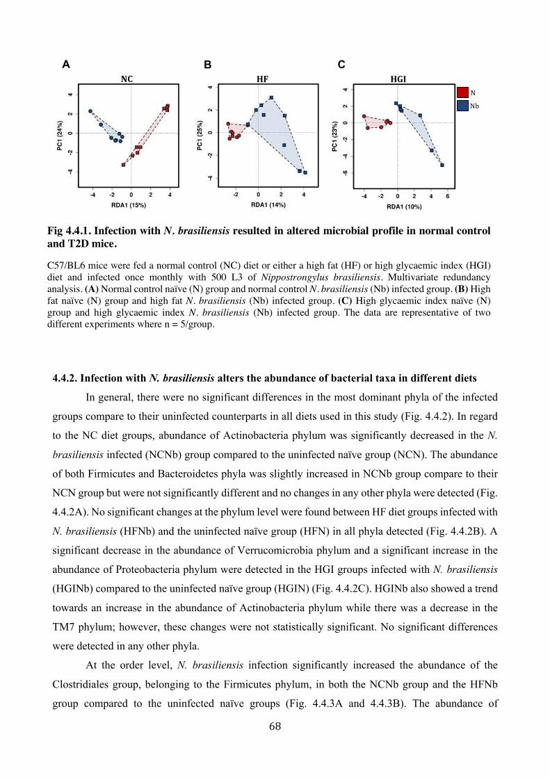
68
4.4.2. Infection with N. brasiliensis alters the abundance of bacterial taxa in different diets
In general, there were no significant differences in the most dominant phyla of the infected
groups compare to their uninfected counterparts in all diets used in this study (Fig. 4.4.2). In regard
to the NC diet groups, abundance of Actinobacteria phylum was significantly decreased in the N.
brasiliensis infected (NCNb) group compared to the uninfected naïve group (NCN). The abundance
of both Firmicutes and Bacteroidetes phyla was slightly increased in NCNb group compare to their
NCN group but were not significantly different and no changes in any other phyla were detected (Fig.
4.4.2A). No significant changes at the phylum level were found between HF diet groups infected with
N. brasiliensis (HFNb) and the uninfected naïve group (HFN) in all phyla detected (Fig. 4.4.2B). A
significant decrease in the abundance of Verrucomicrobia phylum and a significant increase in the
abundance of Proteobacteria phylum were detected in the HGI groups infected with N. brasiliensis
(HGINb) compared to the uninfected naïve group (HGIN) (Fig. 4.4.2C). HGINb also showed a trend
towards an increase in the abundance of Actinobacteria phylum while there was a decrease in the
TM7 phylum; however, these changes were not statistically significant. No significant differences
were detected in any other phyla.
At the order level, N. brasiliensis infection significantly increased the abundance of the
Clostridiales group, belonging to the Firmicutes phylum, in both the NCNb group and the HFNb
group compared to the uninfected naïve groups (Fig. 4.4.3A and 4.4.3B). The abundance of
Fig 4.4.1. Infection with N. brasiliensis resulted in altered microbial profile in normal control and T2D mice.
C57/BL6 mice were fed a normal control (NC) diet or either a high fat (HF) or high glycaemic index (HGI) diet and infected once monthly with 500 L3 of Nippostrongylus brasiliensis. Multivariate redundancy analysis. (A) Normal control naïve (N) group and normal control N. brasiliensis (Nb) infected group. (B) High fat naïve (N) group and high fat N. brasiliensis (Nb) infected group. (C) High glycaemic index naïve (N) group and high glycaemic index N. brasiliensis (Nb) infected group. The data are representative of two different experiments where n = 5/group.
A B
NNb
CNC HF HGI

69
Bifidobacteriales belonging to the Actinobacteria phylum significantly decreased in the NCNb group
compared to the NCN group (Fig. 4.4.3A). N. brasiliensis infection also caused a significant elevation
in the abundance of Desulfovibrionales and Burkholderiales orders belonging to the Proteobacteria
phylum in both the HFNb and HGINb groups, respectively, compared to the uninfected naïve groups
(Fig. 4.4.3B and 4.4.3C). The abundance of both the CW040 order belonging to the TM7 phylum and
the Verrucomicrobiales order belonging to the Verrucomicrobia phylum significantly decreased in
the HGINb group compared to the HGIN group (Fig. 4.4.3C).
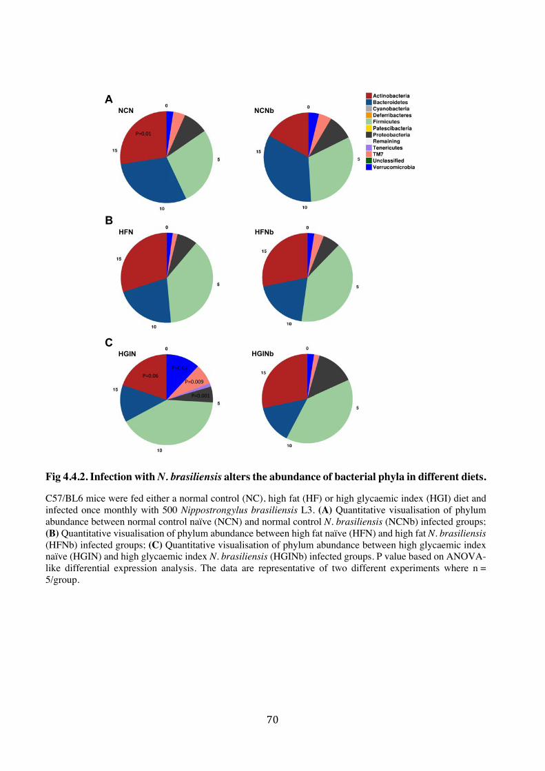
70
A
HFN HFNbB
CHGIN HGINb
NCN NCNb
P=0.01
P=0.02
P=0.001
P=0.009P=0.06
Fig 4.4.2. Infection with N. brasiliensis alters the abundance of bacterial phyla in different diets.
C57/BL6 mice were fed either a normal control (NC), high fat (HF) or high glycaemic index (HGI) diet and infected once monthly with 500 Nippostrongylus brasiliensis L3. (A) Quantitative visualisation of phylum abundance between normal control naïve (NCN) and normal control N. brasiliensis (NCNb) infected groups; (B) Quantitative visualisation of phylum abundance between high fat naïve (HFN) and high fat N. brasiliensis (HFNb) infected groups; (C) Quantitative visualisation of phylum abundance between high glycaemic index naïve (HGIN) and high glycaemic index N. brasiliensis (HGINb) infected groups. P value based on ANOVA-like differential expression analysis. The data are representative of two different experiments where n = 5/group.

71
Fig 4.4.3. Infection with N. brasiliensis alters the abundance of bacterial order in different diets.
C57/BL6 mice were fed normal a control (NC), high fat (HF) or high glycaemic index (HGI) diet and infected once monthly with 500 L3 from Nippostrongylus brasiliensis. (A) Bacterial order abundance between normal control naïve (N) and normal control N. brasiliensis (Nb) infected groups; (B) Bacterial order abundance between high fat naïve (N) and high fat N. brasiliensis (Nb) infected groups; (C) Bacterial order abundance between high glycaemic index naïve (N) and high glycaemic index N. brasiliensis (Nb) infected groups. P value based on ANOVA-like differential expression analysis. The data are representative of two different experiments where n = 5/group. *p < 0.05; **p < 0.01; ***p < 0.00.
A
B
NC
HF HF
CHGI
NC
HGIHGI
Abundancesqrt(TSS)
Abundancesqrt(TSS)
Abundancesqrt(TSS)
Abundancesqrt(TSS)
Abundancesqrt(TSS)
Abundancesqrt(TSS)
Abundancesqrt(TSS)
NNb
**
*
* *
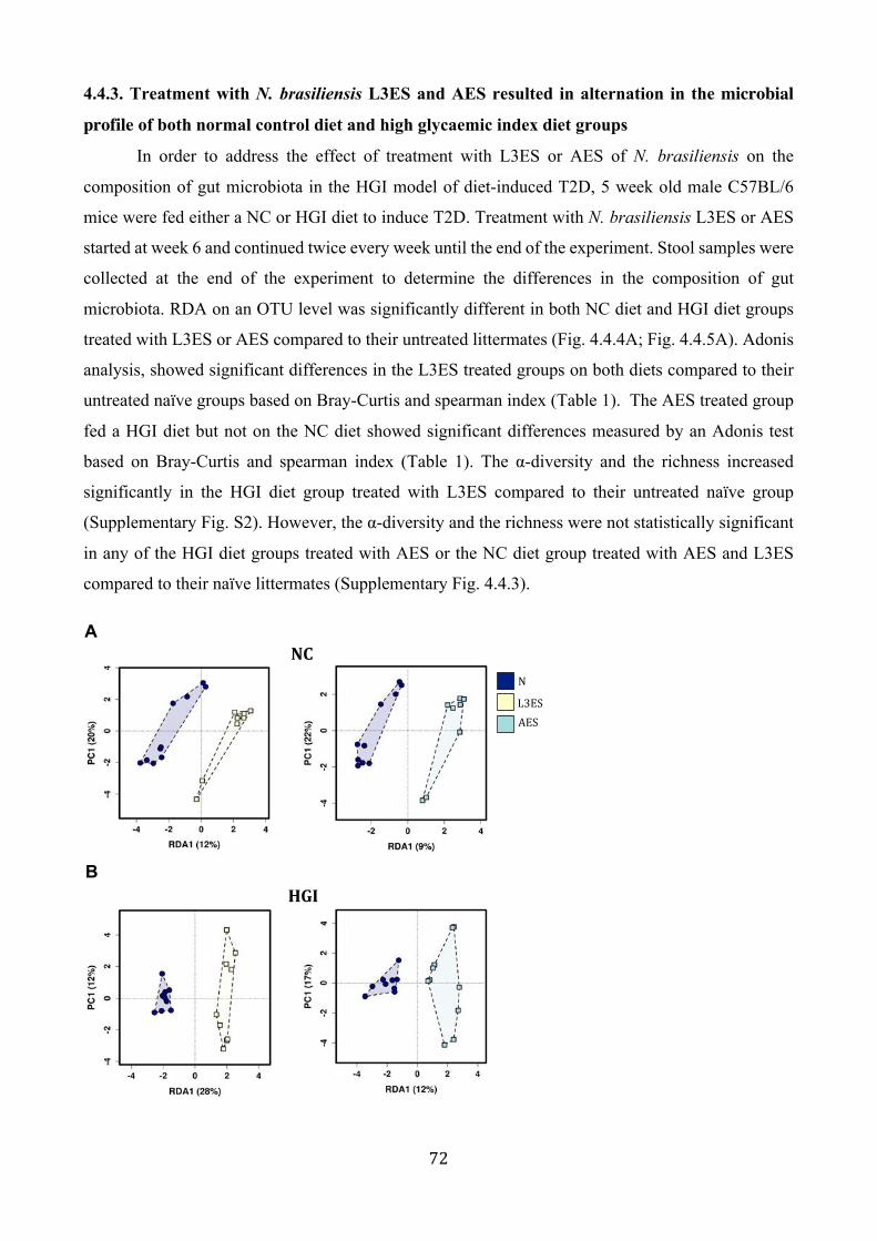
72
4.4.3. Treatment with N. brasiliensis L3ES and AES resulted in alternation in the microbial
profile of both normal control diet and high glycaemic index diet groups
In order to address the effect of treatment with L3ES or AES of N. brasiliensis on the
composition of gut microbiota in the HGI model of diet-induced T2D, 5 week old male C57BL/6
mice were fed either a NC or HGI diet to induce T2D. Treatment with N. brasiliensis L3ES or AES
started at week 6 and continued twice every week until the end of the experiment. Stool samples were
collected at the end of the experiment to determine the differences in the composition of gut
microbiota. RDA on an OTU level was significantly different in both NC diet and HGI diet groups
treated with L3ES or AES compared to their untreated littermates (Fig. 4.4.4A; Fig. 4.4.5A). Adonis
analysis, showed significant differences in the L3ES treated groups on both diets compared to their
untreated naïve groups based on Bray-Curtis and spearman index (Table 1). The AES treated group
fed a HGI diet but not on the NC diet showed significant differences measured by an Adonis test
based on Bray-Curtis and spearman index (Table 1). The α-diversity and the richness increased
significantly in the HGI diet group treated with L3ES compared to their untreated naïve group
(Supplementary Fig. S2). However, the α-diversity and the richness were not statistically significant
in any of the HGI diet groups treated with AES or the NC diet group treated with AES and L3ES
compared to their naïve littermates (Supplementary Fig. 4.4.3).
ANC
BHGI
NL3ESAES

73
4.4.5. Treatment with L3ES or AES of N. brasiliensis alters the abundance of bacterial taxa
L3ES or AES treatment showed no significant changes in the most dominant phyla in
comparison with their untreated littermates in both diets (Fig. 4.4.5). There was only a significant
increase in the abundance of the Verrucomicrobia phylum of the NC diet group treated with L3ES
compared to the naïve un-treated group. No differences in any other phyla were detected (Fig.
4.4.5A). The abundance of the Actinobacteria phylum showed a significant decrease in the HGI diet
groups treated with either L3ES or AES compared to their untreated littermates. Furthermore, the
abundance of the Patescibacteria phylum significantly increased in the AES group fed on a HGI diet
(Fig. 4.4.5B). At the order level, the Coriobacteriales group belonging to Actinobacteria showed a
significant decrease in abundance in the NC and HGI diet groups treated with L3ES, as well as in the
HGI diet group treated with AES (Fig. 4.4.6A and 4.4.6B). Verrucomicrobiales belonging to
Verrucomicrobia significantly increased in the NC and HGI diet groups treated with L3ES or AES,
respectively. However, this order showed a significant decrease in abundance in the HGI diet group
treated with L3ES (Fig. 4.4.6A and 4.4.6B). Desulfovibrionales, Rhodospirillales and
Betaproteobacteriales belonging to the Proteobacteria phylum showed a significant decrease in the
NC diet group treated with L3ES, the NC diet group treated with AES and the HGI diet group treated
with L3ES, respectively, compared to naïve littermates (Fig. 4.4.6A and 4.4.6B). The abundance of
Saccharimonadales belonging to the Patescibacteria phylum and Lactobacillales belonging to the
Firmicutes phylum were significantly higher in the HGI group treated with AES compare to their
untreated littermates (Fig. 4.4.6B).
Fig 4.4.4. Treatment with L3ES or AES of N. brasiliensis resulted in alternation in the microbial profile of both normal control diet and high glycaemic index diet groups.
C57/BL6 mice were fed normal control (NC) or high glycaemic index (HGI) diets and treated twice weekly with L3ES or AES of Nippostrongylus brasiliensis (A) Multivariate redundancy analysis between normal control naïve (N) and normal control N. brasiliensis (L3ES) or (AES) treated groups (B) Multivariate redundancy analysis between high glycaemic naïve (N) and high glycaemic N. brasiliensis (L3ES) or (AES) treated groups. The data are representative of one experiment where n = 10/group.

74
Fig 4.4.5. Treatment with L3ES or AES of N. brasiliensis alters the abundance of bacterial phyla.
C57/BL6 mice were fed normal control (NC) or high glycaemic index (HGI) diets and treated twice weekly with L3ES or AES from Nippostrongylus brasiliensis. (A) Quantitative visualisation of phylum abundance between normal control naïve (N) and normal control N. brasiliensis (L3ES) or (AES) treated groups; (B) Quantitative visualisation of phylum abundance between high glycaemic index naïve (N) and high glycaemic index N. brasiliensis (L3ES) or (AES) treated groups. P value based on ANOVA-like differential expression analysis. The data are representative of one experiment where n = 10/group.
A
BHGI
NC
L3ES AES
N
N
L3ES AES
P=0.009
P=0.002
P=0.001
P=0.0001

75
Fig 4.4.6. Treatment with L3ES or AES of N. brasiliensis alters the abundance of bacterial order.
C57/BL6 mice were fed normal control (NC) or high glycaemic index (HGI) diets and treated twice weekly with L3ES or AES from Nippostrongylus brasiliensis. (A) Bacterial order abundance between normal control naïve (N) and normal control N. brasiliensis (L3ES) or (AES) treated groups; (B) Bacterial order abundance between high glycaemic index naïve (N) and high glycaemic index N. brasiliensis (L3ES) or (AES) treated groups. P value based on ANOVA-like differential expression analysis. The data are representative of one experiment where n = 10/group. *p < 0.05; **p < 0.01; ***p < 0.00.
ANC
Abundancesqrt(TSS)
Abundancesqrt(TSS)
Abundancesqrt(TSS)
Abundancesqrt(TSS)
Abundancesqrt(TSS)
Abundancesqrt(TSS)
Abundancesqrt(TSS)
Abundancesqrt(TSS)
NL3ESAES
Abundancesqrt(TSS)
B
Abundancesqrt(TSS)
Abundancesqrt(TSS)
HGI
*
* *
* * *

76
4.5 Discussion
Changes in environmental factors such as diet can modulate bacterial composition and
metabolic activity (332). These changes can result in a higher intestinal glucose and energy absorption
as well as an increase in the absorption of LPS produced by gut microbiota, which can trigger an
inflammatory immune response leading to the development of T2D (275, 332). Indeed, there is
growing evidence that helminth infection and their ES products alter the composition of the gut
microbial community, conferring protection against immune mediated diseases such as allergic
inflammation (250), CD (251), obesity (254, 255), arthritis (256, 352) and Lupus (341). Here, firstly
we aimed to address the effect of infection with N. brasiliensis on the composition of the gut
microbiota in mice fed with a normal control diet and in two mouse models of diet induced T2D (HF
and HGI). Secondly, we aimed to address the effect of treatment with N. brasiliensis L3ES or AES
on the composition of the gut microbiota in mice fed a normal control diet or a HGI diet that caused
the development of T2D.
No significant changes were observed in α-diversity in response to N. brasiliensis infection
in all diets studied (NC, HF and HGI diet); however, we still found a significant shift in the microbiota
composition at the community level. This was in agreement with other studies with N. brasiliensis
and Hymenolepis diminuta that also found no significant differences in α-diversity between infected
and uninfected groups (246, 253), which suggested inter-individual variation in the microbiota
composition as a result of infection (246). In the latter study, mice on a NC diet infected with N.
brasiliensis showed a significant reduction in the Firmicutes phylum and an increase in
Bacteroidetes/S24-7 and Actinobacteria/Coriobacteriaceae. However, in our study we found that, in
response to infection, the NC group had non-significant increases in both Firmicutes and
Bacteroidetes phyla, while Actinobacteria (in particular the Bifidobacteriales group) abundance was
significantly lower and abundance of order Clostridiales belonging to Firmicutes phylum was
Groups p-value Bray-Curtis p-value spearman indexNCN vs NCNb 0.002 0.0003
HFN vs HFNb 0.0006 0.005
HGIN vs HGINb 0.06 0.03
NCN vs NCL3ES 0.01 0.004
NCN vs NCAES 0.14 0.1
HGIN vs HGIL3ES 0.0003 0.0003
HGIN vs HGIAES 0.0006 0.0003
Table:1 Adonis p-value result based on Bray-Curtis and spearman index

77
significantly higher. While the aforementioned study found that among the Firmicutes phylum
abundance of Clostridiaceae, Peptostreptococcaceae and Turicibacteraceae were significantly
lower, abundance of the Lactobacillaceae was significantly higher in the infected group (246). The
differences between this study and our own study reported herein might be a result of the differences
in the microbiota analysis in terms of 16srRNA amplification and sequencing, as well as differences
in the experimental design in terms of infection strategy and time point for both experiments, as
studies in humans (353, 354) and mice (355) found changes in the composition of gut microbiota
correlating with age. In agreement with our findings, in human studies, individuals with different
helminth infections (i.e. Trichuris spp., Ascaris spp. and hookworm) had lower abundance of the
Bifidobacteriales compared to helminth uninfected individuals (261). Many linked the latter group
with health benefits in T2D (356); however, others have reported them as causing infections (357).
In mouse studies, infection with T. muris or H. polygyrus protected against CD in NOD2-/- deficient
mice (251) and attenuated allergic airway inflammation in mice inoculated with HDM (250), which
was associated with an increase in the abundance of the order Clostridiales (250, 251).
In regard to diabetic groups, infection increased the abundance of the Clostridiales group in
the HF diet group as well as the abundance of Desulfovibrionales and Burkholderiales belonging to
Proteobacteria in the HF and HGI diet groups, respectively. As previously mentioned, in a mouse
study, attenuation of allergic airway inflammation was associated with an increase in the abundance
of Clostridiales (250). It has also been reported that reduction in the abundance of Clostridia was
associated with T2D in humans (236, 339, 340, 358), and an increase in this group was associated
with an improvement in glucose and lipid metabolism (359). In one study, the Proteobacteria phylum
also increased in H. polygyrus-infected mice fed a NC or a HF diet compared to naïve littermates.
This phylum is, in part, responsible for regulating weight gain in HF diet fed mice (254). Abundance
of Desulfovibrionales order, class and family were increased in mouse faecal samples as a result of
infection with Schistosoma heamatobium (360), Ascaris lumbricoides and Trichuris trichiura (361).
Moreover, the abundance of Desulfovibrionales and Burkholderiales groups was lower in obese mice
(362). Cold exposure attenuated diet-induced obesity in mice, which was associated with an increase
in the abundance of Desulfovibrionaceae (363). Diabetic rats treated with insulin had higher relative
abundance of Burkholderiales; however, another study found lower abundance of the
Desulfovibrionales order in diabetic rats when treated with insulin (364). Others have also reported
an increase in the abundance of the Desulfovibrionales order and family in HF compared to NC diet
fed groups (365, 366). Desulfovibrionales are sulphate-reducing bacteria that use hydrogen or other
compounds such as lactate, pyruvate and ethanol as electron donors to produce hydrogen sulphide
(H2S) (367), which is implicated in intestinal disease (368). So, on both sides, many parameters need
to be considered, such as the concentration of sulphide inside the intestine and the ability of IECs to
78
detoxify and to utilise sulphide as an energy source and other mechanisms that might be involved in
the signalling pathway of H2S (368). In our study, the Verrucomicrobiales group belonging to the
Verrucomicrobia phylum and the CW040 group belonging to the TM7 phylum were significantly
lower in the HGI group infected with N. brasiliensis in comparison with uninfected littermates .
Verrucomicrobia and TM7 groups were significantly higher in obese compared to non-obese children
(369), and mice with leptin deficiency (db/db)-induced T2D showed an increase in the abundance of
Verrucomicrobia (370). However, these mice exhibited a decrease in the abundance of
Verrucomicrobia when subjected to an intermittent fasting regime which protected against diabetic
retinopathy (371).
In regard to treatment with N. brasiliensis ES products, while mice on a NC diet treated with
AES or L3ES and those on a HGI diet treated with AES showed non-significant increases in α-
diversity based on Shannon index, those on a HGI diet treated with L3ES had significantly higher α-
diversity. The NC diet group treated with L3ES as well as the HGI diet groups treated with L3ES or
AES had lower abundance of the Actinobacteria/Coriobacteriales group compared to the untreated
controls. The abundance of members of the Coriobacteriaceae family was increased in obese children
and adolescents compare to their normal weight groups (372). Patients with T2D had increased
abundance of Coriobacteriales in comparison with the non-diabetic and pre-diabetic groups (373).
Moreover, T2D patients with cardiovascular complications had higher abundance of the latter group
compare to the diabetic patients without cardiovascular complications (374). Rats fed a fructose-rich
diet had higher Coriobacteriales/Coriobacteriaceae compared to rats fed a control diet (375), however
faecal microbiota transplantation from the latter control group to the high fructose diet group resulted
in decreased Coriobacteriaceae abundance (375). In our study, the Verrucomicrobia/
Verrucomicrobiales group showed a different outcome, as NC diet mice treated with L3ES and HGI
diet mice treated with AES had a higher abundance of this group, however those mice on a HGI diet
treated with L3ES had lower abundance of Verrucomicrobiales than their control groups. In one
study, mice treated with helminth derivative phosphorylcholine TPC had lower abundance of
Verrucomicrobia but higher abundance of Clostridiaceae compared to their control group (341).
Furthermore, as we mentioned previously, in obese people (369) as well as in obese mice, (370)
researchers reported an increase in the abundance of the Verrucomicrobia group. However, another
study found a decrease in the abundance of Verrucomicrobia in both prediabetes and T2D subjects
(376). Verrucomicrobia are mucin degrading bacteria (377) implicated in health and disease. In this
sense, systematic contribution of mucin degrading bacteria in gut homeostasis and dysbiosis has yet
to be investigated (378).
Abundance of Desulfovibrionales, Betaproteobacteriales and Rhodospirillales groups
belonging to proteobacteria was lower in NC diet group treated with L3ES, HGI diet group treated

79
with L3ES and NC diet group treated with AES respectively compare to their untreated littermates.
In a previous study, mice fed with a diet that was rich in fat, fructose and cholesterol had a higher
abundance of Rhodospirillales compared to the control diet fed group (379). Moreover, pre-diabetic
and diabetic subjects had a higher abundance of Betaproteobacteria compared to non-diabetic subjects
(236, 376).
Abundance of Saccharimonadales (belonging to Patescibacteria) and Lactobacillales groups
was significantly higher in the HGI diet group treated with AES compare to their untreated group.
Patescibacteria are anaerobic fermenters, but this newly identified bacterial group has not been well
studied and information on their function is still scarce (380). Lactobacillus on the other hand has
been widely studied, and different Lactobacillus probiotic strains have been reported to improve
parameters related to T2D (381).
Many factors may play a role in modulating the abundance of microbial species in the gut.
The bacterial composition can vary in the same individual due to variation in the physiology, pH and
O2 tension, digesta flow rates in different anatomical regions, as well as between individuals due to
variation in genetic factors, environment and habitual dietary intake (382). These variations can be
factors contributing to the onset of many diseases and may have potential therapeutic implications
(383). Correlation between helminth presence and changes in the microbial composition have already
been mentioned elsewhere throughout, but it is pertinent to note that many studies reported changes
in the composition of the gut microbiota as a result of helminth infection and subsequent improvement
in the outcome of immune mediated diseases. Whether the changes we found in our study are a direct
effect of helminth infection or a consequence of the host’s immune response to the infection, and
whether these changes are essential to confer protection against T2D are yet to be investigated. A
deeper understanding of the interplay between the host-microbiota-helminth triad and other variables
may represent a new therapeutic strategy to reverse the pathological effects of T2D.

80
Supplementary Fig 4.4.1: Infection with N. brasiliensis resulted in altered alpha diversity and microbial richness in normal control and T2D mice.
C57/BL6 mice were fed either a normal control (NC), high fat (HF) or high glycaemic index (HGI) diet and infected once monthly with 500 Nippostrongylus brasiliensis L3. Microbial alpha diversity and richness in the small intestine of N. brasiliensis infected groups (Nb) and uninfected groups (N) in normal control diet (NC) diet, high fat (HF) diet and high glycaemic index (HGI) diet. The data are representative of two different experiments where n = 5/group.
ANC HF HGI
NNb

81
Supplementary Fig 4.4.3: Treatment with N. brasiliensis L3ES and AES resulted in alternation in the alpha diversity and species richness of both normal control diet and high glycaemic index diet groups.
C57/BL6 mice were fed normal control (NC) or high glycaemic index (HGI) diets and treated twice weekly with L3ES or AES of Nippostrongylus brasiliensis. Microbial alpha diversity and richness in the faecal samples of N. brasiliensis (L3ES) and (AES) treated groups and untreated groups (N). (A) In normal control diet (NC). (B) In high glycemic index diet (HGI). The data are representative of one experiment where n = 10/group.
A
NL3ESAES
NC
BHGI

82
Acknowledgments
This work was supported by a program grant from the National Health and Medical Research
Council (NHMRC) [program grant number 1132975] and The Australian Institute of Tropical Health
and Medicine (AITHM) [program grant number SRI40200003]. The funders had no role in study
design, data collection and analysis, decision to publish, or preparation of the manuscript.
83
5. General Discussion
5.1 Overview
DM is widely distributed and considered the fourth leading cause of non-communicable disease
deaths. DM and the associated pathologic sequelae are responsible for about 5.1 million deaths per
year globally and accounted for USD 727 billion of the world’s health expenditure in 2017 (3). It has
been reported that the prevalence of diabetes is higher in urban (279.2 million) than in rural (145.7
million) areas and this gap is predicted to widen to 472.6 million people in urban areas and 156
million in rural areas by 2045, with wide variations in this prevalence worldwide (3). For example,
the Western Pacific region has the highest number of people with diabetes (159 million people),
followed by South-East Asia (82 million), Europe (58 million), and North America (46 million) (3).
Further, it has been shown that T2D occurs in youth, with the highest rates observed in people aged
15–19 (384). Moreover, it has been estimated that 352.1 million people have impaired glucose
tolerance worldwide and nearly half of those are under the age of 50 (3). T2D is characterised by a
persistent elevation in the blood glucose level due to an absolute or relative deficiency in insulin or
resistance to its action (385).
Diabetes is a multifaceted disease where genetics, immunological factors and environmental
factors have a major role in its pathogenesis (386). Appropriate balance between pro-inflammatory
and anti-inflammatory immune responses is essential to maintain whole body homeostasis and to
avoid inflammatory diseases (387). Inflammation is a crucial factor contributing to the development
of T2D (388). In T2D, levels of certain inflammatory molecules are increased, and the components
of the immune system are skewed toward pro-inflammatory subsets (388). It has been proposed that
changed dietary habits, a cleaner environment, vaccination programmes and excessive antibiotic use
have reduced our exposure to many infectious agents and symbiotic microorganisms (including
helminths and gut microbiota) that had an evolutionary relationship with humans, and this
relationship helps in the regulation of immune responses and preservation of homeostasis (389). It
has been suggested that gut microbiota composition has an effect on the pathogenesis of T2D (390).
Lack of exposure to pathogens is thought to increase susceptibility to allergic and autoimmune
diseases like, asthma, IBD and T1D (65). Epidemiological studies as well as experimental studies
have reported an inverse relationship between the presence of helminths and T2D (391). Moreover,
helminth presence has been linked with microbiota diversity and composition which may contribute
to beneficial health effects in the host (392). Hence, alteration of the gut microbiome in the presence
of helminth parasites might contribute to improved metabolic outcomes observed in T2D.

84
Data presented in this dissertation provide evidence that infection with N. brasiliensis and
administration of its ES products from both adult larval stages significantly decreased blood glucose
levels and reduced body weight gain in mice using two different models of diet-induced T2D: HGI,
HF. Specifically, I have shown that N. brasiliensis infection and administration of ES products
resulted in significantly increased systemic eosinophil responses in different organs including MLNs,
AT, and liver. This was associated with an increase in type 2 immune responses measured by IL-4,
Chil3 and Rentla in AT, liver and gut tissue and an increase in the levels of IL-4 and IL-5 in AT and
liver of ES treated groups. I also found that infection with N. brasiliensis or treatment with their ES
products showed a significant difference in the microbial profile of those groups compared to naïve
littermates. Moreover, there were trends towards an increase in the gut microbial diversity and
changes in specific bacterial taxa after helminth infection or treatment with ES products.
5.1.1 N. brasiliensis infection and ES products improve glucose metabolism and regulate the
immune responses associated with T2D.
Helminth infections play a crucial role in the induction of Th2- and Treg-mediated immune
responses in many inflammatory disorders such as IBD, MS, arthritis, asthma and T1D (270).
Similarly, it has been reported that helminth infections showed promising results in patients with
IBD, celiac disease and MS (104, 105, 271). Further, it has been demonstrated that helminths and
their secretions can regulate the host’s immune response, which might have beneficial effects in
promoting or preventing the inflammatory responses associated with immune-mediated diseases
(164).
In our studies, we found that infection with N. brasiliensis or treatment with its ES products
had a beneficial effect against T2D in two different diabetic models. Infection with N. brasiliensis or
treatment with their ES products significantly induced peripheral glucose uptake and specific glucose
sensitivity and reduced body weight gain (see results from Chapter 2 & 3). In line with my studies,
infection with N. brasiliensis decreased weight gain and improved glucose metabolism in two
different models of obese mice, which was associated with induction of Th2 immune responses (136).
Other studies have also reported improvements in glucose tolerance of obese mice after infection with
the filarial helminth L. sigmodontis or administration of soluble adult worm extract which was
associated with an increase in the numbers of CD4+ T cells, eosinophils and AAMs in the AT.
Depletion of eosinophils resulted in impaired glucose tolerance (137). Infection with H. polygyrus
also resulted in decreased body weight gain and improved glucose and lipid metabolism with
increases in Th2/Treg immune responses in the MLNs, AT, liver and gut. (134, 135). SEA from S.
japonicum decreased the level of blood sugar and induced Th2 and Treg immune responses that
protected against T2D in mice (315).
85
5.1.2 Mechanism(s) by which N. brasiliensis and their ES regulate immune responses to T2D.
In the present study I have identified an important role for helminth infection in the induction
of type 2 immune responses by enhancing eosinophil recruitment in MLNs, AT and liver, with
increased expression of IL-4 and markers of M2 MACs Rentla and Chil3 in AT, liver and SI (see
results from Chapter 2). Further, I showed that administration of N. brasiliensis crude ES from adult
and L3 stages also induced eosinophil recruitment in the MLNs, AT and liver, with a significant
increase in the level of IL-5 in the AT and liver (see results from Chapter 3). In line with my results,
N. brasiliensis infectyion resulted in improved insulin sensitivity and lipid metabolism, decrease in
adiposity and increase in energy expenditure in mouse models of obesity (37, 41, 136). Infection with
N. brasiliensis is associated with induction of Th2 immune responses, resulting in the production of
IL‐4, IL‐5, IL‐9, IL‐10, IL‐13, IL-25 and IL-33 and induction of a systemic and localized eosinophilia,
ILC2s and M2 MACs (94, 280-285, 393-395). Infection with N. brasiliensis induces residential
ILC2s that secret and maintain IL-5 leading to local eosinophil accumulation (396). Also, N.
brasiliensis induced ILC2s and CD+4 T cells and expansion of IL-4 and/or IL-13 that maintain M2
MACs (280). N. brasiliensis infection drives signaling via IL-4/IL-13/STAT-6 axis (282, 284), which
has already been reported to be involved in the regulation of lipid metabolism leading to weight loss
in obese mice after N. brasiliensis infection (136).
Eosinophils are involved in the regulation of homeostasis of many tissues including
gastrointestinal tract, lungs, adipose tissue, thymus, uterus and mammary glands (397). Recently,
eosinophils have been implicated in the regulation of glucose metabolism and energy expenditure
(37, 41, 288, 298, 398). IL-5 promotes eosinophil development, activation and survival (397). IL-5
plays an important role in promoting AT eosinophils that maintain adipose AAMs. Eosinophil
accumulation in AT was impaired in IL-5 deficient mice, leading to increased body fat, impaired
glucose tolerance and insulin resistance in obese mice (37, 41). An in vitro study found that IL-4
restored insulin sensitivity in an experimental IR model of 3T3-L1 adipocytes (293). Moreover, IL-4
decreased blood glucose and body weight, which promotes insulin sensitivity and inhibits
accumulation of lipids in AT and reduced the adipocyte layer in the skin in vivo (292, 295). Emerging
studies implicate different immunological pathways in the regulation of glucose metabolism and
energy expenditure. Resident ILC2s in AT increase production of IL-5 and IL-13, which promotes
accumulation of eosinophils and M2 MACs. This reduces inflammation in AT and increases insulin
sensitivity and beiging of AT (41, 399). Another study has highlighted the role of Metrnl in the
recruitment of eosinophils to the AT (398). This induced IL4/IL13, which are required to increase
M2 MAC numbers and induction of AT beiging (298, 398). Moreover, the IL-33/ILC2 axis can
promote beiging in AT and limit adiposity via increasing caloric expenditure that regulates metabolic
homeostasis (290). Signaling via IL-4/ STAT6 regulates nutrient metabolism in the liver and insulin

86
sensitivity and attenuates body weight and AT inflammation (291). Considering these multiple
immunological signaling pathways, a plausible mechanism by which N. brasiliensis improves
glucose metabolism and decreases adiposity might due to local and systemic eosinophilia that leads
to expansion of Th2 cytokines and M2 MACs.
Regarding treatment with N. brasiliensis ES products, I have reported that administration of
AES or L3ES twice weekly at a dose of 1 mg/kg resulted in significantly decreased blood glucose
levels and attenuation of body weight gain. This was associated with an increase in the level of AT
and liver IL-5 and a trend towards an increase in the level of IL-4 in these tissues. Expression of IL-
6 significantly decreased in AT but increased in the liver. As previously discussed, eosinophils play
a role in the increase in levels of IL-5 and/or IL-4 in regulating glucose and lipid metabolism as well
as energy expenditure (37, 41, 399). An in vitro study revealed that treatment of intestinal pig
epithelial cells with T. suis ES products resulted in increased levels of IL-6 and IL-10, with no
secretion of IL-4 in both differentiated and undifferentiated cells (323). Although an increase in the
levels of IL-6 were reported to be involved in the induction of insulin resistance in the AT and liver
and pathogenesis of T2D (324, 325), others have reported a beneficial role for IL-6 in liver
regeneration and maintenance of liver tissue homeostasis as well as protection against liver
inflammation and insulin resistance (326-328). Human PBMC stimulated with a-CD3 and a-CD28
antibodies showed up-regulation in the expression of IL-4 and IL-5 after short term preincubation
with IL-6. However, long term preincubation of these cells with IL-6 led to gradual disappearance of
its effect on IL-4 production but the effect on IL-5 became more pronounced (329). Moreover, IL-6
enhanced in vivo and in vitro expansion and survival of Ag-stimulated CD4+ T cells in the blood,
lymph nodes, spleen, liver and lung, which reduced the level of apoptosis in these cells (330).
Furthermore, splenocytes of TLR2+/+ mice immunised with OVA in the presence of eosinophil-
derived neurotoxin (EDN) produced IL-5, IL-6, IL-10 and IL-13, whereas in the presence of LPS
they produced IFN-γ (331). So, it is more likely that the increase in the levels of IL-6 in the liver that
I observed are a consequence of avoiding apoptosis in this tissue, and which might be mediated via
EDN activating TLR2 and expansion and survival of CD4+ T cells in the liver. The mechanisms by
which IL-6 might enhance a protective effect against T2D has yet to be investigated.
5.1.3 Mechanism(s) by which N. brasiliensis and host microbiota regulate immune responses and
T2D.
The gut microbiota that inhabits the mammalian body plays an essential role in regulating
host metabolism, physiology, nutrition and immune function (400). Changes in environmental factors
such as dietary habits, excessive use of antibiotics, sanitation, mode of birth and feeding pattern all
can have a devastating impact on the composition and diversity of the intestinal microbiota (401).

87
Changes in the composition of the gut microbiota as a result of helminth infections have been reported
by many researchers (244-247, 249, 257, 261). On the other hand, a growing body of evidence
suggests that helminth infections and their ES products can alter the gut microbial community,
leading to protection against immune mediated diseases, such as allergic inflammation, colitis,
obesity, arthritis and lupus (250, 251, 254-256, 341, 352).
My data showed significant differences in the clustering analysis between N. brasiliensis
infected groups compared to naïve groups in all diets (NC, HGI and HF diets). The same result was
found between the groups treated with L3ES or AES and untreated littermates in both NC and HGI
diets. However, no significant differences in α-diversity and richness were detected in the N.
brasiliensis infected groups compared to the uninfected groups, as well as between L3ES or AES
treated groups on a NC diet, or the AES treated group on a HGI diet compared to the untreated groups.
Only mice on a HGI diet and treated with L3ES showed significantly higher α-diversity and richness
when compared with the naïve group; however, I still found significant changes in the community
composition at the phylum and order levels in the infected as well as treated groups compared to
naïve littermates, but each of them showed variability among different diets as well as between the
different treatments (L3ES and AES). Clostridiales increased while Bifidobacteriales decreased in
the NC diet group. Clostridiales and Desulfovibrionales increased in the HF diet group.
Burkholderiales increased, while CW040 and Verrucomicrobiales decreased in the HGI diet group.
Coriobacteriales and Desulfovibrionales decreased, while Verrucomicrobiales increased in the NC
diet group treated with L3ES. Rhodospirillales decreased in the NC diet group treated with AES.
Coriobacteriales, Verrucomicrobiales and Betaproteobacteriales decreased in the HGI group treated
with L3ES. Coriobacteriales decreased while Verrucomicrobiales, Saccharimonadales and
Lactobacillales increased in the HGI diet group treated with AES.
It has been reported that T2D patients had lower abundance of Clostridiales and this group
negatively correlated with glucose and lipid metabolism (236, 339, 340, 358, 359). Moreover, the
microbiota of diabetic group is represented in lower members of Bifidobacterium in comparison with
the healthy group (402). These organisms are reported to be associated with health benefits in T2D
(356); however, a few studies have reported Bifidobacterium to cause infections (357). The
Burkholderiales group was lower in obese mice, and treatment of diabetic rats with insulin increased
the abundance of Burkholderiales (362, 364). I found a conflicting result regarding
Desulfovibrionales and Verrucomicrobiales, although this controversy is also found in the literature.
While some groups have reported a decrease, others have reported an increase in the abundance of
the Desulfovibrionales group when treating obese and diabetic rats with insulin (362, 364, 365).
Furthermore, while some researchers reported an increase in the abundance of the Verrucomicrobia
group in obesity and T2D, others found a decrease in the abundance of this group (369-371, 376). In

88
addition, order CW040 belongs to the TM7 phylum, and this phylum has been found to be higher in
obese children and adolescents (369). A fat, fructose and cholesterol-rich diet has been associated
with a higher abundance of Rhodospirillales (379). Also, a higher abundance of Betaproteobacteriales
has been detected in pre-diabetic and diabetic subjects (236, 376). Saccharimonadales belonging to
the Patescibacteria phylum are anaerobic fermenters and a newly identified bacterial group from
which little is known (380). The effect of many different Lactobacillus probiotic strains has been
reported in their ability to improve parameters related to T2D (381). My result are in line with other
studies conducted in humans and rats that found no changes in α-diversity as a result of infection with
different parasitic nematodes, but still reported some changes in the bacterial communities (253, 403,
404). To explain the variability in the microbiota community composition among different diets as a
result of infection as well as between the different treatments, one might consider the differences in
the dietary substrates. We also should consider the pathways for the metabolism of these substrates
as well as the inter-individual variation in the metabolism they can have and its implication on the
abundance of different microbial species in the gut (405). In my work, the sample size was small,
which allowed greater sampling variability. On the other hand, helminth presence has diverse effects
on the composition of the gut microbiota, with some species having minor effects while others have
greater effect swith potential to protect against immune-mediated diseases. In summary, infection
with N. brasiliensis or treatment with their ES products has no effect on microbial a-diversity;
however, clustering analysis demonstrates the existence of discrete bacterial communities. Many
factors can influence the abundance of microbial species in the gut, such as diet, host physiology and
inflammatory immune responses produced as a consequence of helminth invasion; however, whether
the changes I found in this study are a cause of direct and/or indirect effect of helminth infection or
their ES products or a consequence of host immune response, and whether these changes are essential
to confer protection against T2D are yet to be investigated.
5.2 Conclusion
In conclusion, in this thesis I found that infection with N. brasiliensis or treatment with AES
or L3ES improves glucose metabolism and decreases body weight gain in diet-induced T2D mouse
models. This was associated with an increase in systemic and peripheral eosinophilia along with an
increase in Th2 immune responses, characterised by an increase in IL-4, Rentla and Chil3 in infected
groups and an increase in IL-5 and a trend towards an increase in IL-4 with a decrease in IL-6 in AT
but not in liver of AES and L3ES treated groups. I found no changes in a-diversity after infection or
ES treatment, but did detect a shift in the microbiota at the community level which needs to be further
investigated.

89
5.3 Future perspective
• In my work I demonstrated a role for eosinophils in the improvement of glucose metabolism
and modulating the immune response associated with T2D. Further studies in eosinophil-
depleted mice would be important to elucidate if this pathway is important for the beneficial
effect.
• I reported an involvement of other immune cells including M2 MACs, ILC2s and Tregs, so
these cell types and their roles in protecting against T2D need to be addressed.
• A large-scale study is important to address the role of infection or treatment with ES on the
gut microbiota composition. Also, a microbiota depletion study will also be important to
address whether the gut microbiota is essential for protection against T2D after helminth
infection or treatment with ES products.
• Further work should be aimed at addressing the potential of anti-inflammatory proteins
secreted by N. brasiliensis as a therapeutics modality for T2D and other inflammatory-
mediated diseases.
90
6. References
1. Association AD. Diagnosis and classification of diabetes mellitus. Diabetes care. 2014;37(Supplement 1):S81-S90. 2. Vlad I, Popa AR. Epidemiology of diabetes mellitus: a current review. Romanian Journal of Diabetes Nutrition and Metabolic Diseases. 2012;19(4):433-40. 3. Federation ID. IDF diabetes atlas. Eighth edition ed: International Diabetes Federation; 2017. 4. D'Adamo E, Caprio S. Type 2 diabetes in youth: epidemiology and pathophysiology. Diabetes Care. 2011;34 Suppl 2:S161-5. 5. Prasad RB, Groop L. Genetics of type 2 diabetes-pitfalls and possibilities. Genes (Basel). 2015;6(1):87-123. 6. Li H, Isomaa B, Taskinen MR, Groop L, Tuomi T. Consequences of a family history of type 1 and type 2 diabetes on the phenotype of patients with type 2 diabetes. Diabetes Care. 2000;23(5):589-94. 7. Lyssenko V, Laakso M. Genetic screening for the risk of type 2 diabetes: worthless or valuable? Diabetes Care. 2013;36 Suppl 2:S120-6. 8. Huber A, Menconi F, Corathers S, Jacobson EM, Tomer Y. Joint genetic susceptibility to type 1 diabetes and autoimmune thyroiditis: from epidemiology to mechanisms. Endocr Rev. 2008;29(6):697-725. 9. Bakay M, Pandey R, Hakonarson H. Genes involved in type 1 diabetes: an update. Genes (Basel). 2013;4(3):499-521. 10. Sun X, Yu W, Hu C. Genetics of type 2 diabetes: insights into the pathogenesis and its clinical application. Biomed Res Int. 2014;2014:926713. 11. Hertel JKH, Johansson S, Midthjell K, Nygård O, Njølstad PR, Molven A. Type 2 diabetes genes–Present status and data from Norwegian studies. Norsk epidemiologi. 2013;23(1). 12. Billings LK, Florez JC. The genetics of type 2 diabetes: what have we learned from GWAS? Ann N Y Acad Sci. 2010;1212:59-77. 13. Mitra A, Dewanjee D, Dey B. Mechanistic studies of lifestyle interventions in type 2 diabetes. World J Diabetes. 2012;3(12):201-7. 14. Despres JP. Body fat distribution and risk of cardiovascular disease: an update. Circulation. 2012;126(10):1301-13. 15. Hu FB. Globalization of diabetes: the role of diet, lifestyle, and genes. Diabetes Care. 2011;34(6):1249-57. 16. Carter P, Khunti K, Davies MJ. Dietary Recommendations for the Prevention of Type 2 diabetes: What Are They Based on? J Nutr Metab. 2012;2012:847202. 17. Pickup JC. Inflammation and activated innate immunity in the pathogenesis of type 2 diabetes. Diabetes Care. 2004;27(3):813-23. 18. Pedicino D, Liuzzo G, Trotta F, Giglio AF, Giubilato S, Martini F, et al. Adaptive immunity, inflammation, and cardiovascular complications in type 1 and type 2 diabetes mellitus. J Diabetes Res. 2013;2013:184258. 19. Odegaard JI, Chawla A. Connecting type 1 and type 2 diabetes through innate immunity. Cold Spring Harb Perspect Med. 2012;2(3):a007724. 20. Pirot P, Cardozo AK, Eizirik DL. Mediators and mechanisms of pancreatic beta-cell death in type 1 diabetes. Arq Bras Endocrinol Metabol. 2008;52(2):156-65. 21. Morran MP, Omenn GS, Pietropaolo M. Immunology and genetics of type 1 diabetes. Mt Sinai J Med. 2008;75(4):314-27.

91
22. Eller K, Kirsch A, Wolf AM, Sopper S, Tagwerker A, Stanzl U, et al. Potential role of regulatory T cells in reversing obesity-linked insulin resistance and diabetic nephropathy. Diabetes. 2011;60(11):2954-62. 23. Osborn O, Olefsky JM. The cellular and signaling networks linking the immune system and metabolism in disease. Nat Med. 2012;18(3):363-74. 24. Roszer T. Understanding the Mysterious M2 Macrophage through Activation Markers and Effector Mechanisms. Mediators Inflamm. 2015;2015:816460. 25. Morris DL. Minireview: Emerging Concepts in Islet Macrophage Biology in Type 2 Diabetes. Mol Endocrinol. 2015;29(7):946-62. 26. Thomsen L, Rosendahl A. Polarization of macrophages in metabolic diseases. J Clin Cellular Immunol. 2015;6:313. 27. Lee BC, Lee J. Cellular and molecular players in adipose tissue inflammation in the development of obesity-induced insulin resistance. Biochimica et biophysica acta. 2014;1842(3):446-62. 28. Espinoza-Jimenez A, Peon AN, Terrazas LI. Alternatively activated macrophages in types 1 and 2 diabetes. Mediators Inflamm. 2012;2012:815953. 29. Samaan MC. The macrophage at the intersection of immunity and metabolism in obesity. Diabetol Metab Syndr. 2011;3(1):29. 30. Kraakman MJ, Murphy AJ, Jandeleit-Dahm K, Kammoun HL. Macrophage polarization in obesity and type 2 diabetes: weighing down our understanding of macrophage function? Front Immunol. 2014;5:470. 31. Eguchi K, Manabe I. Macrophages and islet inflammation in type 2 diabetes. Diabetes Obes Metab. 2013;15 Suppl 3:152-8. 32. Samaan MC. The macrophage at the intersection of immunity and metabolism in obesity. Diabetology & metabolic syndrome. 2011;3:29. 33. Zhu L, Su T, Xu M, Xu Y, Li M, Wang T, et al. Eosinophil inversely associates with type 2 diabetes and insulin resistance in Chinese adults. PLoS One. 2013;8(7):e67613. 34. Lloyd CM, Saglani S. Eosinophils in the spotlight: Finding the link between obesity and asthma. Nat Med. 2013;19(8):976-7. 35. Jacobsen EA, Helmers RA, Lee JJ, Lee NA. The expanding role(s) of eosinophils in health and disease. Blood. 2012;120:3882-90. 36. Schipper HS, Prakken B, Kalkhoven E, Boes M. Adipose tissue-resident immune cells: key players in immunometabolism. Trends Endocrinol Metab. 2012;23(8):407-15. 37. Wu D, Molofsky AB, Liang HE, Ricardo-Gonzalez RR, Jouihan HA, Bando JK, et al. Eosinophils sustain adipose alternatively activated macrophages associated with glucose homeostasis. Science. 2011;332(6026):243-7. 38. Saetang J, Sangkhathat S. Role of innate lymphoid cells in obesity and metabolic disease (Review). Mol Med Rep. 2018;17(1):1403-12. 39. Benezech C, Jackson-Jones LH. ILC2 Orchestration of Local Immune Function in Adipose Tissue. Front Immunol. 2019;10:171. 40. Miller AM, Asquith DL, Hueber AJ, Anderson LA, Holmes WM, McKenzie AN, et al. Interleukin-33 induces protective effects in adipose tissue inflammation during obesity in mice. Circulation research. 2010;107(5):650-8. 41. Molofsky AB, Nussbaum JC, Liang HE, Van Dyken SJ, Cheng LE, Mohapatra A, et al. Innate lymphoid type 2 cells sustain visceral adipose tissue eosinophils and alternatively activated macrophages. J Exp Med. 2013;210(3):535-49. 42. Fabbrini E, Cella M, McCartney SA, Fuchs A, Abumrad NA, Pietka TA, et al. Association between specific adipose tissue CD4+ T-cell populations and insulin resistance in obese individuals. Gastroenterology. 2013;145(2):366-74.e1-3.

92
43. Gong F, Wu J, Zhou P, Zhang M, Liu J, Liu Y, et al. Interleukin-22 Might Act as a Double-Edged Sword in Type 2 Diabetes and Coronary Artery Disease. Mediators Inflamm. 2016;2016:8254797. 44. Wang X, Ota N, Manzanillo P, Kates L, Zavala-Solorio J, Eidenschenk C, et al. Interleukin-22 alleviates metabolic disorders and restores mucosal immunity in diabetes. Nature. 2014;514(7521):237-41. 45. Tiemessen MM, Jagger AL, Evans HG, van Herwijnen MJ, John S, Taams LS. CD4+CD25+Foxp3+ regulatory T cells induce alternative activation of human monocytes/macrophages. Proc Natl Acad Sci U S A. 2007;104(49):19446-51. 46. Tang Q, Adams JY, Penaranda C, Melli K, Piaggio E, Sgouroudis E, et al. Central role of defective interleukin-2 production in the triggering of islet autoimmune destruction. Immunity. 2008;28(5):687-97. 47. Kornete M, Mason ES, Piccirillo CA. Immune Regulation in T1D and T2D: Prospective Role of Foxp3+ Treg Cells in Disease Pathogenesis and Treatment. Front Endocrinol (Lausanne). 2013;4:76. 48. Feuerer M, Herrero L, Cipolletta D, Naaz A, Wong J, Nayer A, et al. Lean, but not obese, fat is enriched for a unique population of regulatory T cells that affect metabolic parameters. Nat Med. 2009;15(8):930-9. 49. Aschner P, Horton E, Leiter LA, Munro N, Skyler JS. Practical steps to improving the management of type 1 diabetes: recommendations from the Global Partnership for Effective Diabetes Management. Int J Clin Pract. 2010;64(3):305-15. 50. Singh S, Usman K, Banerjee M. Pharmacogenetic studies update in type 2 diabetes mellitus. World J Diabetes. 2016;7(15):302-15. 51. Inzucchi SE, Bergenstal RM, Buse JB, Diamant M, Ferrannini E, Nauck M, et al. Management of hyperglycemia in type 2 diabetes, 2015: a patient-centered approach: update to a position statement of the American Diabetes Association and the European Association for the Study of Diabetes. Diabetes care. 2015;38(1):140-9. 52. Zaccone P, Fehervari Z, Phillips JM, Dunne DW, Cooke A. Parasitic worms and inflammatory diseases. Parasite Immunol. 2006;28(10):515-23. 53. Strachan DP. Hay fever, hygiene, and household size. BMJ. 1989;299(6710):1259-60. 54. Rook GA. Hygiene hypothesis and autoimmune diseases. Clinical reviews in allergy & immunology. 2012;42(1):5-15. 55. Vercelli D. Mechanisms of the hygiene hypothesis--molecular and otherwise. Curr Opin Immunol. 2006;18(6):733-7. 56. Bettelli E, Oukka M, Kuchroo VK. T(H)-17 cells in the circle of immunity and autoimmunity. Nat Immunol. 2007;8(4):345-50. 57. Caton AJ, Weissler KA. Regulatory cells in health and disease. Immunological reviews. 2014;259(1):5-10. 58. Dejaco C, Duftner C, Grubeck-Loebenstein B, Schirmer M. Imbalance of regulatory T cells in human autoimmune diseases. Immunology. 2006;117(3):289-300. 59. Piccirillo CA. Regulatory T cells in health and disease. Cytokine. 2008;43(3):395-401. 60. Hooper LV, Littman DR, Macpherson AJ. Interactions between the microbiota and the immune system. Science. 2012;336(6086):1268-73. 61. Matamoros S, Gras-Leguen C, Le Vacon F, Potel G, de La Cochetiere MF. Development of intestinal microbiota in infants and its impact on health. Trends Microbiol. 2013;21(4):167-73. 62. Neu J, Rushing J. Cesarean versus vaginal delivery: long-term infant outcomes and the hygiene hypothesis. Clin Perinatol. 2011;38(2):321-31. 63. Trasande L, Blustein J, Liu M, Corwin E, Cox LM, Blaser MJ. Infant antibiotic exposures and early-life body mass. Int J Obes (Lond). 2013;37(1):16-23.

93
64. Girgis NM, Gundra UM, Loke P. Immune regulation during helminth infections. PLoS Pathog. 2013;9(4):e1003250. 65. Garn H, Renz H. Epidemiological and immunological evidence for the hygiene hypothesis. Immunobiology. 2007;212:441-52. 66. Liu AH. Revisiting the hygiene hypothesis for allergy and asthma. J Allergy Clin Immunol. 2015;136(4):860-5. 67. Nyan OA, Walraven GE, Banya WA, Milligan P, Van Der Sande M, Ceesay SM, et al. Atopy, intestinal helminth infection and total serum IgE in rural and urban adult Gambian communities. Clin Exp Allergy. 2001;31(11):1672-8. 68. Koloski NA, Bret L, Radford-Smith G. Hygiene hypothesis in inflammatory bowel disease: a critical review of the literature. World J Gastroenterol. 2008;14(2):165-73. 69. Fleming JO, Cook TD. Multiple sclerosis and the hygiene hypothesis. Neurology. 2006;67(11):2085-6. 70. Maizels RM. Infections and allergy - helminths, hygiene and host immune regulation. Curr Opin Immunol. 2005;17(6):656-61. 71. Jouvin MH, Kinet JP. Trichuris suis ova: testing a helminth-based therapy as an extension of the hygiene hypothesis. J Allergy Clin Immunol. 2012;130(1):3-10; quiz 1-2. 72. Dominguez-Bello MG, Costello EK, Contreras M, Magris M, Hidalgo G, Fierer N, et al. Delivery mode shapes the acquisition and structure of the initial microbiota across multiple body habitats in newborns. Proc Natl Acad Sci U S A. 2010;107(26):11971-5. 73. Penders J, Gerhold K, Stobberingh EE, Thijs C, Zimmermann K, Lau S, et al. Establishment of the intestinal microbiota and its role for atopic dermatitis in early childhood. J Allergy Clin Immunol. 2013;132(3):601-7.e8. 74. Cardwell CR, Stene LC, Joner G, Cinek O, Svensson J, Goldacre MJ, et al. Caesarean section is associated with an increased risk of childhood-onset type 1 diabetes mellitus: a meta-analysis of observational studies. Diabetologia. 2008;51(5):726-35. 75. Thavagnanam S, Fleming J, Bromley A, Shields MD, Cardwell CR. A meta-analysis of the association between Caesarean section and childhood asthma. Clin Exp Allergy. 2008;38(4):629-33. 76. Bonifacio E, Warncke K, Winkler C, Wallner M, Ziegler AG. Cesarean section and interferon-induced helicase gene polymorphisms combine to increase childhood type 1 diabetes risk. Diabetes. 2011;60(12):3300-6. 77. Shaw SY, Blanchard JF, Bernstein CN. Association between the use of antibiotics in the first year of life and pediatric inflammatory bowel disease. Am J Gastroenterol. 2010;105(12):2687-92. 78. Stensballe LG, Simonsen J, Jensen SM, Bonnelykke K, Bisgaard H. Use of antibiotics during pregnancy increases the risk of asthma in early childhood. J Pediatr. 2013;162(4):832-8.e3. 79. Jacobs D, Fox M, Gibbons L, Hermosilla C. Principles of Veterinary Parasitology. Chicester, UNKNOWN: John Wiley & Sons, Incorporated; 2015. 80. Grencis RK. Immunity to helminths: resistance, regulation, and susceptibility to gastrointestinal nematodes. Annual review of immunology. 2015;33:201-25. 81. Maizels RM, Pearce EJ, Artis D, Yazdanbakhsh M, Wynn TA. Regulation of pathogenesis and immunity in helminth infections. J Exp Med. 2009;206(10):2059-66. 82. Anthony RM, Rutitzky LI, Urban JF, Jr., Stadecker MJ, Gause WC. Protective immune mechanisms in helminth infection. Nat Rev Immunol. 2007;7(12):975-87. 83. Kreider T, Anthony RM, Urban JF, Jr., Gause WC. Alternatively activated macrophages in helminth infections. Curr Opin Immunol. 2007;19(4):448-53. 84. Cadman ET, Lawrence RA. Granulocytes: effector cells or immunomodulators in the immune response to helminth infection? Parasite Immunol. 2010;32(1):1-19.

94
85. Taylor MD, van der Werf N, Maizels RM. T cells in helminth infection: the regulators and the regulated. Trends in immunology. 2012;33(4):181-9. 86. Grencis RK. Immunity to helminths: resistance, regulation, and susceptibility to gastrointestinal nematodes. Annu Rev Immunol. 2015;33:201-25. 87. Grencis RK, Humphreys NE, Bancroft AJ. Immunity to gastrointestinal nematodes: mechanisms and myths. Immunological reviews. 2014;260(1):183-205. 88. Camberis M, Le Gros G, Urban J, Jr. Animal Model of Nippostrongylus brasiliensis and Heligmosomoides polygyrus. Current protocols in immunology. 2003;Chapter 19:Unit 19.2. 89. Knott ML, Matthaei KI, Giacomin PR, Wang H, Foster PS, Dent LA. Impaired resistance in early secondary Nippostrongylus brasiliensis infections in mice with defective eosinophilopoeisis. Int J Parasitol. 2007;37(12):1367-78. 90. Chen F, Wu W, Millman A, Craft JF, Chen E, Patel N, et al. Neutrophils prime a long-lived effector macrophage phenotype that mediates accelerated helminth expulsion. Nat Immunol. 2014;15(10):938-46. 91. Siracusa MC, Reece JJ, Urban JF, Jr., Scott AL. Dynamics of lung macrophage activation in response to helminth infection. Journal of leukocyte biology. 2008;84(6):1422-33. 92. Guo L, Huang Y, Chen X, Hu-Li J, Urban JF, Jr., Paul WE. Innate immunological function of TH2 cells in vivo. Nat Immunol. 2015;16(10):1051-9. 93. Huang Y, Guo L, Qiu J, Chen X, Hu-Li J, Siebenlist U, et al. IL-25-responsive, lineage-negative KLRG1(hi) cells are multipotential 'inflammatory' type 2 innate lymphoid cells. Nat Immunol. 2015;16(2):161-9. 94. Harvie M, Camberis M, Le Gros G. Development of CD4 T Cell Dependent Immunity Against N. brasiliensis Infection. Front Immunol. 2013;4:74. 95. Price AE, Liang HE, Sullivan BM, Reinhardt RL, Eisley CJ, Erle DJ, et al. Systemically dispersed innate IL-13-expressing cells in type 2 immunity. Proc Natl Acad Sci U S A. 2010;107(25):11489-94. 96. Knott ML, Matthaei KI, Foster PS, Dent LA. The roles of eotaxin and the STAT6 signalling pathway in eosinophil recruitment and host resistance to the nematodes Nippostrongylus brasiliensis and Heligmosomoides bakeri. Molecular immunology. 2009;46(13):2714-22. 97. Voehringer D, Shinkai K, Locksley RM. Type 2 immunity reflects orchestrated recruitment of cells committed to IL-4 production. Immunity. 2004;20(3):267-77. 98. Reece JJ, Siracusa MC, Southard TL, Brayton CF, Urban JF, Jr., Scott AL. Hookworm-induced persistent changes to the immunological environment of the lung. Infect Immun. 2008;76(8):3511-24. 99. Reece JJ, Siracusa MC, Scott AL. Innate immune responses to lung-stage helminth infection induce alternatively activated alveolar macrophages. Infect Immun. 2006;74(9):4970-81. 100. Aoyama H, Hirata T, Sakugawa H, Watanabe T, Miyagi S, Maeshiro T, et al. An inverse relationship between autoimmune liver diseases and Strongyloides stercoralis infection. The American journal of tropical medicine and hygiene. 2007;76(5):972-6. 101. Daveson AJ, Jones DM, Gaze S, McSorley H, Clouston A, Pascoe A, et al. Effect of hookworm infection on wheat challenge in celiac disease--a randomised double-blinded placebo controlled trial. PLoS One. 2011;6:e17366. 102. McSorley HJ, Gaze S, Daveson J, Jones D, Anderson RP, Clouston A, et al. Suppression of inflammatory immune responses in celiac disease by experimental hookworm infection. PLoS One. 2011;6(9):e24092. 103. Gaze S, McSorley HJ, Daveson J, Jones D, Bethony JM, Oliveira LM, et al. Characterising the mucosal and systemic immune responses to experimental human hookworm infection. PLoS Pathog. 2012;8(2):e1002520. 104. Croese J, Gaze ST, Loukas A. Changed gluten immunity in celiac disease by Necator americanus provides new insights into autoimmunity. Int J Parasitol. 2013;43(3-4):275-82.

95
105. Croese J, Giacomin P, Navarro S, Clouston A, McCann L, Dougall A, et al. Experimental hookworm infection and gluten microchallenge promote tolerance in celiac disease. J Allergy Clin Immunol. 2015;135(2):508-16. 106. Croese J, O'neil J, Masson J, Cooke S, Melrose W, Pritchard D, et al. A proof of concept study establishing Necator americanus in Crohn's patients and reservoir donors. Gut. 2006;55:136-7. 107. Summers RW, Elliott DE, Qadir K, Urban JF, Jr., Thompson R, Weinstock JV. Trichuris suis seems to be safe and possibly effective in the treatment of inflammatory bowel disease. Am J Gastroenterol. 2003;98(9):2034-41. 108. Summers RW, Elliott DE, Urban JFJ, Thompson R, Weinstock JV. Trichuris suis therapy in Crohn's disease. Gut. 2005;54:87-90. 109. Summers RW, Elliott DE, Urban JFJ, Thompson RA, Weinstock JV. Trichuris suis therapy for active ulcerative colitis: a randomized controlled trial. Gastroenterology. 2005;128:825-32. 110. Correale J, Farez MF. The impact of parasite infections on the course of multiple sclerosis. J Neuroimmunol. 2011;233(1-2):6-11. 111. Benzel F, Erdur H, Kohler S, Frentsch M, Thiel A, Harms L, et al. Immune monitoring of Trichuris suis egg therapy in multiple sclerosis patients. J Helminthol. 2012;86(3):339-47. 112. Cheng AM, Jaint D, Thomas S, Wilson JK, Parker W. Overcoming evolutionary mismatch by self-treatment with helminths: current practices and experience. J Evol Med. 2015. 113. Wohlleben G, Trujillo C, Muller J, Ritze Y, Grunewald S, Tatsch U, et al. Helminth infection modulates the development of allergen-induced airway inflammation. Int Immunol. 2004;16(4):585-96. 114. Wilson MS, Taylor MD, O'Gorman MT, Balic A, Barr TA, Filbey K, et al. Helminth-induced CD19+CD23hi B cells modulate experimental allergic and autoimmune inflammation. European journal of immunology. 2010;40(6):1682-96. 115. Wilson MS, Taylor MD, Balic A, Finney CA, Lamb JR, Maizels RM. Suppression of allergic airway inflammation by helminth-induced regulatory T cells. J Exp Med. 2005;202(9):1199-212. 116. Chen Z, Andreev D, Oeser K, Krljanac B, Hueber A, Kleyer A, et al. Th2 and eosinophil responses suppress inflammatory arthritis. Nature communications. 2016;7:11596. 117. Salinas-Carmona MC, de la Cruz-Galicia G, Perez-Rivera I, Solis-Soto JM, Segoviano-Ramirez JC, Vazquez AV, et al. Spontaneous arthritis in MRL/lpr mice is aggravated by Staphylococcus aureus and ameliorated by Nippostrongylus brasiliensis infections. Autoimmunity. 2009;42(1):25-32. 118. Khan WI, Blennerhasset PA, Varghese AK, Chowdhury SK, Omsted P, Deng Y, et al. Intestinal nematode infection ameliorates experimental colitis in mice. Infect Immun. 2002;70(11):5931-7. 119. Hang L, Setiawan T, Blum AM, Urban J, Stoyanoff K, Arihiro S, et al. Heligmosomoides polygyrus infection can inhibit colitis through direct interaction with innate immunity. J Immunol. 2010;185(6):3184-9. 120. Gruden-Movsesijan A, Ilic N, Mostarica-Stojkovic M, Stosic-Grujicic S, Milic M, Sofronic-Milosavljevic L. Mechanisms of modulation of experimental autoimmune encephalomyelitis by chronic Trichinella spiralis infection in Dark Agouti rats. Parasite Immunol. 2010;32(6):450-9. 121. Wu Z, Nagano I, Asano K, Takahashi Y. Infection of non-encapsulated species of Trichinella ameliorates experimental autoimmune encephalomyelitis involving suppression of Th17 and Th1 response. Parasitol Res. 2010;107(5):1173-88. 122. Donskow-Lysoniewska K, Krawczak K, Doligalska M. Heligmosomoides polygyrus: EAE remission is correlated with different systemic cytokine profiles provoked by L4 and adult nematodes. Exp Parasitol. 2012;132(2):243-8.
96
123. Hays R, Esterman A, Giacomin P, Loukas A, McDermott R. Does Strongyloides stercoralis infection protect against type 2 diabetes in humans? Evidence from Australian Aboriginal adults. Diabetes research and clinical practice. 2015;107(3):355-61. 124. Wiria AE, Hamid F, Wammes LJ, Prasetyani MA, Dekkers OM, May L, et al. Infection with Soil-Transmitted Helminths Is Associated with Increased Insulin Sensitivity. PLoS One. 2015;10(6):e0127746. 125. Chen Y, Lu J, Huang Y, Wang T, Xu Y, Xu M, et al. Association of previous schistosome infection with diabetes and metabolic syndrome: a cross-sectional study in rural China. The Journal of clinical endocrinology and metabolism. 2013;98(2):E283-7. 126. Aravindhan V, Mohan V, Surendar J, Muralidhara Rao M, Pavankumar N, Deepa M, et al. Decreased prevalence of lymphatic filariasis among diabetic subjects associated with a diminished pro-inflammatory cytokine response (CURES 83). PLoS Negl Trop Dis. 2010;4(6):e707. 127. Hays R, Esterman A, McDermott R. Type 2 Diabetes Mellitus Is Associated with Strongyloides stercoralis Treatment Failure in Australian Aboriginals. PLoS Negl Trop Dis. 2015;9(8):e0003976. 128. Aravindhan V, Mohan V, Surendar J, Rao MM, Ranjani H, Kumaraswami V, et al. Decreased prevalence of lymphatic filariasis among subjects with type-1 diabetes. The American journal of tropical medicine and hygiene. 2010;83(6):1336-9. 129. Mishra PK, Patel N, Wu W, Bleich D, Gause WC. Prevention of type 1 diabetes through infection with an intestinal nematode parasite requires IL-10 in the absence of a Th2-type response. Mucosal Immunol. 2013;6(2):297-308. 130. Hubner MP, Shi Y, Torrero MN, Mueller E, Larson D, Soloviova K, et al. Helminth protection against autoimmune diabetes in nonobese diabetic mice is independent of a type 2 immune shift and requires TGF-beta. J Immunol. 2012;188(2):559-68. 131. Liu Q, Sundar K, Mishra PK, Mousavi G, Liu Z, Gaydo A, et al. Helminth infection can reduce insulitis and type 1 diabetes through CD25- and IL-10-independent mechanisms. Infect Immun. 2009;77(12):5347-58. 132. Saunders KA, Raine T, Cooke A, Lawrence CE. Inhibition of autoimmune type 1 diabetes by gastrointestinal helminth infection. Infect Immun. 2007;75(1):397-407. 133. Bach JF, Chatenoud L. The hygiene hypothesis: an explanation for the increased frequency of insulin-dependent diabetes. Cold Spring Harb Perspect Med. 2012;2(2):a007799. 134. Su CW, Chen CY, Li Y, Long SR, Massey W, Kumar DV, et al. Helminth infection protects against high fat diet-induced obesity via induction of alternatively activated macrophages. Sci Rep. 2018;8(1):4607. 135. Morimoto M, Azuma N, Kadowaki H, Abe T, Suto Y. Regulation of type 2 diabetes by helminth-induced Th2 immune response. J Vet Med Sci. 2017;78(12):1855-64. 136. Yang Z, Grinchuk V, Smith A, Qin B, Bohl JA, Sun R, et al. Parasitic nematode-induced modulation of body weight and associated metabolic dysfunction in mouse models of obesity. Infect Immun. 2013;81(6):1905-14. 137. Berbudi A, Surendar J, Ajendra J, Gondorf F, Schmidt D, Neumann AL, et al. Filarial Infection or Antigen Administration Improves Glucose Tolerance in Diet-Induced Obese Mice. J Innate Immun. 2016;8(6):601-16. 138. Hussaarts L, Garcia-Tardon N, van Beek L, Heemskerk MM, Haeberlein S, van der Zon GC, et al. Chronic helminth infection and helminth-derived egg antigens promote adipose tissue M2 macrophages and improve insulin sensitivity in obese mice. Faseb j. 2015;29(7):3027-39. 139. Stanley RG, Jackson CL, Griffiths K, Doenhoff MJ. Effects of Schistosoma mansoni worms and eggs on circulating cholesterol and liver lipids in mice. Atherosclerosis. 2009;207(1):131-8. 140. Hewitson JP, Grainger JR, Maizels RM. Helminth immunoregulation: the role of parasite secreted proteins in modulating host immunity. Mol Biochem Parasitol. 2009;167(1):1-11.

97
141. Jenkins SJ, Mountford AP. Dendritic cells activated with products released by schistosome larvae drive Th2-type immune responses, which can be inhibited by manipulation of CD40 costimulation. Infect Immun. 2005;73(1):395-402. 142. Okano M, Satoskar AR, Nishizaki K, Harn DA, Jr. Lacto-N-fucopentaose III found on Schistosoma mansoni egg antigens functions as adjuvant for proteins by inducing Th2-type response. J Immunol. 2001;167(1):442-50. 143. Schramm G, Mohrs K, Wodrich M, Doenhoff MJ, Pearce EJ, Haas H, et al. Cutting edge: IPSE/alpha-1, a glycoprotein from Schistosoma mansoni eggs, induces IgE-dependent, antigen-independent IL-4 production by murine basophils in vivo. J Immunol. 2007;178(10):6023-7. 144. Everts B, Perona-Wright G, Smits HH, Hokke CH, van der Ham AJ, Fitzsimmons CM, et al. Omega-1, a glycoprotein secreted by Schistosoma mansoni eggs, drives Th2 responses. J Exp Med. 2009;206(8):1673-80. 145. Donnelly S, O'Neill SM, Sekiya M, Mulcahy G, Dalton JP. Thioredoxin peroxidase secreted by Fasciola hepatica induces the alternative activation of macrophages. Infect Immun. 2005;73(1):166-73. 146. Goodridge HS, Wilson EH, Harnett W, Campbell CC, Harnett MM, Liew FY. Modulation of macrophage cytokine production by ES-62, a secreted product of the filarial nematode Acanthocheilonema viteae. J Immunol. 2001;167(2):940-5. 147. Du L, Wei H, Li L, Shan H, Yu Y, Wang Y, et al. Regulation of recombinant Trichinella spiralis 53-kDa protein (rTsP53) on alternatively activated macrophages via STAT6 but not IL-4Ralpha in vitro. Cell Immunol. 2014;288(1-2):1-7. 148. Cvetkovic J, Ilic N, Sofronic-Milosavljevic L, Gruden-Movsesijan A. Glycans expressed on Trichinella spiralis excretory-secretory antigens are important for anti-inflamatory immune response polarization. Comp Immunol Microbiol Infect Dis. 2014;37(5-6):355-67. 149. Harrison LC, Honeyman MC, Morahan G, Wentworth JM, Elkassaby S, Colman PG, et al. Type 1 diabetes: lessons for other autoimmune diseases? J Autoimmun. 2008;31(3):306-10. 150. Loukas A, Prociv P. Immune responses in hookworm infections. Clin Microbiol Rev. 2001;14(4):689-703, table of contents. 151. Hsieh GC, Loukas A, Wahl AM, Bhatia M, Wang Y, Williamson AL, et al. A secreted protein from the human hookworm necator americanus binds selectively to NK cells and induces IFN-gamma production. J Immunol. 2004;173(4):2699-704. 152. Phillips C, Coward WR, Pritchard DI, Hewitt CR. Basophils express a type 2 cytokine profile on exposure to proteases from helminths and house dust mites. Journal of leukocyte biology. 2003;73(1):165-71. 153. Balic A, Harcus Y, Holland MJ, Maizels RM. Selective maturation of dendritic cells by Nippostrongylus brasiliensis-secreted proteins drives Th2 immune responses. European journal of immunology. 2004;34(11):3047-59. 154. Holland MJ, Harcus YM, Riches PL, Maizels RM. Proteins secreted by the parasitic nematode Nippostrongylus brasiliensis act as adjuvants for Th2 responses. European journal of immunology. 2000;30(7):1977-87. 155. Uchikawa R, Matsuda S, Arizono N. Suppression of gamma interferon transcription and production by nematode excretory-secretory antigen during polyclonal stimulation of rat lymph node T cells. Infect Immun. 2000;68(11):6233-9. 156. Marsland BJ, Camberis M, Le Gros G. Secretory products from infective forms of Nippostrongylus brasiliensis induce a rapid allergic airway inflammatory response. Immunol Cell Biol. 2005;83(1):40-7. 157. Marcilla A, Martin-Jaular L, Trelis M, de Menezes-Neto A, Osuna A, Bernal D, et al. Extracellular vesicles in parasitic diseases. J Extracell Vesicles. 2014;3:25040.

98
158. Fromm B, Ovchinnikov V, Hoye E, Bernal D, Hackenberg M, Marcilla A. On the presence and immunoregulatory functions of extracellular microRNAs in the trematode Fasciola hepatica. Parasite Immunol. 2017;39(2). 159. Sotillo J, Pearson M, Potriquet J, Becker L, Pickering D, Mulvenna J, et al. Extracellular vesicles secreted by Schistosoma mansoni contain protein vaccine candidates. Int J Parasitol. 2016;46(1):1-5. 160. Samoil V, Dagenais M, Ganapathy V, Aldridge J, Glebov A, Jardim A, et al. Vesicle-based secretion in schistosomes: Analysis of protein and microRNA (miRNA) content of exosome-like vesicles derived from Schistosoma mansoni. Sci Rep. 2018;8(1):3286. 161. de la Torre-Escudero E, Gerlach JQ, Bennett APS, Cwiklinski K, Jewhurst HL, Huson KM, et al. Surface molecules of extracellular vesicles secreted by the helminth pathogen Fasciola hepatica direct their internalisation by host cells. PLoS Negl Trop Dis. 2019;13(1):e0007087. 162. Sotillo J, Sanchez-Flores A, Cantacessi C, Harcus Y, Pickering D, Bouchery T, et al. Secreted Proteomes of Different Developmental Stages of the Gastrointestinal Nematode Nippostrongylus brasiliensis . Mol Cell Proteomics. 2014;13(10):2736-51. 163. Eichenberger RM, Ryan S, Jones L, Buitrago G, Polster R, Montes de Oca M, et al. Hookworm Secreted Extracellular Vesicles Interact With Host Cells and Prevent Inducible Colitis in Mice. Front Immunol. 2018;9:850. 164. Zakeri A, Hansen EP, Andersen SD, Williams AR, Nejsum P. Immunomodulation by Helminths: Intracellular Pathways and Extracellular Vesicles. Front Immunol. 2018;9:2349. 165. Tritten L, Geary TG. Helminth extracellular vesicles in host-parasite interactions. Curr Opin Microbiol. 2018;46:73-9. 166. Liu J, Zhu L, Wang J, Qiu L, Chen Y, Davis RE, et al. Schistosoma japonicum extracellular vesicle miRNA cargo regulates host macrophage functions facilitating parasitism. PLoS Pathog. 2019;15(6):e1007817. 167. Wang L, Li Z, Shen J, Liu Z, Liang J, Wu X, et al. Exosome-like vesicles derived by Schistosoma japonicum adult worms mediates M1 type immune- activity of macrophage. Parasitol Res. 2015;114(5):1865-73. 168. Coakley G, McCaskill JL, Borger JG, Simbari F, Robertson E, Millar M, et al. Extracellular Vesicles from a Helminth Parasite Suppress Macrophage Activation and Constitute an Effective Vaccine for Protective Immunity. Cell reports. 2017;19(8):1545-57. 169. Ferreira I, Smyth D, Gaze S, Aziz A, Giacomin P, Ruyssers N, et al. Hookworm excretory/secretory products induce interleukin-4 (IL-4)+ IL-10+ CD4+ T cell responses and suppress pathology in a mouse model of colitis. Infect Immun. 2013;81(6):2104-11. 170. Cancado GG, Fiuza JA, de Paiva NC, Lemos Lde C, Ricci ND, Gazzinelli-Guimaraes PH, et al. Hookworm products ameliorate dextran sodium sulfate-induced colitis in BALB/c mice. Inflamm Bowel Dis. 2011;17(11):2275-86. 171. Trujillo-Vargas CM, Werner-Klein M, Wohlleben G, Polte T, Hansen G, Ehlers S, et al. Helminth-derived products inhibit the development of allergic responses in mice. Am J Respir Crit Care Med. 2007;175(4):336-44. 172. Dainichi T, Maekawa Y, Ishii K, Zhang T, Nashed BF, Sakai T, et al. Nippocystatin, a cysteine protease inhibitor from Nippostrongylus brasiliensis, inhibits antigen processing and modulates antigen-specific immune response. Infect Immun. 2001;69(12):7380-6. 173. Yang X, Yang Y, Wang Y, Zhan B, Gu Y, Cheng Y, et al. Excretory/secretory products from Trichinella spiralis adult worms ameliorate DSS-induced colitis in mice. PLoS One. 2014;9(5):e96454. 174. Sofronic-Milosavljevic LJ, Radovic I, Ilic N, Majstorovic I, Cvetkovic J, Gruden-Movsesijan A. Application of dendritic cells stimulated with Trichinella spiralis excretory-secretory antigens

99
alleviates experimental autoimmune encephalomyelitis. Medical microbiology and immunology. 2013;202(3):239-49. 175. Kuijk LM, Klaver EJ, Kooij G, van der Pol SM, Heijnen P, Bruijns SC, et al. Soluble helminth products suppress clinical signs in murine experimental autoimmune encephalomyelitis and differentially modulate human dendritic cell activation. Molecular immunology. 2012;51(2):210-8. 176. Buck AH, Coakley G, Simbari F, McSorley HJ, Quintana JF, Le Bihan T, et al. Exosomes secreted by nematode parasites transfer small RNAs to mammalian cells and modulate innate immunity. Nature communications. 2014;5:5488. 177. Matisz CE, Leung G, Reyes JL, Wang A, Sharkey KA, McKay DM. Adoptive transfer of helminth antigen-pulsed dendritic cells protects against the development of experimental colitis in mice. European journal of immunology. 2015;45(11):3126-39. 178. Matisz CE, Faz-Lopez B, Thomson E, Al Rajabi A, Lopes F, Terrazas LI, et al. Suppression of colitis by adoptive transfer of helminth antigen-treated dendritic cells requires interleukin-4 receptor-alpha signaling. Sci Rep. 2017;7:40631. 179. Driss V, El Nady M, Delbeke M, Rousseaux C, Dubuquoy C, Sarazin A, et al. The schistosome glutathione S-transferase P28GST, a unique helminth protein, prevents intestinal inflammation in experimental colitis through a Th2-type response with mucosal eosinophils. Mucosal Immunol. 2016;9(2):322-35. 180. Navarro S, Pickering DA, Ferreira IB, Jones L, Ryan S, Troy S, et al. Hookworm recombinant protein promotes regulatory T cell responses that suppress experimental asthma. Sci Transl Med. 2016;8(362):362ra143. 181. Schnoeller C, Rausch S, Pillai S, Avagyan A, Wittig BM, Loddenkemper C, et al. A helminth immunomodulator reduces allergic and inflammatory responses by induction of IL-10-producing macrophages. J Immunol. 2008;180(6):4265-72. 182. Du L, Tang H, Ma Z, Xu J, Gao W, Chen J, et al. The protective effect of the recombinant 53-kDa protein of Trichinella spiralis on experimental colitis in mice. Digestive diseases and sciences. 2011;56(10):2810-7. 183. Langdon K, Phie J, Thapa CB, Biros E, Loukas A, Haleagrahara N. Helminth-based therapies for rheumatoid arthritis: A systematic review and meta-analysis. International immunopharmacology. 2019;66:366-72. 184. Lund ME, O'Brien BA, Hutchinson AT, Robinson MW, Simpson AM, Dalton JP, et al. Secreted proteins from the helminth Fasciola hepatica inhibit the initiation of autoreactive T cell responses and prevent diabetes in the NOD mouse. PLoS One. 2014;9(1):e86289. 185. Zaccone P, Burton OT, Gibbs S, Miller N, Jones FM, Dunne DW, et al. Immune modulation by Schistosoma mansoni antigens in NOD mice: effects on both innate and adaptive immune systems. Journal of biomedicine & biotechnology. 2010;2010:795210. 186. La Flamme AC, Harvie M, Kenwright D, Cameron K, Rawlence N, Low YS, et al. Chronic exposure to schistosome eggs reduces serum cholesterol but has no effect on atherosclerotic lesion development. Parasite Immunol. 2007;29(5):259-66. 187. Wolfs IM, Stoger JL, Goossens P, Pottgens C, Gijbels MJ, Wijnands E, et al. Reprogramming macrophages to an anti-inflammatory phenotype by helminth antigens reduces murine atherosclerosis. Faseb j. 2014;28(1):288-99. 188. Bhargava P, Li C, Stanya KJ, Jacobi D, Dai L, Liu S, et al. Immunomodulatory glycan LNFPIII alleviates hepatosteatosis and insulin resistance through direct and indirect control of metabolic pathways. Nat Med. 2012;18(11):1665-72. 189. Reynolds Lisa A, Finlay BB. Worming Their Way into the Picture: Microbiota Help Helminths Modulate Host Immunity. Immunity.43(5):840-2. 190. Ottman N, Smidt H, de Vos WM, Belzer C. The function of our microbiota: who is out there and what do they do? Front Cell Infect Microbiol. 2012;2:104.
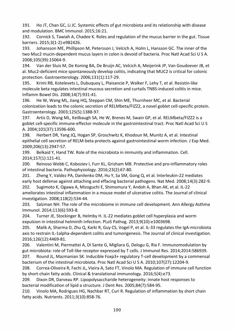
100
191. Ho JT, Chan GC, Li JC. Systemic effects of gut microbiota and its relationship with disease and modulation. BMC Immunol. 2015;16:21. 192. Cornick S, Tawiah A, Chadee K. Roles and regulation of the mucus barrier in the gut. Tissue barriers. 2015;3(1-2):e982426. 193. Johansson ME, Phillipson M, Petersson J, Velcich A, Holm L, Hansson GC. The inner of the two Muc2 mucin-dependent mucus layers in colon is devoid of bacteria. Proc Natl Acad Sci U S A. 2008;105(39):15064-9. 194. Van der Sluis M, De Koning BA, De Bruijn AC, Velcich A, Meijerink JP, Van Goudoever JB, et al. Muc2-deficient mice spontaneously develop colitis, indicating that MUC2 is critical for colonic protection. Gastroenterology. 2006;131(1):117-29. 195. Krimi RB, Kotelevets L, Dubuquoy L, Plaisancie P, Walker F, Lehy T, et al. Resistin-like molecule beta regulates intestinal mucous secretion and curtails TNBS-induced colitis in mice. Inflamm Bowel Dis. 2008;14(7):931-41. 196. He W, Wang ML, Jiang HQ, Steppan CM, Shin ME, Thurnheer MC, et al. Bacterial colonization leads to the colonic secretion of RELMbeta/FIZZ2, a novel goblet cell-specific protein. Gastroenterology. 2003;125(5):1388-97. 197. Artis D, Wang ML, Keilbaugh SA, He W, Brenes M, Swain GP, et al. RELMbeta/FIZZ2 is a goblet cell-specific immune-effector molecule in the gastrointestinal tract. Proc Natl Acad Sci U S A. 2004;101(37):13596-600. 198. Herbert DR, Yang JQ, Hogan SP, Groschwitz K, Khodoun M, Munitz A, et al. Intestinal epithelial cell secretion of RELM-beta protects against gastrointestinal worm infection. J Exp Med. 2009;206(13):2947-57. 199. Belkaid Y, Hand TW. Role of the microbiota in immunity and inflammation. Cell. 2014;157(1):121-41. 200. Reinoso Webb C, Koboziev I, Furr KL, Grisham MB. Protective and pro-inflammatory roles of intestinal bacteria. Pathophysiology. 2016;23(2):67-80. 201. Zheng Y, Valdez PA, Danilenko DM, Hu Y, Sa SM, Gong Q, et al. Interleukin-22 mediates early host defense against attaching and effacing bacterial pathogens. Nat Med. 2008;14(3):282-9. 202. Sugimoto K, Ogawa A, Mizoguchi E, Shimomura Y, Andoh A, Bhan AK, et al. IL-22 ameliorates intestinal inflammation in a mouse model of ulcerative colitis. The Journal of clinical investigation. 2008;118(2):534-44. 203. Salzman NH. The role of the microbiome in immune cell development. Ann Allergy Asthma Immunol. 2014;113(6):593-8. 204. Turner JE, Stockinger B, Helmby H. IL-22 mediates goblet cell hyperplasia and worm expulsion in intestinal helminth infection. PLoS Pathog. 2013;9(10):e1003698. 205. Malik A, Sharma D, Zhu Q, Karki R, Guy CS, Vogel P, et al. IL-33 regulates the IgA-microbiota axis to restrain IL-1alpha-dependent colitis and tumorigenesis. The Journal of clinical investigation. 2016;126(12):4469-81. 206. Valentini M, Piermattei A, Di Sante G, Migliara G, Delogu G, Ria F. Immunomodulation by gut microbiota: role of Toll-like receptor expressed by T cells. J Immunol Res. 2014;2014:586939. 207. Round JL, Mazmanian SK. Inducible Foxp3+ regulatory T-cell development by a commensal bacterium of the intestinal microbiota. Proc Natl Acad Sci U S A. 2010;107(27):12204-9. 208. Correa-Oliveira R, Fachi JL, Vieira A, Sato FT, Vinolo MA. Regulation of immune cell function by short-chain fatty acids. Clinical & translational immunology. 2016;5(4):e73. 209. Dixon DR, Darveau RP. Lipopolysaccharide heterogeneity: innate host responses to bacterial modification of lipid a structure. J Dent Res. 2005;84(7):584-95. 210. Vinolo MA, Rodrigues HG, Nachbar RT, Curi R. Regulation of inflammation by short chain fatty acids. Nutrients. 2011;3(10):858-76.

101
211. Park J, Kim M, Kang SG, Jannasch AH, Cooper B, Patterson J, et al. Short-chain fatty acids induce both effector and regulatory T cells by suppression of histone deacetylases and regulation of the mTOR-S6K pathway. Mucosal Immunol. 2015;8(1):80-93. 212. Round JL, Mazmanian SK. The gut microbiota shapes intestinal immune responses during health and disease. Nature Reviews Immunology. 2009;9(5):313-23. 213. Brown K, DeCoffe D, Molcan E, Gibson DL. Diet-induced dysbiosis of the intestinal microbiota and the effects on immunity and disease. Nutrients. 2012;4(8):1095-119. 214. Herbst T, Sichelstiel A, Schar C, Yadava K, Burki K, Cahenzli J, et al. Dysregulation of allergic airway inflammation in the absence of microbial colonization. Am J Respir Crit Care Med. 2011;184(2):198-205. 215. Trompette A, Gollwitzer ES, Yadava K, Sichelstiel AK, Sprenger N, Ngom-Bru C, et al. Gut microbiota metabolism of dietary fiber influences allergic airway disease and hematopoiesis. Nat Med. 2014;20(2):159-66. 216. Kitajima S, Morimoto M, Sagara E, Shimizu C, Ikeda Y. Dextran sodium sulfate-induced colitis in germ-free IQI/Jic mice. Exp Anim. 2001;50(5):387-95. 217. Chiu CC, Ching YH, Wang YC, Liu JY, Li YP, Huang YT, et al. Monocolonization of germ-free mice with Bacteroides fragilis protects against dextran sulfate sodium-induced acute colitis. Biomed Res Int. 2014;2014:675786. 218. Furusawa Y, Obata Y, Fukuda S, Endo TA, Nakato G, Takahashi D, et al. Commensal microbe-derived butyrate induces the differentiation of colonic regulatory T cells. Nature. 2013;504(7480):446-50. 219. Vael C, Nelen V, Verhulst SL, Goossens H, Desager KN. Early intestinal Bacteroides fragilis colonisation and development of asthma. BMC Pulm Med. 2008;8:19. 220. Scher JU, Sczesnak A, Longman RS, Segata N, Ubeda C, Bielski C, et al. Expansion of intestinal Prevotella copri correlates with enhanced susceptibility to arthritis. Elife. 2013;2:e01202. 221. Liu X, Zou Q, Zeng B, Fang Y, Wei H. Analysis of fecal Lactobacillus community structure in patients with early rheumatoid arthritis. Curr Microbiol. 2013;67(2):170-6. 222. Morgan XC, Tickle TL, Sokol H, Gevers D, Devaney KL, Ward DV, et al. Dysfunction of the intestinal microbiome in inflammatory bowel disease and treatment. Genome Biol. 2012;13(9):R79. 223. Ohkusa T, Koido S. Intestinal microbiota and ulcerative colitis. Journal of infection and chemotherapy : official journal of the Japan Society of Chemotherapy. 2015;21(11):761-8. 224. Vernia P, Annese V, Bresci G, d'Albasio G, D'Inca R, Giaccari S, et al. Topical butyrate improves efficacy of 5-ASA in refractory distal ulcerative colitis: results of a multicentre trial. European journal of clinical investigation. 2003;33(3):244-8. 225. Canani RB, Costanzo MD, Leone L, Pedata M, Meli R, Calignano A. Potential beneficial effects of butyrate in intestinal and extraintestinal diseases. World J Gastroenterol. 2011;17(12):1519-28. 226. Musso G, Gambino R, Cassader M. Obesity, diabetes, and gut microbiota: the hygiene hypothesis expanded? Diabetes Care. 2010;33(10):2277-84. 227. Rabot S, Membrez M, Bruneau A, Gerard P, Harach T, Moser M, et al. Germ-free C57BL/6J mice are resistant to high-fat-diet-induced insulin resistance and have altered cholesterol metabolism. Faseb j. 2010;24(12):4948-59. 228. Lam YY, Ha CW, Campbell CR, Mitchell AJ, Dinudom A, Oscarsson J, et al. Increased gut permeability and microbiota change associate with mesenteric fat inflammation and metabolic dysfunction in diet-induced obese mice. PLoS One. 2012;7(3):e34233. 229. Ley RE, Backhed F, Turnbaugh P, Lozupone CA, Knight RD, Gordon JI. Obesity alters gut microbial ecology. Proc Natl Acad Sci U S A. 2005;102(31):11070-5.

102
230. Bervoets L, Van Hoorenbeeck K, Kortleven I, Van Noten C, Hens N, Vael C, et al. Differences in gut microbiota composition between obese and lean children: a cross-sectional study. Gut Pathog. 2013;5(1):10. 231. Tai N, Wong FS, Wen L. The role of gut microbiota in the development of type 1, type 2 diabetes mellitus and obesity. Rev Endocr Metab Disord. 2015;16(1):55-65. 232. Cox AJ, West NP, Cripps AW. Obesity, inflammation, and the gut microbiota. The lancet Diabetes & endocrinology. 2015;3(3):207-15. 233. Tagliabue A, Elli M. The role of gut microbiota in human obesity: recent findings and future perspectives. Nutr Metab Cardiovasc Dis. 2013;23(3):160-8. 234. Han JL, Lin HL. Intestinal microbiota and type 2 diabetes: from mechanism insights to therapeutic perspective. World J Gastroenterol. 2014;20(47):17737-45. 235. Moreno-Indias I, Cardona F, Tinahones FJ, Queipo-Ortuno MI. Impact of the gut microbiota on the development of obesity and type 2 diabetes mellitus. Front Microbiol. 2014;5:190. 236. Larsen N, Vogensen FK, van den Berg FW, Nielsen DS, Andreasen AS, Pedersen BK, et al. Gut microbiota in human adults with type 2 diabetes differs from non-diabetic adults. PLoS One. 2010;5(2):e9085. 237. Davis-Richardson AG, Triplett EW. A model for the role of gut bacteria in the development of autoimmunity for type 1 diabetes. Diabetologia. 2015;58(7):1386-93. 238. Woting A, Blaut M. The Intestinal Microbiota in Metabolic Disease. Nutrients. 2016;8(4):202. 239. Utzschneider KM, Kratz M, Damman CJ, Hullarg M. Mechanisms Linking the Gut Microbiome and Glucose Metabolism. The Journal of clinical endocrinology and metabolism. 2016;101(4):1445-54. 240. Mutapi F. The gut microbiome in the helminth infected host. Trends in parasitology. 2015;31:405-6. 241. Belkaid Y, Hand TW. Role of the Microbiota in Immunity and Inflammation. Cell. 2014;157:121-41. 242. Kreisinger J, Bastien G, Hauffe HC, Marchesi J, Perkins SE. Interactions between multiple helminths and the gut microbiota in wild rodents. Philos Trans R Soc Lond B Biol Sci. 2015;370(1675). 243. Hayes KS, Bancroft AJ, Goldrick M, Portsmouth C, Roberts IS, Grencis RK. Exploitation of the intestinal microflora by the parasitic nematode Trichuris muris. Science. 2010;328(5984):1391-4. 244. Houlden A, Hayes KS, Bancroft AJ, Worthington JJ, Wang P, Grencis RK, et al. Chronic Trichuris muris Infection in C57BL/6 Mice Causes Significant Changes in Host Microbiota and Metabolome: Effects Reversed by Pathogen Clearance. PLoS One. 2015;10(5):e0125945. 245. Holm JB, Sorobetea D, Kiilerich P, Ramayo-Caldas Y, Estelle J, Ma T, et al. Chronic Trichuris muris Infection Decreases Diversity of the Intestinal Microbiota and Concomitantly Increases the Abundance of Lactobacilli. PLoS One. 2015;10(5):e0125495. 246. Fricke WF, Song Y, Wang AJ, Smith A, Grinchuk V, Mongodin E, et al. Type 2 immunity-dependent reduction of segmented filamentous bacteria in mice infected with the helminthic parasite Nippostrongylus brasiliensis. Microbiome. 2015;3:40. 247. Rausch S, Held J, Fischer A, Heimesaat MM, Kuhl AA, Bereswill S, et al. Small intestinal nematode infection of mice is associated with increased enterobacterial loads alongside the intestinal tract. PLoS One. 2013;8(9):e74026. 248. Reynolds LA, Smith KA, Filbey KJ, Harcus Y, Hewitson JP, Redpath SA, et al. Commensal-pathogen interactions in the intestinal tract: lactobacilli promote infection with, and are promoted by, helminth parasites. Gut Microbes. 2014;5(4):522-32.

103
249. Walk ST, Blum AM, Ewing SA, Weinstock JV, Young VB. Alteration of the murine gut microbiota during infection with the parasitic helminth Heligmosomoides polygyrus. Inflamm Bowel Dis. 2010;16(11):1841-9. 250. Zaiss MM, Rapin A, Lebon L, Dubey LK, Mosconi I, Sarter K, et al. The Intestinal Microbiota Contributes to the Ability of Helminths to Modulate Allergic Inflammation. Immunity. 2015;43:998-1010. 251. Ramanan D, Bowcutt R, Lee SC, Tang MS, Kurtz ZD, Ding Y, et al. Helminth infection promotes colonization resistance via type 2 immunity. Science. 2016;352(6285):608-12. 252. McFarlane AJ, McSorley HJ, Davidson DJ, Fitch PM, Errington C, Mackenzie KJ, et al. Enteric helminth-induced type I interferon signaling protects against pulmonary virus infection through interaction with the microbiota. J Allergy Clin Immunol. 2017. 253. McKenney EA, Williamson L, Yoder AD, Rawls JF, Bilbo SD, Parker W. Alteration of the rat cecal microbiome during colonization with the helminth Hymenolepis diminuta. Gut Microbes. 2015;6(3):182-93. 254. Shimokawa C, Obi S, Shibata M, Olia A, Imai T, Suzue K, et al. Suppression of Obesity by an Intestinal Helminth through Interactions with Intestinal Microbiota. Infect Immun. 2019;87(6). 255. Pace F, Carvalho BM, Zanotto TM, Santos A, Guadagnini D, Silva KLC, et al. Helminth infection in mice improves insulin sensitivity via modulation of gut microbiota and fatty acid metabolism. Pharmacological research. 2018;132:33-46. 256. Doonan J, Tarafdar A, Pineda MA, Lumb FE, Crowe J, Khan AM, et al. The parasitic worm product ES-62 normalises the gut microbiota bone marrow axis in inflammatory arthritis. Nature communications. 2019;10(1):1554. 257. Jenkins TP, Formenti F, Castro C, Piubelli C, Perandin F, Buonfrate D, et al. Author Correction: A comprehensive analysis of the faecal microbiome and metabolome of Strongyloides stercoralis infected volunteers from a non-endemic area. Sci Rep. 2019;9(1):8571. 258. Cantacessi C, Giacomin P, Croese J, Zakrzewski M, Sotillo J, McCann L, et al. Impact of experimental hookworm infection on the human gut microbiota. J Infect Dis. 2014;210(9):1431-4. 259. Giacomin P, Zakrzewski M, Croese J, Su X, Sotillo J, McCann L, et al. Experimental hookworm infection and escalating gluten challenges are associated with increased microbial richness in celiac subjects. Sci Rep. 2015;5:13797. 260. Giacomin P, Zakrzewski M, Jenkins TP, Su X, Al-Hallaf R, Croese J, et al. Changes in duodenal tissue-associated microbiota following hookworm infection and consecutive gluten challenges in humans with coeliac disease. Sci Rep. 2016;6:36797. 261. Lee SC, Tang MS, Lim YA, Choy SH, Kurtz ZD, Cox LM, et al. Helminth colonization is associated with increased diversity of the gut microbiota. PLoS Negl Trop Dis. 2014;8(5):e2880. 262. Molofsky AB, Nussbaum JC, Liang H-E, Van Dyken SJ, Cheng LE, Mohapatra A, et al. Innate lymphoid type 2 cells sustain visceral adipose tissue eosinophils and alternatively activated macrophages. The Journal of experimental medicine. 2013;210:535-49. 263. Wu D, Molofsky AB, Liang H-E, Ricardo-Gonzalez RR, Jouihan HA, Bando JK, et al. Eosinophils sustain adipose alternatively activated macrophages associated with glucose homeostasis. Science. 2011;332:243-7. 264. Goh YP, Henderson NC, Heredia JE, Red Eagle A, Odegaard JI, Lehwald N, et al. Eosinophils secrete IL-4 to facilitate liver regeneration. Proc Natl Acad Sci U S A. 2013;110(24):9914-9. 265. Tomasello E, Bedoui S. Intestinal innate immune cells in gut homeostasis and immunosurveillance. Immunol Cell Biol. 2013;91(3):201-3. 266. Elliott DE, Weinstock JV. Helminth-host immunological interactions: prevention and control of immune-mediated diseases. Annals of the New York Academy of Sciences. 2012;1247:83-96. 267. Maizels RM, McSorley HJ, Smyth DJ. Helminths in the hygiene hypothesis: sooner or later? Clinical and experimental immunology. 2014;177:38-46.

104
268. Anthony RM, Rutitzky LI, Urban JFJ, Stadecker MJ, Gause WC. Protective immune mechanisms in helminth infection. Nature reviews Immunology. 2007;7:975-87. 269. Kreider T, Anthony RM, Urban JFJ, Gause WC. Alternatively activated macrophages in helminth infections. Current opinion in immunology. 2007;19:448-53. 270. Weinstock JV, Elliott DE. Helminth Infections Decrease Host Susceptibility to Immune-Mediated Diseases. The Journal of Immunology. 2014;193(7):3239-47. 271. Helmby H. Human helminth therapy to treat inflammatory disorders - where do we stand? BMC Immunol. 2015;16:12. 272. Wiria AE, Sartono E, Supali T, Yazdanbakhsh M. Helminth infections, type-2 immune response, and metabolic syndrome. PLoS pathogens. 2014;10:e1004140. 273. Larsen N, Vogensen FK, van den Berg FWJ, Nielsen DS, Andreasen AS, Pedersen BK, et al. Gut microbiota in human adults with type 2 diabetes differs from non-diabetic adults. PloS one. 2010;5:e9085. 274. Moreno-Indias I, Cardona F, Tinahones FJ, Queipo-Ortuno MI. Impact of the gut microbiota on the development of obesity and type 2 diabetes mellitus. Frontiers in microbiology. 2014;5:190. 275. Musso G, Gambino R, Cassader M. Obesity, Diabetes, and Gut Microbiota: The hygiene hypothesis expanded? Diabetes Care. 2010;33:2277-84. 276. Reynolds LA, Smith KA, Filbey KJ, Harcus Y, Hewitson JP, Redpath SA, et al. Commensal-pathogen interactions in the intestinal tract: lactobacilli promote infection with, and are promoted by, helminth parasites. Gut microbes. 2014;5:522-32. 277. Walk ST, Blum AM, Ewing SA-S, Weinstock JV, Young VB. Alteration of the murine gut microbiota during infection with the parasitic helminth Heligmosomoides polygyrus. Inflammatory bowel diseases. 2010;16:1841-9. 278. Zhou T, Hu Z, Yang S, Sun L, Yu Z, Wang G. Role of Adaptive and Innate Immunity in Type 2 Diabetes Mellitus. J Diabetes Res. 2018;2018:7457269. 279. Pirola L, Ferraz JC. Role of pro- and anti-inflammatory phenomena in the physiopathology of type 2 diabetes and obesity. World J Biol Chem. 2017;8(2):120-8. 280. Bouchery T, Kyle R, Camberis M, Shepherd A, Filbey K, Smith A, et al. ILC2s and T cells cooperate to ensure maintenance of M2 macrophages for lung immunity against hookworms. Nature communications. 2015;6:6970. 281. Oeser K, Schwartz C, Voehringer D. Conditional IL-4/IL-13-deficient mice reveal a critical role of innate immune cells for protective immunity against gastrointestinal helminths. Mucosal Immunol. 2015;8(3):672-82. 282. Finkelman FD, Shea-Donohue T, Morris SC, Gildea L, Strait R, Madden KB, et al. Interleukin-4- and interleukin-13-mediated host protection against intestinal nematode parasites. Immunological reviews. 2004;201:139-55. 283. Daly CM, Mayrhofer G, Dent LA. Trapping and immobilization of Nippostrongylus brasiliensis larvae at the site of inoculation in primary infections of interleukin-5 transgenic mice. Infect Immun. 1999;67(10):5315-23. 284. Urban JF, Jr., Noben-Trauth N, Donaldson DD, Madden KB, Morris SC, Collins M, et al. IL-13, IL-4Ralpha, and Stat6 are required for the expulsion of the gastrointestinal nematode parasite Nippostrongylus brasiliensis. Immunity. 1998;8(2):255-64. 285. Shin EH, Osada Y, Chai JY, Matsumoto N, Takatsu K, Kojima S. Protective roles of eosinophils in Nippostrongylus brasiliensis infection. Int Arch Allergy Immunol. 1997;114 Suppl 1:45-50. 286. Fisher-Wellman KH, Ryan TE, Smith CD, Gilliam LA, Lin CT, Reese LR, et al. A Direct Comparison of Metabolic Responses to High-Fat Diet in C57BL/6J and C57BL/6NJ Mice. Diabetes. 2016;65(11):3249-61.

105
287. Morris JL, Bridson TL, Alim MA, Rush CM, Rudd DM, Govan BL, et al. Development of a diet-induced murine model of diabetes featuring cardinal metabolic and pathophysiological abnormalities of type 2 diabetes. Biol Open. 2016;5(8):1149-62. 288. Lee EH, Itan M, Jang J, Gu HJ, Rozenberg P, Mingler MK, et al. Eosinophils support adipocyte maturation and promote glucose tolerance in obesity. Sci Rep. 2018;8(1):9894. 289. Withers SB, Forman R, Meza-Perez S, Sorobetea D, Sitnik K, Hopwood T, et al. Eosinophils are key regulators of perivascular adipose tissue and vascular functionality. Sci Rep. 2017;7:44571. 290. Brestoff JR, Kim BS, Saenz SA, Stine RR, Monticelli LA, Sonnenberg GF, et al. Group 2 innate lymphoid cells promote beiging of white adipose tissue and limit obesity. Nature. 2015;519(7542):242-6. 291. Ricardo-Gonzalez RR, Red Eagle A, Odegaard JI, Jouihan H, Morel CR, Heredia JE, et al. IL-4/STAT6 immune axis regulates peripheral nutrient metabolism and insulin sensitivity. Proc Natl Acad Sci U S A. 2010;107(52):22617-22. 292. Elbe-Burger A, Egyed A, Olt S, Klubal R, Mann U, Rappersberger K, et al. Overexpression of IL-4 alters the homeostasis in the skin. J Invest Dermatol. 2002;118(5):767-78. 293. Stafeev IS, Michurina SS, Podkuychenko NV, Vorotnikov AV, Menshikov MY, Parfyonova YV. Interleukin-4 Restores Insulin Sensitivity in Lipid-Induced Insulin-Resistant Adipocytes. Biochemistry (Mosc). 2018;83(5):498-506. 294. Huang X, Liu G, Guo J, Su Z. The PI3K/AKT pathway in obesity and type 2 diabetes. Int J Biol Sci. 2018;14(11):1483-96. 295. Chang YH, Ho KT, Lu SH, Huang CN, Shiau MY. Regulation of glucose/lipid metabolism and insulin sensitivity by interleukin-4. Int J Obes (Lond). 2012;36(7):993-8. 296. Madden KB, Yeung KA, Zhao A, Gause WC, Finkelman FD, Katona IM, et al. Enteric nematodes induce stereotypic STAT6-dependent alterations in intestinal epithelial cell function. J Immunol. 2004;172(9):5616-21. 297. Huang Z, Zhong L, Lee JTH, Zhang J, Wu D, Geng L, et al. The FGF21-CCL11 Axis Mediates Beiging of White Adipose Tissues by Coupling Sympathetic Nervous System to Type 2 Immunity. Cell Metab. 2017;26(3):493-508 e4. 298. Qiu Y, Nguyen KD, Odegaard JI, Cui X, Tian X, Locksley RM, et al. Eosinophils and type 2 cytokine signaling in macrophages orchestrate development of functional beige fat. Cell. 2014;157(6):1292-308. 299. Wang W, Seale P. Control of brown and beige fat development. Nat Rev Mol Cell Biol. 2016;17(11):691-702. 300. Hussaarts L, Garcia-Tardon N, van Beek L, Heemskerk MM, Haeberlein S, van der Zon GC, et al. Chronic helminth infection and helminth-derived egg antigens promote adipose tissue M2 macrophages and improve insulin sensitivity in obese mice. FASEB journal. 2015;29:3027-39. 301. Maizels RM, McSorley HJ. Regulation of the host immune system by helminth parasites. J Allergy Clin Immunol. 2016;138(3):666-75. 302. Logan J, Manda SS, Choi YJ, Field M, Eichenberger RM, Mulvenna J, et al. Comprehensive analysis of human hookworm secreted proteins using a proteogenomic approach. bioRxiv; 2018. 303. McSorley HJ, Maizels RM. Helminth infections and host immune regulation. Clin Microbiol Rev. 2012;25(4):585-608. 304. Hewitson JP, Grainger JR, Maizels RM. Helminth immunoregulation: the role of parasite secreted proteins in modulating host immunity. Molecular and biochemical parasitology. 2009;167:1-11. 305. Ferreira IB, Pickering DA, Troy S, Croese J, Loukas A, Navarro S. Suppression of inflammation and tissue damage by a hookworm recombinant protein in experimental colitis. Clinical & translational immunology. 2017;6(10):e157.
106
306. Sotillo J, Ferreira I, Potriquet J, Laha T, Navarro S, Loukas A, et al. Changes in protein expression after treatment with Ancylostoma caninum excretory/secretory products in a mouse model of colitis. Sci Rep. 2017;7:41883. 307. Wangchuk P, Shepherd C, Constantinoiu C, Ryan RYM, Kouremenos KA, Becker L, et al. Hookworm-Derived Metabolites Suppress Pathology in a Mouse Model of Colitis and Inhibit Secretion of Key Inflammatory Cytokines in Primary Human Leukocytes. Infect Immun. 2019;87(4). 308. Segura M, Su Z, Piccirillo C, Stevenson MM. Impairment of dendritic cell function by excretory-secretory products: a potential mechanism for nematode-induced immunosuppression. European journal of immunology. 2007;37(7):1887-904. 309. Grainger JR, Smith KA, Hewitson JP, McSorley HJ, Harcus Y, Filbey KJ, et al. Helminth secretions induce de novo T cell Foxp3 expression and regulatory function through the TGF-beta pathway. J Exp Med. 2010;207(11):2331-41. 310. Sun Y, Liu G, Li Z, Chen Y, Liu Y, Liu B, et al. Modulation of dendritic cell function and immune response by cysteine protease inhibitor from murine nematode parasite Heligmosomoides polygyrus. Immunology. 2013;138(4):370-81. 311. McInnes IB, Leung BP, Harnett M, Gracie JA, Liew FY, Harnett W. A novel therapeutic approach targeting articular inflammation using the filarial nematode-derived phosphorylcholine-containing glycoprotein ES-62. J Immunol. 2003;171(4):2127-33. 312. Rodgers DT, McGrath MA, Pineda MA, Al-Riyami L, Rzepecka J, Lumb F, et al. The parasitic worm product ES-62 targets myeloid differentiation factor 88-dependent effector mechanisms to suppress antinuclear antibody production and proteinuria in MRL/lpr mice. Arthritis & rheumatology (Hoboken, NJ). 2015;67(4):1023-35. 313. Hams E, Bermingham R, Wurlod FA, Hogan AE, O'Shea D, Preston RJ, et al. The helminth T2 RNase omega1 promotes metabolic homeostasis in an IL-33- and group 2 innate lymphoid cell-dependent mechanism. FASEB J. 2016;30(2):824-35. 314. van den Berg SM, van Dam AD, Kusters PJH, Beckers L, den Toom M, van der Velden S, et al. Helminth antigens counteract a rapid high-fat diet-induced decrease in adipose tissue eosinophils. Journal of molecular endocrinology. 2017;59(3):245-55. 315. Tang CL, Yu XH, Li Y, Zhang RH, Xie J, Liu ZM. Schistosoma japonicum Soluble Egg Antigen Protects Against Type 2 Diabetes in Lepr (db/db) Mice by Enhancing Regulatory T Cells and Th2 Cytokines. Front Immunol. 2019;10:1471. 316. Cho KW, Morris DL, Lumeng CN. Flow cytometry analyses of adipose tissue macrophages. Methods Enzymol. 2014;537:297-314. 317. Nishiyama K, Nakashima H, Ikarashi M, Kinoshita M, Nakashima M, Aosasa S, et al. Mouse CD11b+Kupffer Cells Recruited from Bone Marrow Accelerate Liver Regeneration after Partial Hepatectomy. PLoS One. 2015;10(9):e0136774. 318. Sehmi R, Wardlaw AJ, Cromwell O, Kurihara K, Waltmann P, Kay AB. Interleukin-5 selectively enhances the chemotactic response of eosinophils obtained from normal but not eosinophilic subjects. Blood. 1992;79(11):2952-9. 319. Rothenberg ME, Petersen J, Stevens RL, Silberstein DS, McKenzie DT, Austen KF, et al. IL-5-dependent conversion of normodense human eosinophils to the hypodense phenotype uses 3T3 fibroblasts for enhanced viability, accelerated hypodensity, and sustained antibody-dependent cytotoxicity. J Immunol. 1989;143(7):2311-6. 320. Yamaguchi Y, Suda T, Suda J, Eguchi M, Miura Y, Harada N, et al. Purified interleukin 5 supports the terminal differentiation and proliferation of murine eosinophilic precursors. J Exp Med. 1988;167(1):43-56. 321. Tran GT, Wilcox PL, Dent LA, Robinson CM, Carter N, Verma ND, et al. Interleukin-5 Mediates Parasite-Induced Protection against Experimental Autoimmune Encephalomyelitis:

107
Association with Induction of Antigen-Specific CD4(+)CD25(+) T Regulatory Cells. Front Immunol. 2017;8:1453. 322. Finlay CM, Stefanska AM, Walsh KP, Kelly PJ, Boon L, Lavelle EC, et al. Helminth Products Protect against Autoimmunity via Innate Type 2 Cytokines IL-5 and IL-33, Which Promote Eosinophilia. J Immunol. 2016;196(2):703-14. 323. Parthasarathy G, Mansfield LS. Trichuris suis excretory secretory products (ESP) elicit interleukin-6 (IL-6) and IL-10 secretion from intestinal epithelial cells (IPEC-1). Vet Parasitol. 2005;131(3-4):317-24. 324. Rehman K, Akash MSH, Liaqat A, Kamal S, Qadir MI, Rasul A. Role of Interleukin-6 in Development of Insulin Resistance and Type 2 Diabetes Mellitus. Crit Rev Eukaryot Gene Expr. 2017;27(3):229-36. 325. Sabio G, Das M, Mora A, Zhang Z, Jun JY, Ko HJ, et al. A stress signaling pathway in adipose tissue regulates hepatic insulin resistance. Science. 2008;322(5907):1539-43. 326. Taub R. Liver regeneration: from myth to mechanism. Nat Rev Mol Cell Biol. 2004;5(10):836-47. 327. Matthews VB, Allen TL, Risis S, Chan MH, Henstridge DC, Watson N, et al. Interleukin-6-deficient mice develop hepatic inflammation and systemic insulin resistance. Diabetologia. 2010;53(11):2431-41. 328. Wunderlich FT, Strohle P, Konner AC, Gruber S, Tovar S, Bronneke HS, et al. Interleukin-6 signaling in liver-parenchymal cells suppresses hepatic inflammation and improves systemic insulin action. Cell Metab. 2010;12(3):237-49. 329. Heijink IH, Vellenga E, Borger P, Postma DS, de Monchy JG, Kauffman HF. Interleukin-6 promotes the production of interleukin-4 and interleukin-5 by interleukin-2-dependent and -independent mechanisms in freshly isolated human T cells. Immunology. 2002;107(3):316-24. 330. Rochman I, Paul WE, Ben-Sasson SZ. IL-6 increases primed cell expansion and survival. J Immunol. 2005;174(8):4761-7. 331. Yang D, Chen Q, Su SB, Zhang P, Kurosaka K, Caspi RR, et al. Eosinophil-derived neurotoxin acts as an alarmin to activate the TLR2-MyD88 signal pathway in dendritic cells and enhances Th2 immune responses. J Exp Med. 2008;205(1):79-90. 332. Cani PD, Delzenne NM. Gut microflora as a target for energy and metabolic homeostasis. Curr Opin Clin Nutr Metab Care. 2007;10(6):729-34. 333. Loke P, Lim YA. Helminths and the microbiota: parts of the hygiene hypothesis. Parasite Immunol. 2015;37(6):314-23. 334. Backhed F, Ding H, Wang T, Hooper LV, Koh GY, Nagy A, et al. The gut microbiota as an environmental factor that regulates fat storage. Proc Natl Acad Sci U S A. 2004;101(44):15718-23. 335. Backhed F, Manchester JK, Semenkovich CF, Gordon JI. Mechanisms underlying the resistance to diet-induced obesity in germ-free mice. Proc Natl Acad Sci U S A. 2007;104(3):979-84. 336. Schwiertz A, Taras D, Schafer K, Beijer S, Bos NA, Donus C, et al. Microbiota and SCFA in lean and overweight healthy subjects. Obesity (Silver Spring). 2010;18(1):190-5. 337. Zuo HJ, Xie ZM, Zhang WW, Li YR, Wang W, Ding XB, et al. Gut bacteria alteration in obese people and its relationship with gene polymorphism. World J Gastroenterol. 2011;17(8):1076-81. 338. Zhang H, DiBaise JK, Zuccolo A, Kudrna D, Braidotti M, Yu Y, et al. Human gut microbiota in obesity and after gastric bypass. Proc Natl Acad Sci U S A. 2009;106(7):2365-70. 339. Qin J, Li Y, Cai Z, Li S, Zhu J, Zhang F, et al. A metagenome-wide association study of gut microbiota in type 2 diabetes. Nature. 2012;490(7418):55-60. 340. Karlsson FH, Tremaroli V, Nookaew I, Bergstrom G, Behre CJ, Fagerberg B, et al. Gut metagenome in European women with normal, impaired and diabetic glucose control. Nature. 2013;498(7452):99-103.

108
341. Neuman H, Mor H, Bashi T, Givol O, Watad A, Shemer A, et al. Helminth-Based Product and the Microbiome of Mice with Lupus. mSystems. 2019;4(1). 342. Bolger AM, Lohse M, Usadel B. Trimmomatic: a flexible trimmer for Illumina sequence data. Bioinformatics. 2014;30(15):2114-20. 343. Caporaso JG, Kuczynski J, Stombaugh J, Bittinger K, Bushman FD, Costello EK, et al. QIIME allows analysis of high-throughput community sequencing data. Nature methods. 2010;7(5):335-6. 344. Altschul SF, Gish W, Miller W, Myers EW, Lipman DJ. Basic local alignment search tool. J Mol Biol. 1990;215(3):403-10. 345. Edgar RC, Haas BJ, Clemente JC, Quince C, Knight R. UCHIME improves sensitivity and speed of chimera detection. Bioinformatics. 2011;27(16):2194-200. 346. DeSantis TZ, Hugenholtz P, Larsen N, Rojas M, Brodie EL, Keller K, et al. Greengenes, a chimera-checked 16S rRNA gene database and workbench compatible with ARB. Appl Environ Microbiol. 2006;72(7):5069-72. 347. Edgar RC. Search and clustering orders of magnitude faster than BLAST. Bioinformatics. 2010;26(19):2460-1. 348. McDonald D, Clemente JC, Kuczynski J, Rideout JR, Stombaugh J, Wendel D, et al. The Biological Observation Matrix (BIOM) format or: how I learned to stop worrying and love the ome-ome. Gigascience. 2012;1(1):7. 349. McMurdie PJ, Holmes S. Phyloseq: a bioconductor package for handling and analysis of high-throughput phylogenetic sequence data. Pac Symp Biocomput. 2012:235-46. 350. Love MI, Huber W, Anders S. Moderated estimation of fold change and dispersion for RNA-seq data with DESeq2. Genome biology. 2014;15(12):550. 351. Zakrzewski M, Proietti C, Ellis JJ, Hasan S, Brion MJ, Berger B, et al. Calypso: a user-friendly web-server for mining and visualizing microbiome-environment interactions. Bioinformatics. 2017;33(5):782-3. 352. Ben-Amram H, Bashi T, Werbner N, Neuman H, Fridkin M, Blank M, et al. Tuftsin-Phosphorylcholine Maintains Normal Gut Microbiota in Collagen Induced Arthritic Mice. Front Microbiol. 2017;8:1222. 353. Lozupone CA, Stombaugh J, Gonzalez A, Ackermann G, Wendel D, Vazquez-Baeza Y, et al. Meta-analyses of studies of the human microbiota. Genome Res. 2013;23(10):1704-14. 354. Yatsunenko T, Rey FE, Manary MJ, Trehan I, Dominguez-Bello MG, Contreras M, et al. Human gut microbiome viewed across age and geography. Nature. 2012;486(7402):222-7. 355. Langille MG, Meehan CJ, Koenig JE, Dhanani AS, Rose RA, Howlett SE, et al. Microbial shifts in the aging mouse gut. Microbiome. 2014;2(1):50. 356. Salgaco MK, Oliveira LGS, Costa GN, Bianchi F, Sivieri K. Relationship between gut microbiota, probiotics, and type 2 diabetes mellitus. Appl Microbiol Biotechnol. 2019;103(23-24):9229-38. 357. Butta H, Sardana R, Vaishya R, Singh KN, Mendiratta L. Bifidobacterium: An Emerging Clinically Significant Metronidazole-resistant Anaerobe of Mixed Pyogenic Infections. Cureus. 2017;9(4):e1134. 358. Sato J, Kanazawa A, Ikeda F, Yoshihara T, Goto H, Abe H, et al. Gut dysbiosis and detection of "live gut bacteria" in blood of Japanese patients with type 2 diabetes. Diabetes Care. 2014;37(8):2343-50. 359. Zhou L, Xiao X, Zhang Q, Zheng J, Li M, Yu M, et al. Improved Glucose and Lipid Metabolism in the Early Life of Female Offspring by Maternal Dietary Genistein Is Associated With Alterations in the Gut Microbiota. Front Endocrinol (Lausanne). 2018;9:516. 360. Ajibola O, Rowan AD, Ogedengbe CO, Mshelia MB, Cabral DJ, Eze AA, et al. Urogenital schistosomiasis is associated with signatures of microbiome dysbiosis in Nigerian adolescents. Sci Rep. 2019;9(1):829.

109
361. Rosa BA, Supali T, Gankpala L, Djuardi Y, Sartono E, Zhou Y, et al. Differential human gut microbiome assemblages during soil-transmitted helminth infections in Indonesia and Liberia. Microbiome. 2018;6(1):33. 362. Madsen MSA, Holm JB, Palleja A, Wismann P, Fabricius K, Rigbolt K, et al. Metabolic and gut microbiome changes following GLP-1 or dual GLP-1/GLP-2 receptor agonist treatment in diet-induced obese mice. Sci Rep. 2019;9(1):15582. 363. Zietak M, Kovatcheva-Datchary P, Markiewicz LH, Stahlman M, Kozak LP, Backhed F. Altered Microbiota Contributes to Reduced Diet-Induced Obesity upon Cold Exposure. Cell Metab. 2016;23(6):1216-23. 364. Zhang Q, Yu H, Xiao X, Hu L, Xin F, Yu X. Inulin-type fructan improves diabetic phenotype and gut microbiota profiles in rats. PeerJ. 2018;6:e4446. 365. Zhang C, Zhang M, Wang S, Han R, Cao Y, Hua W, et al. Interactions between gut microbiota, host genetics and diet relevant to development of metabolic syndromes in mice. The ISME journal. 2010;4(2):232-41. 366. Tomas J, Mulet C, Saffarian A, Cavin JB, Ducroc R, Regnault B, et al. High-fat diet modifies the PPAR-γ pathway leading to disruption of microbial and physiological ecosystem in murine small intestine. Proc Natl Acad Sci U S A. 2016;113(40):E5934-E43. 367. Loubinoux J, Valente FM, Pereira IA, Costa A, Grimont PA, Le Faou AE. Reclassification of the only species of the genus Desulfomonas, Desulfomonas pigra, as Desulfovibrio piger comb. nov. International journal of systematic and evolutionary microbiology. 2002;52(Pt 4):1305-8. 368. Blachier F, Davila AM, Mimoun S, Benetti PH, Atanasiu C, Andriamihaja M, et al. Luminal sulfide and large intestine mucosa: friend or foe? Amino Acids. 2010;39(2):335-47. 369. Hou YP, He QQ, Ouyang HM, Peng HS, Wang Q, Li J, et al. Human Gut Microbiota Associated with Obesity in Chinese Children and Adolescents. Biomed Res Int. 2017;2017:7585989. 370. Yu F, Han W, Zhan G, Li S, Jiang X, Wang L, et al. Abnormal gut microbiota composition contributes to the development of type 2 diabetes mellitus in db/db mice. Aging (Albany NY). 2019;11(22):10454-67. 371. Beli E, Yan Y, Moldovan L, Vieira CP, Gao R, Duan Y, et al. Restructuring of the Gut Microbiome by Intermittent Fasting Prevents Retinopathy and Prolongs Survival in db/db Mice. Diabetes. 2018;67(9):1867-79. 372. Nirmalkar K, Murugesan S, Pizano-Zarate ML, Villalobos-Flores LE, Garcia-Gonzalez C, Morales-Hernandez RM, et al. Gut Microbiota and Endothelial Dysfunction Markers in Obese Mexican Children and Adolescents. Nutrients. 2018;10(12). 373. Lambeth SM, Carson T, Lowe J, Ramaraj T, Leff JW, Luo L, et al. Composition, Diversity and Abundance of Gut Microbiome in Prediabetes and Type 2 Diabetes. J Diabetes Obes. 2015;2(3):1-7. 374. Eg Siegel, J Lorenzo Bermejo, and IF, Hasslacher C. Cardiovascular Complications and Composition of the Intestinal Microbiome in Patients with Type 2 Diabetes. International Journal of Diabetes and Clinical Research. 2018;5(2). 375. Di Luccia B, Crescenzo R, Mazzoli A, Cigliano L, Venditti P, Walser JC, et al. Rescue of Fructose-Induced Metabolic Syndrome by Antibiotics or Faecal Transplantation in a Rat Model of Obesity. PLoS One. 2015;10(8):e0134893. 376. Zhang X, Shen D, Fang Z, Jie Z, Qiu X, Zhang C, et al. Human gut microbiota changes reveal the progression of glucose intolerance. PLoS One. 2013;8(8):e71108. 377. Derrien M, Vaughan EE, Plugge CM, de Vos WM. Akkermansia muciniphila gen. nov., sp. nov., a human intestinal mucin-degrading bacterium. International journal of systematic and evolutionary microbiology. 2004;54(Pt 5):1469-76.

110
378. Tailford LE, Crost EH, Kavanaugh D, Juge N. Mucin glycan foraging in the human gut microbiome. Front Genet. 2015;6:81. 379. Brandt A, Hernandez-Arriaga A, Kehm R, Sanchez V, Jin CJ, Nier A, et al. Metformin attenuates the onset of non-alcoholic fatty liver disease and affects intestinal microbiota and barrier in small intestine. Sci Rep. 2019;9(1):6668. 380. Sanchez-Osuna M, Barbe J, Erill I. Comparative genomics of the DNA damage-inducible network in the Patescibacteria. Environ Microbiol. 2017;19(9):3465-74. 381. Bordalo Tonucci L, Dos Santos KM, De Luces Fortes Ferreira CL, Ribeiro SM, De Oliveira LL, Martino HS. Gut microbiota and probiotics: Focus on diabetes mellitus. Crit Rev Food Sci Nutr. 2017;57(11):2296-309. 382. Healey GR, Murphy R, Brough L, Butts CA, Coad J. Interindividual variability in gut microbiota and host response to dietary interventions. Nutr Rev. 2017;75(12):1059-80. 383. Rinninella E, Raoul P, Cintoni M, Franceschi F, Miggiano GAD, Gasbarrini A, et al. What is the Healthy Gut Microbiota Composition? A Changing Ecosystem across Age, Environment, Diet, and Diseases. Microorganisms. 2019;7(1). 384. D'Adamo E, Caprio S. Type 2 diabetes in youth: epidemiology and pathophysiology. Diabetes care. 2011;34 Suppl 2:S161-5. 385. American Diabetes Association. Diagnosis and classification of diabetes mellitus. Diabetes care. 2014;37 Suppl 1:S81-90. 386. Schwartz SS, Epstein S, Corkey BE, Grant SF, Gavin JR, 3rd, Aguilar RB. The Time Is Right for a New Classification System for Diabetes: Rationale and Implications of the beta-Cell-Centric Classification Schema. Diabetes Care. 2016;39(2):179-86. 387. Cicchese JM, Evans S, Hult C, Joslyn LR, Wessler T, Millar JA, et al. Dynamic balance of pro- and anti-inflammatory signals controls disease and limits pathology. Immunological reviews. 2018;285(1):147-67. 388. Donath MY, Shoelson SE. Type 2 diabetes as an inflammatory disease. Nat Rev Immunol. 2011;11(2):98-107. 389. Elliott DE, Weinstock JV. Helminth-host immunological interactions: prevention and control of immune-mediated diseases. Ann N Y Acad Sci. 2012;1247:83-96. 390. Brunkwall L, Orho-Melander M. The gut microbiome as a target for prevention and treatment of hyperglycaemia in type 2 diabetes: from current human evidence to future possibilities. Diabetologia. 2017;60(6):943-51. 391. de Ruiter K, Tahapary DL, Sartono E, Soewondo P, Supali T, Smit JWA, et al. Helminths, hygiene hypothesis and type 2 diabetes. Parasite Immunol. 2017;39(5). 392. Kreisinger J, Bastien G, Hauffe HC, Marchesi J, Perkins SE. Interactions between multiple helminths and the gut microbiota in wild rodents. Philosophical transactions of the Royal Society of London. 2015;370. 393. Hung LY, Lewkowich IP, Dawson LA, Downey J, Yang Y, Smith DE, et al. IL-33 drives biphasic IL-13 production for noncanonical Type 2 immunity against hookworms. Proc Natl Acad Sci U S A. 2013;110(1):282-7. 394. Dent LA, Daly CM, Mayrhofer G, Zimmerman T, Hallett A, Bignold LP, et al. Interleukin-5 transgenic mice show enhanced resistance to primary infections with Nippostrongylus brasiliensis but not primary infections with Toxocara canis. Infect Immun. 1999;67(2):989-93. 395. Al-Dahwi Z, Mayberry LF, Conder GA, Bristol JR. Suppression of extraintestinal and intestinal Nippostrongylus brasiliensis-induced eosinophilia by Eimeria nieschulzi. J Parasitol. 2006;92(5):962-70. 396. Nussbaum JC, Van Dyken SJ, von Moltke J, Cheng LE, Mohapatra A, Molofsky AB, et al. Type 2 innate lymphoid cells control eosinophil homeostasis. Nature. 2013;502(7470):245-8.

111
397. Marichal T, Mesnil C, Bureau F. Homeostatic Eosinophils: Characteristics and Functions. Front Med (Lausanne). 2017;4:101. 398. Rao RR, Long JZ, White JP, Svensson KJ, Lou J, Lokurkar I, et al. Meteorin-like is a hormone that regulates immune-adipose interactions to increase beige fat thermogenesis. Cell. 2014;157(6):1279-91. 399. Lee MW, Odegaard JI, Mukundan L, Qiu Y, Molofsky AB, Nussbaum JC, et al. Activated type 2 innate lymphoid cells regulate beige fat biogenesis. Cell. 2015;160(1-2):74-87. 400. Hooper LV, Littman DR, Macpherson AJ. Interactions Between the Microbiota and the Immune System. Science. 2012;336:1268-73. 401. Voreades N, Kozil A, Weir TL. Diet and the development of the human intestinal microbiome. Front Microbiol. 2014;5:494. 402. Wu X, Ma C, Han L, Nawaz M, Gao F, Zhang X, et al. Molecular characterisation of the faecal microbiota in patients with type II diabetes. Curr Microbiol. 2010;61(1):69-78. 403. Wegener Parfrey L, Jirku M, Sima R, Jalovecka M, Sak B, Grigore K, et al. A benign helminth alters the host immune system and the gut microbiota in a rat model system. PLoS One. 2017;12(8):e0182205. 404. Jenkins TP, Rathnayaka Y, Perera PK, Peachey LE, Nolan MJ, Krause L, et al. Infections by human gastrointestinal helminths are associated with changes in faecal microbiota diversity and composition. PLoS One. 2017;12(9):e0184719. 405. Rowland I, Gibson G, Heinken A, Scott K, Swann J, Thiele I, et al. Gut microbiota functions: metabolism of nutrients and other food components. Eur J Nutr. 2018;57(1):1-24.