Immunostaining Protocols - Emory University School of Medicine

Immunostaining of PD-1/PD-Ls in liver tissues of patients with hepatitis and hepato�ellular �ar�inomahepato�ellular �ar�inoma
Bao-Ju Wang, Jun-Jie Bao, Jun-Zhong Wang, Yang Wang, Min Jiang, Ming-You Xing, Wan-Guang Zhang, Jun-Ying Qi, Michael Roggendorf, Meng-Ji Lu, Dong-Liang Yang
Bao-Ju Wang, Jun-Jie Bao, Jun-Zhong Wang, Yang Wang, Dong-Liang Yang, Division of Clinical Immunology, Tongji Hospital of Tongji Medical College, Huazhong University of Sci-ence and Technology, Wuhan 430030, Hubei Province, ChinaJun-Jie Bao, Department of Clinical Laboratory, Guangzhou Women and Children’s Medical Center, Guangzhou 510055, Guangdong Province, ChinaMin Jiang, Meng-Ji Lu, Department of Microbiology, Tongji Medical College, Huazhong University of Science and Technol-ogy, Wuhan 430015, Hubei Province, ChinaMing-You Xing, Jun-Ying Qi, Department of Infectious Disease, Tongji Hospital of Tongji Medical College, Huazhong University of Science and Technology, Wuhan 430030, Hubei Province, ChinaWan-Guang Zhang, Hepatic Surgery Center, Tongji Hospital of Tongji Medical College, Huazhong University of Science and Technology, Wuhan 430030, Hubei Province, ChinaMichael Roggendorf, Meng-Ji Lu, Institute of Virology, Univer-sity Hospital Essen, Hufelandstrasse 55, 45122 Essen, GermanyAuthor contributions: Wang BJ and Bao JJ performed the ex-periments and data analysis; Wang JZ, Wang Y and Jiang M per- formed the experiments; Xing MY, Zhang WG and Qi JY contrib-uted to collecting the samples and providing the patients informa-tion; Wang BJ, Roggendorf M, Lu MJ and Yang DL contributed to research designation, manuscript preparation and editing.Supported by Grants from the National Mega Research Program of China, No. 2008ZX10002-011; the National Key Basic Re-search Program of China, No. 2001CB510008, 2005CB522901, 2007CB512804 and 2009CB522500; the Deutsche Forschun-gsgemeinschaft (SFB/Transregio 60)Correspondence to: Dong-Liang Yang, MD, �rofessor of Medi- Dong-Liang Yang, MD, �rofessor of Medi-cine, Director, Division of Clinical Immunology, Tongji Hospital of Tongji Medical College, Huazhong University of Science and Technology, Jiefang Avenue 1095, Wuhan 430030, Hubei Prov-ince, China. [email protected]: +86-27-83662894 Fax: +86-27-83662894Received: September 16, 2010 Revised: November 13, 2010Accepted: November 20, 2010�ublished online: July 28, 2011
AbstractAIM: To investigate the expression of programmed
death (�D)-1, �D ligand 1 (�D-L1) and �D-L2 in liver tissues in the context of chronic hepatitis and hepato-cellular carcinoma (HCC).
METHODS: Liver biopsies and HCC specimens from patients were collected and histologically examined. The expression of �D-1, �D-L1, and �D-L2 in biopsy specimens of chronic hepatitis and HCC specimens was evaluated by immunohistochemical staining. The as-sociation between the expression level of �D-1, �D-L1, and �D-L2 and clinical and pathological variables was analyzed statistically.
RESULTS: Expression of �D-1 was found in liver-infiltrating lymphocytes. In contrast, PD-L1 and PD-L2 were expressed in non-parenchyma liver cells and tumor cells. The expression of PD-L1 was significantly correlated with hepatitis B virus infection (1.42 ± 1.165 vs 0.50 ± 0.756, � = 0.047) and with the stage of HCC (7.50 ± 2.121 vs 1.75 ± 1.500 vs 3.00 ± 0.001, � = 0.018). PD-1 and PD-Ls were significantly up-regulated in HCC specimens (1.40 ± 1.536 vs 5.71 ± 4.051, � = 0.000; 1.05 ± 1.099 vs 4.29 ± 3.885, � = 0.004; 1.80 ± 1.473 vs 3.81 ± 3.400, � = 0.020).
CONCLUSION: �D-L1 may contribute to negative regulation of the immune response in chronic hepatitis B. �D-1 and �D-Ls may play a role in immune evasion of tumors.
© 2011 Baishideng. All rights reserved.
Key words: Hepatitis B virus; �rogrammed death-1; �rogrammed death ligands; Hepatitis; Hepatocellular carcinoma
Peer reviewer: Atsushi Nakajima, MD, PhD, Professor, Divi-sion of Gastroenterology, Yokohama City University Hospital 3-9 Fukuura, Kanazawaku, Yokohama 236-0004 Kanagawa, Japan
Wang BJ, Bao JJ, Wang JZ, Wang Y, Jiang M, Xing MY, Zhang
ORIGINAL ARTICLE
World J Gastroenterol 2011 July 28; 17(28): 3322-3329ISSN 1007-9327 (print) ISSN 2219-2840 (online)
© 2011 Baishideng. All rights reserved.
Online Submissions: http://www.wjgnet.com/[email protected]:10.3748/wjg.v17.i28.3322
3322 July 28, 2011|Volume 17|Issue 28|WJG|www.wjgnet.com

Wang BJ et al . �D-1/�D-Ls in viral hepatitis and HCC
WG, Qi JY, Roggendorf M, Lu MJ, Yang DL. Immunostaining of PD-1/PD-Ls in liver tissues of patients with hepatitis and he-patocellular carcinoma. World J Gastroenterol 2011; 17(28): 3322-3329 Available from: URL: http://www.wjgnet.com/1007- 9327/full/v17/i28/3322.htm DOI: http://dx.doi.org/10.3748/wjg.v17.i28.3322
INTRODUCTIONThough active vaccination against hepatitis B virus (HBV) infection is successful, chronic HBV infection remains an important medical issue. There are approximately 400 million individuals chronically infected with HBV world-wide. Chronic HBV infection is one of the major causes of liver cirrhosis and hepatocellular carcinoma (HCC).
It is generally accepted that an appropriate T-cell response to HBV is crucial for viral elimination[1]. It has been suggested that HBV-specific CD8+ T cells have dual functions: the production of antiviral cytokines to down-regulate HBV replication in hepatocytes and the elimination of residual HBV-infected cells by cytotoxic activities[2]. Chronic HBV infection is characterized by weak or absent specific T-cell responses to HBV in pe-ripheral blood. In the liver, low numbers of HBV-specific CD4 and CD8 T cells have been found. However, those T cells show a low expression of interferon (IFN)-γ and low cytotoxic activity[3,4]. Mechanisms leading to de-creased cellular immune responses to HBV are not yet defined. Though there are many approaches suitable for priming specific CTL responses, their ability to break im-mune tolerance mechanisms in chronically infected HBV patients requires further investigation. In this respect, un-derstanding tolerance mechanisms in chronic HBV infec-tion will further help the design of strategies to overcome the unresponsiveness of T cells.
Programmed death (PD)-1 is a member of the CD28 family and is involved in the regulation of T-cell activa-tion[5]. PD-1 is expressed on T cells, B cells, and myeloid cells. Two ligands for PD-1, PD ligand 1 (PD-L1) and PD-L2 (also known as B7-H1 and B7-DC), have been identified and have features as co-stimulatory molecules. A large number of publications have indicated a role for PD-L1 in the negative regulation of T-cell functions and the maintenance of peripheral tolerance[6]. The exact role of PD-L2 requires further definition. In murine liver tis-sues, PD-L1 was found to be expressed on Kupffer cells (KCs) and liver sinusoidal epithelial cells (LSECs)[7]. He-patocytes express constitutively low levels of PD-L1 but show enhanced expression of PD-L1 upon stimulation with interferons[8]. It has also been shown in cell culture that PD-L1 expression on hepatoma cells induces apopto-sis in T-cells. PD-L1 deficiency leads to hepatic accumula-tion and impaired apoptosis of T-cells in a murine mod-el[9], and PD-1 deficiency leads to enhanced proliferation of effector cells in the liver during adenoviral infection[10].
Recent data from other chronic viral infections, such as lymphocytic choriomeningitis virus, human immuno-
deficiency virus, and hepatitis C virus infections, indicate that up-regulation of the PD-1/PD-L1 system may be responsible for the impairment of T-cell function[11-18]. Blocking of PD-L1 led to the rescue of exhausted CD8 T-cells even in the absence of Th functions[15-18]. Kassel et al[19] examined the expression of PD-1, PD-L1, and PD-L2 in patient liver samples and found that there was a direct link between the degree of inflammation and the expression of PD-1/PD-L1. Gao et al[20] found in patients with HCC that overexpression of PD-L1 significantly associates with tumor aggressiveness and postoperative recurrence.
The expression of PD-Ls was further suggested as a mechanism of immune evasion for tumors[21]. Elevated expression of PD-L1 was found in different tumor enti-ties[22-29]. It could be shown that tumor cells expressing PD-L1 were able to induce apoptosis of T-cells. Thus, it is necessary to investigate the expression of PD-L1/2 and PD-1 in the liver in the context of viral hepatitis and HCC.
MATERIALS AND METHODSSpecimensSpecimens of liver tissues were obtained by biopsy or surgery from 20 hepatitis patients (including 12 with HBV infection and 8 with non-viral hepatitis) and 26 HCC patients (including 21 with HBV infection) at Tongji Hos-pital of Tongji Medical College, Huazhong University of Science and Technology, China, between 2006 and 2007 (Tables 1-3). Surgically resected or biopsy specimens were fixed in formalin and embedded in paraffin for routine histological diagnosis, and then embedded in OCT com-pound and snap frozen in liquid nitrogen for immunohis-tochemical analysis. The histological activity index (HAI) was assessed according to the classification of Ishak et al[30], which combines grading and staging scores (Table 2). All patients with hepatitis C virus (HCV) and human im-munodeficiency virus (HIV) infection were excluded. No patient underwent antiviral drug treatment prior to biopsy. Tumors were classified as stage Ⅰ to Ⅲ based on the Chengdu conference[31] and as grade Ⅰ to Ⅳ based on the Edmondson-Steiner Guidelines (Table 3). The Chengdu conference stage classification was based on tumor di-mension and lobar distribution, vascular thrombosis, lymph node metastasis, distant metastasis and Child-Pugh staging. No patient underwent radiation or chemotherapy prior to surgery.
Immunohistochemical staining Four micron sections of the specimens were air-dried for 10 min and then fixed in acetone for 10 min. Endog-enous peroxidase activity was blocked by treatment with 0.3% hydrogen peroxidase in PBS for 30 min at room temperature. Sections were then washed three times in PBS. After blocking nonspecific binding with normal goat serum for 20 min at room temperature, sections
3323 July 28, 2011|Volume 17|Issue 28|WJG|www.wjgnet.com

were incubated with primary antibodies in a humidified chamber at 4℃ overnight. Anti-PD-1 (J116), anti-PD-L1 (MIH1), anti-PD-L2 (MIH18), anti-FoxP3 (236A/E7) and anti-CD3(UCHT1) antibodies were purchased from eBioscience (San Diego, CA) and used as primary an-tibodies at the final concentrations of 5, 10, 5, 10 and 5 μg/mL, respectively. The bound primary antibodies were detected with a Dako Envision™ Kit according to the manufacturer’s instructions, and sections were coun-terstained with hematoxylin. IgG fractions from naïve mice were used instead of the primary antibody as nega-tive controls.
The expression levels of PD-1, PD-L1 and PD-L2 were defined as the quickscore which was calculated ac-cording to the Detre S’s method[32]. In brief, the propor-tion of cells staining positively throughout the section was termed category A and was assigned scores from 1 to 6 (1 = 0%-4%; 2 = 5%-19%; 3 = 20%-39%; 4 = 40%-59%; 5 = 60%-79%; 6 = 80%-100%). The whole section was scanned at low power in order to gauge the general level of intensity throughout. The average inten-sity, corresponding to the presence of negative, weak, intermediate, and strong staining, was given a score from 0 to 3, respectively, and termed category B. The product (A × B) was recorded as the quickscore.
Statistical analysisThe association between the expression level of PD-1, PD-L1, and PD-L2 and clinical and pathological variables was analyzed statistically by the Student’s t test and Cor-relation analysis using SPSS 12.0 software. Values of P < 0.05 were considered to indicate statistical significance, and all tests were two-tailed.
RESULTSImmunostaining of PD-1Typically, lymphocyte infiltration occurred in liver tissues of patients with hepatitis (Figure 1A). In these tissues, lymphocytes were recognized as small mononuclear cells and showed positive staining with anti-PD-1. The pat-tern of staining was consistent with the fact that PD-1 molecules are associated with cell membranes. The PD-1 positive cells showed a scattered pattern in liver tissues. Liver cells were not stained with anti-PD-1, which is con-sistent with the tissue specificity of PD-1 expression. The expression of PD-1 in liver tissues from patients with hepatitis had no relation to age, gender, alanine amino-transferase (ALT) and total bilirubin (TB) level, HAI or etiology (Table 4).
In peritumoral regions of HCC and within tumor tis-sues, PD-1 positive cells accumulated as massive lympho-cyte infiltration took place (Figure 1D and G). Therefore, the expression level of PD-1 was determined by lympho-cyte infiltration and was not dependent on the parameters
3324 July 28, 2011|Volume 17|Issue 28|WJG|www.wjgnet.com
Table 1 Patient information
Patients Age (mean, yr) Sex (M/F) Etiology (viral/non-viral)
Hepatitis 36.6 14/6 12/8HCC 49.3 21/5 21/5Total 43.8 35/11 33/13
HCC: Hepatocellular carcinoma.
Table 2 Characteristics of patient samples with hepatitis
No. Age (yr) Sex Disease, etiology HAI
LB10 35 M Hepatitis, HBV 2LB24 18 M Hepatitis, HBV 2LB18 43 M Hepatitis, HBV 3LB23 33 M Hepatitis, HBV 3LB26 36 F Hepatitis, HBV 3LB11 72 M Hepatitis, HBV 4LB21 40 M Hepatitis, HBV 4LB7 26 M Hepatitis, HBV 4LB9 40 F Hepatitis, HBV 5LB25 25 M Hepatitis, HBV 6LB31 28 M Hepatitis, HBV 6LB8 22 M Hepatitis, HBV 13LB4 36 F Hepatitis 2LB19 56 F Hepatitis 2LB22 28 F Hepatitis 2LB16 43 M Hepatitis 3LB20 42 F Hepatitis 3LB27 19 M Hepatitis 3LB30 55 M Hepatitis 3LB17 35 M Hepatitis 4
HBV: Hepatitis B virus; HAI: Histological activity index.
Table 3 Characteristics of patient samples with hepatocellular carcinoma
No. Sex Age (yr) Disease, etiology Grade Stage
c002 M 71 HCC, HBV Ⅰ Ⅰ
c005 M 42 HCC, HBV Ⅰ Ⅰ
c025 M 34 HCC, HBV Ⅰ Ⅰ
c009 M 58 HCC, HBV Ⅰ Ⅱbc021 M 50 HCC, HBV Ⅰ Ⅱbc042 M 48 HCC, HBV Ⅰ
c045 M 49 HCC, HBV Ⅰ
c014 M 48 HCC, HBV Ⅱ Ⅰ
c046 F 53 HCC, HBV Ⅱ
c023 F 34 HCC, HBV Ⅲ Ⅱac027 M 45 HCC, HBV Ⅲ Ⅱac026 F 18 HCC, HBV Ⅲ Ⅱbc029 M 43 HCC, HBV Ⅲ Ⅱbc043 M 61 HCC, HBV Ⅲ
c044 M 56 HCC, HBV Ⅲ
c020 M 49 HCC, HBV Ⅳ Ⅱbc040 M 52 HCC, HBV Ⅳ
c004 M 64 HCC, HBVc011 M 41 HCC, HBVc019 F 40 HCC, HBVc028 M 50 HCC, HBVc015 M 48 HCC Ⅰ Ⅰ
c001 M 37 HCC Ⅰ Ⅱac003 M 62 HCC Ⅰ Ⅱac007 F 65 HCC Ⅲ Ⅱac022 M 64 HCC Ⅳ Ⅱa
HCC: Hepatocellular carcinoma; HBV: Hepatitis B virus.
Wang BJ et al . �D-1/�D-Ls in viral hepatitis and HCC

Intr
atum
oral
�erit
umor
alH
epat
itis
�D-1 �D-L1 �D-L2
D
CBA
FE
G IH
Figure 1 Immunohistochemical staining of liver tissues from patients with hepatitis and hepatocellular carcinoma for programmed death-1, programmed death-L1, and programmed death-L2. A-C: Programmed death (PD)-1 (A), PD ligand 1 (PD-L1) (B), and PD-L2 (C) expression in liver tissues with hepatitis; D-I: PD-1 (D and G), PD-L1 (E and H), and PD-L2 (F and I) expression in liver tissues with hepatocellular carcinoma (HCC); D-F: Peritumoral region; G-I: Intratumoral region. A, D and G: PD-1 was expressed on the membrane of infiltrated lymphocytes in liver tissues with hepatitis and HCC; B, E and H: PD-L1 was expressed on the membrane of hepatic cells and/or tumor cells in liver tissues with hepatitis and HCC; C, F and I: PD-L2 was expressed on the membrane of hepatic cells and/or tumor cells in liver tissues with hepatitis and HCC. Solid arrows indicate PD-L1+ tumor cells, and dashed arrows indicate PD-L2+ tumor cells. Magnification 200 ×.
3325 July 28, 2011|Volume 17|Issue 28|WJG|www.wjgnet.com
Table 4 Programmed death 1/programmed death ligands expression in liver tissues of patients with hepatitis (mean ± SD)
n PD-1 P PD-L1 P PD-L2 P
Age (yr) < 36.6 12 1.67 ± 1.826 0.297 1.08 ± 1.240 0.873 1.83 ± 1.467 0.905 ≥ 36.6 8 1.00 ± 0.926 1.00 ± 0.926 1.75 ± 1.581Gender Male 14 1.43 ± 1.604 0.903 1.07 ± 1.207 0.898 1.71 ± 1.437 0.702 Female 6 1.33 ± 1.506 1.00 ± 0.894 2.00 ± 1.673ALT level (U/L) Normal (≤ 40) 11 1.45 ± 1.440 0.866 1.27 ± 1.191 0.330 2.00 ± 1.673 0.517 Abnormal (> 40) 9 1.33 ± 1.732 0.78 ± 0.972 1.56 ± 1.236TB level (μmol/L) Normal (≤ 19) 13 1.46 ± 1.613 0.814 0.92 ± 0.862 0.496 1.77 ± 1.691 0.903 Abnormal (> 19) 7 1.29 ± 1.496 1.29 ± 1.496 1.86 ± 1.069HAI score < 4 12 1.00 ± 1.477 0.159 0.67 ± 0.778 0.053 1.50 ± 1.567 0.276 ≥ 4 8 2.00 ± 1.512 1.62 ± 1.302 2.25 ± 1.282Etiology HBV 12 1.83 ± 1.528 0.125 1.42 ± 1.165 0.047 2.08 ± 1.311 0.304 Unknown 8 0.75 ± 1.389 0.50 ± 0.756 1.38 ± 1.685
PD: Programmed death; PD-L: Programmed death ligand; HBV: Hepatitis B virus; ALT: Alanine aminotransferase; TB: Total bilirubin; HAI: Histological activity index.
Wang BJ et al . �D-1/�D-Ls in viral hepatitis and HCC

of age, gender, tumor stage or grade, or HBV infection (Table 5).
PD-1 was expressed on infiltrating lymphocytes (Fi- gure 2A). These lymphocytes were CD3 positive, as both T-cell types were present in liver tissues and HCC tissues
(Figure 2B). In addition, a significant portion of infiltrat-ing lymphocytes were FoxP3 positive, indicating that negative regulation by regulatory T cells is operative in HCC (Figure 3).
Immunostaining of PD-L1 and PD-L2 in hepatitisIt has been reported that PD-L1 and PD-L2 are abun-
3326 July 28, 2011|Volume 17|Issue 28|WJG|www.wjgnet.com
n PD-1 P PD-L1 P PD-L2 P
Age (yr) < 49.3 21 6.45 ± 4.510 0.397 4.56 ± 4.304 0.779 4.73 ± 3.524 0.202 ≥ 49.3 5 4.90 ± 3.872 4.00 ± 3.625 2.80 ± 3.120Gender Male 21 5.60 ± 4.154 0.844 4.45 ± 4.390 0.826 3.67 ± 3.619 0.769 Female 5 6.00 ± 4.147 4.00 ± 3.098 4.17 ± 3.061Stage Ⅰ 5 4.75 ± 2.500 0.252 7.50 ± 2.121 0.0181 2.75 ± 1.893 0.624 Ⅱa 6 7.00 ± 5.612 1.75 ± 1.500 2.60 ± 2.302 Ⅱb 5 1.67 ± 1.528 3.00 ± 0.001 1.33 ± 1.528Grade Ⅰ, Ⅱ 12 5.78 ± 3.632 0.958 6.80 ± 5.495 0.153 4.00 ± 3.775 0.736 Ⅲ, Ⅳ 10 5.67 ± 4.975 2.44 ± 1.878 3.44 ± 3.046Etiology HBV 21 5.60 ± 4.154 0.844 4.69 ± 4.231 0.464 3.73 ± 3.615 0.876 Unknown 5 6.00 ± 4.147 3.00 ± 2.449 4.00 ± 3.098
1The significance among different stages is mainly the difference between stage Ⅰ and Stage Ⅱ (including Ⅱa and Ⅱb) according to the SNK analysis. PD: Programmed death; PD-L: Programmed death ligand; HBV: Hepatitis B virus.
Table 5 Programmed death 1/programmed death ligands expression in liver tissues of patients with hepatocellular carcinoma (mean ± SD)
B
A
Figure 2 Expression of CD3 and programmed death-1 on regular liver sections from a patient with hepatocellular carcinoma. Serial sections were prepared from the same liver sample of a patient with hepatocellular carcinoma; CD3 and programmed death (PD)-1 were stained on these serial sections. CD3 was expressed on the membrane of liver-infiltrating lymphocytes, and the ex-pression of PD-1 had a similar pattern to that of CD3, indicating PD-1 was also expressed on liver-infiltrating lymphocytes. A: PD-1; B: CD3.
B
A
Figure 3 Immunohistochemical staining of liver tissues from patients with hepatocellular carcinoma for FoxP3. FoxP3 was expressed in liver-infiltrating lymphocytes in liver tissues with hepatocellular carcinoma. Liver infiltrating lympho-cytes were mainly FoxP3 positive. A: Intratumoral region; B: Peritumoral region.
Wang BJ et al . �D-1/�D-Ls in viral hepatitis and HCC
dantly expressed on KCs and LSECs[7]. In liver tissues with hepatitis, the expression of PD-L1 and PD-L2 was weakly detectable (Figure 1B and C), and the percentages of PD-L1 and PD-L2 positive cells were found to be only 0.57% and 1.29% of total cell counts, respectively. Cells with PD-L1 and PD-L2 expression were clearly different from lymphocytes and hepatocytes and repre-sented rather KCs and LSECs.
The PD-L1 expression in liver tissues from patients with hepatitis had no relation to age, gender, or ALT and TB level (Table 4). Enhanced PD-L1 expression seemed to be associated with HBV infection, as compared with non-viral hepatitis (Table 4). In contrast, there was no relationship between PD-L2 expression and age, gender, ALT and TB level, or the etiology of hepatitis (Table 4).
The HAI was assessed to evaluate inflammation in the liver according to the classification of Ishak et al[30]. As shown in Table 4, a weak correlation between PD-L1 expression and HAI was found, which is consistent with previous observations that PD-L1 expression can be stimulated by viral infection and further pulse with IFNs[8]. The correlation between HAI and PD-L1 expres-sion might be explained by the production of proinflam-matory cytokines by infiltrating lymphocytes in chronic viral hepatitis. In contrast, there was no relationship be-tween PD-L2 expression and HAI (Table 4).
Immunostaining of PD-L1 and PD-L2 in HCCIt has been reported that PD-L1 is abundantly expressed on cancer cells in various types of tumors. Therefore, 26 HCC specimens and peritumoural tissues prepared from surgery were examined for the expression of PD-L1 and PD-L2. The tumors were classified as stage Ⅰ (n = 5), stage Ⅱa (n = 6), or stage Ⅱb (n = 5) based on the Chengdu conference, and as grade Ⅰ (n = 10), grade Ⅱ (n = 2), grade Ⅲ (n = 7), or grade Ⅳ (n = 3) tumors based on the Edmondson-Steiner Guidelines (Table 3).
In peritumoural tissues, the expression of PD-L1 and PD-L2 was significantly elevated. However, the expres-sion of PD-Ls appeared to still be restricted to cell types like KCs and LESCs (Figure 1E and F). Compared with liver tissues from patients with hepatitis, the expression of PD-Ls was recognizably enhanced in intensity for IHC staining with respective antibodies.
PD-L1 and PD-L2 had focal or scattered expression in 24 (92.6%) and 23 (88.9%) of 26 HCC specimens, re-spectively (Figure 1H and I). Thus, the enhanced expres-sion of PD-Ls was a general phenomenon in HCC. In tumor tissues, tumor cells expressing PD-L1 and PD-L2
were detected. These cells had a different morphological appearance and were characterized by large nuclei sur-rounded by thick cytoplasm (Figure 1H and I). Tumor cells were heterogeneous in regards to the expression of PD-L1 and PD-L2 molecules, and less than 50% of cells in tumor tissues expressed these molecules. Thus, the expression of PD-L1 and PD-L2 did not appear to be regulated by an exogenous inducer for cancer cells, but rather was an intrinsic characteristic of these cancer cells.
The expression of PD-L1 was more pronounced at an early stage of HCC, while there was a lower level of PD-L1 expression with increasing stage of HCC (Table 5). Thus, the expression of PD-L1 appeared to be an early event during tumor progression. In contrast, there was no significant correlation between tumor grade and PD-L1 expression level. In addition, HBV infection had no sig-nificant influence on PD-L1 or PD-L2 expression in HCC (Table 5).
Compared to liver tissues from patients with chronic HBV infection, PD-1, PD-L1, and PD-L2 expression levels in HCC were greatly enhanced (Table 6).
DISCUSSIONIt has been demonstrated that the PD-1/PD-L1/PD-L2 system can deliver a negative signal to T cells and lead to T-cell exhaustion or apoptosis[9,10,33,34]. To assess the role of the PD-1/PD-L1/PD-L2 system in chronic HBV infection and HCC, we analyzed the expression of these molecules in liver tissues from patients with these condi-tions. From the results of this study, we conclude the following: (1) the expression of PD-L1 and PD-L2 is de-tectable on KCs and LSECs in liver tissues with viral and non-viral hepatitis; (2) PD-L1 expression in liver tissues is enhanced in patients with chronic hepatitis B; and (3) PD-1 and PD-Ls expression are significantly enhanced in peritumoral and tumor tissues. The first two findings are in agreement with previous findings[19,20]. It will be interesting to investigate the role of PD-L1 expression in chronic HBV infection. While KCs and LSECs are the cell types expressing PD-L1 and PD-L2 in peritumoral tissues, a portion of tumor cells in HCC gained the abil-ity to express PD-L1 and PD-L2. PD-L1 and PD-L2 expression in tumor tissues occurred in the early stage and may represent an important contribution to immune evasion during tumor progression. Thus, HCC may evade immune control by different mechanisms including nega-tive regulation of T-cells.
PD-1/PD-L1 expression may play a role in viral hepa-
3327 July 28, 2011|Volume 17|Issue 28|WJG|www.wjgnet.com
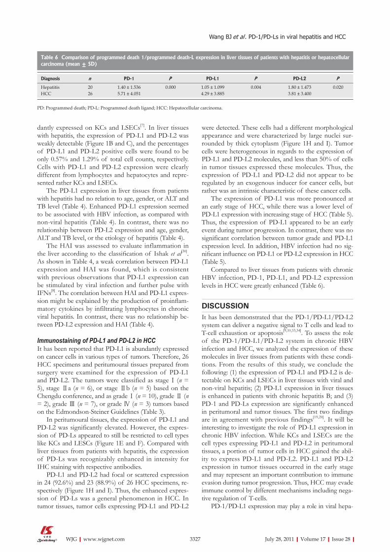
Table 6 Comparison of programmed death 1/programmed death-L expression in liver tissues of patients with hepatitis or hepatocellular carcinoma (mean ± SD)
Diagnosis n PD-1 P PD-L1 P PD-L2 P
Hepatitis 20 1.40 ± 1.536 0.000 1.05 ± 1.099 0.004 1.80 ± 1.473 0.020HCC 26 5.71 ± 4.051 4.29 ± 3.885 3.81 ± 3.400
PD: Programmed death; PD-L: Programmed death ligand; HCC: Hepatocellular carcinoma.
Wang BJ et al . �D-1/�D-Ls in viral hepatitis and HCC

3328 July 28, 2011|Volume 17|Issue 28|WJG|www.wjgnet.com
titis[13,14,17,18]. Upon activation, T-cells express PD-1 and are therefore susceptible to negative signaling by its li-gands[33,34]. In our work, it is evident that liver-infiltrating lymphocytes were positive for PD-1. The activated T-cells may enter liver tissues due to attraction by chemokines. Due to the expression of PD-L1 and PD-L2 on KCs and LSECs in liver tissues and on cancer cells in HCC, liver-infiltrating cells would frequently encounter negative sig-nals and become “exhausted” T-cells.
In chronic viral infection, virus-specific T cells are dysfunctional either because of interaction with regula-tory T cells[35] or interaction between PD-1 and its ligand PD-L1, which results in the down-regulation of T-cell functions[11-14]. Recently, blockage of PD-L1 emerged as an effective measure to promote the proliferation and functions of T-cells in lymphocytic choriomeningitis virus infection of mice[15]. Barber et al[15] demonstrated that vi-rus-specific, exhausted cytotoxic T-lymphocytes regained their ability to proliferate and perform functions such as IFN-γ production and killing of target cells. In HIV- and HCV-positive patients, virus-specific cells also have high expression levels of PD-1, which may explain their dysfunction[12-14,16-18]. Therefore, the blockage of PD-L1 and PD-L2 may restore impaired T-cell functions in in-dividuals with persistent HBV infection and may lead to effective immunological control of viral infection. It is evident that T-cell functions are impaired in patients chronically infected with HBV. In our work, PD-L1 is up-regulated in chronic HBV, suggesting that PD-L1 may play an important role in chronic HBV infection due to the negative control of proliferation and func-tions of liver-infiltrating T-cells. A recent study demon-strated that a blockage of PD-L1 could restore the func-tions of peripheral and intrahepatic T-cells from patients with chronic hepatitis B[36].
Our findings have important implications for immu-notherapeutic approaches to chronic hepatitis and HCC. Until now, the focus of experimental approaches has been to improve the ability of vaccines to induce a broad, strong T-cell response. However, these efforts could be compromised by the fact that specific T cells may be un-able to exert their functions in the liver due to negative regulation, despite effective priming in the peripheral and local lymphoid organs. Thus, future research on immuno-therapeutic approaches should consider the blockage of negative T-cell regulation in combination with immunos-timulation.
Taken together, PD-L1 and PD-L2 were expressed by KCs and LSECs independent of viral and non-viral hepa-titis and were up-regulated by chronic HBV infection and in HCC. The presence of PD-L1 and PD-L2 may lead to the suppression of immune responses and therefore fa-cilitate viral persistence and carcinogenesis. Furthermore, the expression of PD-1, PD-L1, and PD-L2 in HCC was significantly higher than in hepatitis and correlated with the stage of HCC and the number of infiltrating lym-phocytes, indicating the importance of the PD-1/PD-Ls system in tumor development.
COMMENTSBackgroundChronic hepatitis B and hepatocellular carcinoma (HCC) remain the big problem in China and around the world. Immune tolerance characterized with weak or absent specific T-cell responses to hepatitis B virus (HBV) or tumor is responsible for the pathogenesis of hepatitis and tumor. In this content, under-standing tolerance mechanisms in chronic hepatitis B and HCC will further help the design of strategies to overcome the unresponsiveness of T cells. Research frontiersIt has been demonstrated that the programmed death (PD)-1/PD ligand 1 (PD-L1)/PD-L2 system can deliver a negative signal to T cells and lead to T-cell exhaustion or apoptosis. Recent data from other chronic viral infections, such as lymphocytic choriomeningitis virus, human immunodeficiency virus, and hepatitis C virus, and other tumors, such as breast cancer, pancreatic cancer, and non-small cell lung cancer, indicated that up-regulation of the PD-1/PD-L1 system may be responsible for the impairment of T-cell function. In this study, the authors investigated the expression of PD-L1/2 and PD-1 in the liver in the context of chronic hepatitis B and HCC.Innovations and breakthroughsRecent reports have highlighted the importance of PD-1/PD-Ls system as a negative immune regulator in pathogenesis of chronic viral infection and tumors. In this study, the authors found: (1) the expression of PD-L1 was signifi-cantly correlated with HBV infection and with the stage of HCC; (2) PD-1 and PD-Ls were significantly up-regulated in HCC specimens, which indicated for the first time that PD-L1 may contribute to negative regulation of the immune response in chronic hepatitis B; and (3) PD-1 and PD-Ls may play a role in im-mune evasion of HCC.ApplicationsBy identifying the fact that expression of PD-1/PD-Ls is highly up-regulated in hepatitis B and HCC, our study indicated the PD-1/PD-Ls system plays a role in immune tolerance of hepatitis B and HCC, and thus may represent a future strategy for therapeutic intervention in the treatment of patients with hepatitis B or HCC.TerminologyPD-1 is a member of the CD28 family and is involved in the regulation of T-cell activation. PD-1 is expressed on T cells, B cells, and myeloid cells. Two ligands for PD-1, PD-L1 and PD-L2 (also known as B7-H1 and B7-DC), have been identified and have features as co-stimulatory molecules. A large number of publications have indicated a role for PD-L1 in the negative regulation of T-cell functions and the maintenance of peripheral tolerance. The exact role of PD-L2 requires further definition.Peer reviewThis manuscript is impressive in that it investigated the expression of PD-1 and PD-Ls in liver tissue from patients with hepatitis and HCC using immunostaining in detail.
REFERENCES1 Guidotti LG, Chisari FV. Immunobiology and pathogenesis
of viral hepatitis. Annu Rev Pathol 2006; 1: 23-612 Guidotti LG, Rochford R, Chung J, Shapiro M, Purcell R,
Chisari FV. Viral clearance without destruction of infected cells during acute HBV infection. Science 1999; 284: 825-829
3 Ferrari C, Penna A, Sansoni P, Giuberti T, Fiaccadori F. Clon-al analysis of intrahepatic T lymphocytes in chronic active hepatitis. Isolation of a T-cell line specific for hepatitis B core antigen from a patient with serological evidence of exposure to HBV. J Hepatol 1986; 3: 384-392
4 Kakumu S, Ishikawa T, Wakita T, Yoshioka K, Takayanagi M, Tahara H, Kusakabe A. Interferon-gamma production specific for hepatitis B virus antigen by intrahepatic T lym-phocytes in patients with acute and chronic hepatitis B. Am J Gastroenterol 1994; 89: 92-96
5 Keir ME, Butte MJ, Freeman GJ, Sharpe AH. PD-1 and its ligands in tolerance and immunity. Annu Rev Immunol 2008; 26: 677-704
COMMENTS
Wang BJ et al . �D-1/�D-Ls in viral hepatitis and HCC
3329 July 28, 2011|Volume 17|Issue 28|WJG|www.wjgnet.com
6 Subudhi SK, Alegre ML, Fu YX. The balance of immune responses: costimulation verse coinhibition. J Mol Med 2005; 83: 193-202
7 Yamazaki T, Akiba H, Iwai H, Matsuda H, Aoki M, Tanno Y, Shin T, Tsuchiya H, Pardoll DM, Okumura K, Azuma M, Yagita H. Expression of programmed death 1 ligands by mu-rine T cells and APC. J Immunol 2002; 169: 5538-5545
8 Mühlbauer M, Fleck M, Schütz C, Weiss T, Froh M, Blank C, Schölmerich J, Hellerbrand C. PD-L1 is induced in hepato-cytes by viral infection and by interferon-alpha and -gamma and mediates T cell apoptosis. J Hepatol 2006; 45: 520-528
9 Dong H, Zhu G, Tamada K, Flies DB, van Deursen JM, Chen L. B7-H1 determines accumulation and deletion of intrahe-patic CD8(+) T lymphocytes. Immunity 2004; 20: 327-336
10 Iwai Y, Terawaki S, Ikegawa M, Okazaki T, Honjo T. PD-1 inhibits antiviral immunity at the effector phase in the liver. J Exp Med 2003; 198: 39-50
11 Wherry EJ, Ha SJ, Kaech SM, Haining WN, Sarkar S, Kalia V, Subramaniam S, Blattman JN, Barber DL, Ahmed R. Molecu-lar signature of CD8+ T cell exhaustion during chronic viral infection. Immunity 2007; 27: 670-684
12 Elrefaei M, Baker CA, Jones NG, Bangsberg DR, Cao H. Presence of suppressor HIV-specific CD8+ T cells is associat-ed with increased PD-1 expression on effector CD8+ T cells. J Immunol 2008; 180: 7757-7763
13 Golden-Mason L, Palmer B, Klarquist J, Mengshol JA, Castelblanco N, Rosen HR. Upregulation of PD-1 expression on circulating and intrahepatic hepatitis C virus-specific CD8+ T cells associated with reversible immune dysfunc-tion. J Virol 2007; 81: 9249-9258
14 Radziewicz H, Ibegbu CC, Fernandez ML, Workowski KA, Obideen K, Wehbi M, Hanson HL, Steinberg JP, Masopust D, Wherry EJ, Altman JD, Rouse BT, Freeman GJ, Ahmed R, Grakoui A. Liver-infiltrating lymphocytes in chronic human hepatitis C virus infection display an exhausted phenotype with high levels of PD-1 and low levels of CD127 expression. J Virol 2007; 81: 2545-2553
15 Barber DL, Wherry EJ, Masopust D, Zhu B, Allison JP, Sharpe AH, Freeman GJ, Ahmed R. Restoring function in exhausted CD8 T cells during chronic viral infection. Nature 2006; 439: 682-687
16 Freeman GJ, Wherry EJ, Ahmed R, Sharpe AH. Reinvigo-rating exhausted HIV-specific T cells via PD-1-PD-1 ligand blockade. J Exp Med 2006; 203: 2223-2227
17 Penna A, Pilli M, Zerbini A, Orlandini A, Mezzadri S, Sac-chelli L, Missale G, Ferrari C. Dysfunction and functional restoration of HCV-specific CD8 responses in chronic hepati-tis C virus infection. Hepatology 2007; 45: 588-601
18 Urbani S, Amadei B, Tola D, Pedrazzi G, Sacchelli L, Cavallo MC, Orlandini A, Missale G, Ferrari C. Restoration of HCV-specific T cell functions by PD-1/PD-L1 blockade in HCV infection: effect of viremia levels and antiviral treatment. J Hepatol 2008; 48: 548-558
19 Kassel R, Cruise MW, Iezzoni JC, Taylor NA, Pruett TL, Hahn YS. Chronically inflamed livers up-regulate expres-sion of inhibitory B7 family members. Hepatology 2009; 50: 1625-1637
20 Gao Q, Wang XY, Qiu SJ, Yamato I, Sho M, Nakajima Y, Zhou J, Li BZ, Shi YH, Xiao YS, Xu Y, Fan J. Overexpression of PD-L1 significantly associates with tumor aggressiveness and postoperative recurrence in human hepatocellular carci-noma. Clin Cancer Res 2009; 15: 971-979
21 Blank C, Mackensen A. Contribution of the PD-L1/PD-1 pathway to T-cell exhaustion: an update on implications for chronic infections and tumor evasion. Cancer Immunol Immu-nother 2007; 56: 739-745
22 Zhang P, Su DM, Liang M, Fu J. Chemopreventive agents induce programmed death-1-ligand 1 (PD-L1) surface ex-pression in breast cancer cells and promote PD-L1-mediated
T cell apoptosis. Mol Immunol 2008; 45: 1470-147623 Nomi T, Sho M, Akahori T, Hamada K, Kubo A, Kanehiro
H, Nakamura S, Enomoto K, Yagita H, Azuma M, Nakajima Y. Clinical significance and therapeutic potential of the pro-grammed death-1 ligand/programmed death-1 pathway in human pancreatic cancer. Clin Cancer Res 2007; 13: 2151-2157
24 Hamanishi J, Mandai M, Iwasaki M, Okazaki T, Tanaka Y, Yamaguchi K, Higuchi T, Yagi H, Takakura K, Minato N, Honjo T, Fujii S. Programmed cell death 1 ligand 1 and tu-mor-infiltrating CD8+ T lymphocytes are prognostic factors of human ovarian cancer. Proc Natl Acad Sci USA 2007; 104: 3360-3365
25 Inman BA, Sebo TJ, Frigola X, Dong H, Bergstralh EJ, Frank I, Fradet Y, Lacombe L, Kwon ED. PD-L1 (B7-H1) expression by urothelial carcinoma of the bladder and BCG-induced granulomata: associations with localized stage progression. Cancer 2007; 109: 1499-1505
26 Nakanishi J, Wada Y, Matsumoto K, Azuma M, Kikuchi K, Ueda S. Overexpression of B7-H1 (PD-L1) significantly as-sociates with tumor grade and postoperative prognosis in human urothelial cancers. Cancer Immunol Immunother 2007; 56: 1173-1182
27 Ghebeh H, Mohammed S, Al-Omair A, Qattan A, Lehe C, Al-Qudaihi G, Elkum N, Alshabanah M, Bin Amer S, Tulbah A, Ajarim D, Al-Tweigeri T, Dermime S. The B7-H1 (PD-L1) T lymphocyte-inhibitory molecule is expressed in breast can-cer patients with infiltrating ductal carcinoma: correlation with important high-risk prognostic factors. Neoplasia 2006; 8: 190-198
28 Ohigashi Y, Sho M, Yamada Y, Tsurui Y, Hamada K, Ikeda N, Mizuno T, Yoriki R, Kashizuka H, Yane K, Tsushima F, Otsuki N, Yagita H, Azuma M, Nakajima Y. Clinical signifi-cance of programmed death-1 ligand-1 and programmed death-1 ligand-2 expression in human esophageal cancer. Clin Cancer Res 2005; 11: 2947-2953
29 Konishi J, Yamazaki K, Azuma M, Kinoshita I, Dosaka-Akita H, Nishimura M. B7-H1 expression on non-small cell lung cancer cells and its relationship with tumor-infiltrating lymphocytes and their PD-1 expression. Clin Cancer Res 2004; 10: 5094-5100
30 Ishak K, Baptista A, Bianchi L, Callea F, De Groote J, Gudat F, Denk H, Desmet V, Korb G, MacSween RN. Histological grading and staging of chronic hepatitis. J Hepatol 1995; 22: 696-699
31 Sheng JM, Zhao WH, Wu FS, Ma ZM, Feng YZ, Zhou XR, Teng LS. The Chinese classification system compared with TNM staging in prognosis of patients with primary hepatic carcinoma after resection. Hepatobiliary Pancreat Dis Int 2005; 4: 561-564
32 Detre S, Saclani Jotti G, Dowsett M. A “quickscore” method for immunohistochemical semiquantitation: validation for oestrogen receptor in breast carcinomas. J Clin Pathol 1995; 48: 876-878
33 Freeman GJ, Long AJ, Iwai Y, Bourque K, Chernova T, Nishimura H, Fitz LJ, Malenkovich N, Okazaki T, Byrne MC, Horton HF, Fouser L, Carter L, Ling V, Bowman MR, Car-reno BM, Collins M, Wood CR, Honjo T. Engagement of the PD-1 immunoinhibitory receptor by a novel B7 family mem-ber leads to negative regulation of lymphocyte activation. J Exp Med 2000; 192: 1027-1034
34 Leibson PJ. The regulation of lymphocyte activation by in-hibitory receptors. Curr Opin Immunol 2004; 16: 328-336
35 Rouse BT, Sarangi PP, Suvas S. Regulatory T cells in virus infections. Immunol Rev 2006; 212: 272-286
36 Fisicaro P, Valdatta C, Massari M, Loggi E, Biasini E, Sac-chelli L, Cavallo MC, Silini EM, Andreone P, Missale G, Fer-rari C. Antiviral intrahepatic T-cell responses can be restored by blocking programmed death-1 pathway in chronic hepa-titis B. Gastroenterology 2010; 138: 682-693, 693.e1-693.e4
S- Editor Tian L L- Editor O’Neill M E- Editor Zheng XM
Wang BJ et al . �D-1/�D-Ls in viral hepatitis and HCC