Immunopathology - Massachusetts General Hospital
16
333 Chapter Immunopathology Atul K. Bhan, Robert B. Colvin, and Harold F. Dvorak T he application of immunology to clini- cal medicine and the development of immu- nopathology as a specialized field stem largely from the pioneering work of the great immunologist/ pathologist Paul Ehrlich, who received the 1908 Noble Prize for his work on immune responses in infection (1). e introduction of immunofluo- rescence techniques by Dr. Albert Coons in the 1940s set the stage for establishment of immu- nohistochemical techniques and development of flow cytometric analysis of cells. Dr. Coons, who had been trained in internal medicine at the Mas- sachusetts General Hospital (MGH), performed most of his work at Harvard Medical School (HMS), initially in the Department of Microbi- ology and Immunology and later in the Pathology Department. Over the years, immunopathology has applied the knowledge obtained from basic immunology research to achieve a better under- standing of disease pathogenesis, develop sophis- ticated tests for accurate clinical diagnosis, and implement new therapeutic strategies. e history of immunopathology at MGH will be described for three periods: (1) before 1975, when immunopathology concepts were being developed and when the Immunopathol- ogy Laboratory was established; (2) from 1975 to 1991, when the Immunopathology Unit was formed and flourished under the leadership of Dr. Robert T. McCluskey (chapter 14) to become the dominant research unit in MGH Pathology, and when, beginning in the 1980s, the unit rap- idly implemented diagnostic immunohistochem- istry into the routine diagnostic service; and (3) from 1991 to the present, when the unit contin- ued to develop new immunological diagnostic tests and conduct basic research. Early Years: Before e earliest manifestations of the then-nascent field of immunopathology at MGH were the studies of Dr. Louis Dienes, a bacteriologist who was recruited to the Pathology department in 1930 (see figure 21.3; chapters 5 and 21). He showed that simple proteins (ovalbumin) could elicit a “tuber- culin reaction” in the skin if the animal (guinea pig) had previously been sensitized by inoculation of the protein into a tuberculous lesion (2). At the time only products of organisms were thought to trigger this type of response, termed a delayed- type hypersensitivity reaction and later shown to be a T cell mediated reaction. With Dr. Tracy Mallory (chapter 6) in 1932 he characterized the morphology of the lesions and contrasted them with antibody-mediated reactions (Arthus reac- tion). Tuberculin sensitivity could also be pro- duced by sensitization with protein alone (no tuberculous infection), although it was milder (Jones-Mote reaction). Also with Dr. Mallory, in 1937, he showed that tuberculin hypersensitivity developed simultaneously with the mononuclear infiltrate surrounding tuberculous lesions.
Transcript of Immunopathology - Massachusetts General Hospital

333
Chapter
ImmunopathologyAtul K. Bhan, Robert B. Colvin,
and Harold F. Dvorak
The application of immunology to clini-cal medicine and the development of immu-
nopathology as a specialized fi eld stem largely from the pioneering work of the great immunologist/pathologist Paul Ehrlich, who received the 1908 Noble Prize for his work on immune responses in infection (1). Th e introduction of immunofl uo-rescence techniques by Dr. Albert Coons in the 1940s set the stage for establishment of immu-nohistochemical techniques and development of fl ow cytometric analysis of cells. Dr. Coons, who had been trained in internal medicine at the Mas-sachusetts General Hospital (MGH), performed most of his work at Harvard Medical School (HMS), initially in the Department of Microbi-ology and Immunology and later in the Pathology Department. Over the years, immunopathology has applied the knowledge obtained from basic immunology research to achieve a better under-standing of disease pathogenesis, develop sophis-ticated tests for accurate clinical diagnosis, and implement new therapeutic strategies.
Th e history of immunopathology at MGH will be described for three periods: (1) before 1975, when immunopathology concepts were being developed and when the Immunopathol-ogy Laboratory was established; (2) from 1975 to 1991, when the Immunopathology Unit was formed and fl ourished under the leadership of Dr. Robert T. McCluskey (chapter 14) to become the dominant research unit in MGH Pathology,
and when, beginning in the 1980s, the unit rap-idly implemented diagnostic immunohistochem-istry into the routine diagnostic service; and (3) from 1991 to the present, when the unit contin-ued to develop new immunological diagnostic tests and conduct basic research.
Early Years: Before
Th e earliest manifestations of the then-nascent fi eld of immunopathology at MGH were the studies of Dr. Louis Dienes, a bacteriologist who was recruited to the Pathology department in 1930 (see fi gure 21.3; chapters 5 and 21). He showed that simple proteins (ovalbumin) could elicit a “tuber-culin reaction” in the skin if the animal (guinea pig) had previously been sensitized by inoculation of the protein into a tuberculous lesion (2). At the time only products of organisms were thought to trigger this type of response, termed a delayed-type hypersensitivity reaction and later shown to be a T cell mediated reaction. With Dr. Tracy Mallory (chapter 6) in 1932 he characterized the morphology of the lesions and contrasted them with antibody-mediated reactions (Arthus reac-tion). Tuberculin sensitivity could also be pro-duced by sensitization with protein alone (no tuberculous infection), although it was milder (Jones-Mote reaction). Also with Dr. Mallory, in 1937, he showed that tuberculin hypersensitivity developed simultaneously with the mononuclear infi ltrate surrounding tuberculous lesions.
pathology_chap23.indd 333 8/16/11 10:25 AM
Keen Minds to Explore the Dark Continents of Disease
334
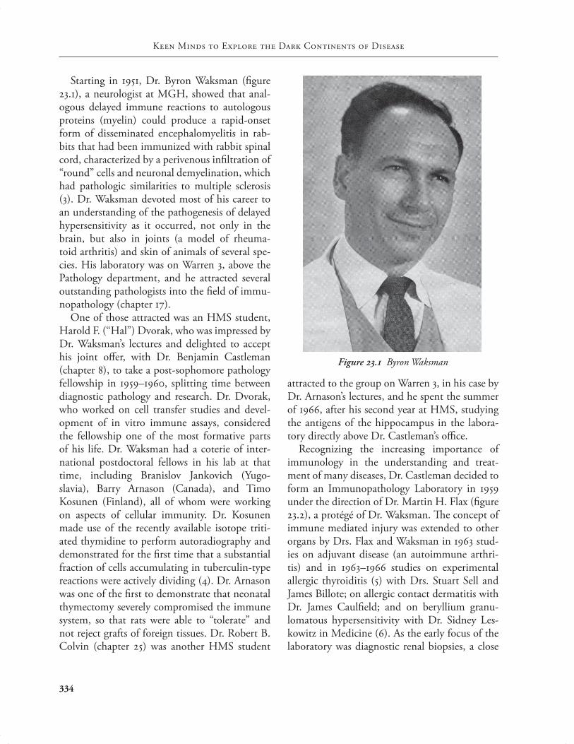
Starting in 1951, Dr. Byron Waksman (fi gure 23.1), a neurologist at MGH, showed that anal-ogous delayed immune reactions to autologous proteins (myelin) could produce a rapid-onset form of disseminated encephalomyelitis in rab-bits that had been immunized with rabbit spinal cord, characterized by a perivenous infi ltration of “round” cells and neuronal demyelination, which had pathologic similarities to multiple sclerosis (3). Dr. Waksman devoted most of his career to an understanding of the pathogenesis of delayed hypersensitivity as it occurred, not only in the brain, but also in joints (a model of rheuma-toid arthritis) and skin of animals of several spe-cies. His laboratory was on Warren 3, above the Pathology department, and he attracted several outstanding pathologists into the fi eld of immu-nopathology (chapter 17).
One of those attracted was an HMS student, Harold F. (“Hal”) Dvorak, who was impressed by Dr. Waksman’s lectures and delighted to accept his joint off er, with Dr. Benjamin Castleman (chapter 8), to take a post-sophomore pathology fellowship in 1959–1960, splitting time between diagnostic pathology and research. Dr. Dvorak, who worked on cell transfer studies and devel-opment of in vitro immune assays, considered the fellowship one of the most formative parts of his life. Dr. Waksman had a coterie of inter-national postdoctoral fellows in his lab at that time, including Branislov Jankovich (Yugo-slavia), Barry Arnason (Canada), and Timo Kosunen (Finland), all of whom were working on aspects of cellular immunity. Dr. Kosunen made use of the recently available isotope triti-ated thymidine to perform autoradiography and demonstrated for the fi rst time that a substantial fraction of cells accumulating in tuberculin-type reactions were actively dividing (4). Dr. Arnason was one of the fi rst to demonstrate that neonatal thymectomy severely compromised the immune system, so that rats were able to “tolerate” and not reject grafts of foreign tissues. Dr. Robert B. Colvin (chapter 25) was another HMS student
attracted to the group on Warren 3, in his case by Dr. Arnason’s lectures, and he spent the summer of 1966, after his second year at HMS, studying the antigens of the hippocampus in the labora-tory directly above Dr. Castleman’s offi ce.
Recognizing the increasing importance of immunology in the understanding and treat-ment of many diseases, Dr. Castleman decided to form an Immunopathology Laboratory in 1959 under the direction of Dr. Martin H. Flax (fi gure 23.2), a protégé of Dr. Waksman. Th e concept of immune mediated injury was extended to other organs by Drs. Flax and Waksman in 1963 stud-ies on adjuvant disease (an autoimmune arthri-tis) and in 1963–1966 studies on experimental allergic thyroiditis (5) with Drs. Stuart Sell and James Billote; on allergic contact dermatitis with Dr. James Caulfi eld; and on beryllium granu-lomatous hypersensitivity with Dr. Sidney Les-kowitz in Medicine (6). As the early focus of the laboratory was diagnostic renal biopsies, a close
Figure 23.1 Byron Waksman
pathology_chap23.indd 334 8/16/11 10:25 AM

Immunopathology
335
relationship developed between the Immunopa-thology Laboratory and nephrologists as well as the Renal Transplantation Unit.
Dr. Vivian Pinn started working with Dr. Flax in 1967, when she joined MGH as a resident in Pathology (see fi gure 7.26). Dr. Pinn moved with Dr. Flax to Tufts University Medical School in 1970, when Dr. Flax was made the Chairman of the Department of Pathology there. She remained involved in immunopathology of the kidney and subsequently became Professor and Chairman of Pathology at Howard University College of Med-icine, the fi rst African American woman to chair an academic pathology department.
Dr. Colvin, who at that time was a resident in Pathology, worked on renal and transplant pathology with Drs. Pinn and Flax and became the resident expert in this area when they left for Tufts. Dr. Colvin was able to resuscitate an ancient Reichert fl uorescent microscope for immunofl uorescence, which had to be done at night, since the room on Warren 1 had no shades on the windows.
Dr. Dvorak had come back to MGH for an internship and residency in Pathology in 1963 and then, working with Dr. Flax, made impor-tant observations regarding immunological unre-sponsiveness induced by intravenous tolerizing antigen. After two years of research at NIH, Dr. Dvorak returned to the MGH in 1967 and began developing and directing immunopathology research. Dr. Dvorak used elegant one-micron Giemsa-stained Epon sections, prepared by his research assistant, Eleanor Manseau, to study the cellular details of delayed-hypersensitivity reac-tions by light microscopy (7). He was, therefore, for the fi rst time able to appreciate infi ltrating basophil leukocytes in tissues and their participa-tion in a type of delayed-hypersensitivity reaction (Jones-Mote reaction) induced by immunization with protein antigens in incomplete Freund’s adjuvant (which were probably what Drs. Dienes and Mallory had created by injecting ovalbumin without tubercle bacillus). Because of its high
basophil content, the reaction was called cuta-neous basophil hypersensitivity (CBH). Simi-lar, basophil-rich reactions were found in other organs and tissues in response to an immune challenge: for example, the eye in ocular hyper-sensitivity and human kidneys undergoing acute allograft rejection (8, 9). Dr. Dvorak remembers showing one-micron Epon slides to Dr. Castle-man; he was impressed but asked for a hematoxy-lin and eosin (H&E) section of the same lesions and had no trouble detecting basophils!
Th ese studies were extended to human cellular immunity when Dr. Dvorak developed poison ivy on Martha’s Vineyard. He biopsied his rash and found it was swarming with basophils. He secured the help of Dr. Colvin and Dr. Martin C. Mihm Jr. of Dermatopathology (chapter 18) to extend these studies to other contact allergens, employing volunteers who were biopsied, includ-ing the investigators. Dr. Ann Dvorak (Hal’s
Figure 23.2 Martin Flax
pathology_chap23.indd 335 8/16/11 10:25 AM

Keen Minds to Explore the Dark Continents of Disease
336
wife), who had done a postdoctoral fellowship in electron microscopy with Dr. Morris Karnovsky at HMS and had helped demonstrate basophils as an important feature of other cellular immunity in guinea pigs, also participated in the studies. Dr. Ann Dvorak showed through ultrastructural studies the activation of microvascular endothe-lial cells and pericytes in the venules from which lymphocytes, basophils, and other infl ammatory cells were extravasating, and the degranulation of both basophils and mast cells as features of cellu-lar immunity. She found that basophils and mast cells in cellular immune reactions underwent not the explosive degranulation characteristic of anaphylaxis, but, rather, a partial type of piece-meal degranulation in which granule contents were released gradually over time by a vesicular transport mechanism. Her concept of piecemeal degranulation is now widely accepted not only in basophils but also in degranulating eosinophils, which she subsequently studied after moving to Beth Israel Hospital in 1979. One important observation made in these studies was that fi brin deposited outside the vessels was related to the induration, a characteristic feature of tubercu-lin hypersensitivity (10). Proof that fi brin was responsible for causing induration came from subsequent studies by Dr. Colvin in two patients with congenital afi brinogenemia; both developed erythematous delayed reactions with a typical cellular infi ltrate but lacked fi brin deposition and induration.
–: The McCluskey Era
As Dr. Dvorak’s studies were progressing, Dr. Robert McCluskey succeeded Dr. Castleman as Chief of Pathology (chapter 13). During Dr. McCluskey’s tenure at Children’s Hospital in Bos-ton (1971–1974), he established immunopathol-ogy research and developed collaborations that continued after his move to MGH. In 1975 Dr. Atul K. Bhan, who had been a postdoctoral fel-low with Dr. McCluskey following completion of Chief Residency in Pathology and a postdoctoral
research fellowship at Children’s Hospital, moved to MGH to pursue immunopathology research. Th e Immunopathology Laboratory at that time was located on the fi rst fl oor of the Warren Build-ing. Dr. Elizabeth Hammond, who graduated from the University of Utah School of Medicine in 1967, joined the lab fi rst as a research fellow in 1972 and subsequently as a staff member in 1974. Dr. Stephen J. Galli, who graduated from HMS in 1973, joined the lab as a research fel-low in 1977. Both Dr. Hammond and Dr. Galli had been residents in Pathology at MGH. At that time as well, Dr. Ann Dvorak was recruited from Tufts to establish a clinical and research electron microscopy laboratory.
In the 1970s MGH administration committed new resources to the Pathology department, add-ing much needed space for research on the fi fth and eighth fl oors of the recently built Cox Build-ing. A committee composed of Drs. Hal and Ann Dvorak, Bhan, Hammond, and Mihm designed the Cox space for research as well as for diagnos-tic immunopathology and electron microscopy. Th e highly functional open design has been copied by the other major research laboratories at MGH (e.g., the Wellman/Th ier, Martin, and Simches buildings). Th e fi fth-fl oor Immunopa-thology Unit was completed by January 1976 and the eighth-fl oor animal facilities in April 1977. Th e only glitch was that the electron microscopes were installed over the massive ventilation fans bolted to the ceiling of the fourth fl oor; the solu-tion was to suspend the microscopes on bungee cords attached to a 3-inch-thick iron plate.
Th e new laboratories in the Cox Building provided the infrastructure for the development of the nationally recognized Immunopathology Unit. Th e unit, directed by Hal Dvorak, was staff ed initially by Drs. Bhan, Hammond, John Long, Galli, Neil Orenstein, and Colvin, who had returned to MGH after working as an experi-mental pathologist at Walter Reed Army Institute of Pathology from 1972 to 1975. Dr. Hammond studied macrophage migration inhibitory factor,
pathology_chap23.indd 336 8/16/11 10:25 AM

Immunopathology
337
and she and Dr. Colvin got their fi rst R01 grant together on the clotting system in delayed-type hypersensitivity. She soon left to accompany her husband to Salt Lake City, however, where she became Professor of Pathology at the University of Utah and Chair of the Department of Pathol-ogy at the LDS Hospital. Dr. Galli and his wife, Anne, continued the work on basophils with Hal Dvorak, demonstrating that purifi ed guinea pig basophils were able to synthesize histamine. Dr. Galli subsequently focused his research on mast cells, and he became one of the world’s leading authorities on mast cell biology; he moved from Beth Israel to become Chair of the Department of Pathology at Stanford University School of Medicine in 1999.
Dr. Hal Dvorak (fi gure 23.3) extended his studies of the immune response to tumors and collaborated with Dr. W. Hallowell Churchill at
Brigham and Women’s Hospital (9). Dr. Dvorak found that supernatants from nearly all human and animal tumors generated intense blue spots in the Evans blue dye permeability assay, whereas those from several normal cells did not (fi gure 23.4). He called this tumor supernatant permeabi-lizing activity vascular permeability factor, or VPF (11). Subsequently, VPF was shown to be a weak endothelial cell mitogen, was renamed vascular endothelial growth factor (VEGF), or, more spe-cifi cally, VEGF-A, and became a therapeutic tar-get in cancer and macular degeneration. Subse-quent studies revealed that VPF/VEGF was over-expressed by most malignant tumors and at lower levels in a number of normal adult tissues. After moving to Beth Israel Hospital in 1979 (taking with him his wife, Ann, and Stephen Galli), Dr. Dvorak continued his work and found that VPF/VEGF had a central role in wound healing and chronic infl ammatory diseases such as psoriasis, rheumatoid arthritis, and induced lymphangio-genesis as well as angiogenesis; others discovered that it was essential for the development of the normal vasculature. Th us, Dr. Dvorak’s discov-ery of VPF resulted not only in the fi nding of a new molecule but also in the elucidation of a process—one that, although not yet completely understood, is responsible for initiating stroma formation and tissue repair following injury. Unfortunately, this same process also facilitates the growth and spread of malignant tumors, as well as having central roles in diabetic retinopa-thy and several types of infl ammatory diseases.
Because of Dr. McCluskey’s research interest in renal diseases, the immunopathogenesis of kid-ney diseases became a major research and diag-nostic focus of the Immunopathology Unit (fi g-ure 23.5). He built a strong research team, includ-ing Dr. Colvin, Dr. Bhan, and Bernard Collins to study mechanisms involved in renal diseases. Dr. Eveline E. Schneeberger, who had worked previously with Dr. McCluskey at Children’s Hospital, joined the team in 1979. Drs. Bhan and McCluskey explored novel mechanisms of
Figure 23.3 Harold Dvorak working in the Immunopathology Laboratory, Cox 5, late 1970s
pathology_chap23.indd 337 8/16/11 10:25 AM

Keen Minds to Explore the Dark Continents of Disease
338
glomerulonephritis. By adoptive transfer experi-ments, they conclusively showed that T cells could induce glomerular injury against a planted glomerular antigen, challenging the dogma that T cells had no role in glomerulonephritis (12). Dr. McCluskey recruited Dr. Lynn Baird to identify the antigen of membranous glomeru-lonephritis, a goal he had long pursued. Th ese studies and a subsequent collaboration with Dr. John Smith led to the identifi cation and clon-ing of an autoantigen located in rat renal tubules and podocytes (megalin), which was the target in Heyman nephritis (13), a rat model of membra-nous glomerulonephritis (MGN); however, this antigen did not prove to be the human autoan-tigen they were seeking. Dr. McCluskey contin-ued to work on this theme after his retirement as chief in 1991 and even secured an NIH grant to support his studies. Dr. Collins and Colvin con-tinue working on the target antigens of MGN in 2011. Collins also took a major initiative in devel-oping and expanding the application of immu-nofl uorescence in human renal biopsies and
developed many tests that were off ered as part of the departmental reference laboratory in renal pathology, including the Western blot assay for the detection of antibodies to glomerular base-ment membrane (GBM), which is the defi nitive diagnostic test for anti-GBM antibodies. Immu-nofl uorescence studies were also applied for the diagnosis of skin disorders, including indirect immunofl uorescence tests for the detection of circulating autoantibodies in patients with bul-lous lesions of the skin.
Dr. John Niles, a nephrologist, started post-doctoral research training with Dr. McCluskey in 1985. Together they investigated the role of anti-neutrophil cytoplasmic autoantibodies (ANCA) in the pathogenesis of Wegener’s granulomato-sis. Th ese studies led to identifi cation of one of the ANCA autoantigens, proteinase-3, and the development of quantitative diagnostic assays. Dr. Niles has achieved international recogni-tion as an expert in ANCA-related diseases and directs the MGH Pathology reference laboratory for ANCA testing (14, 15).
Robert Colvin became the Director of the Immunopathology Unit in 1979. In Dr. Colvin, Dr. McCluskey found a colleague with a com-mon interest in renal pathogenesis and develop-ing a strong diagnostic renal pathology program. While a house offi cer in Pathology at MGH, Dr. Colvin had become interested in renal and trans-plant pathology. Dr. Paul Russell, a distinguished surgeon who established the MGH Transplant Service, became a mentor and longtime friend in this fi eld. From 1971 to 1972, Dr. Colvin was an NIH Research Fellow under the mentorship of Dr. Harold Dvorak, working on the role of the clotting system in T cell mediated immune reactions. Dr. Colvin extended the studies to the role of fi bronectin in infl ammatory reactions and wound healing with a fellow, Dr. Richard Clark; together they defi ned the concept of a “provi-sional matrix” (16). Dr. Colvin worked on the pathogenesis of autoimmune tubulointerstitial diseases, continuing the work on anti-tubular
Figure 23.4 Harold and Ann Dvorak in front of the research poster describing tumor secreted mediators,
Cox 5, late 1970s
pathology_chap23.indd 338 8/16/11 10:25 AM

Immunopathology
339
had close associations with members of the MGH Pathology staff (chapter 7). Both Benja-min Castleman and Walter Putschar had visited AIIMS to help the development of the Pathology department. Dr. Bhan’s research experience while at Children’s Hospital from 1972 to 1975 and col-laborative studies with Dr. Stuart Schlossman at Dana-Farber Cancer Institute set the foundation for his future work on lymphoid cells in nor-mal and diseased states. One of the important fi ndings of these studies was the demonstration that T cells, but not B cells, migrate to rejecting allografts (18).
Th e development of hybridoma-derived mono-clonal antibodies in 1975 had a profound infl u-ence on all aspects of biological sciences, includ-ing immunopathology. One of the immediate eff ects was on the characterization of normal and neoplastic hematopoietic cell lineages. Dr. Bhan performed important early work to identify and characterize lymphoid cell diff erentiation and dendritic cells in the lymphoid tissues and at the infl ammatory sites (19, 20). Dr. Sibrand Poppema, a visiting fellow from the University of Groningen, participated in the initial studies to identify stages of T cell and B cell location and diff erentiation in the lymphoid tissues (19); Dr. Poppema, interna-tionally recognized for his research on Hodgkin’s disease, became Professor of Pathology at the Uni-versity of Groningen in 1995. Dr. George Murphy (chapter 18), a resident in Pathology at MGH and a trainee of Dr. Mihm’s, carried out studies on the analysis of Langerhans cells and histiocytosis X with monoclonal antibodies in collaboration with Drs. Bhan and Terence Harrist (21).
In 1981 the Diagnostic Immunoperoxidase Laboratory was established by Dr. Bhan with the help of Bruce Kaynor, a talented and dedi-cated technician. Dr. Nancy L. Harris (see fi g-ure 24.9), who had joined the Immunopathol-ogy Unit in 1978 as a research fellow, published several important papers with Dr. Bhan on immunohistochemical characterization of lym-phoid cells for the diagnosis and classifi cation of
basement membrane disease and in transplan-tation. His close interactions with Dr. Benedict Cosimi and other members of the renal trans-plant group resulted in seminal observations and publications and recognition of MGH as a major center of renal transplantation. Dr. Colvin was a key member of the team that tried the fi rst therapeutic monoclonal antibody, OKT3, in a patient who developed acute renal allograft rejec-tion on October 20, 1980 (17). Th e team gath-ered around the prototype fl ow cytometer oper-ated by Dr. Colvin the evening after the fi rst dose and observed that all the T cells had disappeared from the circulation within an hour of treatment. Someone in the fl ow cytometry room quoted Dr. John Collins Warren (chapter 1) and said, “Gen-tlemen, this is no humbug!”
Atul Bhan had been trained in anatomic and experimental pathology at the All India Insti-tute of Medical Sciences (AIIMS), New Delhi, under Dr. Vulimiri Ramalingaswami, who had
Figure 23.5 Robert McCluskey (left) with Richard Dickersin (looking through electron microscope) and
Ann Brescia (electron microscopy technician), Cox 5, late 1970s
pathology_chap23.indd 339 8/16/11 10:25 AM

Keen Minds to Explore the Dark Continents of Disease
340
lymphoid malignancies (22). She continued to apply immunohistochemistry to hematological malignancies and achieved national and interna-tional recognition (chapter 16). Subsequently, the application of immunohistochemical techniques extended to all specialties of surgical pathology. Dr. Salim Kabawat, who joined the Immunopa-thology Unit as a research fellow in Dr. Colvin’s laboratory in 1983, described the reactivity of a monoclonal antibody, OC125, produced against an ovarian tumor by Dr. Robert Bast at Dana-Farber Cancer Institute (23). (As a medical stu-dent, Dr. Bast had spent time in MGH Pathol-ogy; chapter 7.) Th e antigen CA125 recognized by the antibody has become an established marker of ovarian tumors, and the serum levels of this antigen are used for diagnosis and monitoring of patients with some forms of ovarian cancer.
Th e refi nement of immunohistochemical techniques and introduction of antigen retrieval methods in the 1980s made it possible to iden-tify an increasing number of tumor markers with antibodies that were preferentially selected for their ability to recognize antigens in routinely fi xed specimens in surgical pathology. As Direc-tor of the Immunohistochemistry (Immunoper-oxidase) Laboratory, Dr. Bhan took a leading role in integrating immunohistochemistry into surgi-cal pathology and selecting panels of antibodies that allowed characterization of tumor cells, not only by their morphologic features, but also by their molecular profi le. His interactions with the Surgical Pathology staff led to the development of strategies for the characterization of a wide spectrum of tumors, including hematopoietic, soft tissue, endocrine, and metastatic epithelial tumors (unknown primary); perhaps the most signifi cant eff ect was in the diagnosis of poorly diff erentiated malignant tumors.
Th e availability of monoclonal antibodies to functionally distinct lymphoid cells had an immediate infl uence on the immunohistochemi-cal characterization of a wide variety of infl am-matory conditions. Th e analysis also included
allograft rejection and tumor infi ltrating lym-phocytes. Many of Dr. Bhan’s studies were sup-ported by an NIH grant to study immunocom-petent cells infi ltrating human breast cancer.
Dr. James T. Kurnick joined the staff of the Immunopathology Unit in 1980, following pathology training at the University of Colo-rado and postdoctoral work in immunology in Denver and Sweden. His work cloning human T lymphocytes, including the fi rst propagation of helper T cells in any species, brought him to the MGH; here he extended his work with normal T cell responses to soluble and cellular antigens, including work propagating activated T lympho-cytes from sites of infl ammation in transplants, myocarditis, rheumatoid arthritis, pyelonephri-tis, and cancers. Th e work led to functional and genetic studies on cells isolated from infl amed tissues, including the demonstration of clonal dominance of T cell infi ltrates in several condi-tions, such as rheumatoid arthritis and tumors (24, 25). Dr. Ivan Stamenkovic (see below) par-ticipated in the studies of rheumatoid arthritis. Dr. Richard Kradin, who had a joint clinical appointment in both Pathology and Medicine (Pulmonary Unit), joined Dr. Kurnick’s research and developed a protocol in which patients with lung cancer were treated with in vitro expanded tumor infi ltrating lymphocytes (fi gure 23.6). Th is led to the treatment of advanced melanomas and non–small cell lung cancers with infusion of autologous tumor infi ltrating lymphocytes (26). Dr. Kurnik’s work on the immune response to cancers has continued in recent years with the realization that tumors can escape immune detection and destruction by the reversible loss of targeted diff erentiation antigens (27). Following a decade in the Immunopathogy Unit on Cox 5, Dr. Kurnick led a departmental expansion to the new research facilities (Pathology Research at MGH East) in Charlestown.
Dr. Kradin continued his work in pulmonary immunology and interstitial lung diseases (28, 29), addressing the T cell, dendritic cell, and
pathology_chap23.indd 340 8/16/11 10:25 AM

Immunopathology
341
macrophage responses to inhaled antigens and migratory properties of dendritic cells, especially to local lymph nodes, and edited a textbook, Immunopathology of Lung Disease. Five of Dr. Kradin’s laboratory research fellows, including Drs. Patricia Finn and Steven Dubinett, subse-quently pursued successful research careers and are currently department chairmen in the United States and internationally.
Th e Flow Cytometry Laboratory was started in 1978 by Dr. Colvin with a gift of a FACS II cell sorter from the Arthur D. Little Company. Ini-tially it was housed in an unused animal room on Cox 8 before it moved to new facilities on War-ren 5. Flow cytometry was used initially in the evaluation of tumor ploidy as well as for detec-tion of lymphocyte surface antigens with mono-clonal antibodies. In the early 1980s, through col-laboration with Ortho Diagnostics, a prototype automated fl ow analyzer was installed and used for pioneering work to characterize circulating T cell subsets with the new monoclonal antibod-ies. Among the research fellows working with Dr. Colvin whose careers were infl uenced by their fl ow cytometry studies were Dr. Andrew Lazarov-its, who created the monoclonal antibody Act-I to α4β7 integrin and later became a Professor of
Medicine in Ottawa; Robert Burton, collabora-tor in the early OKT3 studies, who went from being a fellow at MGH to Professor of Surgery at Newcastle, Australia; and Anthony Warrens, now a Professor of Medicine at Hammersmith Hos-pital in London. Dr. Frederic I. Preff er, who had research training at Roswell Park Memorial Insti-tute at Buff alo, joined the Immunopathology Unit as Dr. Bhan’s postdoctoral fellow in 1982. He made fl ow cytometry his fi eld and found the technology fascinating (30); he later described his experience as “love at fi rst sight.” He was chosen to direct the Clinical Flow Cytometry Lab, which was established 1985, and he directs it to this day. He also directs the Research Flow Cytometry Core, which has numerous state-of-the-art fl ow sorters and analyzers, and a premier research/cell sorting lab in the MGH research campus in Charlestown as well as a facility in the Simches Building on the main campus (31, 32).
Dr. Nadine Cerf-Bensussan, a postdoctoral research fellow from France, joined Dr. Bhan’s laboratory in 1981 to pursue research in mucosal immunology. She identifi ed a molecule expressed exclusively by intraepithelial lymphocytes (IEL) in rats (33) and subsequently in human IEL (αΕβ7, CD103), an integrin that is involved in the interaction with intestinal epithelial cells by binding to E-cadherin. Dr. Cerf-Bensussan is now an internationally recognized mucosal immunologist. Dr. Gary Russell, a pediatric gas-troenterologist working with Dr. Bhan, extended these studies by making several similar antibodies against mucosal lymphocytes propagated from intestinal biopsies from patients with celiac dis-ease (34). Monoclonal antibodies specifi c to rat Kupff er cells made in Dr. Bhan’s laboratory with the help of Drs. Richard Moscicki (who had been a research fellow in the unit) and Kurt Bloch, Director of Clinical Immunology, confi rmed the hypothesis that cells express unique functionally important molecules depending on their loca-tion and environment. Dr. Masafumi Maruiwa, a postdoctoral fellow from Japan in Dr. Bhan’s
Figure 23.6 Richard Kradin (left) and James Kurnick, Immunopathology Laboratory, Cox 5, 1980s
pathology_chap23.indd 341 8/16/11 10:25 AM

Keen Minds to Explore the Dark Continents of Disease
342
laboratory, demonstrated in collaboration with Dr. Amin Arnaout of the MGH Renal Unit that one of these antibodies recognized a unique com-plement receptor on Kupff er cells (35).
Th e infl uence of immunopathology on diag-nostic pathology grew in the 1970s and 1980s. Th e U.S. and Canadian Academy of Pathology (USCAP) invited Drs. McCluskey and Colvin to organize a special course entitled “Immunopatho-logic Techniques in Diagnostic Pathology.” Th e course was fi rst given by the members of the unit at the annual meeting of the society in Boston in 1976 and subsequently every two years from 1977 to 1987. Th e success of the course led to the pub-lication in 1988 of a textbook entitled Diagnostic Immunopathology, edited by Drs. Colvin, Bhan, and McCluskey. A second edition of the book, containing a more comprehensive review of immu-nopathologic mechanisms and diseases, transplan-tation, immunohistochemistry of tumors, and techniques, was published in 1995 (36).
Immunopathology was approved by the American Board of Medical Specialties in 1983. Dr. McCluskey was among the fi rst to be certi-fi ed and become a member of the test committee. Subsequently, both Drs. Colvin and Bhan were certifi ed and served on the test committee. By 1999 the American Board of Pathology stopped issuing Special Certifi cation in Immunopathol-ogy; since every trainee in pathology needed to know immunological mechanisms and immu-nology-based laboratory tests, the Board decided to include immunopathology as a part of Ana-tomic and Clinical Pathology certifi cation.
Th e NIH general postdoctoral research train-ing program that started in the Pathology depart-ment in 1957 served as a forum for enlisting train-ees in immunopathology as well as other areas. Support for research training in immunopathol-ogy beginning in 1980 was largely through an NIH institutional training grant titled “Immu-nology and Tumor Biology,” submitted originally by Harold Dvorak; Dr. Colvin was the fi rst pro-gram director. Th e fi ve concurrent trainees were
selected primarily from the MGH Pathology Training Program and clinical services, but there were also individuals trained at other institutions. Early trainees included Drs. Galli, Harris, James Faix, Raymond Sobel, Dobri Kiprov, Th eodore Mayer, Kradin, Andrew Rosenberg, Moscicki, Preff er, Niles, and Johnson Wong. Dr. Sobel (chapter 17) joined the Immunopathology Unit in 1981 as a research fellow with Dr. Colvin, after completing neuropathology training at Stanford. Th e focus of his research was immune responses in the brain, including experimental allergic encephalomyelitis (EAE), which extended Dr. Waksman’s work (37). Dr. Sobel returned to Stanford in 1992.
to Present
In 1991 Dr. Colvin succeeded Dr. McCluskey as Chief of the Pathology Service at MGH (fi g-ure 23.7). Dr. Colvin appointed Dr. Bhan the Director of the Immunopathology Unit, which at this time was composed of research laborato-ries staff ed by Drs. Bhan (mucosal immunology), Kradin (pulmonary immunology and infl amma-tion), Niles (ANCA), Schneeberger (permeability properties of the air/blood barrier in the lung), Preff er (fl ow cytometry), Wong (immunother-apy of HIV infection and transplant rejection), and Colvin (transplantation immunology). Dr. G. Richard Dickersin, who had succeeded Ann Dvorak as Director of the Diagnostic Electron Microscopy Laboratory (chapter 16), shared the electron microscopic area of the Cox 5 laboratory space with Dr. Schneeberger.
Dr. Colvin continued to be involved in trans-plant research; among his notable research con-tributions were the identifi cation of the diagnos-tic criteria for acute antibody-mediated rejection and the discovery of chronic antibody-mediated rejection, using peritubular capillary deposi-tion of the complement fragment, C4d, as a key marker (38, 39, 40, 41). His criteria have been accepted by the Banff schema and have become incorporated into standard clinical practice. Dr.
pathology_chap23.indd 342 8/16/11 10:25 AM

Immunopathology
343
Colvin was one of the initial authors on the Banff classifi cation in 1993, and he continues as a leader of this biannual international meeting that pro-motes scientifi c progress and standardization of practice in transplantation. In studies with Dr. Paul Russell, Dr. Colvin showed that antibody is suffi cient to initiate chronic vascular rejection in mouse heart transplants, without a necessary par-ticipation of T cells (42). More recently, Dr. Col-vin has collaborated with Drs. Sachs and Cosimi on a series of preclinical trials of induction of tol-erance to kidney allografts by combined donor bone marrow transplantation. He also has an NIH-funded program on regulatory T cells in mouse kidney graft acceptance and in vivo imag-ing of graft-infi ltrating cells.
Th e establishment of liver transplantation pro-gram in 1983 extended Dr. Bhan’s collaboration with Dr. Jules Dienstag (MGH Gastrointestinal Unit), with whom he had investigated the immu-nopathogenesis of primary biliary cirrhosis and viral hepatitis (43). Th is collaboration also led to Dr. Bhan’s participation in an NIH-sponsored multicenter clinical trial to evaluate therapy for hepatitis C.
Th e development of knockout and transgenic mice in the early 1990s revolutionized the way immunologically mediated reactions and diseases could be studied. Dr. Bhan’s collaboration with Dr. Peter Mombaerts in Dr. Susumu Tonegawa’s laboratory at MIT led to the recognition that colitis could develop spontaneously in mice
Figure 23.7 Farewell to Robert Colvin as Director of Immunopathology Unit upon becoming Chief of Pathology, Cox 5 staff , 1991. Front row, seated, left to right: Atul Bhan,
Robert Colvin, Richard Dickersin, Eveline Schneeberger. First row standing: Bruce Kaynor, Chen-Kwen Hsuing, Guoli Pan, Wei Lin. Second row standing: Luba Zugachin (behind Atul Bhan), Ruth Manozzi, Wei Jia Xia, Colleen Hagan (partially hidden), Marcia Levy, unidentifi ed, Chris Howard. Th ird row standing: John Niles, Masafumi Mauiwa, Gary Russell (partially hidden behind Wei Jia Xia), Richard Kradin, Bernard Collins, Volker Nickelit (behind Marcia Levy), Malgorzata Stronska, unidentifi ed, Gertrude Fondren
(behind Chris Howard). Back row: Fred Preff er (behind Gary Russell), Ray Sobel (behind and right of M. Stronska).
pathology_chap23.indd 343 8/16/11 10:25 AM

Keen Minds to Explore the Dark Continents of Disease
344
defi cient in T cells (44). Similar observations in other immunodefi cient mice formed the basis for developing models of human infl ammatory bowel disease (IBD) and contributed to the for-mulation of the widely accepted hypothesis that the two major forms of IBD, namely, Crohn’s disease and ulcerative colitis, develop owing to dysregulated immune responses to normally resi-dent enteric microfl ora. Dr. Atsushi Mizoguchi and his wife, Dr. Emiko Mizoguchi, postdoctoral fellows in Dr. Bhan’s laboratory, performed stud-ies in these T cell receptor knockout mice, which resulted in long-term research support from NIH. Th e most signifi cant fi ndings included the recognition of a pathogenic role of cytokines in colitis, the demonstration of a suppressive eff ect of appendectomy on colitis development (45), and the identifi cation of regulatory B cells and their suppressive role in chronic mucosal infl am-mation (46). Dr. Bhan helped Dr. Daniel Podol-sky, head of the MGH Gastrointestinal Unit, establish an NIH-funded Center for the Study of Infl ammatory Bowel Disease (CSIBD) at MGH in 1990. Th is is a nationally and internationally recognized research center that helps investiga-tors in their IBD-related research and provides pilot feasibility grants.
Dr. Schneeberger’s research focused on two themes supported by NIH grants. Th e fi rst relates to examination of the molecular structure and regulation of tight junctions as they per-tain to permeability properties of the air/blood barrier in the lung (47). Th e second area is the biology of dendritic cells in the lung (48). Th e studies include analysis of tricellulin in regulating the migration of dendritic cells across airway and alveolar epithelium in an ovalbumin-induced model of asthma in mice. She also has partici-pated in the diagnostic renal pathology service for many years.
Dr. Johnson Wong, who had been trained in Immunology/Allergy at MGH, joined the Immu-nopathology Unit as Dr. Colvin’s research fellow from 1983 to 1985. He became an independent
investigator and continued his research in the new immunopathology facilities on Warren 5 until 2002, when he returned to the Clinical Immu-nology Unit. He developed anti-CD3:anti-CD8 bifunctional antibodies to eliminate the replica-tion component of virus and improvement of immunologic defi cit in HIV-1 infected patients, and as a therapeutic protocol for prolongation of allograft survival with low systemic toxicity. Dr. James A. MacLean, who had also been trained in Immunology/Allergy at MGH, participated in these studies as a research fellow in the Immuno-pathology Unit from 1989 to 1992 (49).
In 1994 the Residency Review Committee for the Accreditation Council for Graduate Medi-cal Education approved the Clinical Training Program in Immunopathology, directed by Dr. Bhan. Th e comprehensive training program included rotations through the Clinical Immu-nology Laboratory under the supervision of Dr. Kurt Bloch and, for molecular biologic training, the Pathology Research Laboratory at MGH East under the supervision of Ivan Stamenkovic. Th e fi rst trainee of the program was Dr. R. Neal Smith, who entered after his residency in Pathol-ogy at MGH. He is now Associate Professor of Pathology; his primary interests are in renal pathology and transplantation. As a research fel-low he had participated in the IBD studies in Dr. Bhan’s laboratory and helped in the identifi ca-tion of regulatory B cells. As a faculty member he became an investigator at the Harvard Center for Islet Transplantation and provided pathology services to the MGH Transplantation Biology Research Center. Dr. Shamila Mauiyyedi, who was a postdoctoral fellow with Dr. Colvin, work-ing on research related to transplantation, com-pleted immunopathology training in 1999, after which she joined the faculty of the University of Texas at Houston Medical School.
Dr. Stamenkovic, who had graduated from the University of Geneva School of Medicine in 1978, and had been a research fellow in the Immunopa-thology Unit and Molecular Biology Department
pathology_chap23.indd 344 8/16/11 10:25 AM

Immunopathology
345
from 1985 to 1988, carried out work on CD44’s eff ect on tumor growth and migration (50) and the role of CD22 in lymphocyte adhesion and activa-tion (51). He was appointed Director of Pathol-ogy Research at MGH East in 1994. He went to Switzerland in 2001 as Professor of Experimental Pathology at the University of Lausanne, where he became chairman of the department in 2009. In the late 1990s immunopathology research at the Pathology research laboratory was carried out by Drs. McCluskey (fi gure 23.8), Giuseppe Andres, and James Kurnick. Dr. Andres, a visiting Professor of Pathology, had a long collaborative research relationship with Dr. McCluskey dating from their days together in Buff alo (chapter 14). Th eir collaborations continued at MGH East and involved the study of pathogenic mechanisms in a model of anti-GBM nephritis in rats.
In 2000 the Immunopathology Unit was moved to the renovated fi fth fl oor of the Warren Building. Dr. Schneeberger’s lab moved to the Pathology Research Laboratory at Charlestown.
Dr. James Stone, who had been recruited from Brigham and Women’s Hospital to lead Cardio-vascular Pathology (chapter 16), joined the group. In 2005 Drs. Bhan’s and Stone’s research labora-tories moved to the eighth fl oor of the Richard B. Simches Research Center. Th is move allowed Dr. Atsushi Mizoguchi to develop an indepen-dent research laboratory. Dr. Colvin’s labora-tory expanded and, as the Immunopathology Research Unit, moved to newly renovated space on the eighth fl oor of the Th ier Building.
Over this period, the Immunopathology Unit, with its busy immunohistochemistry, immu-nofl uorescence, fl ow cytometry, and ANCA laboratories, became primarily engaged in diag-nostic testing. Th ese diagnostic activities grew dramatically in volume and sophistication. Th e clinical volume of the Clinical Flow Cytometry Laboratory increased from 44 to 9,000 cases per year. Th ese studies include eight-color analyses of lymphoid cells, leukocytes, and stem cells in normal, immunodefi cient, and malignant states.
Figure 23.8 Robert McCluskey’s eightieth birthday celebration, 2003. Left to right: Atul Bhan, Bernard Collins, Robert McCluskey, Eveline Schneeberger.
pathology_chap23.indd 345 8/16/11 10:25 AM

Keen Minds to Explore the Dark Continents of Disease
346
Th e immunofl uorescence studies, both direct and indirect (for detection of circulating autoan-tibodies), continued to expand; they now involve more than 1,000 cases each year. Th e renal biopsy reference laboratory, under Dr. Colvins’s direc-tion and Collins’s technical supervision, provides light, immunofl uorescence, and electron micros-copy services for 400–500 renal biopsies per year, and the unit performs serological testing on over 8,000 ANCA and 800 anti-glomerular basement membrane antibodies samples. Th e Immuno-histochemistry (Immunoperoxidase) Laboratory, under Dr. Bhan’s direction for most of this period, grew from a menu of fewer than 10 immunoper-oxidase stains in 1981 to over 200 stains in 2007, when the laboratory became fully automated. Th e most signifi cant eff ect of immunohistochemistry on tumor diagnosis and classifi cation happened in the 1990s and led to numerous publications and presentations by both staff and trainees at national and international meetings.
Conclusion
Th e Immunopathology Unit has functioned for more than 30 years as a center that has brought together physicians and researchers interested not only in basic research but also in develop-ing diagnostic tests. Perhaps its most important legacy has been the training of young patholo-gists and clinicians who have succeeded in both research and clinical practice. Th eir eff orts will no doubt keep the fi eld of immunopathology a vibrant component of patient care and medical inquiry for many years to come.
References
1. Gill TJ III, Ward PA. Immunopathology. Hyper-sensitivity or tolerance? JAMA 238:1921–1923, 1977.
2. Swartz MN, Dvorak HF. Obituary: Dr. Louis Dienes. J Infect Dis 130:89–91, 1974.
3. Waksman BH, Adams RD. Infectious leu-koencephalitis. A critical comparison of cer-tain experimental and naturally-occurring viral
leukoencephalitides with experimental allergic encephalomyelitis. J Neuropathol Exp Neurol 21:491–518, 1962.
4. Kosunen TU, Waksman BH, Flax MH, Tihen WS. Radioautographic study of cellular mecha-nisms in delayed hypersensitivity. I. Delayed reac-tions to tuberculin and purifi ed proteins in the rat and guinea pig. Immunology 6:276–290, 1963.
5. Flax MH. Experimental allergic thyroiditis in the guinea pig. II. Morphologic studies of develop-ment of disease. Lab Invest 12:199–213, 1963.
6. Dvorak HF, Simpson BA, Flax MH, Leskowitz S. Th e fate of antigen in delayed hypersensitivity skin reactions. J Immunol 104:718–727, 1970.
7. Dvorak HF, Dvorak AM, Simpson BA, Richer-son HB, Leskowitz S, Karnovsky MJ. Cutaneous basophil hypersensitivity. II. A light and electron microscopic description. J Exp Med 132:558–582, 1970.
8. Dvorak HF, Mihm MC Jr., Dvorak AM, Barnes BA, Manseau EJ, Galli SJ. Rejection of fi rst-set skin allografts in man. Th e microvasculature is the critical target of the immune response. J Exp Med 150:322–337, 1979.
9. Dvorak HF, Dvorak AM, Churchill WH. Immu-nologic rejection of diethylnitrosamine-induced hepatomas in strain 2 guinea pigs. Participation of basophilic leukocytes and macrophage aggregates. J Exp Med 137:751–775, 1973.
10. Colvin RB, Johnson RA, Mihm MC Jr., Dvorak HF. Role of the clotting system in cell-mediated hypersensitivity. I. Fibrin deposition in delayed skin reactions in man. J Exp Med 138:686–698, 1973.
11. Senger DR, Galli SJ, Dvorak AM, Perruzzi CA, Harvey VS, Dvorak HF. Tumor cells secrete a vas-cular permeability factor that promotes accumula-tion of ascites fl uid. Science 219:983–985, 1983.
12. Bhan AK, Schneeberger EE, Collins AB, McClus-key RT. Evidence for a pathogenic role of a cell-mediated immune mechanism in experimental glomerulonephritis. J Exp Med 148:246–260, 1978.
13. Raychowdhury R, Niles JL, McCluskey RT, Smith JA. Autoimmune target in Heymann nephritis is a glycoprotein with homology to the LDL receptor. Science 244:1163–1165, 1989.
14. Niles JL, McCluskey RT, Ahmad MF, Arnaout MA. Wegener’s granulomatosis autoantigen is a
pathology_chap23.indd 346 8/16/11 10:25 AM

Immunopathology
347
novel neutrophil serine proteinase. Blood 74:1888–1893, 1989.
15. Niles JL, Pan GL, Collins AB, Shannon T, Skates S, Fienberg R, Arnaout MA, McCluskey RT. Antigen-specifi c radioimmunoassays for anti-neu-trophil cytoplasmic antibodies in the diagnosis of rapidly progressive glomerulonephritis. J Am Soc Nephrol 2:27–36, 1991.
16. Clark RA, Lanigan JM, Della Pelle P, Manseau E, Dvorak HF, Colvin RB. Fibronectin and fi brin provide a provisional matrix for epidermal cell migration during wound reepithelialization. J Invest Dermatol 79:264–269, 1982.
17. Cosimi AB, Colvin RB, Burton RC, Rubin RH, Goldstein G, Kung PC, Hansen WP, Delmonico FL, Russell PS. Use of monoclonal antibodies to T-cell subsets for immunologic monitoring and treatment in recipients of renal allografts. NEJM 305:308–314, 1981.
18. Bhan AK, Reinisch CL, Levey RH, McClus-key RT, Schlossman SF. T-cell migration into allografts. J Exp Med 141:1210–1215, 1975.
19. Bhan AK, Reinherz EL, Poppema S, McCluskey RT, Schlossman SF. Location of T cell and major histocompatibility complex antigens in the human thymus. J Exp Med 152:771–782, 1980.
20. Bhan AK, Nadler LM, Stashenko P, McCluskey RT, Schlossman SF. Stages of B cell diff erentiation in human lymphoid tissue. J Exp Med 154:737–749, 1981.
21. Murphy GF, Bhan AK, Sato S, Harrist TJ, Mihm MC Jr. Characterization of Langerhans cells by the use of monoclonal antibodies. Lab Invest 45:465–468, 1981.
22. Harris NL, Nadler LM, Bhan AK. Immunohisto-logic characterization of two malignant lympho-mas of germinal center type (centroblastic/centro-cytic and centrocytic) with monoclonal antibod-ies. Follicular and diff use lymphomas of small-cleaved-cell type are related but distinct entities. Am J Pathol 117:262–272, 1984.
23. Kabawat SE, Bast RC Jr., Bhan AK, Welch WR, Knapp RC, Colvin RB. Tissue distribution of a coelomic-epithelium-related antigen recognized by the monoclonal antibody OC125. Int J Gynecol Pathol 2:275–285, 1983.
24. Kurnick JT, Kradin RL, Blumberg R, Schnee-berger EE, Boyle LA. Functional characterization
of T lymphocytes propagated from human lung carcinomas. Clin Immunol Immunopathol 38:367–380, 1986.
25. Stamenkovic I, Stegagno M, Wright KA, Krane SM, Amento EP, Colvin RB, Duquesnoy RJ, Kur-nick JT. Clonal dominance among T-lymphocyte infi ltrates in arthritis. Proc Natl Acad Sci 85:1179–1183, 1988.
26. Kradin RL, Kurnick JT, Lazarus DS, Preff er FI, Dubinett SM, Pinto CE, Giff ord J, Davidson E, Grove B, Callahan RJ, et al. Tumour-infi ltrating lymphocytes and interleukin-2 in treatment of advanced cancer. Lancet 333:577–580, 1989.
27. Kurnick JT, Ramirez-Montagut T, Boyle LA, Andrews DM, Pandolfi F, Durda PJ, Butera D, Dunn IS, Benson EM, Gobin SJ, van den Elsen PJ. A novel autocrine pathway of tumor escape from immune recognition. Melanoma cell lines produce a soluble protein that diminishes expres-sion of the gene encoding the melanocyte lineage melan-A/MART-1 antigen through down-modu-lation of its promoter. J Immunol 167:1204–1211, 2001.
28. Kradin RL, Divertie MB, Colvin RB, Ramirez J, Ryu J, Carpenter HA, Bhan AK. Usual interstitial pneumonitis is a T-cell alveolitis. Clin Immunol Immunopathol 40:224–235, 1986.
29. Xia W, Pinto CE, Kradin RL. Th e antigen-presenting activities of Ia+ dendritic cells shift dynamically from lung to lymph node after an airway challenge with soluble antigen. J Exp Med 181:1275–1283, 1995.
30. Preff er FI, Colvin RB, Leary CP, Boyle LA, Tua-zon TV, Lazarovits AI, Cosimi AB, Kurnick JT. Two-color fl ow cytometry and functional analy-sis of lymphocytes cultured from human renal allografts. Identifi cation of a Leu-2+3+ subpopula-tion. J Immunol 137:2823–2830, 1986.
31. Preff er FI, Dombkowski D, Sykes M, Scadden D, Yang YG. Lineage-negative side-population (SP) cells with restricted hematopoietic capacity circulate in normal human adult blood. Immuno-phenotypic and functional characterization. Stem Cells 20:417–427, 2002.
32. Preff er F, Dombkowski D. Advances in com-plex multiparameter fl ow cytometry technology. Applications in stem cell research. Cytometry Part B, Clinical Cytometry 76:295–314, 2009.
pathology_chap23.indd 347 8/16/11 10:25 AM

Keen Minds to Explore the Dark Continents of Disease
348
33. Cerf-Bensussan N, Guy-Grand D, Lisowska-Grospierre B, Griscelli C, Bhan AK. A monoclonal antibody specifi c for rat intestinal lymphocytes. J Immunol 136:76–82, 1986.
34. Russell GJ, Parker CM, Cepek KL, Mandelbrot DA, Sood A, Mizoguchi E, Ebert EC, Brenner MB, Bhan AK. Distinct structural and functional epitopes of the alpha E beta 7 integrin. Eur J Immunol 24:2832–2841, 1994.
35. Maruiwa M, Mizoguchi A, Russell GJ, Narula N, Stronska M, Mizoguchi E, Rabb H, Arnaout MA, Bhan AK. Anti-KCA-3, a monoclonal antibody reactive with a rat complement C3 receptor, dis-tinguishes Kupff er cells from other macrophages. J Immunol 150:4019–4030, 1993.
36. Colvin RB, Bhan AK, McCluskey RT. Diagnostic Immunopathology, 2nd ed. New York: Raven Press, 1995.
37. Sobel RA, Blanchette BW, Bhan AK, Colvin RB. Th e immunopathology of experimental aller-gic encephalomyelitis. I. Quantitative analysis of infl ammatory cells in situ. J Immunol 132:2393–2401, 1984.
38. Colvin RB, Cohen AH, Saiontz C, Bonsib S, Buick M, Burke B, Carter S, Cavallo T, Haas M, Lindblad A, Manivel JC, Nast CC, Salomon D, Weaver C, Weiss M. Evaluation of pathologic cri-teria for acute renal allograft rejection. Reproduc-ibility, sensitivity, and clinical correlation. J Am Soc Nephrol 8:1930–1941, 1997.
39. Collins AB, Schneeberger EE, Pascual MA, Said-man SL, Williams WW, Tolkoff -Rubin N, Cosimi AB, Colvin RB. Complement activation in acute humoral renal allograft rejection. Diagnostic sig-nifi cance of C4d deposits in peritubular capillar-ies. J Am Soc Nephrol 10:2208–2214, 1999.
40. Mauiyyedi S, Della Pelle P, Saidman S, Collins AB, Pascual M, Tolkoff -Rubin NE, Williams WW, Cosimi AB, Schneeberger EE, Colvin RB. Chronic humoral rejection. Identifi cation of anti-body-mediated chronic renal allograft rejection by C4d deposits in peritubular capillaries. J Am Soc Nephrol 12:574–582, 2001.
41. Smith RN, Kawai T, Boskovic S, Nadazdin O, Sachs DH, Cosimi AB, Colvin RB. Four stages and lack of stable accommodation in chronic
alloantibody-mediated renal allograft rejection in Cynomolgus monkeys. Am J Transplantation 8:1662–1672, 2008.
42. Hirohashi T, Uehara S, Chase CM, Della Pelle P, Madsen JC, Russell PS, Colvin RB. Complement independent antibody-mediated endarteritis and transplant arteriopathy in mice. Am J Transplan-tation 10:510–517, 2010.
43. Wands JR, Dienstag JL, Bhan AK, Feller ER, Issel-bacher KJ. Circulating immune complexes and complement activation in primary biliary cirrho-sis. NEJM 298:233–237, 1978.
44. Mombaerts P, Mizoguchi E, Grusby MJ, Glim-cher LH, Bhan AK, Tonegawa S. Spontaneous development of infl ammatory bowel disease in T cell receptor mutant mice. Cell 75:274–282, 1993.
45. Mizoguchi A, Mizoguchi E, Chiba C, Bhan AK. Role of appendix in the development of infl am-matory bowel disease in TCR-alpha mutant mice. J Exp Med 184:707–715, 1996.
46. Mizoguchi A, Mizoguchi E, Takedatsu H, Blum-berg RS, Bhan AK. Chronic intestinal infl amma-tory condition generates IL-10-producing regula-tory B cell subset characterized by CD1d upregula-tion. Immunity 16:219–230, 2002.
47. Hou J, Renigunta A, Konrad M, Gomes AS, Schneeberger EE, Paul DL, Waldegger S, Good-enough DA. Claudin-16 and claudin-19 interact and form a cation-selective tight junction com-plex. J Clin Invest 118:619–628, 2008.
48. Schneeberger EE, Vu Q, LeBlanc BW, Doerschuk CM. Th e accumulation of dendritic cells in the lung is impaired in CD18-/- but not in ICAM-1-/- mutant mice. J Immunol 164:2472–2478, 2000.
49. MacLean JA, Su Z, Guo Y, Sy MS, Colvin RB, Wong JT. Anti-CD3:anti-IL-2 receptor bispecifi c monoclonal antibody. Targeting of activated T cells in vitro. J Immunol 150:1619–1628, 1993.
50. Bartolazzi A, Peach R, Aruff o A, Stamenkovic I. Interaction between CD44 and hyaluronate is directly implicated in the regulation of tumor development. J Exp Med 180:53–66, 1994.
51. Sgroi D, Koretzky GA, Stamenkovic I. Regula-tion of CD45 engagement by the B-cell receptor CD22. Proc Natl Acad Sci 92:4026–4030, 1995.
pathology_chap23.indd 348 8/16/11 10:25 AM