Immunological aspects of acute myelogenous leukemiabora.uib.no/bitstream/1956/2151/6/Main Thesis_E...
59
Immunological aspects of acute myelogenous leukemia Elisabeth Ersvær Dissertation for the degree philosophiae doctor (PhD) at the University of Bergen, 2007.
Transcript of Immunological aspects of acute myelogenous leukemiabora.uib.no/bitstream/1956/2151/6/Main Thesis_E...

Immunological aspects of acute myelogenous leukemia
Elisabeth Ersvær
Dissertation for the degree philosophiae doctor (PhD)
at the University of Bergen, 2007.

ISBN 978-82-308-0303-5 Bergen, Norway 2007
Printed by Allkopi Ph: +47 55 54 49 40

ii
Scientific environment
This thesis was initiated in 2004 and was conducted at Section for Hematology, Institute of
Medicine, University of Bergen. The financial support was provided by the Norwegian Cancer
Society.


iii
Acknowledgements
A number of people have been instrumental in bringing this thesis to its proper end:
I will first and foremost acknowledge my supervisor Professor Øystein Bruserud. I appreciate that he
gave me the opportunity to take my PhD in the interesting fields of hematology and immunology. It
is a real pleasure to work and communicate with such a structured, supportive and polite person, not
to mention his impressive knowledge.
My co-supervisor Dr. Bjørn Tore Gjertsen fearlessly accepted me as a master student some years
back and I am grateful for that. He taught me how to work independently, but at any time, his useful
and enthusiastic advices have been available to me during all these years.
I am most grateful to the members of the ‘Hematologisk forskningslaboratorium’ and office co-
workers for excellent social, professional and helpful surroundings; Eirik Bratland, Nils Glenjen,
Kimberley Hatfield, Aina Kvinnsland, Astrid Olsnes, Kristin Paulsen, Anita Ryningen, Camilla
Stapnes, and Anette Bøe Wolff. I would especially like to thank Aina Kvinnsland and Kristin Paulsen
for the invaluable technical assistance, Anita Ryningen for flow cytometry guidance, and Eirik
Bratland and Anette Bøe Wolff for proof-reading this thesis.
In particularly, I want to thank all the people that make the surroundings of the A, B and C corridor
to an enjoyable place to work.
Finally, I would like to thank the most important persons in my life; my always supportive family,
my friends, and last but by no means least, my partner Trond Falk Lorentzen.
Bergen, September 2006.
Elisabeth Ersvær

iv
Table of contents ABBREVIATIONS --------------------------------------------------------------------------------------------------------- 1
SUMMARY ------------------------------------------------------------------------------------------------------------------ 2
LIST OF PAPERS ---------------------------------------------------------------------------------------------------------- 3
INTRODUCTION----------------------------------------------------------------------------------------------------------- 4 1. ACUTE MYELOGENOUS LEUKEMIA ------------------------------------------------------------------------------- 4
1.1. Diagnosis and classification ----------------------------------------------------------------------------- 4 I. Diagnosis -----------------------------------------------------------------------------------------------------------------4 II. Diagnostic tools----------------------------------------------------------------------------------------------------------5 III. Prognostic features ------------------------------------------------------------------------------------------------------7
1.2. The treatment of AML------------------------------------------------------------------------------------- 8 I. Remission induction therapy-------------------------------------------------------------------------------------------9 II. Postremission therapy------------------------------------------------------------------------------------------------- 10
1.3. Additional biological characteristics of the AML cells----------------------------------------------11 2. THE IMMUNE SYSTEM --------------------------------------------------------------------------------------------13
2.1. The innate immune system ------------------------------------------------------------------------------13 2.2. The adaptive immune system----------------------------------------------------------------------------14
I. Antigen processing and presentation ------------------------------------------------------------------------------- 14 II. T cell maturation ------------------------------------------------------------------------------------------------------ 14 III. T helper cells----------------------------------------------------------------------------------------------------------- 15 IV. Cytotoxic T cells ------------------------------------------------------------------------------------------------------ 16 V. Regulatory T cells ----------------------------------------------------------------------------------------------------- 17 VI. B cells ------------------------------------------------------------------------------------------------------------------- 17
3. THE IMMUNE SYSTEM IN AML PATIENTS ----------------------------------------------------------------------19 3.1. The immune system in untreated AML-----------------------------------------------------------------19
I. Cellular innate immunity in AML ---------------------------------------------------------------------------------- 19 II. Cellular immunity in AML------------------------------------------------------------------------------------------- 20 III. Humoral immunity in AML------------------------------------------------------------------------------------------ 22 IV. Leukemia derived factors affecting the immune system--------------------------------------------------------- 23
3.2. The immune system in AML patients after chemotherapy-------------------------------------------24 I. Effects of conventional chemotherapy on the cellular immune system---------------------------------------- 24 II. The immune system after autologous and allogeneic stem cell transplantation ------------------------------ 26
AIMS OF THE THESIS --------------------------------------------------------------------------------------------------27
METHODS AND SUBJECTS -------------------------------------------------------------------------------------------28
SUMMARY OF THE RESULTS ---------------------------------------------------------------------------------------29
GENERAL DISCUSSION------------------------------------------------------------------------------------------------34 The bone marrow as an immunological compartment -------------------------------------------------------------------- 34 IFN-γ in patients with AML -------------------------------------------------------------------------------------------------- 35 Naive versus memory T cells------------------------------------------------------------------------------------------------- 36 The possibly use of PEP005 in AML --------------------------------------------------------------------------------------- 36 Disease-induced alterations in the cellular immune system in AML and multiple myeloma patients ------------- 38 Autoantibodies in patients with malignant disorders---------------------------------------------------------------------- 38 Concluding remarks ----------------------------------------------------------------------------------------------------------- 40
REFERENCES -------------------------------------------------------------------------------------------------------------42
PAPERS I-V-----------------------------------------------------------------------------------------------------------------53

1
Abbreviations allo-HSCT Allogeneic hematopoietic stem cell transplantation AML Acute myelogenous leukemia ALL Acute lymphoblastic leukemia APC Antigen presenting cell BCR B cell receptor BM Bone marrow cMPO Cytoplasmatic myeloperoxidase CR Complete remission DCs Dendritic cells FAB French-American-British Flt3 Fms-like tyrosine kinase 3 GVH Graft-versus-host GVL Graft-versus-leukemia HDAC High dose cytarabine HLA Human leukocyte antigen IFN� Interferon gamma Ig Immunoglobulin ITD Internal tandem duplications LAAs Leukemia associated antigens mDC Myeloid derived DC MDS Myelodysplastic syndrome MHC Major histocompatibility complex mIg membrane Ig MM Multiple myeloma NK Natural killer cells PB Peripheral blood PBL Peripheral blood lymphocytes PBMC Peripheral blood mononuclear cells pDC lymphoid-derived DC PKC Protein kinase C pMHC peptide-MHC complex SLE Systemic lupus erythematosus SR Spontaneous remission TAAs Tumor associated antigens TC cell T cytotoxic cell TCR T cell receptor TGF� Transforming growth factor TH cell T helper cell Treg cell Regulatory T cell VEGF Vascular endothelial growth factor
2
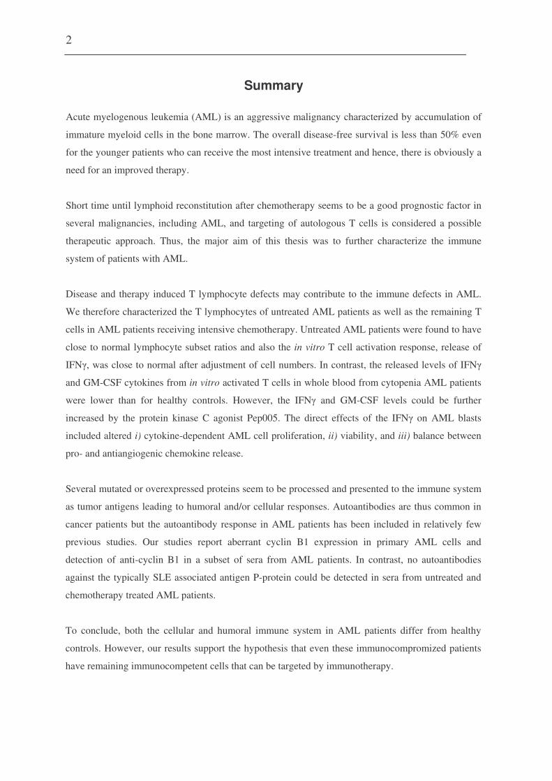
Summary
Acute myelogenous leukemia (AML) is an aggressive malignancy characterized by accumulation of
immature myeloid cells in the bone marrow. The overall disease-free survival is less than 50% even
for the younger patients who can receive the most intensive treatment and hence, there is obviously a
need for an improved therapy.
Short time until lymphoid reconstitution after chemotherapy seems to be a good prognostic factor in
several malignancies, including AML, and targeting of autologous T cells is considered a possible
therapeutic approach. Thus, the major aim of this thesis was to further characterize the immune
system of patients with AML.
Disease and therapy induced T lymphocyte defects may contribute to the immune defects in AML.
We therefore characterized the T lymphocytes of untreated AML patients as well as the remaining T
cells in AML patients receiving intensive chemotherapy. Untreated AML patients were found to have
close to normal lymphocyte subset ratios and also the in vitro T cell activation response, release of
IFN�, was close to normal after adjustment of cell numbers. In contrast, the released levels of IFN�
and GM-CSF cytokines from in vitro activated T cells in whole blood from cytopenia AML patients
were lower than for healthy controls. However, the IFN� and GM-CSF levels could be further
increased by the protein kinase C agonist Pep005. The direct effects of the IFN� on AML blasts
included altered i) cytokine-dependent AML cell proliferation, ii) viability, and iii) balance between
pro- and antiangiogenic chemokine release.
Several mutated or overexpressed proteins seem to be processed and presented to the immune system
as tumor antigens leading to humoral and/or cellular responses. Autoantibodies are thus common in
cancer patients but the autoantibody response in AML patients has been included in relatively few
previous studies. Our studies report aberrant cyclin B1 expression in primary AML cells and
detection of anti-cyclin B1 in a subset of sera from AML patients. In contrast, no autoantibodies
against the typically SLE associated antigen P-protein could be detected in sera from untreated and
chemotherapy treated AML patients.
To conclude, both the cellular and humoral immune system in AML patients differ from healthy
controls. However, our results support the hypothesis that even these immunocompromized patients
have remaining immunocompetent cells that can be targeted by immunotherapy.

3
List of Papers
Paper I
Ersvær E, Hampson P, Wendelbo Ø, Lord J, Gjertsen BT, Bruserud Ø. Circulating T cells derived
from patients with untreated acute myelogenous leukemia are heterogeneous and can be activated
through the CD3/TCR complex (2006)
Paper II
Ersvær E, Hampson P, Hatfield K, Ulvestad E, Wendelbo Ø, Lord J, Gjertsen BT, Bruserud Ø. T
cells remaining after intensive chemotherapy for acute myelogenous leukemia show a broad
cytokine release profile including high levels of interferon-� that can be further increased by protein
kinase C agonist PEP005. (2006)
Paper III
Ersvær E, Skavland J, Ulvestad E, Gjertsen BT, Bruserud O. Effects of interferon gamma on native
human acute myelogenous leukaemia cells (2006) Cancer Immunol Immunother. Apr 13; [Epub
ahead of print]
Paper IV
Ersvær E, Bertelsen L-T, Espenes LC, Bredholt T, Bøe SO, Iversen BM, Bruserud Ø, Ulvestad E,
Gjertsen BT. Characterization of ribosomal P autoantibodies in relation to cell destruction and
autoimmune disease (2004) Scand J Immunol. Jul-Aug;60(1-2):189-98
Paper V
Ersvær E, Zhang JY, McCormack E, Ånensen N, Tan EM, Gjertsen BT, Bruserud Ø. Cyclin B1 is
commonly expressed in the cytoplasm of primary human acute myelogenous leukemia cells and
serves as a leukemia associated antigen associated with autoantibody response in a subset of
patients (2006).

4
INTRODUCTION
1. Acute myelogenous leukemia
Acute myelogenous leukemia (AML) is a hematological malignancy characterized by clonal
proliferation of immature myeloid precursors and an arrest in the maturation of these cells (1). This
results in accumulation of leukemic blasts in the bone marrow (BM) and eventually in the peripheral
blood (PB); other tissues are usually not affected. The bone marrow infiltration causes a decrease in
the production and thereby a reduction of the peripheral blood levels of mature myeloid cells;
platelets (resulting in thrombocytopenia and eventually hemorrhages), erythrocytes (anemia) and
neutrophils (granulocytopenia and eventually infections). The initial presentation of patients with
AML is with symptoms often related to this pancytopenia (i.e., thrombocytopenia, anemia, and
neutropenia) and the symptoms can include weakness, fatigue, infections and/or bleedings
(hemorrhage). The incidence of AML in Norway is approximately 125 new cases per year.
1.1. Diagnosis and classification
I. Diagnosis
The definitive diagnosis of AML requires demonstration of three diagnostic components:
o The presence of more than 30% (the older The French-American-British (FAB) standard (2, 3))
or 20% (the newer WHO standard (4)) leukemic blasts in a bone marrow aspirate.
o The detection of myeloid differentiation either by histochemistry or by analysis of myeloid
membrane molecules usually by flow cytometry.
o Sub-classification according to (i) the FAB that is based on morphology and histochemistry; or
(ii) a more detailed classification based on morphological characteristics together with clinical
history (de novo or secondary leukemia) and genetic analysis in accordance with the new WHO
classification (Table 1).

5
II. Diagnostic tools
The infiltration of leukemic blasts in the bone marrow is validated cytologically in a bone
marrow smear to identify the blasts and highlight their nuclear and cytoplasmic morphology (4).
AML is a heterogeneous disease and the classification into appropriate variant is a part of the
diagnosis. According to the morphological FAB classification (2, 3), AML can be divided into eight
subtypes (M0-M7). The subtypes differ with respect to the myeloid lineage involved and the degree
of leukemic cell differentiation. Recently, the World Health Organization (WHO) modified the FAB
classification (4, 5) and grouped AML in four categories; i) AML with recurrent genetic
abnormalities, ii) AML with multilineage dysplasia, iii) AML and myelodysplastic syndromes
(MDS), therapy related, and iv) AML, not otherwise categorized (reflecting the old FAB
classification subtypes M0-M7). The WHO classification is summarized in Table 1. An additional
group of acute leukemia of ambigous lineage is defined, these are rare leukemias and account for less
than 4% of all cases of acute leukemia.
Table 1. WHO classification of acute myelogenous leukemia (4, 5). AML with recurrent genetic abnormalities
o AML with t(8;21)(q22;q22), (AML1/ETO) o AML with abnormal BM eosinophils and inv(16)(p13q22) or
t(16;16)(p13;q22), (CBF�/MYH11) o APL with t(15;17)(q22;q12), (PML/RAR�) and variants o AML with 11q23 (MLL) abnormalities
AML with multilineage dysplasia
o Following MDS or MDS/MPD o Without antecedent MDS or MDS/MPD, but with dysplasia in at least
50% of cells in 2 or more myeloid lineage AML and MDS, therapy related
o Alkylating agent/radiation – related type o Topoisomerase II inhibitor – related type o Others
AML, not otherwise categorized
AML minimally differentiated AML without maturation AML with maturation Acute myelomonocytic leukemia Acute monoblastic/acute monocytic leukemia Acute erythroid leukemia (erytroid/leukemia and pure erythroid) Acute megakaryoblastic leukemia Acute basophilic leukemia Acute panmyelosis with myelofibrosis Myeloid sarcoma

6
Cytochemistry and light microscopy (primarily Wright-Giemsa or May-Grünwald-Giemsa
staining) is the principle method for the diagnosis and FAB-subclassification of AML (1). To require
the diagnosis all cases of AML, except M0, must stain positive for MPO and Sudan Black. Other
stains that can be briefly mentioned are non-specific esterase (AML-M2, M4 or M5) and
chloroacetate esterase (late myeloblast and early promyelocyte stage AML-M3) (1).
Immunophenotyping (i.e. flowcytometric analysis of differentiation-associated molecule
expression) analysis plays a central role especially in separating between minimally differentiated
acute myeloid leukemia and acute lymphoblastic leukemia (6). Immunophenotyping is generally
performed by flow cytometry, but also by immunohistochemistry on slides, and the value of
particular markers may differ depending on the technique. All hematopoietic cells express the
common leucocyte antigen (CD45) and 40-60% of the cases will express the stem cell marker CD34
(1). Antigens most frequently used for myeloid lineage assessment are the highly specific myeloid
antigens CD117 (c-kit) and cytoplasmic myeloperoxidase (cMPO). Other less specific myeloid
antigens like CD13, CD33 and CD15, can also occur in acute lymphoblastic leukemia (ALL); and
lymphoid markers like CD2 may also be abberantly expressed in AML (1). In addition to
determining myeloid lineage, immunophenotyping can identify monocytic, erythroid, or
megakaryocytic differentiation. Markers related to lineage differentiation include CD14 and CD11b
in AML-M4 and M5, glycophorin A in AML-M6 and platelet glycoproteins CD41, CD42 and CD61
in AML-M7 (1).
Cytogenetic analysis shows that 50-70% of patients with de novo AML have chromosomal
abnormalities like translocation (t), deletion (del), inversion (inv) or aneuploidy (i.e. abnormal
numbers of specific chromosomes or chromosome sets) (1). Trisomy 8 is the most common
numerical cytogenetic abnormalities found in AML. Specific cytogentic abnormalities (i) can be
closely or uniquely associated with morphology and WHO-classification subsets; (ii) may be
associated with previous chemotherapy in the WHO-subclasses associated with previous treatment
with alkylating agents or topoisomerase II inhibitors; and (iii) can have additional diagnostic (t(8;21)
and inv (16) classified as AML independent of the blast count according to the WHO criteria),
7
prognostic and therapeutic importance. Cytogenetic analysis includes the well-established routine
analysis of G-Banded chromosomes, as well as molecular cytogenetics such as fluorescent in situ
hybridization (FISH) and comparative genomic hybridization (CGH). In addition to cytogenetic
analysis the PCR-based techniques are also applied.
III. Prognostic features
If AML remain untreated, most patients will die during days or weeks depending mostly on the
levels of blasts in the peripheral blood and bone marrow complications. The median survival for
patients only receiving supportive therapy is only 3-4 months, and only exceptional patients survive
for more than one year. Complete remission (CR) rates after intensive chemotherapy vary from 10 to
80%, approximately 10-20% of patients will have refractory disease and first line therapies will fail
to achieve CR, and 5-20% of those treated will die during induction therapy (see below). This
treatment-related mortality depends on the age and the general health condition of the patients and
individuals above 60 years of age will usually not receive the most intensive chemotherapy with
high-dose cytarabine. A large proportion of those in CR will relapse usually within 1-2 years, and
their overall survival is less than 10% (1). It is generally accepted that patients with primary
refractory disease AML relapse after intensive chemotherapy cannot be cured by conventional
chemotherapy but only by allogeneic stem cell transplantation (7).
Age is a highly significant prognostic value as prognosis worsens with increased age. This is due
to the increased treatment-related mortality in elderly patients and an increased frequency of high-
risk karyotypic abnormalities. Response to the initial induction therapy and thus the time taken to
achieve blast clearance is an important indicator to outcome (1). Failure to clear leukemic blasts by
day 16 is a poor prognostic marker as reported by the German AMLCG group (8).
Approximately 60% of AML patients have an abnormal karyotype and this is highly predictive
of response (9, 10):
o Approximately 25% of patients have favourable cytogentics, i.e. t(15;17), inv(16), t(16;16) and
t(8;21). These patients have a 5 year disease-free survival of around 65-80%.
8
o A smaller subset of patients (approximately 10%) will have adverse cytogenetics that include
aneuploidy (-7, -5-5q), abnormalities of 3q or a complex karyotype. These patients have a 5 year
survival of 10-20%.
o The remaining will have intermediate-risk cytogenetics, many of them with a normal karyotype,
and a 5 year survival of 30-40% (1).
In-frame internal tandem duplication (ITD) mutations of the Fms-like tyrosine kinase 3 (flt-3), exons
14-15, have been found in 15-30% of cases of AML. These mutations result in dimerization and
constitutive activation of the receptor (11). ITD mutations have been shown to be an independent
poor prognostic factor in several studies (1). In addition, there has been reported a point mutation of
codon 835 of flt3 in 7-8% of cases of de novo AML (12), resulting in the upregulation of the function
of the kinase domain. The prognostic significance of the point mutation remains uncertain although
certain investigators have reported an adverse effect (1). On the contrary of the poor prognosis
marker ITD-flt3, the mutations of Nucleophosmin (NPM), exon-12, occur in 40-50% of AML with
normal karyotype and are predictors of favourable prognosis especially for patients without Flt3-ITD
(13-16).
Gene expression profiling (GEP) by DNA microarray of AML is becoming more and more
established and seem to be valuable, not only for diagnosing different cytogenetic subtypes but also
for discovering novel AML subclasses (17). Several studies have emphasized that gene expression
signature can be associated with prognosis (17-19).
1.2. The treatment of AML
The initial treatment of AML is remission induction therapy that aims to reduce the leukemic cells to
below the cytologically detectable level of approximately 109 cells. This treatment is followed by a
postinduction or “remission consolidation” therapy, consisting of one or more courses of
chemotherapy or stem cell transplantation. The goal of the postinduction therapy is to eradicate

9
residual leukemia cells and thereby prevent later relapse. The treatment-related mortality is relatively
low for consolidation therapy with conventional chemotherapy and autologous stem cell
transplantation, whereas allotransplantation has a higher treatment-related mortality. The decision
whether or not to recommend an allotransplantation is often based on the following criteria:
o The cytogentic abnormalities (20) and karyotype (21) of the AML cells and whether they are
associated with a high/intermediate/low risk of later relapse.
o The patients age and general health; allotransplatation is usually not recommended for patients
above 55-60 years of age.
o Whether an HLA-matched sibling donor is available or an HLA-matched unrelated donor has to
be used; the transplantation-related mortality is higher when unrelated donors are used.
I. Remission induction therapy
The most common induction remission regime is cytarabine given by continuous intravenous
(IV) infusions (100 mg/m2 per day) for seven days plus daunorubicin (45 to 60 mg/m2 by intravenous
short-time infusion) daily for the first three days (the “7+3” regimen). Approximately 60 to 80
percent of patients achieve a CR with this regimen (22-24). Typically, the side effects of this
treatment are severe bone marrow suppression (myelosuppression), injury of the mucosal lining of
mouth and throat (mucosities), and diarrhea. Alternative intensification regimen with intensification
of induction therapy or addition of potentially non-cross-resistant drugs have been explored (25-30),
but the relative benefits of these strategies or of long-term maintenance therapy are not firmly clear
(31).
Supportive care, like transfusions and antibiotics, has reduced treatment related mortality for
both young and elderly patients. Initial uncontrolled trials using GM-CSF or G-CSF suggested
decrease in the duration of neutropenia after remission induction chemotherapy (1). However, data
from more recent large controlled trials are variable (32, 33) and thus does not firmly support the
previous conclusions drawn from the uncontrolled trials. Other cytokines and growth factors to be
used as supportive care are under investigation but are not used in routine clinical practice (34).
10
G-CSF has also been used to sensitize (prime) leukemic blasts and thereby increase the
effects of chemotherapy (35, 36), either through enhanced leukemic cell uptake of chemotherapeutic
agents (35) and/or by driving resting cells into cell cycle (36). Some of these studies suggest
improved overall survival in the subset of patients with intermediate-risk karyotypes (37, 38).
II. Postremission therapy
In general, there are three choices for postremission therapy: conventional consolidation
chemotherapy, allogeneic hematopoietic cell transplantation (HCT), or autologous HCT.
Consolidation chemotherapy can be the same chemotherapy regimen used for remission
induction or potentially non-cross-resistant drugs. High dose cytarabine (HDAC) seems to provide
the best survival, at least for good and intermediate prognosis patients (24). This intensive
consolidation therapy results in significantly longer survival than less intensive maintenance therapy
alone (true for patients below 60 years of age) and if several courses of consolidation chemotherapy
are given, survival at two or three years is 35 to 50 percent for young and middle-aged adults who
have achieved CR (22, 24, 39, 40). HDAC cannot be used for patients above 60 years of age due to
an unacceptable risk of severe neurological toxicity.
Bone marrow transplantation usually requires that the recipient’s own bone marrow is
destroyed (myeloblative therapy) or partly destroyed (nonmyeloblative therapy) prior to
transplantation. Both peripheral blood mobilized and bone marrow stem cells can be used for
transplantation. Allogeneic hematopoietic stem cell transplantation (allo-HSCT) involves related
(preferably an human leukocyte antigens (HLA)-identical sibling or an HLA-matched relative) or
unrelated donor with the same HLA as the recipient. HLA genes can be categorized into two major
types; Type I (e.g. HLA-A, HLA-B and HLA-C) and Type II (e.g. HLA-DR, HLA-DQB1). If there is
no complete match, a partially matched donor can be considered. However, this last procedure will
increase the risk of graft rejection or severe graft-versus-host (GVH) disease. One beneficial
therapeutic component of allo-HCT is that the donor T cells may produce a specific graft-versus-
leukemia (GVL) immune response, which may contribute to the eradication of remaining leukemia

11
cells. This immune response has been correlated with improved disease-free survival. Long-term
disease free survival in adult patients receiving allo-HSCT in first CR is approximately 45-65% (41-
46). In contrast, survival following allo-HCT in patients with relapsed AML is 35% or less (47). In
autologous hematopoietic cell transplantation (auto-HCT) the patients’ own hematopoietic stem cells
(HSC) are isolated, stored and returned to the body after myeloablative therapy. Treatment-related
morbidity and mortality are low (�5%), thereby allowing it’s use in patients up to 70 years of age,
while relapse rates are relatively high (30-50%). Overall outcomes after auto-HCT are not clearly
better than for patients who receive conventional chemotherapy (48-50).
1.3. Additional biological characteristics of the AML cells
Human AML cells are characterized by (i) constitutive cytokine release; (ii) expression of a wide
range of cytokine receptors, including several hematopoietic growth factors; (iii) expression of
various adhesion molecules involved in the crosstalk between AML cells, extracellular matrix
elements and nonleukemic neighbouring cells; (iv) alteration in intracellular signaling pathways,
especially in pathways involved in regulation of apoptosis (51-54). These biological characteristics
are probably important for leukemogenesis, and these characteristics also seem to be of clinical
importance as prognostic parameters:
o Previous studies suggest that autocrine proliferation, i.e. spontaneous in vitro proliferation due to
autocrine/spontaneous/constitutive release of growth factors, is an adverse prognostic parameter
associated with an increased relapse risk and decreased survival (51).
o The constitutive release of angioregulatory mediators seems to have a prognostic impact. A
recent publication described that expression of Angiopoietin-2 was associated with a good
prognosis (55), whereas VEGF release and high systemic levels of the angioregulatory molecules
VEGF and endostatin seem to be associated with an adverse prognosis (56).
o Intracellular signaling events seem to affect prognosis: (i) specific intracellular phosphoresponses
to exogenous cytokines seem to be associated with prognosis (52); and (i) the intracellular

12
balance between pro- and antiapoptotic signaling seem to influence the risk of relapse after
intensive chemotherapy (54).
Taken together these observations suggest that leukemogenesis and chemosensitivity depend both on
intracellular AML cell characteristics as well as the extracellular interactions between AML cells and
matrix molecules or neighbouring nonleukemic cells.
13
2. The immune system
The immune system (57, 58) may be divided into two major compartments; innate immunity that is
antigen non-specific and adaptive immunity that involves the antigen specific humoral and cellular
arms of the immune system. However, the innate and adaptive responses are a highly cooperative
system, increasing the efficiency of immune responsiveness.
2.1. The innate immune system
Innate immunity (59, 60) constitute the antigen-independent immune mechanism generally involving
i) surface barriers including antimicrobial peptides, ii) mononuclear phagocytes (e.g. monocytes and
macrophages), iii) polymorphonuclear phagocytes (i.e. neutrophil, eosinophil and basophil), iv)
natural killer (NK) cells , v) dendritic cells (DCs), and vi) complement activation. NK cells and DCs
are especially important in tumor immunology and immunotherapy.
NK cells are lymphocytes capable of both directly killing of target cells and production of
immunoregulatory cytokines (61, 62). Mature NK cells are primarily found in PB (10-15% of total
lymphocytes), and typically they express the low affinity receptor for the Fc portion of IgG (CD16),
CD56 and CD161 (61, 62). Phenotypically distinct NK cell populations have been suggested to
represent independent subsets specialized to primarily mediate one or more NK cell functions based
on different levels of spontaneous cytotoxicity or of cytokines produced (61, 62).
DCs are professional antigen presenting cells (APC) and a central player in all immune
responses, both innate and adaptive (63). By phagocytosis, endocytosis, pinocytosis, or receptor-
mediated uptake, DCs capture antigens for immune presentation. After capture of the foreign
material, DCs mature and transport the antigens to lymphoid follicles to deliver it to the B
lymphocytes and to present antigenic peptides to the T lymphocytes. As a result, the specific immune
responses are induced (63). In PB, there has been described at least two DC subsets; the myeloid-

14
derived CD11c+CD123- DCs (mDCs) and lymphoid-derived CD11c-CD123+ DCs (pDCs). pDC seem
to support the generation of a TH2 response, while mDCs predominantly support a TH1 response (see
below) (64-66).
2.2. The adaptive immune system
Humoral (involving B cells) and cell-mediated (involving T cells) immunity constitute the antigen-
dependent immune mechanisms (67).
I. Antigen processing and presentation
The function of the APCs is to present the antigen to the B and T lymphocytes. B cells interact
directly with the antigen, via their cell surface B cell receptor (BCR; consisting of membrane
immunoglobulin (Ig) and Ig�/Igß heterodimer), to differentiate into cells that produce antigen-
specific antibodies (67). Unlike B cells, antigenic peptides are presented to T cells in complex with
HLA molecules from the major histocompatibility complex (MHC) genome area class I or class II.
Of the nine classical MHC-genes the HLA-A, B and C genes belong to class I, while six HLA-D
genes belong to the class II. Generally, class II MHC presents peptides derived from proteolysis of
extracellular antigens in endosomal-type compartments, while class I MHC presents peptides
primarily originating from intracellular degradation of proteins in the cytosol (57, 58, 67).
II. T cell maturation
Progenitor double-negative (CD4-CD8-) T cells from the bone marrow enter the thymus and
rearrange the T cell receptor (TCR) genes to become TCR��+ CD4+CD8- or CD4-CD8+ T cells (the
majority) or TCR��+ CD4+CD8- or CD4-CD8+ T cells (67). Positive selection in the thymus
eliminates T cells unable to recognize self-MHC (i.e. MHC-restriction) and negative selection
eliminates thymocytes with high-affinity receptors for self-MHC molecules alone or autoantigen plus
15
self-MHC (i.e. self-tolerance) (67). Less than 5% of T cells in humans are TCR��+ T cells. In
general, �� T cells are not MHC restricted, and most do not express the CD4 and CD8 coreceptors. ��
T cells seem to recognize epitopes and bind to these in much the same way as immunoglobulin
receptors of B cells (67).
III. T helper cells
The generation of both humoral immune responses (by B cells) and cell-mediated
cytotoxicity (by cytotoxic T (TC) cells) depends on the activation of CD4+ T helper (TH) cells. Initial
TH cell activation is initiated by the interaction of the TCR-CD3 complex (68) with the processed
antigenic peptide bound to a class II MHC molecule on the surface of an APC. TCRs are cell surface
heterodimers consisting of either disulfide-linked �- and �- or �- and �-chains. Each TCR chain is
composed of variable and constant Ig-like extracellular domains, a transmembrane domain and a
short cytoplasmic tail. The �� TCRs bind peptides-MHC (pMHC) with low affinity (~1-100 µM)
through complementary-determining regions (CDRs) in their variable domains (69). The interaction
of the TCR-CD3 complex with the antigenic-peptide MHC molecule is followed by binding of
coreceptor CD4 to the invariant regions of the MHC molecule, leading to the assembly of a signaling
complex. CD4-associated Lck, a protein tyrosine kinase, phosphorylates the immune-receptor
tyrosine-based activation motifs (ITAMs) of CD3 z-chain and thus create a docking site for ZAP70
(67). ZAP70 then phosphorylates adaptor molecules that recruit components from several signaling
pathways like PkC-mediated pathways, CA2+- mediated pathways and small G-protein mediated
pathways like Ras and Rac. Finally, these events lead to changes in gene expression by several
transcription factors (i.e. NFAT, NF-kB, and Elk) (67).
Naive cells require more than the initial interactions, described above, to be fully activated
and subsequently proliferate into effector cells. An antigen nonspecific co-stimulatory signal is
provided by interactions between CD28 on the T cell and members of the B7 family (B7-1 and B7-2)
on the APC. The ligands for B7 are CD28 and CTLA-4 (CD152) which both are expressed on T cells
and act antagonistically (67). That is, signaling through CD28 delivers a positive co-stimulatory
16
signal to the T cell while signaling through CTLA-4 is inhibitory and down-regulates the activation
of T cells. If CD28 co-stimulatory signaling occurs, T cells are triggered into G1 phase of the cell
cycle, transcription of the gene for IL-2 and the �-chain of the high-affinity IL-2 receptor (CD25) are
induced. The subsequent secretion of IL-2 and its binding to the IL-2 receptor induces the activated
naive T cells to proliferate and differentiate into long-lived memory or short-lived effector T cell
populations (67). TH CD4+ effector cells form two subpopulations: the TH1 subset (IL-12R�1�2+)
secretes IL-2, IFN� and TNF-�, and generally aid the activation of cytotoxic T lymphocytes, while
the TH2 subset (IL-12R�1+) secretes IL-4, IL-5, IL-6 and IL-10, and is generally important as helper
cells for B cell activation (57). The cytokines produced by the two subsets also have a cross-
regulatory role, i.e. cytokines secreted from activated TH2 cells will down-regulate the TH1 cells in the
neighborhood and vice versa (67).
IV. Cytotoxic T cells
CD8+ T cells recognize antigen presented by MHC class I and function as TC cells (67).
Activation of resting TC involves first of all TCR stimuli and then secondly stimulation with
cytokines, especially IL-2, most probably from activated TH cells. Naive TC does not express IL-2
receptors on their surface but antigen stimulation increases the expression of the IL-2 receptor and
ensures that only the cells recognizing the antigen will become activated (67).
Like the CD4+ TH1 and TH2 subsets, effector CD8+ T cell subsets can also be identified; TC1
and TC2 (70) . The proximal signal for inducing TC1 and TC2 seem to be IL-12 and IL-4 cytokines,
respectively. The CD8+ T cell subsets are suggested to differ in homing as TC1 is found to express CC
chemokine receptor 5 (CCR5) and TC2 express CCR4 (70).
Cytokine-induced killer (CIK) cells are a unique and rare (1-5% in uncultured PBMC)
population of cytotoxic T lymphocytes with a characteristic CD3+CD56+ phenotype. CIK cells are
non-MHC-restricted in target cell recognition and killing (71).

17
V. Regulatory T cells
Tolerance is generally divided into central and peripheral tolerance (72). Central tolerance is
the clonal deletion and inactivation of self-reactive immature lymphocytes during differentiation (see
above). Peripheral tolerance concerns mature circulating lymphocytes and it is proposed that
regulatory T cells are responsible for inducing and maintaining peripheral tolerance. The classical
regulatory T cells are the thymus-derived CD4+CD25+FOXP3+ Treg cells. However, several
phenotypically distinct regulatory T-cell populations of both CD4+ and CD8+ subset have been
suggested. Possibly, Treg cells can mediate tolerance by suppressing self-antigen-reactive T cells by
various mechanisms, including:
o inducing B7-H4 expression by APCs, which in turn induce T cell cycle arrest through B7-H4;
o directly killing of target cells such as T cells and APCs through perforin- or granzyme B-
dependent pathways;
o CTLA4+ Treg cells can induce indoleamine 2,3-dioxygenase (IDO) expression by APCs which in
turn suppress T cell activation by reducing tryptophan;
o Release of IL-10 and transforming growth factor (TGF�) and thereby direct inhibition of T cell
activation and suppression of APC function by decreasing their expression of MHC molecules,
CD80, CD86, and IL-12 (72).
VI. B cells
Antigen-independent maturation of B cells, involving Ig gene rearrangements, occurs in the
bone marrow and generates mature naive B cells expressing membrane IgM (mIgM) and mIgD with
a single antigenic specificity (67). During maturation, the self-reactive B cells are eliminated by
negative selection. The activation, proliferation and differentiation of naive B cells occur in the
periphery and require the presence of antigen. The antigen recognition molecule of the B cell is the
surface Ig that associates with the heterodimer Ig-�/Ig-�, thus forming the BCR. The B cell co-
receptor (complex of CD19, CD21 and CD81) provides stimulatory modifying signals that enhance
B cell responses. B cell activation through antigen binding to BCR, can occur by two different routes

18
depending on the nature of antigen, one route is dependent on TH cells and the other is not. Antigens
that can activate B cells in the absence of direct interaction with TH cells are known as thymus-
independent (TI) antigens. TI antigens can be further divided into two groups; (i) TI-1 antigens that
are truly T cell independent and give a weaker response with no memory cells formed and IgM as the
predominant antibody; and (ii) TI-2 antigens that require TH cytokines both for efficient B-cell
proliferation and for class switch to isotypes other than IgM. The B cell response to thymus-
dependent (TD) antigens requires the direct contact with TH cells. The TD-antigens bind to mIg on B
cells and become internalized by receptor-mediated endocytosis, processed into peptides, and
presented in MHC class II molecules. Once TH cells recognize the MHC-peptide, the two cells form a
T-B conjugate. This leads to TH cell cytokine release as well as upregulation of CD40L to provide an
essential signal for T cell dependent and antigen-specific B cell activation. Once activated, B cells
begin to express membrane receptors for IL-2, IL-4, IL-5 as well as other cytokines. These receptors
bind the TH cytokines and thus induce differentiation with Ig class switching and development of
antibody-secreting plasma cells, memory B cells and affinity maturation (67).

19
3. The immune system in AML patients
It is well known that AML patients are immunocompromized and have an increased risk of
infections. These patients often have neutropenia initially due to the disease and later eventually due
to intensive chemotherapy. However, as will be seen from the studies reviewed below these patients
can also have other disease- or therapy-induced immune dysfunctions. One would in addition expect
that there will be differences between patients due to for example differences in chemotherapy
regimen, different AML cell phenotypes and age-dependent alterations of the immune system.
3.1. The immune system in untreated AML
I. Cellular innate immunity in AML
Elevated levels of cells with NK phenotype have been found in AML (73). The CD3-CD56+
NK cells were found significantly increased at diagnosis both in the blood and bone marrow. In
contrast, CD16+CD2+ and CD16+CD2- NK cells were only increased in the blood whereas there were
normal levels in the bone marrow. When the authors divided the AML cases into two groups
according to the absolute number of circulating NK cells, the patients with the highest levels also
showed an increased proportion of circulating leukemic blasts. In contrast to elevated NK cell levels,
several other studies report of NK cell dysfunction (74-76) or impeded NK cell maturation (77) in
patients with cancer and leukemia. In addition, it has been suggested that leukemic cells display a
more inhibitory AB killer cell immunoglobulin-like receptor (KIR) phenotype, compared to healthy
controls, in favor of escape from NK cell immunity (78). It is difficult to make a firm conclusion

20
from these studies, but it is justified to state that both qualitative as well as quantitative disturbances
in the NK cell system can be detected in cancer patients and probably also in AML patients.
DCs are central in the presentation of tumor antigens to the adaptive arms of the immune
system. There are several reports of DC defects in cancer patients (reviewed in (79)). Abnormal
frequencies as well as abnormal differentiation and/or maturation possibly caused by tumor derived
factors, are some of the dysfunctions described (79). A number of studies throw light on the
generation of leukemic DCs in the context of immunotherapy, but there are few studies describing
the remaining normal DCs in AML. Mohty et al. (80) reported a quantitative imbalance in circulating
blood myeloid DCs (MDCs) and plasmacytoid monocytes (PDCs) in 70% of the AML patients.
II. Cellular immunity in AML
Altered ratios of TH1 versus TH2 or TC1 versus TC2 cells have been observed in several
malignancies (81-90). However, a general decreased T cell number without such shifts has also been
reported (91, 92), as well as close to normal or increased T-cell numbers (93, 94). A possible
explanation for this variation is that there are differences between malignancies and possibly also
between individual cancer patients.
An abnormal CD4:CD8 ratio has been reported in several malignancies, and this can be due
to increased or decreased absolute levels of CD8+ cells (95-97) and/or increased or reduced levels of
the CD4+ cells (97-99). In patients with head and neck cancer increased CD4:CD8 ratio was observed
in lymph node lymphocytes versus tumor-infiltrating lymphocytes and peripheral blood lymphocytes
(PBLs). In these cases the aberrant ratio was attributable to both a significant enrichment in CD4+ T
cells as well as a decrease in CD8+ T cells (97). In contrast, analysis of patients with non-small cell
lung cancer revealed that tumor infiltrating lymphocytes displayed a lower CD4:CD8 ratio than the
PBLs (100). In peripheral blood of patients with multiple myeloma the percentage of CD3+ and CD8+
cells was within the normal range while the percentage of CD4+ cells was slightly reduced for a small
subset of patients. On the contrary, in bone marrow of myeloma patients the percentage of CD4+ was
profoundly reduced, leading to an altered CD4:CD8 ratio in all multiple myeloma patients (99).

21
There are some studies suggesting that cancer patients have more memory and less naive T
cells applicable for PBL or tumor infiltrating lymphocytes or both (101-104). In hematological
malignancies there has also been suggested decreased memory T helper cells (105, 106).
There are a few reports regarding the T lymphocyte subsets in patients with de novo AML.
Vidriales and colleagues (73) reported (i) increased T lymphocytes with NK activities (CD3+CD56+)
in PB but normal levels in BM, (ii) normal distribution of CD4+CD45RA+ (putative naïve) and
CD4+CD29+ (putative memory) cells in the PB, and (iii) increased distribution of the cytotoxic subset
CD8+CD57+ within the CD8+ cells (73). A more recent report (107) outlined that the absolute
numbers of CD8+ and CD8- (putative CD4+) CD3+ T cells in whole blood of 13 patients with AML
were similar to those of healthy controls. However, there was a tendency of higher numbers of CD8+
T cells in the patients compared with healthy controls, which was also mirrored in the lower
CD4:CD8 ratios (107). These investigators (107) also measured the intracellular cytokine levels of
the whole-blood lymphocytes in the absence of in vitro stimulation. They did not find any significant
changes in the IL-4, IL-10, IL-12 or IFN� levels in the cell subsets derived from AML patients
compared with healthy individuals and thus suggesting normal TH1 and TH2 profile. However, a trend
towards higher absolute numbers and percentages of CD8+ and CD8- lymphocytes with detectable IL-
10, IL-12 and IFN� was observed for the AML patients compared with the healthy controls. Again it
is difficult to reach a firm conclusion, and this is partly due to the question of patient heterogeneity
and the low number of patients in some of the studies.
Treg cells control autoimmune T cell reactivity in the periphery and may also suppress
immune responses against cancer cells (72). An increased number of Treg cells has been reported for
patients with ovarian cancer, lung cancer, breast cancer, gastrointestinal malignancies and lymphoma
(108-114). Wang et al. reported significantly higher proportions of CD4+CD25high Treg cells in AML
patients compared with healthy controls (115). These cells were (i) CD45-RA-, CD69-, CD45-RO+,
CD95+, intercellular CTLA-4+ ; (ii) secreted low levels of TNF-� and IL-10 and did not release IL-2,
IL-4, IL-5 and or IFN�; and (iii) behaved as Treg cells by inhibiting CD4+CD25- T cell proliferation
and cytokine production during in vitro activation (115).

22
III. Humoral immunity in AML
There are sporadic reports of spontaneous remission (SR) of cancer, including leukemia and
myelodysplasia (116, 117), often but not necessarily following bacterial infections or blood
transfusions (118). Some authors have suggested that the mechanism leading to SR is an underlying
humoral immunologic response (116), although there are also other possibilities (119, 120).
However, spontaneous remission in cancer is rare, despite the presence of naturally serum antibodies
against tumor-associated antigens (TAAs) in 14-31 per cent of patients (121). It is not known why
these antibodies against cancer are not more effective. On the other hand, the use of antibody therapy
in cancer treatment suggests that cancer-reactive antibodies can mediate strong anti-cancer effects
(122). However, despite the encouraging experience with antibody therapy in certain malignancies it
is at present hard to believe that humoral immune responses alone can be responsible for spontaneous
remissions in cancer patients.
There is an expanding list of autoantibodies against tumor-associated antigens (TAAs) that
can be detected in cancer patients, including anti-oncoprotein (HER-2/neu), anti-tumor suppression
antigen (P53), anti-proliferation associated antigens (cyclin A, cyclin B1, and CDKs), anti-
onconeural antigens (Hu, and Yo), and anti-cancer/testis antigens (NY-ESO-1 and MAGE-1), and
others (see (123) for review). The identification of a cancer autoantibody signature may become
useful as a diagnostic and prognostic specimen (124-126), but their possible role in the immune
defence against cancer need to be further investigated.
The number of autoantibodies known to occur in AML patients is few compared to other
malignancies. However, autoantibodies against the following antigens have been detected at an
increased frequency in AML patients compared with healthy controls: Wilms tumor gene product
(WT1) (127, 128), single-stranded DNA (129), anticardiolipin antibodies (ACA) (130), the M-phase
phosphoprotein 11 (MPP11) (131), receptor for hyaluronan acid-mediated motility (RHAMM) (132),
MAZ (133), PASD1 (134), and Rhamm-like protein (135). Such antibodies have been reported for 20

23
to 100 per cent of the AML patients examined. Some of these autoantibodies have been detected in
either conditions associated with tissue destruction such as systemic lupus erythematosus (SLE)
(ssDNA) (129) or infections (ACA) (130). Some studies have in addition reported the disappearance
of autoantibodies in AML patients when they reach hematological remission (128, 136), possibly as a
consequence of a lower AML cell burden and decreased antigenic stimulation leading to decreased
autoantibody production.
IV. Leukemia derived factors affecting the immune system
Numerous immunosuppressive factors like TGF-�, IL-10, vascular endothelial growth factor
(VEGF) and sFas-L have been detected in tumor micoenvironments (137). Such tumor-derived
factors can for instance induce apoptosis in T cells (138-140) or inhibit a Th1 response (141) and
thereby contribute to local immunosuppression. The AML microenvironment has also been shown to
inhibit T cell cytotoxic activity (142, 143), and AML culture supernatants show a TGF-�, IL-10 and
VEGF independent inhibition of T cell activation, T cell proliferation and Th1 cytokine production
(143). AML supernatants have also been shown to affect major signaling pathways involved in T cell
activation and proliferation;
o reduced nuclear translocation of NFATc and NF-�B;
o delayed activation of c-Jun N-terminal kinase 1/2;
o no phosphorylation of pRb, cyclin-dependent kinase 6/4-cyclin D, and of p130 was found;
o no induction of c-Myc, cyclin D3, and p107.
However, calcium mobilization, extracellular signal-regulated kinase 1/2, p38, and STAT5 remained
unaffected (143).
Misoguchi et al. (144) were the first to suggest that immune dysfunction in cancer patients
was due to an altered composition of the T-cell receptor signaling complex. Today, decreased
expression of CD3- � (CD3- zeta) in T cells have been demonstrated in several malignancies (145-
148). Reduced CD3-� expression in tumor infiltrating lymphocytes even seems to be an independent
24
prognostic factor for patients with oral carcinoma (149). Furthermore, Buggins et al. (150) reported
abnormal expression of CD3-� in 64 % out of 46 myeloid leukemia patients examined (of which 11
were acute) and successful remission induction was associated with recovery of CD3-� expression.
Moreover, the CD3-� associated protein tyrosine kinases (p56lck, p59fyn, and ZAP-70) showed
variable but often reduced expression in these patients (150). Finally, in a murine AML model (151)
there were seen reduced responses (proliferation and IL-2 secretion) to mitogenic anti-CD3-� but not
to PMA/ionomycin (which is TCR independent) as early as one week following the injection of
leukemic cells, whereas loss of CD3-� protein expression and signaling abnormalities (calcium
mobilization and tyrosine kinase activity) were detected only in advanced disease (4 weeks after
injection). It was also shown in vitro that leukemia-derived factor(s) stimulated splenic macrophages
to secrete a second soluble factor(s) that caused the loss of CD3-� (151). Thus, altered intracellular
signaling during T cell activation probably contributes to the immunodeficiency in AML.
3.2. The immune system in AML patients after chemotherapy
I. Effects of conventional chemotherapy on the cellular immune system
Patients receiving conventional intensive chemotherapy or myeloblative treatment prior to auto-SCT,
encounter a post-treatment period of severe cytopenia, including T lymphopenia (152, 153).
Hematopoietic reconstitution (neutrophils, monocytes, platelets) often occurs relatively early after
chemotherapy compared with lymphoid reconstitution (153).
For patients receiving conventional chemotherapy most peripheral leukocytes are T
lymphocytes, whereas B lymphocytes and monocytes show a wide variation among patients and
usually represent less than 10% of the cells (154). Most of the circulating T cells in these patients
express the activation markers HLA-DR as well as CD25 and CD69 (153). Following chemotherapy
there seems to an absence of CD45RA+CD4+ T cells, all the remaining CD4+ T cells expressed the
CD45RO+ isoform (152, 153).

25
There has also been some reports regarding functional evaluation of T cells, these have
included the examination of cytokine release and cytokine responsiveness of the in vitro expanded
clonogenic T cell minority derived from AML patients with therapy-induced cytopenia (155-159).
Circulating CD4+ and CD8+ TCR��+ T cells include a minor subset of clonogenic cells (155) that
secrete a range of immunoregulatory cytokines (156). The frequency of these clonogenic T cells is
often reduced compared to healthy individuals (155).
Reports of functional analysis of peripheral T cells other than the clonogenic minority are
scarce. However, there are a few studies regarding the proliferative response of T cell derived from
patients with leucopenia evaluated in a whole blood assay (154, 160). In this assay T cells were
found to have proliferative responsiveness equal to healthy controls in the presence of optimal
costimulation with anti-CD3 + anti-CD28, but responses were significantly reduced in the AML
patients with anti-CD3 stimulation only. Furthermore, the responses were significantly lower for
ALL than for the AML patients (154).
Patients receiving chemotherapy for malignant disorders seem to develop a CD4+ T
lymphopenia together with high serum level of the pleiotropic cytokine IL-7 (161, 162). In contrast,
patients with untreated AML and severe chemotherapy-induced leucopenia with CD4+ T
lymphopenia showed decreased IL-7 serum levels, and the detection of circulating IL-7-responsive T
cells indicated that variations in systemic IL-7 levels are functionally important and may contribute
to an additional qualitative T cell defect in T lymphopenic AML patients (163). Other studies have
demonstrated that administration of recombinant interleukin-7 to humans could selectively increase
total CD4+ and CD8+ T lymphocytes together with decrease in the percentage of CD4+ Treg cells
(164).
Very few studies have examined the T cell system in AML patients after hematopoietic
reconstitution. Long-lasting T cell defects can occur after intensive chemotherapy for other
malignancies, especially in adult patients (152). One would expect that similar effects may occur
after AML therapy.

26
II. The immune system after autologous and allogeneic stem cell
transplantation
The recirculation pattern of leukocyte subpopulations during the first 24h after auto-HSCT differs
between leukocyte subsets (165). The number of CD3+ T lymphocytes increased during this period,
whereas CD56+ NK cells decreased rapidly and remained low throughout the observation period. B
lymphocyte levels were also low during the observation period (165). The early reconstitution of T
lymphocyte response after both auto- and allo-BMT is mainly due to the peripheral expansion of
mature T cells transferred with the graft (166, 167). The recovery of polyclonal T lymphocytes
occurs gradually, and complete reconstitution of humoral and cellular immunity may take more than
one year (168). Similar to the observations in patients only receiving intensive chemotherapy, there
seem to be an absence of CD45RA+CD4+ T cells early after auto-transplantation with a remaining
population of CD4+CD45RO+ circulating T lymphocytes (152, 153, 169). One study has suggested
that BM-resident memory T cells are resistant to both pretransplant chemotherapy and ex vivo
pharmacological purging and thus may contribute to the immune reconstitution after auto-BMT
(170).
The studies in this thesis did not include allografted patients and thus a detailed review of
reconstitution after allografting is therefore not included in this presentation. Anyhow, recently, the
CD4+ helper T cell recovery after allo-HSCT have been characterized for patients with various
hematological malignancies (171). In short, the early recovery of CD4+ T cells at 3 months was a
favorable prognostic factor together with higher CD34+ cell transplant dose in terms of overall
survival and non-relapse mortality (171). Also, early immune recovery has been suggested enhanced
following blood stem cell allografting compared with BM allografting (172).

27
Aims of the thesis
New and less toxic therapeutic strategies are needed to improve survival after chemotherapy and also
as an alternative to chemotherapy for elderly patients, and for this reason immunotherapy is
considered in AML. A characterization of the immune system in AML patients is therefore important
as a basis for the design of clinical therapeutic approaches.
The aims of the studies were:
o to characterize the circulating T cells in patients with untreated AML
o to further characterize the remaining T cells in AML patients receiving intensive chemotherapy,
including the T cell release of IFNγ as well as other cytokines known to affect primary human
AML cells
o to characterize in detail the functional effects of the T cell derived cytokine IFN� on primary
human AML cells for a large group of consecutive patients
o to investigate the humoral immune response against autoantigens in AML patients, including a
more detailed investigation of whether cyclin B1 should be regarded as a leukemia-associated
antigen in AML

28
Methods and subjects
Unless otherwise stated we included unselected patients in our studies, i.e. either randomly selected
patients or consecutive patients. For our studies of AML cells we investigated cells that were stored
frozen in liquid nitrogen. The advantage of this approach are that (i) cells from the same patient can
be available for several experiments; (ii) an extensive biological characterization is possible; and (iii)
large groups of patients can be investigated within a reasonable time and in the same experimental
setup. However, two major disadvantages are present: the cells have a decreased viability after
storage and even the viable cells have alterations especially in the membrane molecule expression
(173-176)
Our in vitro cultures were usually prepared in serum-free medium (177). However, we regard the
serumfree Stem Span medium as suboptimal for fibroblasts, osteoblasts and bone marrow stromal
cells (178) and for this reason cocultutures of AML cells and these nonleukemic cells were prepared
in serum-containing Stem Span as described in these studies.
When investigating T cell responses for patients with chemotherapy-induced cytopenia we used a
whole blood assay that has been characterized in detail in previous studies (154, 160). The cells were
then activated by using well-characterized T cell stimulatory signals. The major disadvantage of this
assay is that the number of T cells per culture well is not determined. The major advantage is that
relatively large experiments can be designed even when only small blood samples can be collected. It
can be argued that the use of a whole blood assay is a disadvantage because the culture conditions
will differ between patients, e.g. autologous serum is included in the cultures and the number of other
cells in peripheral blood will differ. On the other hand, it can also be regarded as an advantage that
the immunocompetent cells are investigated in their natural immunoregulatory network by using
diluted whole blood.

29
Summary of the results
Paper I
Ersvaer E, Hampson P, Wendelbo O, Lord J, Gjertsen BT, Bruserud O. Circulating T cells derived
from patients with untreated acute myelogenous leukemia are heterogeneous and can be activated
through the CD3/TCR complex (2006)
Objectives. T lymphocyte defects may contribute to the immune defects in acute myelogenous
leukemia (AML). We characterized the T cell system for a large group of untreated AML patients.
Methods. T lymphocyte subsets were analyzed by flow cytometry for 45 patients. The in vitro
interferon (IFN) γ release in response to stimulation with anti-CD3 + anti-CD28 in the presence of
autologous AML cells was examined for 32 consecutive patients.
Results. The majority of circulating lymphocytes were CD3+ T cell, and CD19+ B cells usually
constituted <10% of the lymphocytes. Most T cells expressed the αβ T cell receptor (TCRαβ+), and
only a minority of the cells was TCRγδ+. Both CD4+ and CD8+ T cells were detected, the CD4:CD8
ratio showed a wide variation but was usually >1.0. The majority of CD4+ T cells were CD45RA+,
whereas most CD8+ T cells were CD45R0+. The T cells could be stimulated to release interferon-γ
(IFNγ) in response to anti-CD3 + anti-CD28 even in the presence of excess autologous AML blasts,
and for a subset (15 of 45) of patients these IFNγ levels could be further increased by the protein
kinase C (PKC) δ agonist Pep005.
Conclusions. Circulating T cells in patients with untreated AML are mainly CD4+ or CD8+ TCRαβ+;
both CD45RA+ and CD45R0+ can be detected, and these cells can be activated through the CD3/TCR
complex even in the presence of excess AML cells. For a subset of patients the T cell responsiveness
can be further increased by targeting PKC.

30
Paper II
Ersvaer E, Hampson P, Hatfield K, Ulvestad E, Wendelbo O, Lord J, Gjertsen BT, Bruserud O. T
cells remaining after intensive chemotherapy for acute myelogenous leukemia show a broad
cytokine release profile including high levels of interferon-� that can be further increased by protein
kinase C agonist PEP005. (2006)
Objectives. Several cytokines are released during T cell activation, including the potentially
antileukemic interferon-γ (IFNγ) and the hematopoietic growth factor granulocyte-macrophage
colony-stimulating factor (GM-CSF) that enhance proliferation and inhibit apoptosis of acute
myelogenous leukemia (AML) cells. In the present study we investigated the release of IFNγ and
GM-CSF by circulating T cells in AML patients with chemotherapy-induced cytopenia.
Methods. T cells were activated with anti-CD3 plus eventually anti-CD28, and the cells were
investigated in a whole blood assay in the presence of their natural cytokine network. We examined
63 samples derived from 16 AML patients during 28 chemotherapy cycles. Activation potential of
protein kinase C agonist Pep005 was also explored.
Results. Activated T cells showed a broad cytokine release profile, but IFNγ and GM-CSF levels
showed a significant correlation and were generally higher than the other cytokine levels. IFNγ and
GM-CSF levels were associated with high CD4:CD8 ratio, low age and no ongoing chemotherapy,
whereas duration of neutropenia or kind of chemotherapy were not similarly associated. The levels of
both cytokines were lower than for healthy controls but higher than for multiple myeloma patients
with cytopenia following autotransplantation. The cytokine levels could be further increased by the
protein kinase C agonist Pep005.
Conclusions. We conclude that remaining T cells after intensive AML therapy show a broad
cytokine release profile including high and significantly correlated levels of potentially antileukemic
IFNγ and the AML growth factor GM-CSF. The final effect of an AML-initiated T cell cytokine
response will then depend on the functional characteristics of the AML cells because the distribution
of IFNγ and GM-CSF receptors differ between AML cells of different patients.

31
Paper III
Ersvaer E, Skavland J, Ulvestad E, Gjertsen BT, Bruserud O. Effects of interferon gamma on native
human acute myelogenous leukaemia cells (2006) Cancer Immunol Immunother. Apr 13; [Epub
ahead of print]
Objectives. T cell targeting immunotherapy is now considered as a possible strategy in acute
myelogenous leukemia (AML), and IFNγ release may then contribute to the antileukemic effects. We
investigated effects of IFNγ on native human AML cells.
Methods. The expression of IFNγ receptor � chain and intracellular Stat and Erk1/2 phosphorylation
after IFN� stimulation were analyzed by flow cytometry. Exogenous IFN� was added to in vitro
cultures of AML blasts, and the effect on proliferation (3H-thymidine incorporation), apoptosis
(Annexin-V and PI) and cytokine release (ELISA) was measured
Results. Normal T cells could be activated to release IFNγ in the presence of AML cells.
Furthermore, high levels of CD119 (IFNγ receptor � chain) expression were observed for all 39
patients examined. Receptor expression was decreased after exposure to exogenous IFNγ, and
receptor ligation caused Stat1 phosphorylation but no phosphorylation of the alternative messengers
Erk1/2. The effect of exogenous IFNγ on AML blast proliferation was dependent on the local
cytokine network and IFNγ (i) inhibited proliferation in the presence of exogenous IL1β, GM-CSF,
G-CSF and SCF; (ii) had divergent effects in the presence of IL3 and Flt3 (65 patients examined);
(iii) inhibited proliferation in the presence of endothelial cells but had divergent effects in the
presence of fibroblasts, osteoblasts and normal stromal cells (65 patients examined). IFNγ increased
stress-induced (spontaneous) in vitro apoptosis as well as cytarabine-induced apoptosis only for a
subset of patients. Furthermore, IFNγ decreased the release of proangiogenic CXCL8 and increased
the release of antiangiogenic CXCL9-11.
Conclusions. We conclude that IFNγ can be released in the presence of native human AML cells and
affect AML cell proliferation, regulation of apoptosis and the balance between pro- and
antiangiogenic chemokine release.

32
Paper IV
Ersvaer E, Bertelsen LT, Espenes LC, Bredholt T, Boe SO, Iversen BM, Bruserud O, Ulvestad E,
Gjertsen BT. Characterization of ribosomal P autoantibodies in relation to cell destruction and
autoimmune disease (2004) Scand J Immunol. Jul-Aug;60(1-2):189-98
Objectives. Autoantibodies against the ribosomal P proteins are related to cell death and tissue
destruction and are frequently exhibited in patients with systemic lupus erythematosus (SLE). We
attempted to explore the effect of tissue destruction on the induction of anti-P autoantibodies and
analysed antibody levels for patients with autoimmune disorders and acute myelogenous leukemia
patients.
Methods. We searched for anti-P autoantibodies by enzyme-linked immunosorbent assay in 201
antinuclear antibody (ANA)-positive individuals, in 10 patients with treated kidney SLE and in 45
untreated acute leukaemia patients. The autoantibody reactivity was further characterized using one-
and two-dimensional immunoblot analysis and immunofluorescence.
Results. Anti-P were detected in 5.5% (11/201) of ANA-positive individuals, but not in kidney-
affected SLE patients or in patients with leukaemia. Seven of 11 anti-P-positive patients had SLE
(3/11), primary Sjogrens's syndrome (1/11) and other autoimmune diseases (3/11). An association
between disease activity and anti-P was suggested by follow-up examinations in one SLE patient,
supported by the absence of anti-P autoantibodies in the 10 treated kidney SLE patients. Anti-P
autoantibodies were detected by immunoblot in one patient with SLE indicating anti-P2
predominance and in the patient with Sjogrens's syndrome indicating anti-P1 predominance.
Diverging humoral responses in these ANA- and anti-P-positive patients were further illustrated by
immunofluorescence, elucidating varying nuclear reactivity and anti-P pattern. Anti-P could not be
detected for any leukemia patients before therapy; a smaller patient group was also investigated
following intensive chemotherapy but anti-P could not be detected for these patients either.
Conclusion. The observation of anti-P in individuals with active autoimmune disease, but not in
patients with chemotherapy-induced cell damage, suggests that anti-P antibodies are part of a specific
disease process, and not elicited as a response to cell destruction per se.

33
Paper V
Ersvaer E, Zhang JY, McCormack E, Anensen N, Tan EM, Gjertsen BT, Bruserud O. Cyclin B1 is
commonly expressed in the cytoplasm of primary human acute myelogenous leukemia cells and
serves as a leukemia associated antigen associated with autoantibody response in a subset of
patients (2006).
Objectives. Cyclin B1 is involved in cell cycle regulation of both normal and malignant cells.
Cytoplasmic expression of cyclin B1 in epithelial malignancies is associated with a specific T cell
response and presumably also a humoral immune response. We therefore investigated the protein
expression of cyclin B1 in native human acute myelogenous leukemia (AML) cells and the
occurrence of cyclin B1 specific antibodies in AML patients.
Methods. Level of intracellular cyclin B1 expression was measured by flow cytometry, while
confocal laser microscopy indicated the cyclin B1 localization within the naive and dendritic AML
cells. Serum antibodies were analyzed by ELISA.
Results. AML cell expression of cyclin B1 was detected for all 42 patients investigated by flow
cytometry; but the percentage of cyclin B1 positive cells showed a wide intra patient variation.
Confocal laser microscopy demonstrated that most of these patients (32/42-76%) showed abnormal
cytoplasmic expression. Furthermore, both cytoplasmic and nuclear expression was maintained after
14 days of in vitro culture and differentiation of the AML cells in the direction of a dendritic cell
phenotype. AML engraftment in NOD/LtSz-Prkdcscid/B2m-/- mice showed no clear correlation with
cyclin B1 expression. Cyclin B1 specific serum antibodies could be detected for 8 out of 66
consecutive patients with untreated AML. Fourteen of these patients of these patients were in
addition tested later following intensive chemotherapy; three of them had detectable antibody levels
that decreased following treatment.
Conclusions. Our studies demonstrate that primary human AML cells show aberrant cytoplasmic
expression of cyclin B1 for a majority of patients; this expression pattern has been associated with
induction of specific immune responses in patients with epithelial malignancies and a specific
humoral immune response was also detected for a subset of patients with untreated AML.
34
General discussion
The bone marrow as an immunological compartment
AML is a bone marrow disease, and the bone marrow is a part of the lymphocyte
recirculation network. Recently the bone marrow was described in mice to function as a secondary
lymphoid organ, providing a microenvironment for priming of T cell responses (179). Naive T cells
can thus home to the bone marrow as a part of their normal recirculatory pathway and become
primed by CD11c+ dendritic cells in the stroma to generate primary and memory T cell responses
(179). These observations are further supported by a study describing a secondary in vivo bone
marrow T cell response induced by a tumor cell vaccine of mice (180). By using peptide/MHC-
tetrameric complexes clonal expansion of antigen-specific T cells could be detected during the
primary response in BM and during the secondary response in the peritoneal cavity and BM (180).
Feuerer et al. have also shown that BM stroma in mice constitutively expressed ICAM-1, VCAM-1,
MadCAM-1, and P-selectin adhesion molecules relevant for homing of blood T lymphocytes to the
bone marrow, and also bone marrow expression of the T cell co-stimulatory molecule CD80 (181). In
the same study, CD3+ T cells were detected together with BM resident CD11c+ DCs, often enriched
in follicle-like structures in the BM parenchyma. Interactions between transferred antigen specific
transgenic CD4+ T cells and antigen loaded BM-DCs then formed multicellular clusters in situ in
BM, generated lymphoblasts and lead to clonal T cell expansion within such clusters. The great
majority of BM-CD4+ T cells had a memory phenotype suggesting that the BM microenvironment
facilitates maintenance of CD4+ memory (181).
As the bone marrow of patients with AML will be severely compromised by the presence of
large numbers of AML blasts, it is possible that reduced T cell responsiveness in this environment
could contribute to the immunodeficiency that accompanies AML.

35
IFN-γγγγ in patients with AML
IFN� (182) is a lymphokine released especially by activated TH1, TC1 and NK cells. Binding of IFN�
to the IFN� receptor complex on target cell transmits signals to the cytoplasm and nucleus through
the Jak-Stat1 mechanism as well as Stat1 independent pathways (183, 184). In a recent study it was
demonstrated that intracellular signaling events after IFN� receptor ligation could be used in
prognostic classification of AML patients (52). In the in vitro culture experiments exploring the
effects of IFN� on AML blasts (paper III), we showed that exogenous IFNγ could affect AML cell
proliferation, regulation of apoptosis and the balance between pro- and antiangiogenic chemokine
release. We would emphasize that these alterations probably are caused by direct effects of IFNγ
effects on the AML cells because we used highly enriched leukemic cell populations in our studies.
IFN� is supposed to induce the expression of MHC class II on the surfaces of various cell
types and thereby probably converting them to nonprofessional APCs. The presentation of leukemia-
associated antigens to T cells may then be improved, but it has also been suggested that MHC II
restricted antigen presentation by this excess of non-professional APCs may result in antigen-specific
T cell anergy or induction of Treg cells (182). Also, a recently study concluded that IFN� appeared to
have a negative regulatory role of the in vivo DC function (185). Thus, further investigations have to
clarify whether local IFNγ release will enhance or downregulate antileukemic T cell reactivity.
The in vivo effects of IFN� on malignant cells seem to differ and both suppression of
tumorigenicity (182, 186) and enhancement of tumor growth or metastatic potential (182, 187) has
been described. These differences can be caused by differences in the direct effects of IFNγ on the
malignant cells, but indirect effects mediated via stromal cells may also be important. We
characterized the direct effects of IFNγ on AML cells, but a recent experimental study in mice
suggested that the crosstalk between leukemic and immunocompetent may be more important than
the direct effects (188). In this model Stat-1, which is the major intracellular target for IFNγ receptor
ligation in AML, is regarded as a tumor suppressor and considered as a key regulator of the
surveillance of developing malignancies. In contrast to these previous studies, these authors
described that Stat-1 acted as a tumor promoter for leukemia development, and absence of Stat-1 was
36
then associated with decreased MHC class I expression and thereby more efficient recognition of
leukemia cells by NK cells. These experimental observations illustrate the importance of
investigating potential antileukemic immune reactivity within the natural immunoregulatory network.
Naive versus memory T cells
Several studies (189-191) suggest that naive and memory T cells cannot be estimated by using only
the typically CD45RA and CD45RO markers, respectively, and for an accurate estimation one has to
use additional markers (i.e. CD62L, CD11a, CD27). However, de Rosa et al. report that when a third
marker was included to identify naive cells, a population of cells were found to be discordant.
Functional studies and analysis of the Vβ repertoire suggest that the discordant cells appeared to be
non-naive (190). Sallusto et al. (191) described that CCR7 identifies a subset of CD8+ memory cells
that coexpress CD62L and de Rosa et al. (190) described that truly naive CD8+ cells had to be
identified by three additional markers similar to CD4+ naive cells. Thus, the frequency of naive T
cells among CD8+ and CD4+ CD45RA+ cells show a wide variation, and would be expected to vary
even more in patients than for healthy individuals. From our study regarding the T cell expression of
CD45RA and CD45RO in untreated AML patients (paper I), there can only be concluded that the
AML patients are heterogeneous with regard to circulating CD45RA/RO cells, and this heterogeneity
in membrane molecule expression probably reflects heterogeneity with regard to frequencies of
memory and naive T cells. However, a proper examination of this question would be to combine
CD4/CD8 with at least 3 additional markers to eliminate discordant cells, and then to investigate the
Vβ repertoire of these cells. The CD45RA+ and CD45RO+ populations are heterogeneous in healthy
individuals, and there is no reason to expect a less complex situation in patients.
The possibly use of PEP005 in AML
A recent study suggested that the active agent in the sap of Euphorbia peplus, ingenol 3-angelate
(PEP005), had antileukemic activity that was mediated through the delta isoform of PKC (192).
PEP005 induced apoptosis in AML cell lines as well as in primary AML cells, whereas normal
hematopoietic progenitors were resistant to PEP005-induced apoptosis. However, the study included
37
primary AML cells only from a relatively small number of patients, and it will be important to
investigate these effects in a larger group of well-characterized unselected patients. Preliminary
studies of AML xenografts using the NB4 cell line in NOD/SCID/�2m-/- mice show no prolonged
survival for mice receiving PEP005. These animals die due to leukemic infiltrates in the brain but the
tumor burden in abdominal lymph nodes and ovaries seem to be reduced (personal communication
with Dr. E. Mc Cormack and Dr. KQ. Wang). A possible explanation for these observations could be
that the antileukemic effect is not observed in the central nervous system due to the blood-brain
barrier and thereby lower drug concentrations in this compartment.
The antileukemic action of PEP005 was proposed to be mediated through the activation of
PKC-� (192). PKC is a family of 11 isoenzymes of which 8 show in vitro responsiveness to PEP005
(classical �, �1, �II, � and novel �, �, , PKCs) (193). PKC seem to have emerging and diverse roles
in immune cell signaling (194), however the effect of PEP005 on T cell functions have not been
examined previously. Our results suggest that PEP005 enhances the in vitro activation of T cells
derived from patients with chemotherapy induced cytopenia (paper II), but an enhancement was
observed only for a minority of patients when testing T cells derived from patients with untreated
AML. This difference could be caused either by (i) the presence of IL2, this was added to the cultures
only when testing cytopenic patients, or (ii) different culture conditions and especially the presence
of peripheral blood components when testing cytopenic patients with the whole blood assay. The
presence of AML cells probably does not have any major influence on the overall results because the
effect of Pep005 was similar when we compared healthy controls and patients with untreated AML.
However, differences in the AML cell phenotype, especially the constitutive release of
immunoregulatory cytokines, may be important for certain patients and contribute to the variation
between patients.
Taken together these studies suggest that Pep005 should be tried in cancer therapy because
(i) the drug seems to have direct antileukemic effects; (ii) the drug seems to enhance T cell reactivity
and may therefore also enhance antileukemic immune reactivity. However, it remains to be clarified
whether Pep005 will affect chemotaxis of immunocompetent cells and thereby their local recruitment
38
to the cancer cell compartments. These future studies have to include the effect of this drug on the
AML cells release of T cell chemotactic chemokines.
Disease-induced alterations in the cellular immune system in AML and multiple
myeloma patients
MM is a clonal B-cell malignancy characterised by an excess of mature plasma cells in the BM, and
the large majority of patients has monoclonal immunoglobulin protein (complete molecules or only
light chains) in serum and/or urine and usually develop decreased normal immunoglobulin (Ig)
levels. The cellular immune system of patients with multiple myeloma has been examined
previously, and several differences were then observed compared with our and others observation in
AML patients:
o a decreased CD4+/CD8+ ratio is usually observed in circulating T lymphocytes of patients with
MM (95, 195), whereas in AML patients the CD4/CD8 ratio close to normal ((107) and paper I);
o myeloma patients seem to have elevated TH1/TH2 ratio (88), whereas Panoskaltsis et al. (107)
described TH1 and TH2 cytokine profiles close to normal in AML patients.
In our comparison of myeloma and AML patients (paper II) we describe a difference in T cell
cytokine release between these patients. Treatment-induced alterations probably contribute to this
difference because our AML patients receive intensive chemotherapy alone, whereas the myeloma
patients receive a different chemotherapy regimen in combination with autologous stem cell support.
Autoantibodies in patients with malignant disorders
It is well-established that autoimmunity can be associated with cancer, and one of the forms of its
expression is the development of autoantibodies and eventually autoimmune disease (196). Several
mutated or overexpressed proteins seem to be processed and presented to the immune system as
tumor antigens leading to humoral or cellular responses (125). One of the most extensively studied
cancer-associated antigens are p53 (197), but more recent studies have demonstrated that
39
autoantibodies agains a wide range of autoantigens can be detected in patients with malignant
disorders (125, 198). Zhang et al. investigated a group of 174 cancer patients for autoantibodies
against 7 known autoantigens, each autoantibody could then be detected in 10-20% of the patients
but approximately half of the patients had at least one autoantobody (125). Thus, cancer-associated
humoral autoimmunity is a common phenomenon, but autoimmune disease is uncommon (196).
Phage display is a technique in which bacteriophages are engineered to fuse a foreign peptide
or protein with their capside proteins and hence expose or display it on their surface. The
combination of phage display technology and immunoscreening can also be used to identify
antibody-binding peptides (199, 200). Examination of peptide phage libraries allows the screening
for autoantibodies without knowledge about the parental antigen (201), and it is then possible to
profile specific immune responses in patient sera and identify new autoantigens. A recent study
described the identification of peptides and cDNA encoded proteins that are recognized by breast
cancer IgG antibodies but not by antibodies from healthy controls, and the reactivity of some of these
antibodies seemed to be linked to survival (202).
The detection of autoantibodies may become important in future clinical medicine:
o Detection of autoantibodies or autoimmune disease may be the first sign of cancer (196).
o Analysis of autoantibodies may become useful in the screening for cancer and possibly cancer-
specific autoantibody profiles can be identified.
o Although the clinical importance of autoantibody detection is uncertain, at least some studies
suggest that detection of autoantibodies may have a prognostic impact. Detection of p53
autoantibodies in breast cancer patients seem to be associated with high-grade tumors and poor
survival (197), whereas a small clinical study suggested that antibodies against a specific peptide
was associated with long survival (202).
o Antibody “fingerprints” to TAAs are proposed as a future biomarker for early cancer diagnosis
and may thereafter function as guidance for individualized therapeutic interventions and to
predict response to therapy or risk of relapse (203).

40
We examined AML patient sera for antibodies against cyclin B1 and the ribosomal p protein. We
have described the novel findings of abnormal cytoplasmatic distribution related to constitutive
expression of cyclin B1 in AML blasts, as well as the detection of a specific humoral immune
response against cyclin B1 in sera from a minor subset of AML patients (paper V). In contrast, we
did not detect autoantibodies against the SLE associated ribosomal P proteins in sera from untreated
AML patients nor in sera from AML patients with chemotherapy-induced cell damage (paper IV).
p53 antibodies in breast cancer seem to be associated with aggressive disease and poor survival.
AML is one of the most aggressive malignancies with a median survival of only 3-4 months for
patients only receiving supportive therapy. Even though other autoantibodies seem to be associated
with aggressive disease, the frequency of AML patients with anti-cyclin B1 was similar to the
frequency previously observed for less aggressive malignancies like prostate, gastric, breast,
colorectal, lung, and hepatocellular cancers (198). This low frequency was observed even though a
major part of AML patients show cytoplasmic cyclin B1 expression that has been associated with
autoimmunity in other malignancies (204, 205). Furthermore, our analysis of AML patients receiving
intensive chemotherapy suggest that neither the development of cyclin B1 nor ribosomal p protein
antibodies is dependent of extensive tissue damage, at least not for immunocompromized AML
patients receiving intensive chemotherapy. It seems more likely that at least the presence of anti-P
antibodies are a part of a specific autoimmune disease process rather than a response to cell
destruction, and this is also supported by proposals of SLE as a disease with an apoptotic clearance
deficiency (206) and association of anti-P with organ-specific kidney (nephritis) disease in SLE
(207).
Concluding remarks
The overall disease-free survival for patients with AML is less than 50% even for the younger
patients who can receive the most intensive therapy, and there is obviously a need for a more
effective therapy. Immunotherapy (e.g. targeting of autologous T cells) is considered as a possible
therapeutic approach. Short time until lymphoid reconstitution after chemotherapy seems to be a

41
good prognostic factor in several malignancies, including AML. Both the cellular and humoral
immune system in AML patients differ from healthy controls. The main aim of this thesis was
therefore to characterize the immune system of patients with AML. Our results substantiate the
hypothesis that even these immunocompromized patients have remaining immunocompetent cells
that can be targeted by immunotherapy.
42
References
1. Smith M, Barnett M, Bassan R, Gatta G, Tondini C, Kern W. Adult acute myeloid leukaemia. Crit Rev Oncol Hematol 2004;50(3):197-222. 2. Bennett JM, Catovsky D, Daniel MT, Flandrin G, Galton DA, Gralnick HR, et al. Proposals for the classification of the acute leukaemias. French-American-British (FAB) co-operative group. Br J Haematol 1976;33(4):451-8. 3. Bennett JM, Catovsky D, Daniel MT, Flandrin G, Galton DA, Gralnick HR, et al. Proposed revised criteria for the classification of acute myeloid leukemia. A report of the French-American-British Cooperative Group. Ann Intern Med 1985;103(4):620-5. 4. Harris NL, Jaffe ES, Diebold J, Flandrin G, Muller-Hermelink HK, Vardiman J, et al. World Health Organization classification of neoplastic diseases of the hematopoietic and lymphoid tissues: report of the Clinical Advisory Committee meeting-Airlie House, Virginia, November 1997. J Clin Oncol 1999;17(12):3835-49. 5. Vardiman JW, Harris NL, Brunning RD. The World Health Organization (WHO) classification of the myeloid neoplasms. Blood 2002;100(7):2292-302. 6. Jaffe ES, Harris NL, Stein H, Vardiman JW, (Eds.). World Health Organization Classification of Tumours. Pathology & Genetics of Tumours of Haematopoietic and Lymphoid Tissues. Lyon: IARC Press; 2001. 7. Johny A, Song KW, Nantel SH, Lavoie JC, Toze CL, Hogge DE, et al. Early stem cell transplantation for refractory acute leukemia after salvage therapy with high-dose etoposide and cyclophosphamide. Biol Blood Marrow Transplant 2006;12(4):480-9. 8. Kern W, Haferlach T, Schoch C, Loffler H, Gassmann W, Heinecke A, et al. Early blast clearance by remission induction therapy is a major independent prognostic factor for both achievement of complete remission and long-term outcome in acute myeloid leukemia: data from the German AML Cooperative Group (AMLCG) 1992 Trial. Blood 2003;101(1):64-70. 9. Grimwade D, Walker H, Oliver F, Wheatley K, Harrison C, Harrison G, et al. The importance of diagnostic cytogenetics on outcome in AML: analysis of 1,612 patients entered into the MRC AML 10 trial. The Medical Research Council Adult and Children's Leukaemia Working Parties. Blood 1998;92(7):2322-33. 10. Grimwade D, Walker H, Harrison G, Oliver F, Chatters S, Harrison CJ, et al. The predictive value of hierarchical cytogenetic classification in older adults with acute myeloid leukemia (AML): analysis of 1065 patients entered into the United Kingdom Medical Research Council AML11 trial. Blood 2001;98(5):1312-20. 11. Yokota S, Kiyoi H, Nakao M, Iwai T, Misawa S, Okuda T, et al. Internal tandem duplication of the FLT3 gene is preferentially seen in acute myeloid leukemia and myelodysplastic syndrome among various hematological malignancies. A study on a large series of patients and cell lines. Leukemia 1997;11(10):1605-9. 12. Yamamoto Y, Kiyoi H, Nakano Y, Suzuki R, Kodera Y, Miyawaki S, et al. Activating mutation of D835 within the activation loop of FLT3 in human hematologic malignancies. Blood 2001;97(8):2434-9. 13. Dohner K, Schlenk RF, Habdank M, Scholl C, Rucker FG, Corbacioglu A, et al. Mutant nucleophosmin (NPM1) predicts favorable prognosis in younger adults with acute myeloid leukemia and normal cytogenetics: interaction with other gene mutations. Blood 2005;106(12):3740-6. 14. Schnittger S, Schoch C, Kern W, Mecucci C, Tschulik C, Martelli MF, et al. Nucleophosmin gene mutations are predictors of favorable prognosis in acute myelogenous leukemia with a normal karyotype. Blood 2005;106(12):3733-9. 15. Verhaak RG, Goudswaard CS, van Putten W, Bijl MA, Sanders MA, Hugens W, et al. Mutations in nucleophosmin (NPM1) in acute myeloid leukemia (AML): association with other gene abnormalities and previously established gene expression signatures and their favorable prognostic significance. Blood 2005;106(12):3747-54.

43
16. Thiede C, Koch S, Creutzig E, Steudel C, Illmer T, Schaich M, et al. Prevalence and prognostic impact of NPM1 mutations in 1485 adult patients with acute myeloid leukemia (AML). Blood 2006;107(10):4011-20. 17. Bullinger L, Valk PJ. Gene expression profiling in acute myeloid leukemia. J Clin Oncol 2005;23(26):6296-305. 18. Valk PJ, Verhaak RG, Beijen MA, Erpelinck CA, Barjesteh van Waalwijk van Doorn-Khosrovani S, Boer JM, et al. Prognostically useful gene-expression profiles in acute myeloid leukemia. N Engl J Med 2004;350(16):1617-28. 19. Yagi T, Morimoto A, Eguchi M, Hibi S, Sako M, Ishii E, et al. Identification of a gene expression signature associated with pediatric AML prognosis. Blood 2003;102(5):1849-56. 20. Bloomfield CD, Lawrence D, Byrd JC, Carroll A, Pettenati MJ, Tantravahi R, et al. Frequency of prolonged remission duration after high-dose cytarabine intensification in acute myeloid leukemia varies by cytogenetic subtype. Cancer Res 1998;58(18):4173-9. 21. Byrd JC, Ruppert AS, Mrozek K, Carroll AJ, Edwards CG, Arthur DC, et al. Repetitive cycles of high-dose cytarabine benefit patients with acute myeloid leukemia and inv(16)(p13q22) or t(16;16)(p13;q22): results from CALGB 8461. J Clin Oncol 2004;22(6):1087-94. 22. Stone RM, Mayer RJ. Treatment of the newly diagnosed adult with de novo acute myeloid leukemia. Hematol Oncol Clin North Am 1993;7(1):47-64. 23. Bishop JF. The treatment of adult acute myeloid leukemia. Semin Oncol 1997;24(1):57-69. 24. Mayer RJ, Davis RB, Schiffer CA, Berg DT, Powell BL, Schulman P, et al. Intensive postremission chemotherapy in adults with acute myeloid leukemia. Cancer and Leukemia Group B. N Engl J Med 1994;331(14):896-903. 25. Buchner T, Hiddemann W, Wormann B, Loffler H, Gassmann W, Haferlach T, et al. Double induction strategy for acute myeloid leukemia: the effect of high-dose cytarabine with mitoxantrone instead of standard-dose cytarabine with daunorubicin and 6-thioguanine: a randomized trial by the German AML Cooperative Group. Blood 1999;93(12):4116-24. 26. Buchner T, Hiddemann W, Berdel WE, Wormann B, Schoch C, Fonatsch C, et al. 6-Thioguanine, cytarabine, and daunorubicin (TAD) and high-dose cytarabine and mitoxantrone (HAM) for induction, TAD for consolidation, and either prolonged maintenance by reduced monthly TAD or TAD-HAM-TAD and one course of intensive consolidation by sequential HAM in adult patients at all ages with de novo acute myeloid leukemia (AML): a randomized trial of the German AML Cooperative Group. J Clin Oncol 2003;21(24):4496-504. 27. Estey EH, Thall PF, Cortes JE, Giles FJ, O'Brien S, Pierce SA, et al. Comparison of idarubicin + ara-C-, fludarabine + ara-C-, and topotecan + ara-C-based regimens in treatment of newly diagnosed acute myeloid leukemia, refractory anemia with excess blasts in transformation, or refractory anemia with excess blasts. Blood 2001;98(13):3575-83. 28. Russo D, Malagola M, de Vivo A, Fiacchini M, Martinelli G, Piccaluga PP, et al. Multicentre phase III trial on fludarabine, cytarabine (Ara-C), and idarubicin versus idarubicin, Ara-C and etoposide for induction treatment of younger, newly diagnosed acute myeloid leukaemia patients. Br J Haematol 2005;131(2):172-9. 29. Bishop JF, Lowenthal RM, Joshua D, Matthews JP, Todd D, Cobcroft R, et al. Etoposide in acute nonlymphocytic leukemia. Australian Leukemia Study Group. Blood 1990;75(1):27-32. 30. Bishop JF, Matthews JP, Young GA, Szer J, Gillett A, Joshua D, et al. A randomized study of high-dose cytarabine in induction in acute myeloid leukemia. Blood 1996;87(5):1710-7. 31. Castaigne S, Chevret S, Archimbaud E, Fenaux P, Bordessoule D, Tilly H, et al. Randomized comparison of double induction and timed-sequential induction to a "3 + 7" induction in adults with AML: long-term analysis of the Acute Leukemia French Association (ALFA) 9000 study. Blood 2004;104(8):2467-74. 32. Schiffer CA. Hematopoietic growth factors as adjuncts to the treatment of acute myeloid leukemia. Blood 1996;88(10):3675-85. 33. Ozer H, Armitage JO, Bennett CL, Crawford J, Demetri GD, Pizzo PA, et al. 2000 update of recommendations for the use of hematopoietic colony-stimulating factors: evidence-based, clinical practice guidelines. American Society of Clinical Oncology Growth Factors Expert Panel. J Clin Oncol 2000;18(20):3558-85.

44
34. Archimbaud E, Ottmann OG, Yin JA, Lechner K, Dombret H, Sanz MA, et al. A randomized, double-blind, placebo-controlled study with pegylated recombinant human megakaryocyte growth and development factor (PEG-rHuMGDF) as an adjunct to chemotherapy for adults with de novo acute myeloid leukemia. Blood 1999;94(11):3694-701. 35. Reuter C, Auf der Landwehr U, Schleyer E, Zuhlsdorf M, Ameling C, Rolf C, et al. Modulation of intracellular metabolism of cytosine arabinoside in acute myeloid leukemia by granulocyte-macrophage colony-stimulating factor. Leukemia 1994;8(2):217-25. 36. Baer MR, Bernstein SH, Brunetto VL, Heinonen K, Mrozek K, Swann VL, et al. Biological effects of recombinant human granulocyte colony-stimulating factor in patients with untreated acute myeloid leukemia. Blood 1996;87(4):1484-94. 37. Lowenberg B, van Putten W, Theobald M, Gmur J, Verdonck L, Sonneveld P, et al. Effect of priming with granulocyte colony-stimulating factor on the outcome of chemotherapy for acute myeloid leukemia. N Engl J Med 2003;349(8):743-52. 38. Hiddemann W, Kern W, Heinecke A, Sauerland C, Buchner T. Priming with G-CSF in acute myeloid leukemia: preliminary data of the AMLCG. Ann Hematol 2004;83 Suppl 1:S53-4. 39. Devine SM, Larson RA. Acute leukemia in adults: recent developments in diagnosis and treatment. CA Cancer J Clin 1994;44(6):326-52. 40. Cassileth PA, Lynch E, Hines JD, Oken MM, Mazza JJ, Bennett JM, et al. Varying intensity of postremission therapy in acute myeloid leukemia. Blood 1992;79(8):1924-30. 41. Keating S, de Witte T, Suciu S, Willemze R, Hayat M, Labar B, et al. The influence of HLA-matched sibling donor availability on treatment outcome for patients with AML: an analysis of the AML 8A study of the EORTC Leukaemia Cooperative Group and GIMEMA. European Organization for Research and Treatment of Cancer. Gruppo Italiano Malattie Ematologiche Maligne dell'Adulto. Br J Haematol 1998;102(5):1344-53. 42. Frassoni F, Labopin M, Gluckman E, Prentice HG, Vernant JP, Zwaan F, et al. Results of allogeneic bone marrow transplantation for acute leukemia have improved in Europe with time--a report of the acute leukemia working party of the European group for blood and marrow transplantation (EBMT). Bone Marrow Transplant 1996;17(1):13-8. 43. Cassileth PA, Harrington DP, Appelbaum FR, Lazarus HM, Rowe JM, Paietta E, et al. Chemotherapy compared with autologous or allogeneic bone marrow transplantation in the management of acute myeloid leukemia in first remission. N Engl J Med 1998;339(23):1649-56. 44. Blaise D, Maraninchi D, Michallet M, Reiffers J, Jouet JP, Milpied N, et al. Long-term follow-up of a randomized trial comparing the combination of cyclophosphamide with total body irradiation or busulfan as conditioning regimen for patients receiving HLA-identical marrow grafts for acute myeloblastic leukemia in first complete remission. Blood 2001;97(11):3669-71. 45. Greinix HT, Nachbaur D, Krieger O, Eibl M, Knobl P, Kalhs P, et al. Factors affecting long-term outcome after allogeneic haematopoietic stem cell transplantation for acute myelogenous leukaemia: a retrospective study of 172 adult patients reported to the Austrian Stem Cell Transplantation Registry. Br J Haematol 2002;117(4):914-23. 46. Suciu S, Mandelli F, de Witte T, Zittoun R, Gallo E, Labar B, et al. Allogeneic compared with autologous stem cell transplantation in the treatment of patients younger than 46 years with acute myeloid leukemia (AML) in first complete remission (CR1): an intention-to-treat analysis of the EORTC/GIMEMAAML-10 trial. Blood 2003;102(4):1232-40. 47. Grigg AP, Szer J, Beresford J, Dodds A, Bradstock K, Durrant S, et al. Factors affecting the outcome of allogeneic bone marrow transplantation for adult patients with refractory or relapsed acute leukaemia. Br J Haematol 1999;107(2):409-18. 48. Tsimberidou AM, Stavroyianni N, Viniou N, Papaioannou M, Tiniakou M, Marinakis T, et al. Comparison of allogeneic stem cell transplantation, high-dose cytarabine, and autologous peripheral stem cell transplantation as postremission treatment in patients with de novo acute myelogenous leukemia. Cancer 2003;97(7):1721-31. 49. Woods WG, Neudorf S, Gold S, Sanders J, Buckley JD, Barnard DR, et al. A comparison of allogeneic bone marrow transplantation, autologous bone marrow transplantation, and aggressive chemotherapy in children with acute myeloid leukemia in remission. Blood 2001;97(1):56-62.

45
50. Nathan PC, Sung L, Crump M, Beyene J. Consolidation therapy with autologous bone marrow transplantation in adults with acute myeloid leukemia: a meta-analysis. J Natl Cancer Inst 2004;96(1):38-45. 51. Bruserud O, Gjertsen BT, Foss B, Huang TS. New strategies in the treatment of acute myelogenous leukemia (AML): in vitro culture of aml cells--the present use in experimental studies and the possible importance for future therapeutic approaches. Stem Cells 2001;19(1):1-11. 52. Irish JM, Hovland R, Krutzik PO, Perez OD, Bruserud O, Gjertsen BT, et al. Single cell profiling of potentiated phospho-protein networks in cancer cells. Cell 2004;118(2):217-28. 53. Bruserud O, Foss B. Serum thrombopoietin levels in acute leukemia patients with chemotherapy-induced cytopenia--inverse correlation between serum levels and platelet counts. Leukemia 1998;12(10):1653-4. 54. Del Principe MI, Del Poeta G, Venditti A, Buccisano F, Maurillo L, Mazzone C, et al. Apoptosis and immaturity in acute myeloid leukemia. Hematology 2005;10(1):25-34. 55. Loges S, Heil G, Bruweleit M, Schoder V, Butzal M, Fischer U, et al. Analysis of concerted expression of angiogenic growth factors in acute myeloid leukemia: expression of angiopoietin-2 represents an independent prognostic factor for overall survival. J Clin Oncol 2005;23(6):1109-17. 56. Hatfield KJ, Olsnes AM, Gjertsen BT, Bruserud O. Antiangiogenic therapy in acute myelogenous leukemia: targeting of vascular endothelial growth factor and interleukin 8 as possible antileukemic strategies. Curr Cancer Drug Targets 2005;5(4):229-48. 57. Delves PJ, Roitt IM. The immune system. Second of two parts. N Engl J Med 2000;343(2):108-17. 58. Delves PJ, Roitt IM. The immune system. First of two parts. N Engl J Med 2000;343(1):37-49. 59. Tosi MF. Innate immune responses to infection. J Allergy Clin Immunol 2005;116(2):241-9; quiz 250. 60. Medzhitov R, Janeway C, Jr. Innate immunity. N Engl J Med 2000;343(5):338-44. 61. Perussia B, Chen Y, Loza MJ. Peripheral NK cell phenotypes: multiple changing of faces of an adapting, developing cell. Mol Immunol 2005;42(4):385-95. 62. Farag SS, VanDeusen JB, Fehniger TA, Caligiuri MA. Biology and clinical impact of human natural killer cells. Int J Hematol 2003;78(1):7-17. 63. Rossi M, Young JW. Human dendritic cells: potent antigen-presenting cells at the crossroads of innate and adaptive immunity. J Immunol 2005;175(3):1373-81. 64. Liu YJ, Kanzler H, Soumelis V, Gilliet M. Dendritic cell lineage, plasticity and cross-regulation. Nat Immunol 2001;2(7):585-9. 65. MacDonald KP, Munster DJ, Clark GJ, Dzionek A, Schmitz J, Hart DN. Characterization of human blood dendritic cell subsets. Blood 2002;100(13):4512-20. 66. Shortman K, Liu YJ. Mouse and human dendritic cell subtypes. Nat Rev Immunol 2002;2(3):151-61. 67. Goldsby RA, Kindt TJ, Osborne BA, Kuby J. Immunology. 5th ed: W.H. Freeman and company; 2003. 68. Alarcon B, Gil D, Delgado P, Schamel WW. Initiation of TCR signaling: regulation within CD3 dimers. Immunol Rev 2003;191:38-46. 69. Rudolph MG, Stanfield RL, Wilson IA. How TCRs bind MHCs, peptides, and coreceptors. Annu Rev Immunol 2006;24:419-66. 70. Woodland DL, Dutton RW. Heterogeneity of CD4(+) and CD8(+) T cells. Curr Opin Immunol 2003;15(3):336-42. 71. Linn YC, Hui KM. Cytokine-induced killer cells: NK-like T cells with cytotolytic specificity against leukemia. Leuk Lymphoma 2003;44(9):1457-62. 72. Zou W. Regulatory T cells, tumour immunity and immunotherapy. Nat Rev Immunol 2006;6(4):295-307. 73. Vidriales MB, Orfao A, Lopez-Berges MC, Gonzalez M, Hernandez JM, Ciudad J, et al. Lymphoid subsets in acute myeloid leukemias: increased number of cells with NK phenotype and normal T-cell distribution. Ann Hematol 1993;67(5):217-22.

46
74. Costello RT, Sivori S, Marcenaro E, Lafage-Pochitaloff M, Mozziconacci MJ, Reviron D, et al. Defective expression and function of natural killer cell-triggering receptors in patients with acute myeloid leukemia. Blood 2002;99(10):3661-7. 75. Pierson BA, Miller JS. CD56+bright and CD56+dim natural killer cells in patients with chronic myelogenous leukemia progressively decrease in number, respond less to stimuli that recruit clonogenic natural killer cells, and exhibit decreased proliferation on a per cell basis. Blood 1996;88(6):2279-87. 76. Sibbitt WL, Jr., Bankhurst AD, Jumonville AJ, Saiki JH, Saiers JH, Doberneck RC. Defects in natural killer cell activity and interferon response in human lung carcinoma and malignant melanoma. Cancer Res 1984;44(2):852-6. 77. Richards JO, Chang X, Blaser BW, Caligiuri MA, Zheng P, Liu Y. Tumor growth impedes natural-killer-cell maturation in the bone marrow. Blood 2006;108(1):246-52. 78. Verheyden S, Bernier M, Demanet C. Identification of natural killer cell receptor phenotypes associated with leukemia. Leukemia 2004;18(12):2002-7. 79. Pinzon-Charry A, Maxwell T, Lopez JA. Dendritic cell dysfunction in cancer: a mechanism for immunosuppression. Immunol Cell Biol 2005;83(5):451-61. 80. Mohty M, Jarrossay D, Lafage-Pochitaloff M, Zandotti C, Briere F, de Lamballeri XN, et al. Circulating blood dendritic cells from myeloid leukemia patients display quantitative and cytogenetic abnormalities as well as functional impairment. Blood 2001;98(13):3750-6. 81. Ito N, Nakamura H, Tanaka Y, Ohgi S. Lung carcinoma: analysis of T helper type 1 and 2 cells and T cytotoxic type 1 and 2 cells by intracellular cytokine detection with flow cytometry. Cancer 1999;85(11):2359-67. 82. Pellegrini P, Berghella AM, Del Beato T, Cicia S, Adorno D, Casciani CU. Disregulation in TH1 and TH2 subsets of CD4+ T cells in peripheral blood of colorectal cancer patients and involvement in cancer establishment and progression. Cancer Immunol Immunother 1996;42(1):1-8. 83. Angevin E, Kremer F, Gaudin C, Hercend T, Triebel F. Analysis of T-cell immune response in renal cell carcinoma: polarization to type 1-like differentiation pattern, clonal T-cell expansion and tumor-specific cytotoxicity. Int J Cancer 1997;72(3):431-40. 84. Sato M, Goto S, Kaneko R, Ito M, Sato S, Takeuchi S. Impaired production of Th1 cytokines and increased frequency of Th2 subsets in PBMC from advanced cancer patients. Anticancer Res 1998;18(5D):3951-5. 85. Goto S, Sato M, Kaneko R, Itoh M, Sato S, Takeuchi S. Analysis of Th1 and Th2 cytokine production by peripheral blood mononuclear cells as a parameter of immunological dysfunction in advanced cancer patients. Cancer Immunol Immunother 1999;48(8):435-42. 86. Tabata T, Hazama S, Yoshino S, Oka M. Th2 subset dominance among peripheral blood T lymphocytes in patients with digestive cancers. Am J Surg 1999;177(3):203-8. 87. Zhang XL, Komada Y, Chipeta J, Li QS, Inaba H, Azuma E, et al. Intracellular cytokine profile of T cells from children with acute lymphoblastic leukemia. Cancer Immunol Immunother 2000;49(3):165-72. 88. Ogawara H, Handa H, Yamazaki T, Toda T, Yoshida K, Nishimoto N, et al. High Th1/Th2 ratio in patients with multiple myeloma. Leuk Res 2005;29(2):135-40. 89. Luczynski W, Stasiak-Barmuta A, Krawczuk-Rybak M, Malinowska I. Assessment of selected co-stimulatory, adhesion and activatory molecules and cytokines of Th(1)/Th(2) balance in acute lymphoblastic leukemia in children. Arch Immunol Ther Exp (Warsz) 2005;53(4):357-63. 90. Reuben JM, Lee BN, Johnson H, Fritsche H, Kantarjian HM, Talpaz M. Restoration of Th1 cytokine synthesis by T cells of patients with chronic myelogenous leukemia in cytogenetic and hematologic remission with interferon-alpha. Clin Cancer Res 2000;6(5):1671-7. 91. Campbell MJ, Scott J, Maecker HT, Park JW, Esserman LJ. Immune dysfunction and micrometastases in women with breast cancer. Breast Cancer Res Treat 2005;91(2):163-71. 92. Kuss I, Hathaway B, Ferris RL, Gooding W, Whiteside TL. Imbalance in absolute counts of T lymphocyte subsets in patients with head and neck cancer and its relation to disease. Adv Otorhinolaryngol 2005;62:161-72. 93. Mellstedt H, Choudhury A. T and B cells in B-chronic lymphocytic leukaemia: Faust, Mephistopheles and the pact with the Devil. Cancer Immunol Immunother 2006;55(2):210-20.
47
94. Isoda A, Yokohama A, Matsushima T, Tsukamoto N, Nojima Y, Karasawa M. The naive T-lymphocyte compartment is well preserved in patients with chronic myelogenous leukaemia in chronic phase. Br J Haematol 2002;119(4):949-55. 95. Koike M, Sekigawa I, Okada M, Matsumoto M, Iida N, Hashimoto H, et al. Relationship between CD4(+)/CD8(+) T cell ratio and T cell activation in multiple myeloma: reference to IL-16. Leuk Res 2002;26(8):705-11. 96. Cartei G, Sala PG, Sanzari M, Ceschia V, Clocchiatti L, Sibau A, et al. Reduced lymphocyte subpopulations in patients with advanced or disseminated melanoma. J Am Acad Dermatol 1993;28(5 Pt 1):738-44. 97. Snyderman CH, Heo DS, Johnson JT, D'Amico F, Barnes L, Whiteside TL. Functional and phenotypic analysis of lymphocytes in head and neck cancer. Arch Otolaryngol Head Neck Surg 1991;117(8):899-905. 98. Kaver I, Pecht M, Trainin N, Greenstein A, Braf Z. T lymphocyte subsets and function in the peripheral blood of patients with urological cancer. Oncology 1992;49(2):108-13. 99. Redoglia V, Boccadoro M, Battaglio S, Dianzani U, Massaia M, Pileri A. Multiple myeloma: altered CD4/CD8 ratio in bone marrow. Haematologica 1990;75(2):129-31. 100. Viale M, Ferrini S, Serrano S, Serrano D, Ardizzoni A, Nicolin A. Peripheral blood and tumor infiltrating lymphocytes in non-small cell lung cancer: analysis at the population and clonal level. Tumori 1990;76(5):488-94. 101. Hueman MT, Stojadinovic A, Storrer CE, Dehqanzada ZA, Gurney JM, Shriver CD, et al. Analysis of naive and memory CD4 and CD8 T cell populations in breast cancer patients receiving a HER2/neu peptide (E75) and GM-CSF vaccine. Cancer Immunol Immunother 2006. 102. Tsukishiro T, Donnenberg AD, Whiteside TL. Rapid turnover of the CD8(+)CD28(-) T-cell subset of effector cells in the circulation of patients with head and neck cancer. Cancer Immunol Immunother 2003;52(10):599-607. 103. Atanackovic D, Block A, de Weerth A, Faltz C, Hossfeld DK, Hegewisch-Becker S. Characterization of effusion-infiltrating T cells: benign versus malignant effusions. Clin Cancer Res 2004;10(8):2600-8. 104. Prado-Garcia H, Aguilar-Cazares D, Flores-Vergara H, Mandoki JJ, Lopez-Gonzalez JS. Effector, memory and naive CD8+ T cells in peripheral blood and pleural effusion from lung adenocarcinoma patients. Lung Cancer 2005;47(3):361-71. 105. van der Horst FA, van der Marel A, den Ottolander GJ, Kluin-Nelemans HC. Decrease of memory T helper cells (CD4+ CD45R0+) in hairy cell leukemia. Leukemia 1993;7(1):46-50. 106. Atanackovic D, Panse J, Schafhausen P, Faltz C, Bartels K, Boeters I, et al. Peripheral T cells of patients with B cell non-Hodgkin's lymphoma show a shift in their memory status. Leuk Res 2005;29(9):1019-27. 107. Panoskaltsis N, Reid CD, Knight SC. Quantification and cytokine production of circulating lymphoid and myeloid cells in acute myelogenous leukaemia. Leukemia 2003;17(4):716-30. 108. Wolf AM, Wolf D, Steurer M, Gastl G, Gunsilius E, Grubeck-Loebenstein B. Increase of regulatory T cells in the peripheral blood of cancer patients. Clin Cancer Res 2003;9(2):606-12. 109. Woo EY, Chu CS, Goletz TJ, Schlienger K, Yeh H, Coukos G, et al. Regulatory CD4(+)CD25(+) T cells in tumors from patients with early-stage non-small cell lung cancer and late-stage ovarian cancer. Cancer Res 2001;61(12):4766-72. 110. Ichihara F, Kono K, Takahashi A, Kawaida H, Sugai H, Fujii H. Increased populations of regulatory T cells in peripheral blood and tumor-infiltrating lymphocytes in patients with gastric and esophageal cancers. Clin Cancer Res 2003;9(12):4404-8. 111. Liyanage UK, Moore TT, Joo HG, Tanaka Y, Herrmann V, Doherty G, et al. Prevalence of regulatory T cells is increased in peripheral blood and tumor microenvironment of patients with pancreas or breast adenocarcinoma. J Immunol 2002;169(5):2756-61. 112. Woo EY, Yeh H, Chu CS, Schlienger K, Carroll RG, Riley JL, et al. Cutting edge: Regulatory T cells from lung cancer patients directly inhibit autologous T cell proliferation. J Immunol 2002;168(9):4272-6.

48
113. Marshall NA, Christie LE, Munro LR, Culligan DJ, Johnston PW, Barker RN, et al. Immunosuppressive regulatory T cells are abundant in the reactive lymphocytes of Hodgkin lymphoma. Blood 2004;103(5):1755-62. 114. Sasada T, Kimura M, Yoshida Y, Kanai M, Takabayashi A. CD4+CD25+ regulatory T cells in patients with gastrointestinal malignancies: possible involvement of regulatory T cells in disease progression. Cancer 2003;98(5):1089-99. 115. Wang X, Zheng J, Liu J, Yao J, He Y, Li X, et al. Increased population of CD4(+)CD25(high), regulatory T cells with their higher apoptotic and proliferating status in peripheral blood of acute myeloid leukemia patients. Eur J Haematol 2005;75(6):468-76. 116. Mitterbauer M, Fritzer-Szekeres M, Mitterbauer G, Simonitsch I, Knobl P, Rintelen C, et al. Spontaneous remission of acute myeloid leukemia after infection and blood transfusion associated with hypergammaglobulinaemia. Ann Hematol 1996;73(4):189-93. 117. Kappauf HW. Spontaneous remission of cancer - enigma and paradigm. Onkologie 2006;29(4):129-30. 118. Paul R, Remes K, Lakkala T, Pelliniemi TT. Spontaneous remission in acute myeloid leukaemia. Br J Haematol 1994;86(1):210-2. 119. Tsavaris N, Kopterides P, Kosmas C, Siakantaris M, Patsouris E, Pangalis G. Spontaneous remission of acute myeloid leukemia associated with GnRH agonist treatment. Leuk Lymphoma 2006;47(3):557-60. 120. Fozza C, Bellizzi S, Bonfigli S, Campus PM, Dore F, Longinotti M. Cytogenetic and hematological spontaneous remission in a case of acute myelogenous leukemia. Eur J Haematol 2004;73(3):219-22. 121. Tan EM, Shi FD. Relative paradigms between autoantibodies in lupus and autoantibodies in cancer. Clin Exp Immunol 2003;134(2):169-77. 122. Cheson BD. Monoclonal antibody therapy of chronic lymphocytic leukemia. Cancer Immunol Immunother 2006;55(2):188-96. 123. Abu-Shakra M, Buskila D, Ehrenfeld M, Conrad K, Shoenfeld Y. Cancer and autoimmunity: autoimmune and rheumatic features in patients with malignancies. Ann Rheum Dis 2001;60(5):433-41. 124. Bradford TJ, Wang X, Chinnaiyan AM. Cancer immunomics: using autoantibody signatures in the early detection of prostate cancer. Urol Oncol 2006;24(3):237-42. 125. Zhang JY, Casiano CA, Peng XX, Koziol JA, Chan EK, Tan EM. Enhancement of antibody detection in cancer using panel of recombinant tumor-associated antigens. Cancer Epidemiol Biomarkers Prev 2003;12(2):136-43. 126. Draghici S, Chatterjee M, Tainsky MA. Epitomics: serum screening for the early detection of cancer on microarrays using complex panels of tumor antigens. Expert Rev Mol Diagn 2005;5(5):735-43. 127. Wu F, Oka Y, Tsuboi A, Elisseeva OA, Ogata K, Nakajima H, et al. Th1-biased humoral immune responses against Wilms tumor gene WT1 product in the patients with hematopoietic malignancies. Leukemia 2005;19(2):268-74. 128. Elisseeva OA, Oka Y, Tsuboi A, Ogata K, Wu F, Kim EH, et al. Humoral immune responses against Wilms tumor gene WT1 product in patients with hematopoietic malignancies. Blood 2002;99(9):3272-9. 129. Kostiala AA, Gripenberg M, Elonen E, Gripenberg G, Kostiala I. Follow-up of antibodies against single-stranded DNA in patients with haematological malignancies. Clin Exp Immunol 1985;61(1):15-23. 130. Stasi R, Stipa E, Masi M, Oliva F, Sciarra A, Perrotti A, et al. Antiphospholipid antibodies: prevalence, clinical significance and correlation to cytokine levels in acute myeloid leukemia and non-Hodgkin's lymphoma. Thromb Haemost 1993;70(4):568-72. 131. Greiner J, Ringhoffer M, Taniguchi M, Hauser T, Schmitt A, Dohner H, et al. Characterization of several leukemia-associated antigens inducing humoral immune responses in acute and chronic myeloid leukemia. Int J Cancer 2003;106(2):224-31.

49
132. Greiner J, Ringhoffer M, Taniguchi M, Schmitt A, Kirchner D, Krahn G, et al. Receptor for hyaluronan acid-mediated motility (RHAMM) is a new immunogenic leukemia-associated antigen in acute and chronic myeloid leukemia. Exp Hematol 2002;30(9):1029-35. 133. Greiner J, Ringhoffer M, Simikopinko O, Szmaragowska A, Huebsch S, Maurer U, et al. Simultaneous expression of different immunogenic antigens in acute myeloid leukemia. Exp Hematol 2000;28(12):1413-22. 134. Guinn BA, Bland EA, Lodi U, Liggins AP, Tobal K, Petters S, et al. Humoral detection of leukaemia-associated antigens in presentation acute myeloid leukaemia. Biochem Biophys Res Commun 2005;335(4):1293-304. 135. Chen G, Zhang W, Cao X, Li F, Liu X, Yao L. Serological identification of immunogenic antigens in acute monocytic leukemia. Leuk Res 2005;29(5):503-9. 136. James SE, Dean CJ, Alexander P. Failure to detect autologous antibodies in the remission sera of patients with AML: complications introduced by the presence of rheumatoid factor. Br J Cancer 1980;42(3):385-91. 137. Chouaib S, Asselin-Paturel C, Mami-Chouaib F, Caignard A, Blay JY. The host-tumor immune conflict: from immunosuppression to resistance and destruction. Immunol Today 1997;18(10):493-7. 138. Rabinowich H, Reichert TE, Kashii Y, Gastman BR, Bell MC, Whiteside TL. Lymphocyte apoptosis induced by Fas ligand- expressing ovarian carcinoma cells. Implications for altered expression of T cell receptor in tumor-associated lymphocytes. J Clin Invest 1998;101(11):2579-88. 139. Gastman BR, Yin XM, Johnson DE, Wieckowski E, Wang GQ, Watkins SC, et al. Tumor-induced apoptosis of T cells: amplification by a mitochondrial cascade. Cancer Res 2000;60(24):6811-7. 140. Buzyn A, Petit F, Ostankovitch M, Figueiredo S, Varet B, Guillet JG, et al. Membrane-bound Fas (Apo-1/CD95) ligand on leukemic cells: A mechanism of tumor immune escape in leukemia patients. Blood 1999;94(9):3135-40. 141. Maeda H, Shiraishi A. TGF-beta contributes to the shift toward Th2-type responses through direct and IL-10-mediated pathways in tumor-bearing mice. J Immunol 1996;156(1):73-8. 142. Lim SH, Worman CP, Jewell A, Goldstone AH. Production of tumour-derived suppressor factor in patients with acute myeloid leukaemia. Leuk Res 1991;15(4):263-8. 143. Buggins AG, Milojkovic D, Arno MJ, Lea NC, Mufti GJ, Thomas NS, et al. Microenvironment produced by acute myeloid leukemia cells prevents T cell activation and proliferation by inhibition of NF-kappaB, c-Myc, and pRb pathways. J Immunol 2001;167(10):6021-30. 144. Mizoguchi H, O'Shea JJ, Longo DL, Loeffler CM, McVicar DW, Ochoa AC. Alterations in signal transduction molecules in T lymphocytes from tumor-bearing mice. Science 1992;258(5089):1795-8. 145. Finke JH, Zea AH, Stanley J, Longo DL, Mizoguchi H, Tubbs RR, et al. Loss of T-cell receptor zeta chain and p56lck in T-cells infiltrating human renal cell carcinoma. Cancer Res 1993;53(23):5613-6. 146. Matsuda M, Petersson M, Lenkei R, Taupin JL, Magnusson I, Mellstedt H, et al. Alterations in the signal-transducing molecules of T cells and NK cells in colorectal tumor-infiltrating, gut mucosal and peripheral lymphocytes: correlation with the stage of the disease. Int J Cancer 1995;61(6):765-72. 147. Renner C, Ohnesorge S, Held G, Bauer S, Jung W, Pfitzenmeier JP, et al. T cells from patients with Hodgkin's disease have a defective T-cell receptor zeta chain expression that is reversible by T-cell stimulation with CD3 and CD28. Blood 1996;88(1):236-41. 148. Rossi E, Matutes E, Morilla R, Owusu-Ankomah K, Heffernan AM, Catovsky D. Zeta chain and CD28 are poorly expressed on T lymphocytes from chronic lymphocytic leukemia. Leukemia 1996;10(3):494-7. 149. Reichert TE, Day R, Wagner EM, Whiteside TL. Absent or low expression of the zeta chain in T cells at the tumor site correlates with poor survival in patients with oral carcinoma. Cancer Res 1998;58(23):5344-7.
50
150. Buggins AG, Hirst WJ, Pagliuca A, Mufti GJ. Variable expression of CD3-zeta and associated protein tyrosine kinases in lymphocytes from patients with myeloid malignancies. Br J Haematol 1998;100(4):784-92. 151. Hirst W, Buggins A, Mufti G. Central role of leukemia-derived factors in the development of leukemia-associated immune dysfunction. Hematol J 2001;2(1):2-17. 152. Mackall CL. T-cell immunodeficiency following cytotoxic antineoplastic therapy: a review. Oncologist 1999;4(5):370-8. 153. Mackall CL, Fleisher TA, Brown MR, Magrath IT, Shad AT, Horowitz ME, et al. Lymphocyte depletion during treatment with intensive chemotherapy for cancer. Blood 1994;84(7):2221-8. 154. Wendelbo O, Nesthus I, Sjo M, Paulsen K, Ernst P, Bruserud O. Functional characterization of T lymphocytes derived from patients with acute myelogenous leukemia and chemotherapy-induced leukopenia. Cancer Immunol Immunother 2004;53(8):740-7. 155. Bruserud O, Ulvestad E, Berentsen S, Bergheim J, Nesthus I. T-lymphocyte functions in acute leukaemia patients with severe chemotherapy-induced cytopenia: characterization of clonogenic T-cell proliferation. Scand J Immunol 1998;47(1):54-62. 156. Bruserud O. Cellular immune responses in acute leukaemia patients with severe chemotherapy-induced leucopenia; characterization of the cytokine repertoire of clonogenic T cells. Cancer Immunol Immunother 1998;46(4):221-8. 157. Bruserud O, Ulvestad E. Cytokine responsiveness of mitogen-activated T cells derived from acute leukemia patients with chemotherapy-induced leukopenia. J Interferon Cytokine Res 2000;20(11):947-54. 158. Bruserud O, von Volkman HL, Ulvestad E. The cellular immune system of patients with acute leukemia and severe chemotherapy-induced leukopenia: characterization of T lymphocyte subsets responsive to IL-16 and IL-17. Acta Haematol 2000;104(2-3):80-91. 159. Bruserud O, Ulvestad E, Halstensen A, Berentsen S, Bergheim J, Nesthus I. Interleukin 4 responses in acute leukaemia patients with severe chemotherapy-induced leucopenia. Eur J Haematol 1997;59(5):269-76. 160. Wendelbo O, Bruserud O. Functional evaluation of proliferative T cell responses in patients with severe T lymphopenia: characterization of optimal culture conditions and standardized activation signals for a simple whole blood assay. J Hematother Stem Cell Res 2003;12(5):525-35. 161. Mackall CL, Fleisher TA, Brown MR, Andrich MP, Chen CC, Feuerstein IM, et al. Age, thymopoiesis, and CD4+ T-lymphocyte regeneration after intensive chemotherapy. N Engl J Med 1995;332(3):143-9. 162. Fry TJ, Connick E, Falloon J, Lederman MM, Liewehr DJ, Spritzler J, et al. A potential role for interleukin-7 in T-cell homeostasis. Blood 2001;97(10):2983-90. 163. Wendelbo O, Glenjen N, Bruserud O. Interleukin-7 (IL-7) in patients receiving intensive chemotherapy for acute myelogenous leukemia: studies of systemic IL-7 Levels and IL-7 responsiveness of circulating T lymphocytes. J Interferon Cytokine Res 2002;22(10):1057-65. 164. Rosenberg SA, Sportes C, Ahmadzadeh M, Fry TJ, Ngo LT, Schwarz SL, et al. IL-7 administration to humans leads to expansion of CD8+ and CD4+ cells but a relative decrease of CD4+ T-regulatory cells. J Immunother 2006;29(3):313-9. 165. Hokland P, Meyer K, Ellegaard J, Nielsen B, Hokland M. Distinct circulation patterns in peripheral blood of leukocyte subpopulations during the first 24 hours following autologous bone marrow transplantation. J Hematother 1996;5(6):647-54. 166. Mackall CL, Bare CV, Granger LA, Sharrow SO, Titus JA, Gress RE. Thymic-independent T cell regeneration occurs via antigen-driven expansion of peripheral T cells resulting in a repertoire that is limited in diversity and prone to skewing. J Immunol 1996;156(12):4609-16. 167. Storek J, Witherspoon RP, Storb R. T cell reconstitution after bone marrow transplantation into adult patients does not resemble T cell development in early life. Bone Marrow Transplant 1995;16(3):413-25. 168. Guillaume T, Rubinstein DB, Symann M. Immune reconstitution and immunotherapy after autologous hematopoietic stem cell transplantation. Blood 1998;92(5):1471-90.
51
169. Zambelli A, Montagna D, Da Prada GA, Maccario R, Zibera C, Moretta A, et al. Evaluation of infectious complications and immune recovery following high-dose chemotherapy (HDC) and autologous peripheral blood progenitor cell transplantation (PBPC-T) in 148 breast cancer patients. Anticancer Res 2002;22(6B):3701-8. 170. Casorati G, Locatelli F, Pagani S, Garavaglia C, Montini E, Lisini D, et al. Bone marrow-resident memory T cells survive pretransplant chemotherapy and contribute to early immune reconstitution of patients with acute myeloid leukemia given mafosfamide-purged autologous bone marrow transplantation. Exp Hematol 2005;33(2):212-8. 171. Kim DH, Sohn SK, Won DI, Lee NY, Suh JS, Lee KB. Rapid helper T-cell recovery above 200 x 10 6/l at 3 months correlates to successful transplant outcomes after allogeneic stem cell transplantation. Bone Marrow Transplant 2006;37(12):1119-28. 172. Abrahamsen IW, Somme S, Heldal D, Egeland T, Kvale D, Tjonnfjord GE. Immune reconstitution after allogeneic stem cell transplantation: the impact of stem cell source and graft-versus-host disease. Haematologica 2005;90(1):86-93. 173. Bruserud O, Ryningen A, Wergeland L, Glenjen NI, Gjertsen BT. Osteoblasts increase proliferation and release of pro-angiogenic interleukin 8 by native human acute myelogenous leukemia blasts. Haematologica 2004;89(4):391-402. 174. Sasnoor LM, Kale VP, Limaye LS. Prevention of apoptosis as a possible mechanism behind improved cryoprotection of hematopoietic cells by catalase and trehalose. Transplantation 2005;80(9):1251-60. 175. Sasnoor LM, Kale VP, Limaye LS. A combination of catalase and trehalose as additives to conventional freezing medium results in improved cryoprotection of human hematopoietic cells with reference to in vitro migration and adhesion properties. Transfusion 2005;45(4):622-33. 176. Sasnoor LM, Kale VP, Limaye LS. Supplementation of conventional freezing medium with a combination of catalase and trehalose results in better protection of surface molecules and functionality of hematopoietic cells. J Hematother Stem Cell Res 2003;12(5):553-64. 177. Bruserud O, Gjertsen BT, von Volkman HL. In vitro culture of human acute myelogenous leukemia (AML) cells in serum-free media: studies of native AML blasts and AML cell lines. J Hematother Stem Cell Res 2000;9(6):923-32. 178. Bruserud O, Tronstad KJ, Berge R. In vitro culture of human osteosarcoma cell lines: a comparison of functional characteristics for cell lines cultured in medium without and with fetal calf serum. J Cancer Res Clin Oncol 2005;131(6):377-84. 179. Feuerer M, Beckhove P, Garbi N, Mahnke Y, Limmer A, Hommel M, et al. Bone marrow as a priming site for T-cell responses to blood-borne antigen. Nat Med 2003;9(9):1151-7. 180. Mahnke YD, Schirrmacher V. Characteristics of a potent tumor vaccine-induced secondary anti-tumor T cell response. Int J Oncol 2004;24(6):1427-34. 181. Feuerer M, Beckhove P, Mahnke Y, Hommel M, Kyewski B, Hamann A, et al. Bone marrow microenvironment facilitating dendritic cell: CD4 T cell interactions and maintenance of CD4 memory. Int J Oncol 2004;25(4):867-76. 182. Billiau A. Interferon-gamma: biology and role in pathogenesis. Adv Immunol 1996;62:61-130. 183. Qing Y, Stark GR. Alternative activation of STAT1 and STAT3 in response to interferon-gamma. J Biol Chem 2004;279(40):41679-85. 184. Ramana CV, Gil MP, Schreiber RD, Stark GR. Stat1-dependent and -independent pathways in IFN-gamma-dependent signaling. Trends Immunol 2002;23(2):96-101. 185. Wu X, Hou W, Sun S, Bi E, Wang Y, Shi M, et al. Novel function of IFN-gamma: negative regulation of dendritic cell migration and T cell priming. J Immunol 2006;177(2):934-43. 186. Shankaran V, Ikeda H, Bruce AT, White JM, Swanson PE, Old LJ, et al. IFNgamma and lymphocytes prevent primary tumour development and shape tumour immunogenicity. Nature 2001;410(6832):1107-11. 187. Hanada T, Kobayashi T, Chinen T, Saeki K, Takaki H, Koga K, et al. IFNgamma-dependent, spontaneous development of colorectal carcinomas in SOCS1-deficient mice. J Exp Med 2006;203(6):1391-7.

52
188. Kovacic B, Stoiber D, Moriggl R, Weisz E, Ott RG, Kreibich R, et al. STAT1 acts as a tumor promoter for leukemia development. Cancer Cell 2006;10(1):77-87. 189. Song K, Rabin RL, Hill BJ, De Rosa SC, Perfetto SP, Zhang HH, et al. Characterization of subsets of CD4+ memory T cells reveals early branched pathways of T cell differentiation in humans. Proc Natl Acad Sci U S A 2005;102(22):7916-21. 190. De Rosa SC, Herzenberg LA, Herzenberg LA, Roederer M. 11-color, 13-parameter flow cytometry: identification of human naive T cells by phenotype, function, and T-cell receptor diversity. Nat Med 2001;7(2):245-8. 191. Sallusto F, Lenig D, Forster R, Lipp M, Lanzavecchia A. Two subsets of memory T lymphocytes with distinct homing potentials and effector functions. Nature 1999;401(6754):708-12. 192. Hampson P, Chahal H, Khanim F, Hayden R, Mulder A, Assi LK, et al. PEP005, a selective small-molecule activator of protein kinase C, has potent antileukemic activity mediated via the delta isoform of PKC. Blood 2005;106(4):1362-8. 193. Kedei N, Lundberg DJ, Toth A, Welburn P, Garfield SH, Blumberg PM. Characterization of the interaction of ingenol 3-angelate with protein kinase C. Cancer Res 2004;64(9):3243-55. 194. Tan SL, Parker PJ. Emerging and diverse roles of protein kinase C in immune cell signalling. Biochem J 2003;376(Pt 3):545-52. 195. San Miguel JF, Gonzalez M, Gascon A, Moro MJ, Hernandez JM, Ortega F, et al. Lymphoid subsets and prognostic factors in multiple myeloma. Cooperative Group for the Study of Monoclonal Gammopathies. Br J Haematol 1992;80(3):305-9. 196. Vedeler CA, Antoine JC, Giometto B, Graus F, Grisold W, Hart IK, et al. Management of paraneoplastic neurological syndromes: report of an EFNS Task Force. Eur J Neurol 2006;13(7):682-90. 197. Soussi T. p53 Antibodies in the sera of patients with various types of cancer: a review. Cancer Res 2000;60(7):1777-88. 198. Koziol JA, Zhang JY, Casiano CA, Peng XX, Shi FD, Feng AC, et al. Recursive partitioning as an approach to selection of immune markers for tumor diagnosis. Clin Cancer Res 2003;9(14):5120-6. 199. Dybwad A, Forre O, Kjeldsen-Kragh J, Natvig JB, Sioud M. Identification of new B cell epitopes in the sera of rheumatoid arthritis patients using a random nanopeptide phage library. Eur J Immunol 1993;23(12):3189-93. 200. Sioud M, Hansen MH. Profiling the immune response in patients with breast cancer by phage-displayed cDNA libraries. Eur J Immunol 2001;31(3):716-25. 201. Cekaite L, Haug O, Myklebost O, Aldrin M, Ostenstad B, Holden M, et al. Analysis of the humoral immune response to immunoselected phage-displayed peptides by a microarray-based method. Proteomics 2004;4(9):2572-82. 202. Hansen MH, Ostenstad B, Sioud M. Antigen-specific IgG antibodies in stage IV long-time survival breast cancer patients. Mol Med 2001;7(4):230-9. 203. Casiano CA, Mediavilla-Varela M, Tan EM. Tumor-associated antigen arrays for the serological diagnosis of cancer. Mol Cell Proteomics 2006. 204. Suzuki H, Graziano DF, McKolanis J, Finn OJ. T cell-dependent antibody responses against aberrantly expressed cyclin B1 protein in patients with cancer and premalignant disease. Clin Cancer Res 2005;11(4):1521-6. 205. Yu M, Zhan Q, Finn OJ. Immune recognition of cyclin B1 as a tumor antigen is a result of its overexpression in human tumors that is caused by non-functional p53. Mol Immunol 2002;38(12-13):981-7. 206. Munoz LE, Gaipl US, Franz S, Sheriff A, Voll RE, Kalden JR, et al. SLE--a disease of clearance deficiency? Rheumatology (Oxford) 2005;44(9):1101-7. 207. Reichlin M. Serological correlations with nephritis in systemic lupus erythematosus. Clin Immunol 2005;117(1):12-4.

53
PAPERS I-V